Carpal Tunnel Syndrome Caused by Lipofibromatous Hamartoma of the Median Nerve
←
→
Page content transcription
If your browser does not render page correctly, please read the page content below
Case Report
J Korean Neurosurg Soc 63 (5) : 664-670, 2020
https://doi.org/10.3340/jkns.2020.0082                                                                                         pISSN 2005-3711 eISSN 1598-7876

Carpal Tunnel Syndrome Caused by Lipofibromatous
Hamartoma of the Median Nerve
Youn-Tae Roh,1 Seok-Whan Song,2 Changhoon Jeong,3 Younghoon Kang,3 Il-Jung Park3
Department of Orthopaedic Surgery,1 H Plus Yangji Hospital, Seoul, Korea
Department of Orthopaedic Surgery,2 Yeouido St. Mary’s Hospital, College of Medicine, The Catholic University of Korea, Seoul, Korea
Department of Orthopaedic Surgery, 3 Bucheon St. Mary’s Hospital, College of Medicine, The Catholic University of Korea, Seoul, Korea

Lipofibromatous hamartoma (LFH) is a rare tumor of the peripheral nerves, which usually involves the median nerve. The authors
reported on two rare cases of carpal tunnel syndrome due to LFH of the median nerve. A 49-year-old female patient complained
of the mass and symptoms consistent with LFH. Magnetic resonance imaging (MRI) showed typical LFH findings. The symptoms
were successfully ameliorated with carpal tunnel release and external neurolysis. A 37-year-old female patient complained of
weakening thumb abduction and the mass where the MRI showed atypical findings. Opponensplasty and debulking operations
were performed after which thumb abduction was improved; however, neurological sequelae remained. LFH of the median nerve is
managed on a case-by-case basis as treatment guidelines are not very clearly defined yet. However, the less invasive treatment such
as carpal tunnel release and external neurolysis than more aggressive surgical treatment should be recommended as a treatment
option.

Key Words : Lipofibromatous hamartoma · Median nerve · Carpal tunnel syndrome · Magnetic resonance imaging.

INTRODUCTION                                                                             ported since Mason first described a case of LFH occurring in
                                                                                         the median nerve in 19534,8,11-13). However, the cause and treat-
   Lipofibromatous hamartoma (LFH) is a rare tumor of pe-                                ment for LFH have not yet been clearly defined. We reported
ripheral nerves that is characterized by an excessive prolifera-                         two cases of secondary carpal tunnel syndrome (CTS) due to
tion of fibroadipose tissue, which infiltrates the epineural and                         LFH on the median nerve.
perineural elements of peripheral nerves. The most commonly
affected nerve is the median nerve, but it is also known to af-
fect the radial nerve, ulnar nerve, and brachial plexus in the                          CASE REPORT
upper extremities2). LFH is considered congenital in origin
and has been commonly associated with macrodactyly and                                   Case 1
other conditions at birth4). A few cases of LFH have been re-                              A 49-year-old woman presented to the hospital with numb-

• Received : March 17, 2020 • Revised : April 9, 2020 • Accepted : April 14, 2020
•A ddress for reprints : Il-Jung Park
  Department of Orthopaedic Surgery, Bucheon St. Mary’s Hospital, College of Medicine, The Catholic University of Korea, 327 Sosa-ro, Bucheon 14647, Korea
  Tel : +82-32-340-7034, Fax : +82-32-340-2671, E-mail : jikocmc@naver.com, ORCID : https://orcid.org/0000-0001-8262-4287

T his is an Open Access article distributed under the terms of the Creative Commons Attribution Non-Commercial License (http://creativecommons.org/licenses/by-nc/4.0)
which permits unrestricted non-commercial use, distribution, and reproduction in any medium, provided the original work is properly cited.

664       Copyright © 2020 The Korean Neurosurgical Society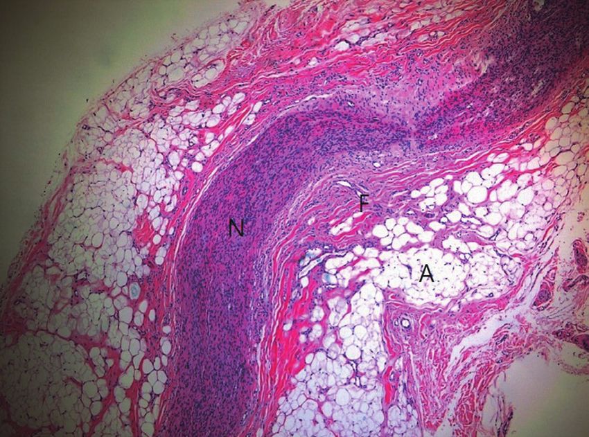
Lipofibromatous Hamartoma of Median Nerve | Roh YT, et al.

ness of the thumb, index, and middle fingers of the right hand                 served.
without any history of trauma. The patient also complained of                    Radiographs did not reveal clear shading of soft tissue
masses in the palm and wrist, which were first observed 7 years                masses and no abnormalities were observed in the bones and
ago after an injection treatment for the numbness of the hand,                 the adjacent joints. Electromyogram tests to identify neuro-
and they gradually increased in size. The patient described the                logical abnormalities indicated CTS of a moderate degree in
injection site as 2 cm above the wrist crease, but she could not               the American Association of Electrodiagnostic Medicine
remember the components of the injection. Physical examina-                    (AAEM) classification. Magnetic resonance imaging (MRI)
tion revealed a mass of 2×3 cm in the thenar area of the right                 was conducted to determine the appearance of the mass. The
palm and another mass of 3×4 cm in the proximal region of the                  coronal MRI scan revealed fusiform swelling of the median
right radiocarpal joint (Fig. 1). The shape and size of the fingers            nerve from the distal forearm to the distal palmar area, and
were all normal. Hypoesthesia of the thumb, index finger, and                  the carpal tunnel median nerve was pressed into an hourglass-
middle finger was observed. Although Tinel’s sign of the carpal                shape by the transverse carpal ligament. In the axial cut, the
tunnel was positive, no atrophy of the thenar muscle was ob-                   mass was shown as an oval shape, and the T1-weighted image

 A                                                                            B

Fig. 1. The clinical photographs (A and B) show a mass from the distal forearm to the palm of the hand (white arrows).

 A                                                         B                                         C

Fig. 2. Magnetic resonance imaging findings. A : The axial T1-weighted image shows the displacement of the flexor tendon and palmar protrusion of
the flexor retinaculum due to an enlarged median nerve (white arrow). B and C : The sagittal and coronal sections demonstrate the fusiform or hourglass
enlargement of the median nerve (white arrows) with low-signal intensity nerve bundles embedded in hyperintense adipose tissue.

                                                                                                   J Korean Neurosurg Soc 63 (5) : 664-670         665
J Korean Neurosurg Soc 63 | September 2020
had a “coaxial cable-like” appearance due to a low-intensity Case 2
nerve fascicle within the high-intensity adipose tissues. In the A 37-year-old woman was admitted due to muscle weakness
coronal and sagittal cuts, the mass had a “spaghetti-like” im- in her left thumb and a mass in her left forearm. The patient
age (Fig. 2). reported having had surgery, the details of which she could
Due to the tumor being on the median nerve and the result- not remember, on her left wrist when she was a child. Since
ing diagnosis of secondary CTS, surgical treatment was decid- then, she had developed thumb abduction weakness and ex-
ed. A 12-cm zigzag longitudinal incision was made alongside perienced dysesthesia. Physical examination confirmed the
the mass, from the distal forearm to the palmar area. After 3 cm post-surgical incision wound alongside the right palmer
dissecting the subcutaneous tissues, the mass was identified side of the thenar crease. Severe atrophy of the thenar muscle
distally along the normal median nerve in the proximal re- and a 4.5×10 cm mass on the palmar side of the distal forearm
gion. No infiltrations into the surrounding tissues were ob- was observed. The left thumb, index, and middle fingers had
served. The outer membrane of the mass was formed along hypoesthesia. Tinel’s sign test was positive on the carpal tun-
the epineurium of the median nerve and a bright yellow sau- nel. There was a weakness of thumb opposition.
sage-like mass was exposed. The mass was severely com-
pressed in the carpal tunnel. In the area of carpal tunnel, we
made an additional curved incision parallel to the thenar
crease approximately 6 cm in length. The transverse carpal
ligament was carefully released along the ulnar border, avoid-
ing damage to the median nerve and its recurrent branch. We
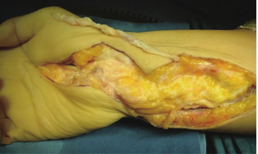
tried to make an incision on the epineurium and perform in-
ternal neurolysis, but the mass and median nerve could not be
separated (Fig. 3). A small portion of the epineurium was ex-
cised for biopsy. The final histological examination confirmed
the diagnosis of LFH (Fig. 4). Numbness disappeared after the
operation. There was no change in the size of the mass
throughout the 12-month post-operative follow-up examina-
tions, but the patient was satisfied with the absence of numb-
ness. Fig. 4. Photomicrography demonstrates the normal nerve fascicle
surrounded by fibroblasts and mature adipocytes (HE staining, ×40). N :
nerve fascicle, F : fibroblast, A : adipoblast.
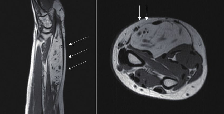
A B
Fig. 3. Intraoperative photographs. A : The enlarged median nerve with fibroadipose tissue proliferation. B : After external neurolysis.
666 https://doi.org/10.3340/jkns.2020.0082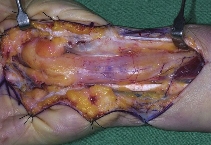
Lipofibromatous Hamartoma of Median Nerve | Roh YT, et al.

   Simple radiography showed an increased shading of soft tis-                  Surgical treatment was determined under the diagnosis of
sues. An electromyogram test indicated CTS of a severe degree                 severe CTS caused by a mass in the median nerve resulting in
in the AAEM classification. An MRI was performed to con-                      a disorder of thumb opposition. A 14-cm zigzag longitudinal
firm the mass properties. In the sagittal and coronal section of              incision was made alongside the mass, and then peeled to ex-
the MRI scan, ‘fusiform swelling’ of the median nerve was                     pose the palmar fascia. The palmaris longus tendon was dis-
observed. In addition, in the axial T1-weighted image at the                  sected up to the midpalmar portion, while leaving the palmar
distal forearm level, a nerve fascicle with low signal intensity              aponeurosis attached for the longest length possible. After dis-
was located eccentrically inside the mass, and the fat tissue                 secting the subcutaneous tissues, the mass was identified dis-
with high signal intensity protruded from the nerve and ex-                   tally along the normal median nerve in the proximal region.
panded to adjacent muscles (Fig. 5).                                          In the area of carpal tunnel, we made an additional curved in-

 A                                                                 B

Fig. 5. Magnetic resonance imaging finding. A : The sagittal section demonstrates fusiform enlargement of the median nerve (white arrows). B : The
axial T1-weighted image shows the distribution of low-intensity nerve bundles was eccentric (white arrows).

 A                                                                           B

Fig. 6. Intraoperative photographs. A : The opponensplasty using the palmaris longus tendon and palmar aponeurosis and the debulking operation are per-
formed. B : Resected tissues are observed.

                                                                                                  J Korean Neurosurg Soc 63 (5) : 664-670          667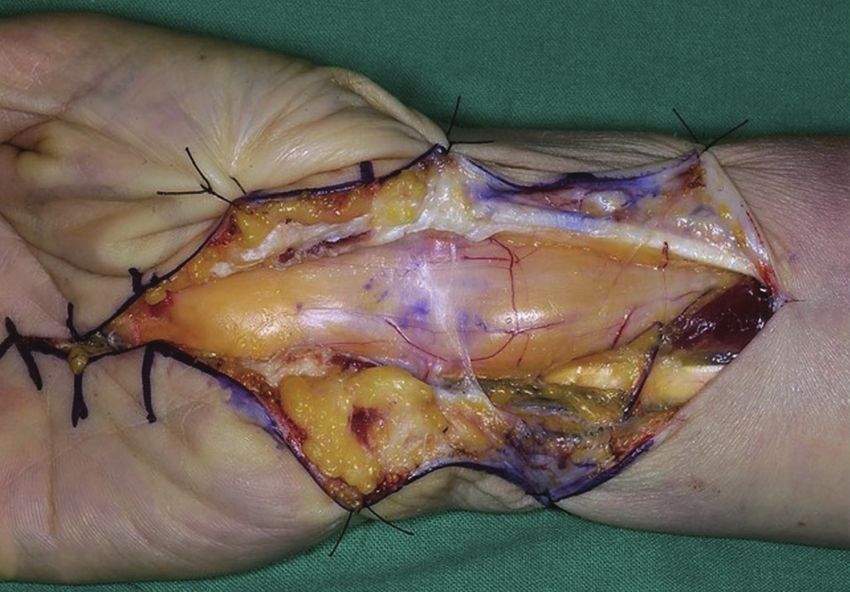
J Korean Neurosurg Soc 63 | September 2020

cision parallel to the thenar crease. The transverse carpal liga-    of injection on her right wrist 7 years before the operation,
ment was carefully released along the ulnar border, avoiding         and the second had surgery on her left wrist in childhood.
damage to the median nerve and its recurrent branch. Care            However, it is very difficult to determine how injection or sur-
should be taken to avoid injury to the superficial palmar ar-        gery is involved in the development of LFH.
tery arch. Then, an internal neurolysis and debulking opera-            The most common symptoms in patients with LFH are en-
tion was performed to remove the yellow tumor tissue as              larging masses on the distal forearm, wrist, palm, or digits13).
much as possible by peeling off the mass while paying special        Tumors gradually increase in size and neuropathy from medi-
attention not to damage nerve fascicles. Subsequently, the pal-      an nerve compression presents in varying degrees. The chief
maris longus tendon was extruded through the subcutaneous            complaints of the patients in this report were numbness of the
tunnel to the radial side of the metacarpophalangeal joint of        thumb, index, and middle fingers and weakness of thumb ab-
the thumb and sutured to the abductor pollicis brevis attach-        duction. The complaint about the development of mass is
ment (Fig. 6). While the thumb was abducted, the first meta-         subsequently reported.
carpal and the metacarpal bone were fixed with Kirschner                Differential diagnosis should include other tumors on the
wire and splint was applied. The final histological examina-         median nerve such as ganglion cysts, lipomas, and traumatic
tion of the resected tumor tissue confirmed the diagnosis of         neuromas. Neurofibromas and schwannomas, which may be-
LFH. After the surgery, the patient wore a thumb splint for          come malignant, could also present in a similar manner. Dif-
4 weeks and a brace for 4 weeks. The Kirschner wire was re-          ferentiation from malignant conditions, such as liposarcoma
moved 8 weeks after the surgery. No increase in the size of the      and malignant peripheral nerve sheath tumor, is also neces-
mass showed 10 months after the surgery. The patient said            sary. In LFH, fibrofatty tissue infiltrates the nerve fascicle,
that thumb abduction was improved greatly after the opera-           making it difficult to separate the tumors and the nerves,
tion, however, the hypoesthesia of the thumb and index finger        while schwannoma is encapsulated and can be easily isolated
persisted.                                                           from the normal nerves. However, neurofibromatosis is diffi-
                                                                     cult to distinguish visually from LFH with macrodactyly, as
                                                                     both are not encapsulated and exhibit mesenchymal over-
DISCUSSION                                                           growth following a nerve distribution. However, the lack of
                                                                     neurocutaneous manifestations and family history allows the
   LFH is a rare condition in which normal forms of adipose          differentiation of LFH from neurofibromatosis. Moreover, the
and fibrous tissues diffusely infiltrate the peripheral nerve13).    two can be further distinguished histologically, as neurofibro-
This condition occurs in patients at a relatively young age. Of      matosis elicits tumefaction of the nerve while LFH is associat-
the reported cases, approximately 71% of patients presented          ed with the proliferation of fibrofatty tissue, leading to infil-
with LFH before the age of 30 and 33% of patients developed          tration between the nerve fibers11,13).
LFH immediately after birth or within the first year. Macro-            Simple radiographs do not reveal any abnormalities in LFH
dactyly around the median nerve was present in 30% of LFH            other than the expansion of the soft tissues. In LFH with mac-
patients2). However, there was no association with family his-       rodactyly, phalangeal bony hypertrophy may be observed. MRI
tory13). Several studies report that pressure from the flexor ret-   is a critical tool for the diagnosis of LFH. MRI findings that are
inaculum and transverse carpal ligament causes microtrauma           characteristics of LFH include displacement of the flexor reti-
to the median nerve, leading to LFH4,9). Reports of trauma be-       naculum and adjacent tendons due to hypertrophy of the me-
fore the onset of symptoms and the early age of onset support        dian nerve viewed in the axial cut; the appearance of coaxial
the speculation of posttraumatic and congenital etiologies for       cables with low-intensity nerve bundles embedded within high-
LFH3,13). Previous trauma to the affected area, such as lacera-      intensity adipose tissue along the median nerve as viewed in the
tion before the onset of symptoms or surgeries for other types       T1-weighted images; and an hourglass appearance caused by
of tumors was observed in 8% of the patients while another           fusiform swelling of the median nerve and compression by the
14% of LFH patients had received surgery to the affected area        transverse carpal ligament, as well as spaghetti-shaped nerve
for similar conditions. In our cases, the first case had a history   bundles surrounded by adipose tissues inside the median

668     https://doi.org/10.3340/jkns.2020.0082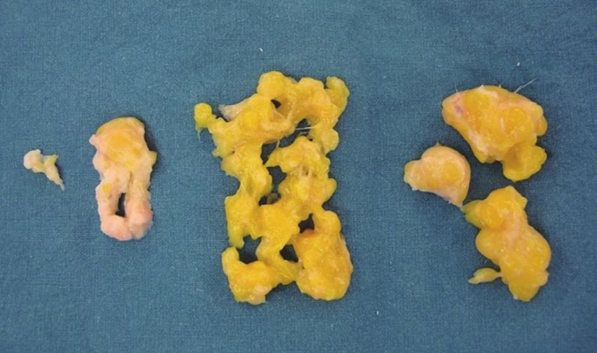
Lipofibromatous Hamartoma of Median Nerve | Roh YT, et al.

nerves as viewed in the coronal and sagittal cuts. Not all LFH       thoughts. Expectant management can be offered for patients
have the same MRI findings. Toms et al.14) reported three cases      with asymptomatic swelling without neurologic impairment
in which, unlike other LFH, nerve bundles were eccentrically         and open carpal tunnel release is the mainstay of treatment for
located. In their cases, the tumors had large cross-sections of      the patients with neurological abnormalities. Microsurgical
2.2, 4.5, and 17 cm2, respectively. As in our second case of a       dissection is also possible but generates disappointing results.
large LFH, the nerve bundle may be eccentrically located in the         Patients with asymptomatic tumors can be observed with-
MRI scan, not in a coaxial cable shape14).                           out intervention. However, spontaneous regression of the tu-
   Biopsy provides a definitive diagnosis of LFH. Histological       mor is uncommon and a gradual increase in size leads to
analysis shows a normal appearance of nerve fibers while fi-         compressive neuropathy. Carpal tunnel release may be per-
broblasts and mature adipocytes separate the nerve fascicles         formed as a preventive measure in asymptomatic patients
and infiltrate the space between the epineurium and perineu-         with a large tumor. And external neurolysis, in addition to
rium. Although routine nerve biopsy is very helpful for diag-        carpal tunnel release, can be a good remedy even for patients
nosis, it is not recommended as functional sequelae may be a         with large tumors13). The chief complaint in case 1 was the
consequence. The biopsy can be performed on a non-lethal             numbness of the hand. Therefore, the patient was very satis-
nerve branch or through limited epineurotomy2). The biopsy           fied with carpal tunnel release and external neurolysis only.
was performed only on parts of the epineurium in case 1.
   Treatment options for median nerve LFH include a wide
range of options, from close observation to complete nerve re-       CONCLUSION
section. In the absence of any neurologic symptoms, close ob-
servation has been advocated10). However, most authors advo-            These cases demonstrates the surgical treatment of LFH of
cate carpal tunnel release to decompress the median nerve in         the median nerve. Symptoms of compressive neuropathy of
symptomatic patients1,4). Complete tumor excision can elimi-         the median nerve do not require routine MRI. However, MRI
nate the risk of malignancy and the likelihood of a recur-           examination is required for diagnosis when a mass is present
rence6). However, sensory and functional impairments and             in the wrist, similar to the current cases, and the MRI find-
formation of painful neuroma may arise post-operatively, and         ings may allow a presurgical diagnosis of LFH. LFH infiltrates
this procedure is rarely recommended for the treatment of be-        into the nerves of the normal adipose tissues and the fibrous
nign LFH. Silverman and Enzinger12) reported persistent neu-         tissues, making it difficult to separate the tumor from the
rologic abnormality in one out of five patients who underwent        nerves. Moreover, the biopsy of the tumor or its removal may
incisional biopsy and four out of 12 patients who underwent          aggravate neurological symptoms. Because of the rarity of the
excisional biopsy. Debulking the tumor has unpredictable             entity, the treatment of LFH is guided by the severity of the
outcomes with some authors reporting minimal functional              symptoms on a case-by-case basis. The conservative approach
sequelae and others documenting permanent neurologic defi-           such as carpal tunnel release and external neurolysis should
cit5,7,15). In our second case, the tumor tissue was excised with    be recommended as treatment options.
great care not to damage the nerve fascicle. However, at the fi-
nal follow-up, the patient complained that hypoesthesia of the
thumb, index finger, and the sensory distribution area of the        CONFLICTS OF INTEREST
median nerve remained. Thus, current treatment options have
moved toward a more conservative approach. “Conservative               No potential conflict of interest relevant to this article was
approach” does not mean a non-surgical approach. It refers to        reported.
the less invasive treatment such as carpal tunnel release and
external neurolysis than more aggressive surgical treatment
(e.g., complete tumor excision, debulking surgery, invasive in-      INFORMED CONSENT
traneural dissection). Tahiri et al.13) also mentioned the con-
servative approach as follows in a similar way to the authors'         Informed consent was obtained from all individual partici-

                                                                                      J Korean Neurosurg Soc 63 (5) : 664-670    669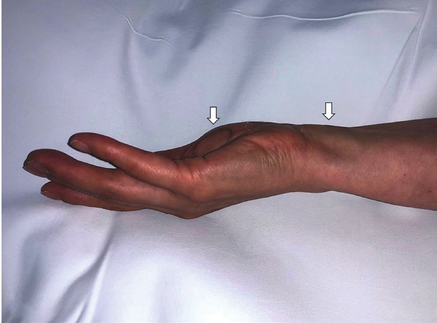
J Korean Neurosurg Soc 63 | September 2020

pants included in this study.                                                       of the digital branches of the median nerve presenting as carpal tun-
                                                                                    nel syndrome: a rare case report with review of the literature. Indian J
                                                                                    Pathol Microbiol 59 : 96-98, 2016
                                                                               4.   Al-Jabri T, Garg S, Mani GV : Lipofibromatous hamartoma of the median
AUTHOR CONTRIBUTIONS                                                                nerve. J Orthop Surg Res 5 : 71, 2010
                                                                               5.   Clavijo-Alvarez JA, Price M, Stofman GM : Preserved neurologic function
     Data curation : SWS, IJP                                                       following intraneural fascicular dissection and nerve graft for digital and
     Formal analysis : YK                                                           median nerve lipofibromatous hamartoma. Plast Reconstr Surg 125 :
     Methodology : YK                                                               120e-122e, 2010
                                                                               6.   Hoellwarth JS, Goitz RJ : Lipofibromatous hamartoma of the palmar cu-
     Project administration : CJ
                                                                                    taneous branch of the median nerve. J Hand Microsurg 10 : 109-112,
     Writing - original draft : YTR
                                                                                    2018
     Writing - review & editing : IJP                                          7.   Kitridis D, Dionellis P, Xarchas K, Givissis P : Giant median nerve due to
                                                                                    hamartoma causing severe carpal tunnel syndrome. J Orthop Case
                                                                                    Rep 8 : 57-60, 2018

ORCID                                                                          8.   Mason ML : Proceedings of the American Society for Surgery of the
                                                                                    Hand: presentation of cases. J Bone Joint Surg Am 35 : 273-275,
                                                                                    1953
     Youn-Tae Roh           https://orcid.org/0000-0003-3203-091X
                                                                               9.   Mohammed Saeed MA, Dawood AA, Mahmood HM : Lipofibromatous
     Seok-Whan Song         https://orcid.org/0000-0002-8578-4697                   hamartoma of the median nerve with macrodactyly of middle finger. J
     Changhoon Jeong        https://orcid.org/0000-0003-3451-2875                   Clin Orthop Trauma 10 : 1077-1081, 2019
     Younghoon Kang         https://orcid.org/0000-0002-8732-1485             10.   Nardella D, Sohawon S, Carlier A : Lipofibromatous hamartoma of the
     Il-Jung Park           https://orcid.org/0000-0001-8262-4287                   median nerve. Three case reports. J Plast Reconstr Aesthet Surg 62 :
                                                                                    e314-e317, 2009
                                                                              11.   Razzaghi A, Anastakis DJ : Lipofibromatous hamartoma: review of early
●
     Acknowledgements                                                               diagnosis and treatment. Can J Surg 48 : 394-399, 2005
                                                                              12.   Silverman TA, Enzinger FM : Fibrolipomatous hamartoma of nerve. A
  We thank Chang Deok Weon, a medical photographer of                               clinicopathologic analysis of 26 cases. Am J Surg Pathol 9 : 7-14,
Bucheon St. Mary’s hospital, the Catholic University of Korea                       1985
for helping in preparing the photo.                                           13.   Tahiri Y, Xu L, Kanevsky J, Luc M : Lipofibromatous hamartoma of the
                                                                                    median nerve: a comprehensive review and systematic approach to
                                                                                    evaluation, diagnosis, and treatment. J Hand Surg Am 38 : 2055-
                                                                                    2067, 2013
References                                                                    14.   Toms AP, Anastakis D, Bleakney RR, Marshall TJ : Lipofibromatous ham-
                                                                                    artoma of the upper extremity: a review of the radiologic findings for 15
    1. Afshar A : Carpal tunnel syndrome due to lipofibromatous hamartoma           patients. AJR Am J Roentgenol 186 : 805-811, 2006
       of the median nerve. Arch Iran Med 13 : 45-47, 2010                    15.   Ulrich D, Ulrich F, Schroeder M, Pallua N : Lipofibromatous hamartoma
    2. Agarwal S, Haase SC : Lipofibromatous hamartoma of the median nerve.         of the median nerve in patients with macrodactyly: diagnosis and treat-
       J Hand Surg Am 38 : 392-397; quiz 397, 2013                                  ment of a rare disease causing carpal tunnel syndrome. Arch Orthop
    3. Agrawal R, Garg C, Agarwal A, Kumar P : Lipofibromatous hamartoma            Trauma Surg 129 : 1219-1224, 2009

670         https://doi.org/10.3340/jkns.2020.0082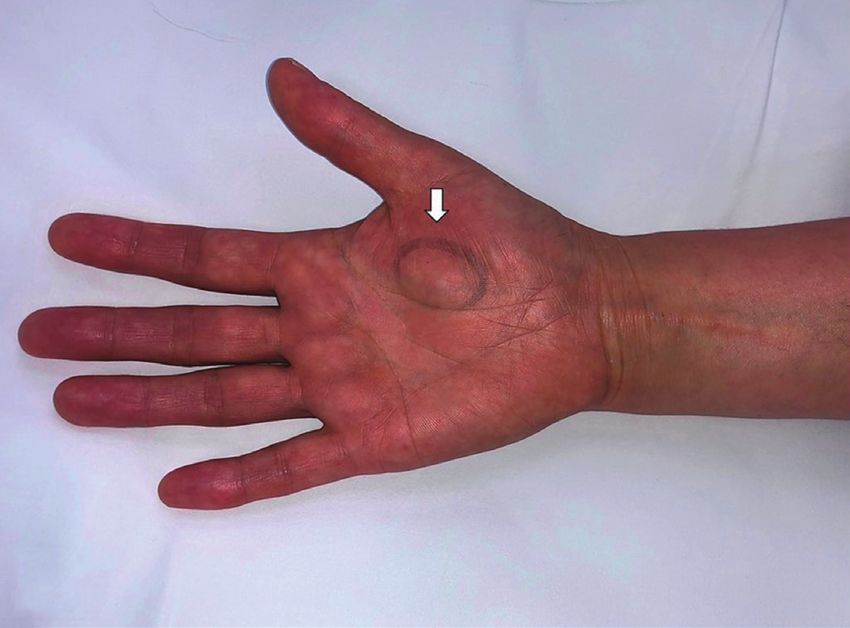
You can also read