Well-differentiated abdominal liposarcoma: experience of a tertiary care center
←
→
Page content transcription
If your browser does not render page correctly, please read the page content below
Karadayi et al. World Journal of Surgical Oncology (2015) 13:166
DOI 10.1186/s12957-015-0580-z                                                                                                    WORLD JOURNAL OF
                                                                                                                                 SURGICAL ONCOLOGY

 RESEARCH                                                                                                                                    Open Access

Well-differentiated abdominal liposarcoma:
experience of a tertiary care center
Kursat Karadayi1,4, Caglar Yildiz2*, Savas Karakus2, Atilla Kurt1, Birkan Bozkurt1, Sinan Soylu1, Ayse A Cicekli3,
Reyhan Egilmez3 and Ali Cetin2

  Abstract
  Background: We presented abdominal liposarcoma cases diagnosed and managed in a tertiary care center and
  also conducted a literature review on main features of this tumor.
  Methods: Chart reviews of eight cases were conducted, and clinical, surgical, histopathological, and follow-up data
  were recorded.
  Results: Overall, complete surgical resection was performed with adjacent organ resection in 25% of cases, and
  radiotherapy was not administered. Recurrence was developed in only one case and died after 2 years and 3
  months, and other cases are under follow-up without recurrence. Histopatological examinations revealed findings
  of well-differentiated liposarcoma.
  Conclusions: According to our surgical experience, the surgical margin positivity may not be a determining factor
  for the survival of patients with well-differentiated liposarcoma, and in the absence of macroscopic invasion,
  adjacent organ resection may not be required. Radiotherapy may not be preferred when complete resection of
  abdominal mass was achieved.
  Keywords: Liposarcoma, Abdomen, Surgical management

Background                                                                         are uncommon [3]. Surgical treatment is the main modal-
Liposarcoma of abdomen is a rare lesion consisted of ma-                           ity in the therapy of retroperitoneal liposarcomas [3]. How-
lign fat cells and accounts for approximately 20% of all                           ever, to our best knowledge, there is a small number of
mesenchymal malignancies in adults. These lesions may be                           case report or series related to abdominal liposarcoma.
found in different organs, typically occurs in either the ret-                     The knowledge related to the clinical features and course
roperitoneum or the extremities [1]. Abdominal liposarco-                          of retroperitoneal liposarcoma is mainly limited single-
mas are generally located in retroperitoneum; it is often                          institutional experiences. We presented cases with abdom-
difficult to collect pathological samples nonsurgically. Sur-                      inal liposarcoma managed in a tertiary care center and also
gical exploration is needed for the final pathologic diagno-                       conducted a literature review on presentation, manage-
sis. Retroperitoneal sarcomas tend to be high grade, except                        ment, and outcomes of these patients.
liposarcomas, they tend to be low to intermediate grade.
Histologic grade is the main factor for survival rates in pa-
tients with liposarcomas [2]. Symptoms are usually non-                            Methods
specific, and they do not appear until the tumor becomes                           Chart reviews of eight cases managed with liposarcoma
very large, a painless abdominal mass that enlarges in a                           at the General Surgery Service of our tertiary care uni-
long period of time in an adult is most common history of                          versity hospital were conducted between December 2011
the patients. Metastases at the time of initial presentation                       and January 2014. This study was approved by the
                                                                                   Human Ethics Committee of our university. Clinical in-
                                                                                   formation regarding the age, gender, clinical findings,
* Correspondence: dr_caglaryildiz@yahoo.com                                        imaging findings, surgical procedure, histopathology of
2
 Department of Obstetrics and Gynecology, Faculty of Medicine, Cumhuriyet
University, Sivas 58140, Turkey                                                    the tumor, follow-up findings, overall survival were re-
Full list of author information is available at the end of the article             corded. Any imaging studies that had been obtained
                                      © 2015 Karadayi et al.; licensee BioMed Central. This is an Open Access article distributed under the terms of the Creative
                                      Commons Attribution License (http://creativecommons.org/licenses/by/4.0), which permits unrestricted use, distribution, and
                                      reproduction in any medium, provided the original work is properly credited. The Creative Commons Public Domain
                                      Dedication waiver (http://creativecommons.org/publicdomain/zero/1.0/) applies to the data made available in this article,
                                      unless otherwise stated.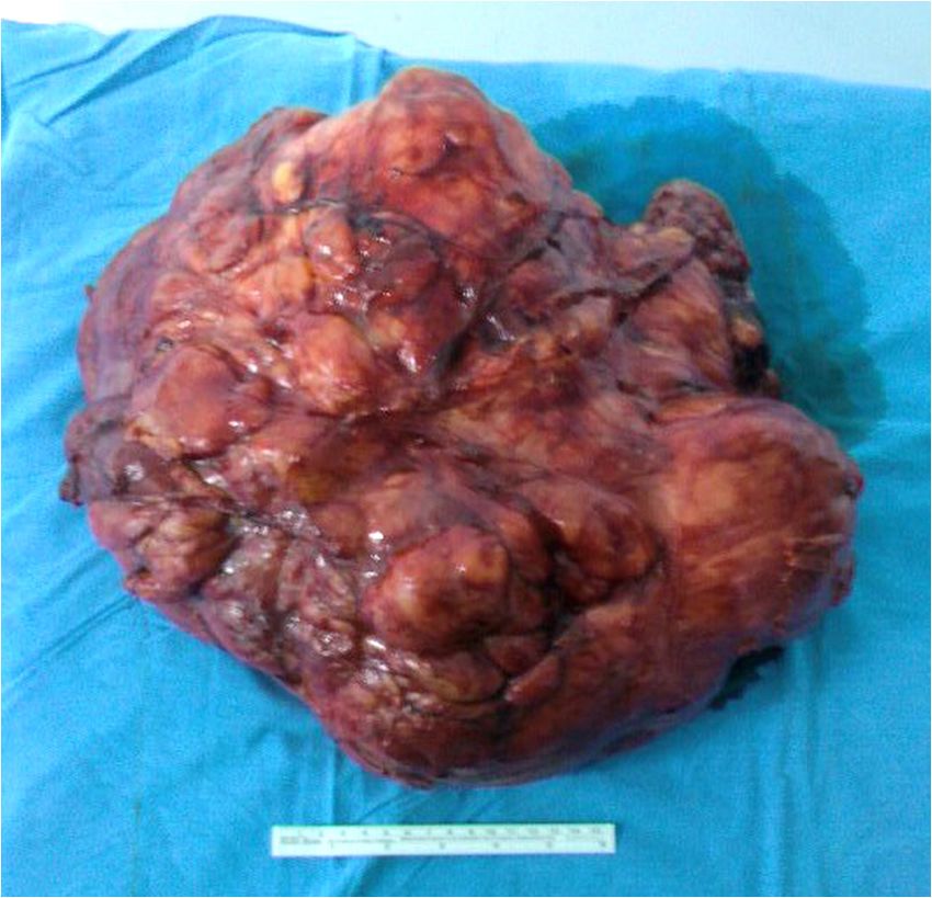
Karadayi et al. World Journal of Surgical Oncology (2015) 13:166 Page 2 of 5
preoperatively were also reviewed. Computerized tomog-
raphy was used for imaging of abdomen in all patients
(Figure 1). A midline abdominal incision and sharp and
blunt dissection of the mass (Figures 2 and 3) was per-
formed and then intraoperative frozen section technique
was used to evaluate the margins of the tumoral mass for
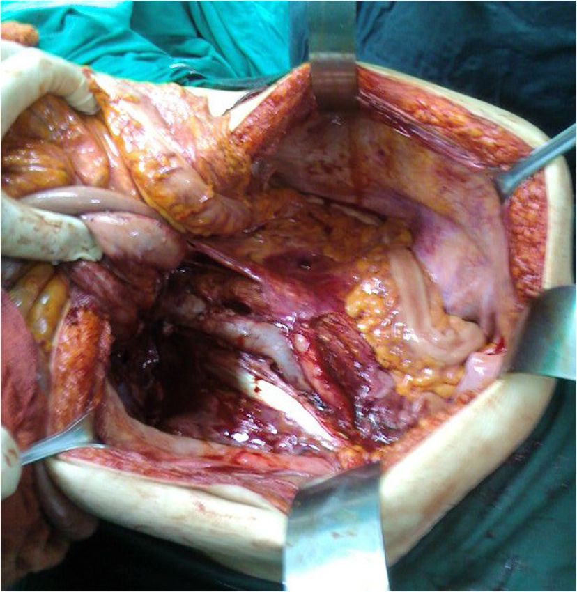
all cases. The organs that involved macroscopically by the
tumor were excised (Figure 4). Tissue specimens were
processed by the hematoxylin-eosin staining method.
All the patients were followed up at 1, 3, 6, and 12
months and 6-month period in second year. Physical
and ultrasonographic examinations with laboratory tests
were performed in the third month, and a computerized
tomography was performed in the sixth month after sur- Figure 2 A midline abdominal incision dissection of the mass.
gery. The median follow-up of 21 months (min.15,
max.24) with the shortest follow-up of 15 months and
longest follow-up of 24 months. hemicolectomy, ileal resection, and partial bladder resec-
tion were added due to local invasion, and histopatho-
Results logical diagnosis was liposarcoma, well-differentiated, and
Table 1 presents selected demographic, clinical, and in- dedifferentiated (mixed) (Figure 5). The blood loss during
traoperative features of cases. Of eight patients, four pa- surgery was acceptable (150 to 220 mL), mean time for
tients were female and median age was 61.5 (21 to 73). surgical procedure was 140 min, and the duration of hos-
Overall, the most important clinical finding is palpable pital stay ranged from 4 to 6 days.
abdominal mass and abdominal pain. During computed In almost all cases, histopatological examinations re-
tomography (CT) examination, we observed a large ab- vealed findings of well-differentiated and grade 1 liposar-
dominal mass about 28 to 50 cm in the largest size with coma. Surgical margins were defined as positive in 75%
heterogeneous structure displacing adjacent organ and tis- of cases and necrosis was detected in 25% of cases.
sues. Complete resection of mass was adequate as surgical Recurrence has occurred in only one case and died
management in cases 1, 3, 5 to 8. In case 2, right nephrec- due to renal failure. Intraoperatively, in case 2, vena cava
tomy and right ureter resection were performed due to tu- inferior injury was developed and repaired. Four patients
moral invasion, and a second surgery was also required for had minor complication according to Clavien-Dindo
local recurrence; however, histopathological diagnosis was classification [4].
the same with other cases except case 4. In case 4, right
Figure 1 Computerized tomography was used for imaging of
abdomen in all patients. Figure 3 Sharp and blunt dissection of the mass.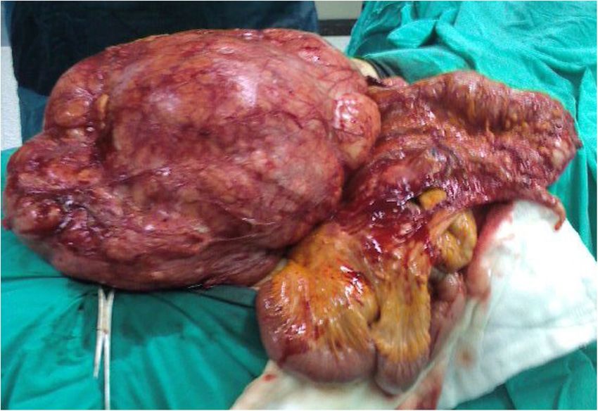
Karadayi et al. World Journal of Surgical Oncology (2015) 13:166                                                      Page 3 of 5

                                                                   well-differentiated liposarcoma, myxoid and/or round cell,
                                                                   and pleomorphic. In rare circumstances, lesions can have a
                                                                   combination of morphologic types; these are classified as
                                                                   combined or mixed-type liposarcomas. The most recent
                                                                   World Health Organization classification of soft tissue tu-
                                                                   mors recognizes five categories of liposarcomas: well differ-
                                                                   entiated, which includes the adipocytic, sclerosing, and
                                                                   inflammatory subtypes; dedifferentiated; myxoid; round cell;
                                                                   and pleomorphic [11,12].
                                                                      Well-differentiated (low-grade) liposarcomas are the
                                                                   most common types of liposarcomas, followed by dediffer-
                                                                   entiated liposarcomas. The amount of lipid inside the
                                                                   cells, the mucoid lipid, and the degree of cell differenti-
                                                                   ation are the essential of the classification. Myxoid, round
                                                                   cell, and pleomorphic liposarcomas are rare in the retro-
                                                                   peritoneum. Myxoid and round cell liposarcomas share
                                                                   the same reciprocal translocation t(12,16)(q13; p11), in
                                                                   which the CHOP gene is inserted adjacent to a novel gene
 Figure 4 The organs that involved macroscopically by the tumor    called FUS or TLS (translocated in liposarcoma). While
 were excised.                                                     no specific chromosomal translocations have been identi-
                                                                   fied in well differentiated/dedifferentiated liposarcomas,
                                                                   amplification of MDM2 and CDK4 is very frequent in
Discussion                                                         these subtypes, and their identification may be useful diag-
Eight patients with abdominal liposarcoma were presented           nostically. Furthermore, overexpression of MDM2 and
in this case series. Overall, complete surgical resection was      CDK4 is also being exploited for therapeutic gain [12,13].
achieved with adjacent organ resection in 25% of cases.               Liposarcomas are often asymptomatic until they reach to
We observed recurrence in only one case who died within            large size in a long period of time before producing any
2 years and 3 months, and other cases are under follow-up          symptoms, and the complaints of patients are mainly re-
without recurrence. After median follow-up of 21 months            lated to direct invasion or compression of other adjacent
(min.15, max.24), all cases were alive without recurrence          organs. There are no significant laboratory abnormalities
except case 2 who survived 2 years and 3 months after first        in the earlier stages and have often grown to a large size by
surgery. Although radiotherapy was not administered to             the time they are identified using a diagnostic modality
our cases, there was no recurrence except case 2.                  such as US or CT. CT of the abdomen is the most useful
   Liposarcoma is the most common mesenchymal tumor                tool in the imaging of retroperitoneum. CT of the chest is
of the retroperitoneal space but continues to pose a chal-         very important for evaluating the lungs, as they are the first
lenge with regard to diagnosis, prediction of clinical behav-      site of metastasis in the most of cases. A CT scan allows
ior, and treatment of disease recurrence within the                not only assessment of the tumor’s location and its rela-
abdominal/retroperitoneal space [5]. The major problem of          tionship to adjacent organs but also identification of meta-
soft tissue sarcomas being present in the extremities like         static lesions in the liver or peritoneal cavity [14]. CT is
retroperitoneum, chest wall, head and neck, and subcuta-           less sensitive to motion artifact than magnetic resonance
neous tissues is the other sites can be affected. Sarcomas         image (MRI); because of this property, CT defines the ana-
are a rare and heterogeneous group of malignant tumors             tomic relationship of the tumor to other abdominal organs
of mesenchymal origin that comprise all adult and child-           better than the MRI. Liposarcoma of the abdominal region,
hood malignancies, respectively 1% and 12% [6,7]. Sarco-           although rare, needs to be differentially diagnosed from
mas constitutes one third of malignant tumors that arise in        other abdominal tumors. Its symptoms are nonspecific,
the retroperitoneum, and approximately 10% to 15% of soft          and its diagnosis is intriguing. CT-scan or MRI are the
tissue sarcomas arise in the retroperitoneum [8,9]. Mesen-         most useful tools for investigation and evaluation of retro-
chymal cells of muscle, fat, and connective tissues are the        peritoneal mass [14]. Surgery is the mainstay of treatment
origins of sarcomas that arise from retroperitoneum.               for these cases and is curative with no recurrence following
   The most commonly encountered histologic subtypes of            complete surgical excision [15]. The final diagnosis should
retroperitoneal sarcoma are liposarcoma (41%), leiomyosar-         always be confirmed with histopathology of the specimen.
coma (28%), malignant fibrous histiocytoma (7%), fibrosar-         We think that although in limited number, these described
coma (6%), and malignant peripheral nerve sheath tumor             cases can contribute to the great amount of controversy re-
(3%) [10]. Liposarcoma occurs in three main biologic forms:        garding surgical management of abdominal liposarcoma.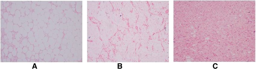
Karadayi et al. World Journal of Surgical Oncology (2015) 13:166                                                                           Page 4 of 5

Table 1 Patient demographics and clinical and intraoperative findings
Patient number        Age (y)   Gender   Clinical findings            CT imaging findings           Surgical procedures          Survival after surgery
Case 1                43        F        Palpable mass at left        A 16 × 10 × 18-cm             Complete resection of        Alive 21 months
                                         lower quadrant               heterogeneous pelvic          mass without additional
                                                                      mass that extends up          organ resection
                                                                      to the right lower
                                                                      quadrant of the
                                                                      abdomen.
Case 2                63        F        Palpable mass of             A 50 × 40 × 28-cm             During first surgery,        Recurrence 2 years
                                         abdomen, general             heterogeneous                 complete resection of        later after first
                                         abdominal pain               abdominal mass                mass with additional right   surgery and died
                                                                      compressing aorta             nephrectomy and right        due to renal failure
                                                                      and vena cava inferior.       ureter resection.            3 months later
                                                                                                                                 after second surgery
                                                                                                    During second surgery
                                                                                                    performed for local
                                                                                                    recurrence 2 years later,
                                                                                                    right hemicolectomy,
                                                                                                    cholecystectomy,
                                                                                                    ileal resection.
Case 3                73        F        General abdominal pain       A 21 × 15 × 5-cm              Complete resection of        Alive 15 months
                                                                      heterogeneous                 mass without additional
                                                                      retroperitoneal mass.         organ resection
Case 4                61        M        Palpable mass of abdomen,    A 15 × 8 × 11-cm              Right hemicolectomy,         Alive 20 months
                                         general abdominal pain       heterogeneous                 ileal resection, partial
                                                                      retroperitoneal mass.         bladder resection
Case 5                28        M        General abdominal pain,      A 30 × 18 × 27-cm             Complete resection of        Alive 24 months
                                         abdominal swelling           heterogeneous                 mass without additional
                                                                      septated abdominal            organ resection
                                                                      mass that displaces
                                                                      the adjacent organs.
Case 6                62        F        Upper abdominal pain         A 19 × 10 × 18-cm             Complete resection of        Alive 22 months
                                                                      heterogeneous pelvic          mass without additional
                                                                      mass that extends             organ resection
                                                                      above the umbilicus.
Case 7                66        M        General abdominal pain,      A 24 × 20 × 12-cm             Complete resection of        Alive 21 months
                                         abdominal swelling           heterogeneous                 mass without additional
                                                                      retroperitoneal mass.         organ resection
Case 8                21        M        General abdominal pain,      A 23 × 10 × 15-cm cystic      Complete resection of        Alive 15 months
                                         abdominal swelling           septated abdominal            mass without additional
                                                                      mass at right lower           organ resection
                                                                      quadrant of the abdomen.
Y, year; F, female; M, male.

   Surgery is indispensable for treatment of abdominal                       many cases, it is not possible to perform complete resec-
liposarcoma. Complete surgical removal of retroperitoneal                    tion because of the tumor being too large or invasive to
tumor is the most effective treatment and has a significant                  organs around it or its relation with the great vessels.
effect on the survival rate. The primary aim of the surgery                    There is continuing research and debate on the use of
is complete resection with negative margins. However, in                     intraoperative radiotherapy (RT), adjuvant RT, preoperative

  Figure 5 Histopatological examination of mixed-type liposarcoma tissue in case 4. (A) Well-differentiated component (HEX100). (B) Myxoid component
  (HEX100). (C) Undifferentiated component (HEX100).
Karadayi et al. World Journal of Surgical Oncology (2015) 13:166 Page 5 of 5
RT, preoperative intensity-modulated RT (IMRT), pre- Authors’ contributions
operative chemoradiotherapy, preoperative chemotherapy, KK, CY, SK, AK, BB, SS, AC collected and analyzed the clinical data and
drafted the manuscript. RE and AAC carried out the histopathological
and adjuvant chemotherapy. Many studies have shown that and morphometric investigations. All authors read and approved the
a significant number of patients experience prolonged final manuscript.
disease-free survival when all grossly evident recurrent dis-
Author details
ease can be resected. Chemotherapy or radiotherapy ad- 1
Department of Surgery, Faculty of Medicine, Cumhuriyet University, Sivas
ministration is still controversial in the treatment of locally 58140, Turkey. 2Department of Obstetrics and Gynecology, Faculty of
recurrent disease [14]. A phase III randomized controlled Medicine, Cumhuriyet University, Sivas 58140, Turkey. 3Department of
Pathology, Faculty of Medicine, Cumhuriyet University, Sivas 58140, Turkey.
trial has been completed yet about radiotherapy in patients 4
The Division of Surgical Oncology, Faculty of Medicine, Cumhuriyet
with primary soft tissue sarcoma of the retroperitoneum or University, Sivas 58140, Turkey.
pelvis [16].
Received: 16 September 2014 Accepted: 8 April 2015
Because of the huge sizes of most retroperitoneal sar-
comas, the size of the tumor is not a predictor for sur-
vival of the disease. Tumor grade has been reported as a References
1. Oishi H, Moriyama S, Kotera K, Miura K, Masuzaki H. First case of liposarcoma
significant factor in some studies, with the weight of evi- arising from the fallopian tube: case report and review of the literature.
dence supporting shorter recurrence-free and overall J Obstet Gynaecol Res. 2008;34:713–6.
survival for patients with high-grade tumors [17,18]. Me- 2. McCallum OJ, Burke 2nd JJ, Childs AJ, Ferro A, Gallup DG. Retroperitoneal
liposarcoma weighing over one hundred pounds with review of the
tastasis potential of liposarcomas is very low; because of literature. Gynecol Oncol. 2006;103:1152–4.
their tendency for local recurrence in the retroperito- 3. Erzen D, Sencar M, Novak J. Retroperitoneal sarcoma: 25 years of experience
neum/mediastinum and spermatic cord, these same tu- with aggressive surgical treatment at the Institute of Oncology. Ljubljana
J Surg Oncol. 2005;91:1–9.
mors are referred to as well-differentiated liposarcomas 4. Clavien PA, Barkun J, de Oliveira ML, Vauthey JN, Dindo D, Schulick RD, et al.
in these locations [7,19]. Well-differentiated liposarco- The Clavien-Dindo classification of surgical complications: five-year
mas are low-grade tumors; compared to dedifferentiated experience. Ann Surg. 2009;250:187–96.
5. Singer S, Antonescu CR, Riedel E, Brennan MF. Histologic subtype and
high-grade liposarcomas, they have lower recurrence margin of resection predict pattern of recurrence and survival for
rates, the potential to metastasize, and dedifferentiated retroperitoneal liposarcoma. Ann Surg. 2003;238:358–70.
liposarcomas have a six-fold higher risk of death [5,19]. 6. Miller RW, Young Jr JL, Novakovic B. Childhood cancer. Cancer. 1995;75:395.
7. Fletcher CDM, Bridge JA, Hogendoorn PCW, Mertens F. World Health
Overall, 5-year survival for well-differentiated subtypes Organization Classification of tumours of soft tissue and bone. 4th ed. Lyon:
is 90%, while 5-year survival for pleomorphic subtypes IARC Press; 2013.
is only 30% to 50%. Dedifferentiated and myxoid/round 8. Pisters PW. Soft tissue sarcoma. In: Norton JA, Bollinger RR, Chang AE, et al.,
editors. Surgery: basic science and clinical evidence. New York: Springer; 2001.
cell subtypes have 5-year survival rates of 75% and 60% to 9. Lawrence Jr W, Donegan WL, Natarajan N, Mettlin C, Beart R, Winchester D.
90%, respectively [2]. Well-differentiated liposarcomas Adult soft tissue sarcomas: a pattern of care survey of the American College
may recur locally, but metastatic potential is low com- of Surgeons. Ann Surg. 1987;205:349.
10. Raut CP, Pisters PW. Retroperitoneal sarcomas: combined-modality treatment
pared with pleomorphic liposarcomas that have high approaches. J Surg Oncol. 2006;94:81.
metastatic potential, and pleomorphic liposarcomas have 11. Schwartz RA, Trovato MJ, Centurion SA. Liposarcoma. Available at:
shorter survival than well-differentiated liposarcomas [5]. emedicine.medscape.com (Accessed on January 2014).
12. Conyers R, Young S, Thomas DM. Liposarcoma: molecular genetics and
therapeutics. Sarcoma. 2011;2011:483154.
13. Italiano A, Bianchini L, Gjernes E, Keslair F, Ranchere-Vince D, Dumollard JM,
Conclusions et al. Clinical and biological significance of CDK4 amplification in well-differentiated
In summary, abdominal liposarcomas are low-grade tu- and dedifferentiated liposarcomas. Clin Cancer Res. 2009;15:5696.
mors in general and surgical excision with wide margin is 14. Windham TC, Pisters PW. Retroperitoneal sarcomas. Cancer Control.
2005;12:36–43.
decreasing the effectiveness of the radiotherapy, increasing 15. Joshi RM, Gangurde GK, Talathi NP, Telavane PP, Singh R, Hanamshetti SR,
the adverse effects especially on intestines. In our cases, al- et al. Large retroperitoneal liposarcoma - a series of five cases. Indian J Surg.
though surgical margin was reported as positive in 75% of 2013;75:64–8.
16. Pisters PWT, O'Sullivan B. Surgery with or without radiation therapy in
patients, because of the absence of macroscopic invasion, treating patients with primary soft tissue sarcoma of the retroperitoneum or
adjacent organ resection was not performed. According to pelvis. NCT00091351, Clinical Trials.gov. Available at http://www.cancer.gov/
our surgical experience, in patients with retroperitoneal clinicaltrials. Accessed on March 30, 2015.
17. Hassan I, Park SZ, Donohue JH, Nagorney DM, Kay PA, Nasciemento AG,
liposarcoma, surgical resection can be completed consid- et al. Operative management of primary retroperitoneal sarcomas: a
erably easy than expected although its size can be huge. In reappraisal of an institutional experience. Ann Surg. 2004;239:244–50.
almost all cases, absence of invasion to adjacent tissues 18. Cheifetz R, Catton CN, Kandel R, O’Sullivan B, Couture J, Swallow CJ. Rescent
progress in the management of retroperitoneal sarcoma. Sarcoma.
and organs is another advantage during surgical manage- 2001;5:17–26.
ment, adjacent organ resection should not be necessary 19. Dei Tos AP, Pedeutour F. Atypical lipomatous tumour. In: Fletcher CDM, Bridge
because of the low invasion potential of these tumors. JA, Hogendoorn PCW, Mertens F, editors. World Health Organization
Classification of tumours of soft tissue and bone. 4th ed. Lyon: IARC; 2013. p. 33.
Competing interests
The authors declare that they have no competing interest.You can also read