Haemochromatosis in a kidney transplant recipient: a case report - BMC ...
←
→
Page content transcription
If your browser does not render page correctly, please read the page content below
Zakrocka et al. BMC Nephrology      (2021) 22:201
https://doi.org/10.1186/s12882-021-02416-9

 CASE REPORT                                                                                                                                       Open Access

Haemochromatosis in a kidney transplant
recipient: a case report
Izabela Zakrocka* , Iwona Baranowicz-Gąszczyk and Wojciech Załuska

  Abstract
  Background: Iron overload is inevitably related to chronic kidney disease (CKD) treatment. Haemochromatosis
  leads to multiorgan damage and is associated with increased mortality. Primary haemochromatosis is the most
  common autosomal recessive disease in white populations. In most cases, the classic form of hereditary
  haemochromatosis is caused by mutations, mainly C282Y and H63D, in the haemochromatosis gene (HFE).
  Secondary haemochromatosis can be triggered by iron administration and blood transfusions. Haemochromatosis is
  rarely reported in kidney transplant recipients. Atypical factors may evoke haemochromatosis in patients without
  HFE mutations or other standard risk factors.
  Case presentation: In the current study, we present a patient who started to have haemochromatosis symptoms
  after kidney transplantation. A 37-year-old man after kidney transplantation from a deceased donor was admitted to
  the hospital due to high serum ferritin levels and impaired graft function. The patient’s past medical history
  included arterial hypertension, embolization of both renal arteries and necrosis of the left femoral head.
  Glomerulonephritis was suspected as a cause of CKD; however, severe kidney failure was diagnosed, kidney biopsy
  was not performed, and the patient started intermittent haemodialysis. While on dialysis to treat anaemia, the
  patient had received erythropoietin and iron intravenously, and the maximal serum ferritin level was 2115 ng/ml.
  After kidney transplantation, ferritin levels started to increase rapidly, with a maximum level of 9468 ng/ml one and
  a half years after surgery. His genetic study showed HFE C282Y heterozygosity. Symptoms of haemochromatosis,
  such as skin hyperpigmentation, elevated activity of aminotransferases, impaired glucose tolerance and heart failure,
  were observed. Therapeutic phlebotomy was started, and 36 procedures were performed. After treatment, graft
  function significantly improved, most haemochromatosis symptoms resolved, and the serum ferritin level
  significantly decreased.
  Conclusions: Haemochromatosis can occur in heterozygotic HFE patients after kidney transplantation. Iron
  administration, infections, type of immunosuppression and liver dysfunction should be considered potential triggers
  of haemochromatosis in this group of patients.
  Keywords: Haemochromatosis, Kidney transplant, Case report, Iron, Tacrolimus

Background                                                                            pericarditis, diabetes, thyroid gland dysfunction, hypogo-
Haemochromatosis is an iron storage disorder leading to                               nadotropic hypogonadism, immunosuppression and skin
multiple organ damage and premature death [1]. Heavy                                  discolouration [1, 2]. Primary haemochromatosis is the
iron overload leads to various complications, including                               most common autosomal recessive disorder in northern
liver cirrhosis, arthritis, cardiomyopathy, arrhythmias,                              populations, with a prevalence of 1 in 300 individuals
                                                                                      [2]. Mutations in HFE, including C282Y and H63D, are
* Correspondence: izabela.zakrocka@umlub.pl                                           responsible for most cases of primary haemochromatosis
Department of Nephrology, Medical University of Lublin, Jaczewskiego street
8, 20-090 Lublin, Poland                                                              [2]. Men are affected two to three times more often than

                                       © The Author(s). 2021 Open Access This article is licensed under a Creative Commons Attribution 4.0 International License,
                                       which permits use, sharing, adaptation, distribution and reproduction in any medium or format, as long as you give
                                       appropriate credit to the original author(s) and the source, provide a link to the Creative Commons licence, and indicate if
                                       changes were made. The images or other third party material in this article are included in the article's Creative Commons
                                       licence, unless indicated otherwise in a credit line to the material. If material is not included in the article's Creative Commons
                                       licence and your intended use is not permitted by statutory regulation or exceeds the permitted use, you will need to obtain
                                       permission directly from the copyright holder. To view a copy of this licence, visit http://creativecommons.org/licenses/by/4.0/.
                                       The Creative Commons Public Domain Dedication waiver (http://creativecommons.org/publicdomain/zero/1.0/) applies to the
                                       data made available in this article, unless otherwise stated in a credit line to the data.
Zakrocka et al. BMC Nephrology (2021) 22:201 Page 2 of 5
women and start to present haemochromatosis symp- kidney transplantation of 44% and 2115 ng/ml, respect-
toms in the fifth decade [3]. Whereas the majority of pa- ively (Fig. 1), but no symptoms of iron overload were
tients with haemochromatosis are homozygotes for found. Kidney transplantation was performed without
C282Y, less than 5% are C282Y or H63D heterozygotes significant complications, but a slight impairment of
[2]. Iron disorders caused by other gene mutations, such graft function was observed (creatinine, 1.6 mg/dl − 1.99
as haemojuvelin, hepcidin antimicrobial peptide, trans- mg/dl). For immunosuppression, the patient received
ferrin receptor-2 and ferroportin, are very rare and occur mycophenolate mofetil, tacrolimus, and prednisone.
at a younger age [1]. Although heterozygotic C282Y pa- Two months after kidney transplantation, the level of
tients do not generally develop haemochromatosis symp- ferritin was 5624 ng/ml, with concomitant anaemia
toms, under some conditions, such as liver steatosis, (haemoglobin concentration, 10.4 g/dl), TS 89% and no
diabetes or excessive alcohol consumption, these symp- iron supplements. CMV infection and urinary tract in-
toms may become more prevalent [4, 5]. Iatrogenic iron fection caused by Enterococcus faecalis were diagnosed
overload and blood transfusions are the main causes of and treated appropriately. Eighteen months after kidney
secondary haemochromatosis in patients with CKD [6]. transplantation, the haemochromatosis suspicion was
Since ferritin is an acute phase protein, its high serum raised, since the ferritin concentration rose to 9468 ng/
level can be observed not only after excessive iron ad- ml and signs of liver damage were observed. The activity
ministration but also in patients with liver injury, sys- level of alanine aminotransferase was 50–134 U/l (nor-
temic inflammatory conditions, haemoglobinopathies mal range < 41 U/l), aspartate aminotransferase was 40–
and haemodialysis treatment [7, 8]. However, transferrin 59 U/l (normal range < 34 U/l), gamma-glutamyl trans-
saturation (TS) above 45% and a serum ferritin concen- ferase was 384 U/l (normal range < 50 U/l), alkaline
tration above 300 ng/ml or unexplained multiorgan phosphatase was 134 U/l (limit 129 U/l) and amylase was
damage with liver involvement indicate the need for 85 U/l (limit 53 U/l). Infection markers (including CMV
haemochromatosis testing [1]. The presence of HFE mu- DNA) and cancer markers were negative. Gastroscopy
tation is related to reduced responsiveness to the treat- and colonoscopy did not reveal any pathology. The
ment [9]. Despite common clinical problem, reports haptoglobin concentration was within the normal range,
about haemochromatosis in kidney transplant patients excluding intravascular haemolysis. Wilson’s disease was
are rare [9, 10]. This study presents a case of a kidney ruled out after ceruloplasmin level analysis. The patient
transplant recipient with haemochromatosis diagnosed reported poor exercise tolerance, darkening of the skin
after kidney transplantation and treated through thera- was observed on physical examination, and impaired
peutic phlebotomy. glucose tolerance was diagnosed. Computed tomography
of the abdomen revealed liver steatosis (Fig. 2 A), and
Case presentation the transplanted kidney located in the right iliac fossa
A 37-year-old recipient of a renal transplant from a de- had no pathologies (Fig. 2 B). In echocardiography, left
ceased donor was admitted to the Nephrology Clinic ventricular hypertrophy was found, with an ejection frac-
due to high serum ferritin levels and impaired graft tion of 62%. Due to calcifications observed in the left an-
function. CKD was diagnosed 3 years prior to trans- terior descending artery, the patient underwent
plantation, and glomerulonephritis was suspected; how- percutaneous coronary intervention. Since a low haemo-
ever, CKD progressed to its end stage, and globin level persisted (10.9 g/dl – 11.3 g/dl), darbopoetin
haemodialysis was started. Arterial hypertension, sec- treatment was started. Because hepatic laboratory test
ondary anaemia and hyperparathyroidism were diag- results improved, liver biopsy was not performed. Gen-
nosed. The patient underwent bilateral renal artery etic tests revealed that the patient was heterozygous for
embolization due to resistant hypertension. As a compli- C282Y. After haematological consultation, the decision
cation of renal angioembolization, patient was diagnosed was made to start therapeutic phlebotomy. The patient
with left femoral head necrosis, which was successfully received necessary information about the proposed
treated conservatively, patient did not report any symp- methods of treatment and their complications. When in-
toms in later observation. No gastrointestinal tract dis- formed consent was obtained from the patient, phlebot-
eases were diagnosed, alcohol dependence was excluded, omy was started, and the patient tolerated the procedure
and tests for hepatitis B virus, hepatitis C virus, cyto- very well. After 36 phlebotomies, the ferritin concentra-
megalovirus (CMV) and Epstein-Barr virus infection tion decreased to 1921 ng/ml, TS decreased to 46%, the
were negative. During 33 months of dialysis, the patient haemoglobin level increased to 15.1 g/dl and kidney
received 8200 mg of iron intravenously (from 200 mg to function significantly improved (creatinine 1.3 mg/dl). At
500 mg per month) and two blood units for the treat- the time of writing this manuscript, graft function
ment of secondary anaemia. His TS and ferritin level remained stable, and most symptoms of haemochroma-
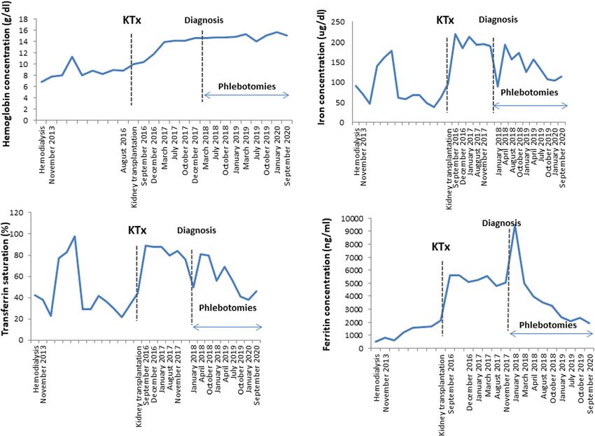
varied, with maximal values reported shortly before tosis gradually deteriorated after phlebotomy.Zakrocka et al. BMC Nephrology (2021) 22:201 Page 3 of 5 Fig. 1 Haemoglobin concentration (g/dl), iron concentration (μg/dl), transferrin saturation (%) and ferritin concentration (ng/ml) in 7 year follow up. Kidney transplantation was performed in August 2016 Fig. 2 Computed tomography (CT) of the abdomen. Liver steatosis (white arrows) (Fig. 2 A). Transplanted kidney in the right iliac fossa (white arrow), 116 × 62 mm size, without abnormalities (Fig. 2 B)
Zakrocka et al. BMC Nephrology   (2021) 22:201                                                                               Page 4 of 5

Discussion and conclusions                                    [15]. The goal of the therapy is lowering serum ferritin
Iron overload is an important problem in kidney trans-        levels below 50 ng/ml, with haemoglobin levels main-
plant recipients. Supplementation with iron secondary to      tained above 11 g/dl [1]. Most symptoms of haemo-
erythropoiesis-stimulating agent administration in pa-        chromatosis, including chronic fatigue and liver damage,
tients with CKD or on haemodialysis may have serious          were shown to disappear after phlebotomy; however, ar-
clinical implications. According to Lorenz et al., high       thropathy, pancreatic endocrine dysfunction and hypo-
serum ferritin levels and TS were observed in 9.4% of         gonadism have a less robust response to this therapy [1,
patients after kidney transplantation [9]. Among them         2]. Similarly, in our patient, haemochromatosis symp-
36.3% of patients had mutations in HFE. On the other          toms diminished after multiple phlebotomies, serum fer-
hand, Ramirez et al. reported that the prevalence of HFE      ritin was significantly lowered, and haemoglobin levels
mutations, although high in the general population, was       improved.
not always associated with laboratory and clinical find-         A limitation of our study is that only HFE mutations
ings leading to a haemochromatosis diagnosis [11]. Add-       were analysed, since testing of other genes was not avail-
itionally,    patients    with    evident     criteria   of   able in our laboratory. However, because non-HFE mu-
haemochromatosis after kidney transplantation may             tations occur in different age groups, they were not
show neither of the mutations related to the disease          taken under consideration for our patient.
[10]. Moreover, some patients may even have high TS or           Our study presents the case of a post-kidney trans-
ferritin levels, suggesting iron overload, although they      plantation patient in whom haemochromatosis symp-
did not receive the maximal iron dose during anaemia          toms became clinically apparent after kidney
treatment [9]. Systemic inflammation, malignancies, liver     transplantation. Repeated phlebotomies significantly im-
damage, viral infections (caused by hepatitis B virus,        proved the haemochromatosis course and prevented dis-
hepatitis C virus, CMV) and alcohol consumption are           ease      complications.     Since      reports     about
claimed to be responsible for the iron overload pheno-        haemochromatosis in kidney transplant recipients are
type [9] and in patients with C282Y/H63D heterozygos-         rare and their results remain inconclusive, more infor-
ity [1]. A similar observation was made by Yaprak et al.      mation is needed about haemochromatosis pathogenesis
[12]. In a study by Jorge et al., it was suggested that       in this group of patients. Special care is necessary for
normalization of kidney endocrine function, especially in     CKD and kidney transplant patients to prevent iron
terms of increased erythropoietin production, and a lack      overload and its consequences.
of uraemic toxins may rapidly evoke haemochromatosis
[10]. Other factors, such as surgery itself and immuno-       Acknowledgements
suppressive drug administration, may also lead to im-         Not applicable.
mune imbalance and iron metabolism abnormalities. In
a recent study, Akhtar et al. observed tacrolimus-            Authors’ contributions
induced iron overload in the livers of Wistar rats, mostly    IZ wrote the manuscript and conducted the literature review. IZ, IBG and WZ
                                                              participated in the clinical care of the patient. IZ, IBG and WZ interpreted
due to increased expression of hepcidin and immuno-
                                                              results under clinical perspective. All authors read and approved the final
logical system activation [13]. In our patient, one of the    manuscript.
risk factors related to haemochromatosis manifestation
was liver steatosis, which was diagnosed after kidney         Funding
transplantation, probably as a side effect of immunosup-      None.
pression. Second, infections after kidney transplantation,
both viral and bacterial, could be potential triggers of      Availability of data and materials
haemochromatosis in the present case. Iron administra-        The datasets used and analysed during the current study are available from
tion, although at high doses, did not evoke a haemo-          the corresponding author on reasonable request.
chromatosis phenotype in our patient when he was
haemodialyzed, despite elevated serum ferritin levels and     Declarations
high TS.
                                                              Ethics approval and consent to participate
   Phlebotomy is the method of choice in the treatment        Not applicable.
of haemochromatosis. Other therapies, namely, erythro-
cytapheresis and iron binders such as desferroxamine
                                                              Consent for publication
mesylate or deferasirox, are considered alternative treat-    Written informed consent was obtained from the patient. A copy of the
ments [2]. In patients with liver cirrhosis or hepatocellu-   consent form is available for review and can be provided on request.
lar carcinoma, liver transplantation may be the only
method of treatment [14]. Phlebotomy procedures are           Competing interests
safe and effective, with tiredness as a main side effect      The authors declare that they have no competing interests.
Zakrocka et al. BMC Nephrology (2021) 22:201 Page 5 of 5
Received: 25 January 2021 Accepted: 24 May 2021
References
1. Murphree CR, Nguyen NN, Raghunathan V, Olson SR, DeLoughery T, Shatzel
JJ. Diagnosis and management of hereditary haemochromatosis. Vox Sang.
2020;115(4):255–62. https://doi.org/10.1111/vox.12896 .
2. Assi TB, Baz E. Current applications of therapeutic phlebotomy. Blood
Transfus. 2014;12(Suppl 1):s75–83.
3. Lim DR, Vidyasankar G, Phua C, Borgaonkar M. Clinical penetrance of
hereditary hemochromatosis-related end-organ damage of C282Y
homozygosity, A Newfoundland Experience. Clin Transl Gastroenterol. 2020;
11(11):e00258. https://doi.org/10.14309/ctg.0000000000000258 .
4. Walsh A, Dixon JL, Ramm GA, Hewett DG, Lincoln DJ, Anderson GJ, et al.
The clinical relevance of compound heterozygosity for the C282Y and H63D
substitutions in hemochromatosis. Clin Gastroenterol Hepatol. 2006;4(11):
1403–10. https://doi.org/10.1016/j.cgh.2006.07.009 .
5. Gurrin LC, Bertalli NA, Dalton GW, Osborne NJ, Constantine CC, McLaren CE,
et al. HFE C282Y/H63D compound heterozygotes are at low risk of
hemochromatosis-related morbidity. Hepatology. 2009;50(1):94–101. https://
doi.org/10.1002/hep.22972 .
6. Rostoker G, Vaziri ND, Fishbane S. Iatrogenic Iron overload in Dialysis
patients at the beginning of the 21st century. Drugs. 2016;76(7):741–57.
https://doi.org/10.1007/s40265-016-0569-0 .
7. Cullis JO, Fitzsimons EJ, Griffiths WJH, Tsochatzis E, Thomas DW.
Investigation and management of a raised serum ferritin. Br J Haematol.
2018;181(3):331–40. https://doi.org/10.1111/bjh.15166 .
8. Branten AJW, Swinkels DW, Klasen IS, Wetzels JFM. Serum ferritin levels are
increased in patients with glomerular diseases and proteinuria. Nephrol Dial
Transplant. 2004;19(11):2754–60. https://doi.org/10.1093/ndt/gfh454 .
9. Lorenz M, Kletzmayr J, Huber A, Hörl WH, Sunder-Plassmann G, Födinger M.
Iron overload in kidney transplants: prospective analysis of biochemical and
genetic markers. Kidney Int. 2005;67(2):691–7. https://doi.org/10.1111/j.1
523-1755.2005.67129.x .
10. Jorge C, Branco P, Domingos F, Gaspar A, Lebre L, Machado D. Renal
transplantation and evolution of hemochromatosis: a clinical case report.
Transplant Proc. 2000;32(8):2613–4. https://doi.org/10.1016/S0041-1345(00)01
807-8 .
11. Ramírez E, Pacho A, Morales JM, De Castro I, Paz-Artal E, Morales P.
Frequency and influence of hemochromatosis gene mutations in kidney
transplant recipients with or without hepatitis C virus infection. Transplant
Proc. 2009;41(6):2422–4. https://doi.org/10.1016/j.transproceed.2009.06.021 .
12. Yaprak M, Çeltik A, Turan I, Nart D, Turan MN, Sezer TÖ, et al. Rare cause of
weight loss in a kidney transplant recipient: Iron overload. Ren Fail. 2014;
36(1):119–22. https://doi.org/10.3109/0886022X.2013.832860 .
13. Akhtar T, Ali G, Sheikh N. Immunosuppressant-induced oxidative stress and
Iron: a paradigm shift from systemic to intrahepatic abnormalities. Oxidative
Med Cell Longev. 2020;8675275:1–10. https://doi.org/10.1155/2020/8675275.
14. Katsarou MS, Papasavva M, Latsi R, Drakoulis N. Hemochromatosis:
hereditary hemochromatosis and HFE gene. Vitam Horm. 2019;110:201–22.
https://doi.org/10.1016/bs.vh.2019.01.010 .
15. Prabhu A, Cargill T, Roberts N, Ryan JD. Systematic review of the clinical
outcomes of Iron reduction in hereditary hemochromatosis. Hepatology.
2020;72(4):1469–82. https://doi.org/10.1002/hep.31405 .
Publisher’s Note
Springer Nature remains neutral with regard to jurisdictional claims in
published maps and institutional affiliations.You can also read