Quelling the neuroinflammatory cytokine storm with Bioelectrics - Massachusetts General Hospital
←
→
Page content transcription
If your browser does not render page correctly, please read the page content below
Quelling the neuroinflammatory cytokine
storm with Bioelectrics
                Christopher G. Wilson, Ph.D.
                Professor, Physiology and Pediatrics
                Lawrence D. Longo, M.D. Center for Perinatal Biology

                                       Turning Points: From Healthy Cells and Systems to
                                                  Neurological Disease States
                                                        August 4th, 2020
Acknowledgements
                  Loma Linda
‣ Jonathan Abdala, Rhaya Johnson, Vadim
   Gospodarev, Brad Cacho, Tyler Hillman,
   Lianne Pak, Lorraine Siebold, Billy Wang
‣ Jane Huang, Jovicarole Raya, Beau Young, Earl
  Lee, Abby Dobbins, Melisa Custer, Noah
  Osman, Kathleen Conner (CSUSB, UCR)
  Michael Morikone (CSUSB, U Nebraska)
‣ Arlin Blood, Sean Wilson (CPB)
‣ Stephen Ashwal (LLU Peds Neurology)

                      CWRU
‣ Peter MacFarlane, Cathy Mayer, Abdelmadjid
  Belkadi, Julie Di Fiore, Kannan Balan, Prabha Kc
‣ Richard Martin
‣ Ken Loparo, TED Dick, Frank Jacono,
‣ Michael DeGeorgia
‣ Peter Thomas, Casey Diekman (NJIT)

Funding: R01-HL081622 (NHLBI),
R03-HD064380 (NICHD), R21-HD092941-01 (NICHD),
NNH16ZTT001N-FG (NASA)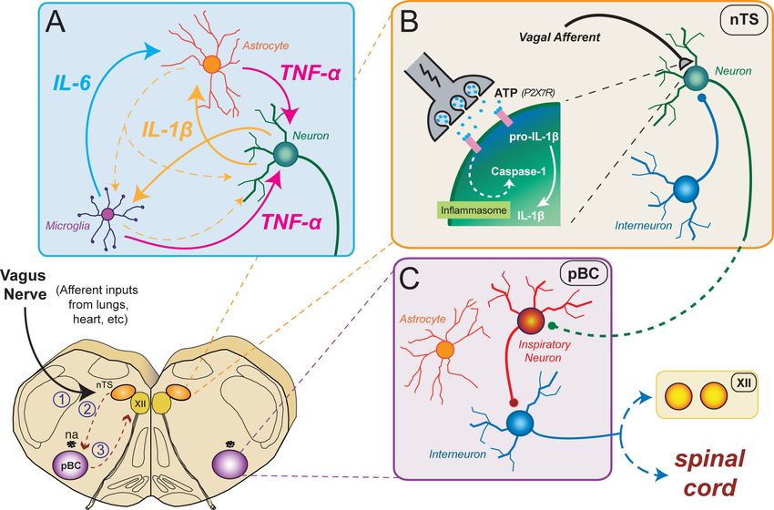
Outline

 ‣ Using neonatal rodent models to understand premature
   breathing patterns in humans

 ‣ Understanding how neuroinflammation alters brainstem
   neural networks and modulates autonomic control circuits

 ‣ Using vagus nerve stimulation (VNS) to prevent central
   neuroinflammation
Premature babies and respiratory control
 ‣ In the U.S. and U.K., 8–18% of all births (>500,000 babies/year!) are
   premature (< 37 weeks gestational age).

 ‣ Respiratory problems are common, particularly infant respiratory distress
   syndrome (IRDS) and chronic lung disease (bronchopulmonary dysplasia).

 ‣ Neurological problems include apnea of prematurity, hypoxic-ischemic
   encephalopathy (HIE), retinopathy of prematurity (ROP), intraventricular
   hemorrhage (IVH).

 ‣ Premature babies are susceptible to infection, including sepsis,
   pneumonia, and urinary tract infection.

 ‣ Infection frequently manifests as respiratory perturbations—like apnea,
   tachypnea, and/or periodic breathing.
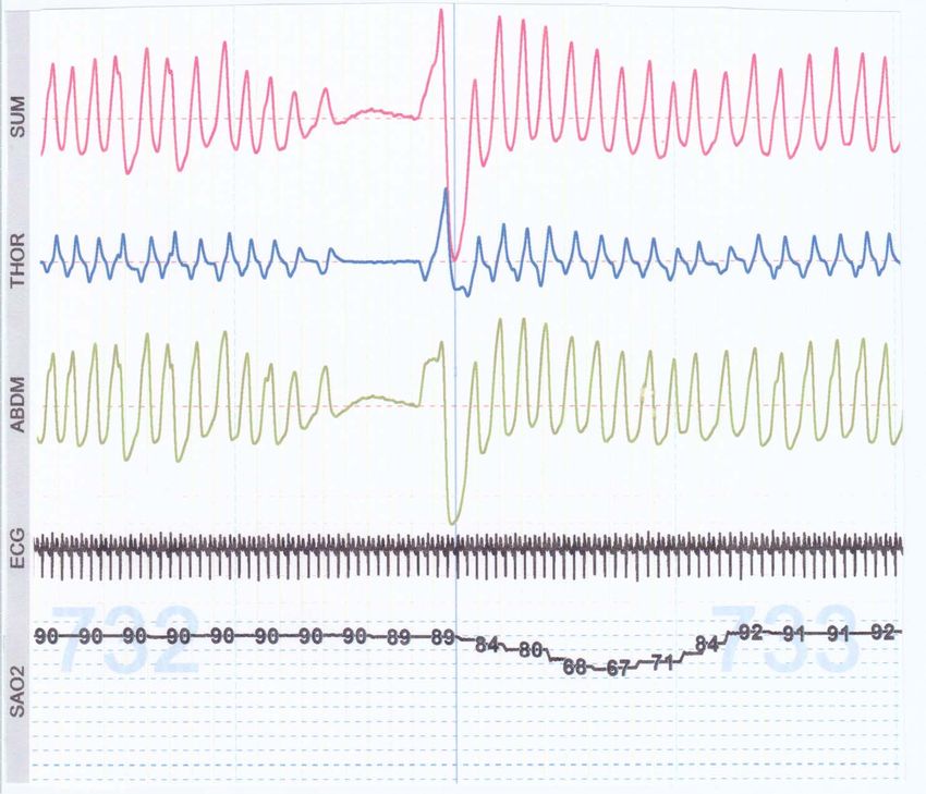
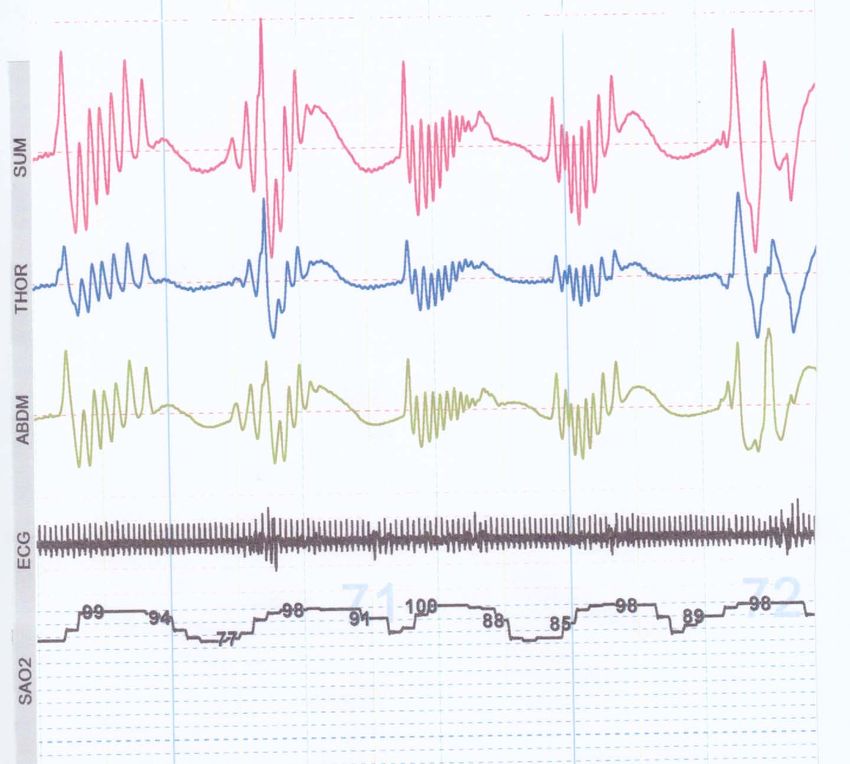
Inductance plethysmography—apnea of prematurity
Inductance plethysmography—periodic breathing
Respiratory Reflexes and Neonatal Apnea

                 IMMATURITY

enhanced             altered               hypoxic
inhibitory         hypercapnic            depression
 reflexes           responses

                     APNEA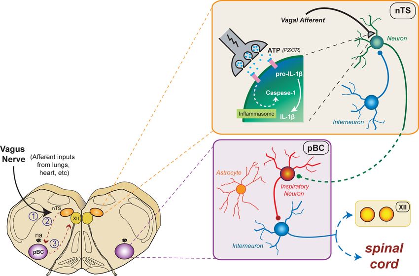
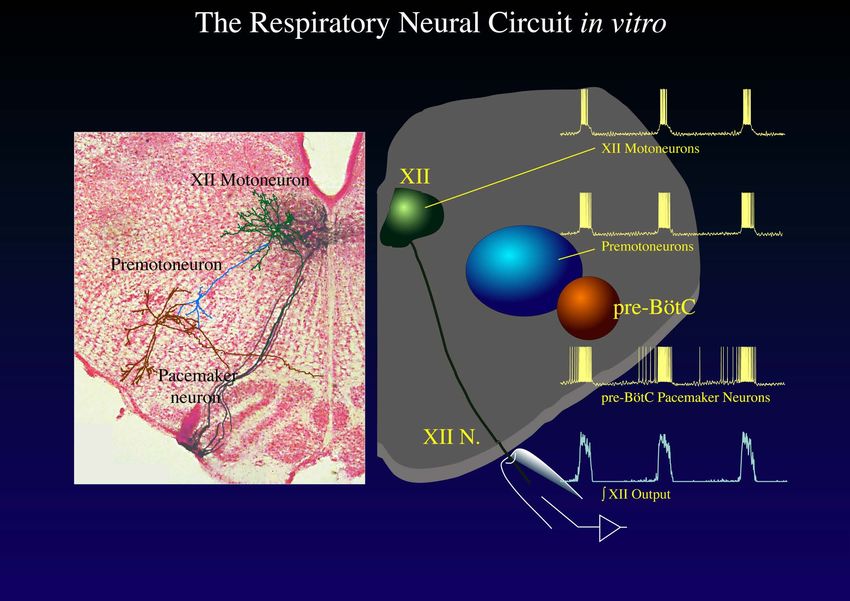
Breathing rhythm originates in the medulla oblongata

                               preBötzinger Complex!
Sagittal section of brainstem
Koizumi et al. J Neuroscience, 2008
Koizumi et al. J Neuroscience, 2008
Morphology of inspiratory-related neurons in the brainstem

                                                   Koizumi, et al., 2008, J Neurosci
Maturation affects firing pattern and connectivity
Smith et al. Resp Physiol, 2000Regions involved in breathing control
This is (sort of!) how apnea of prematurity is treated….

                      http://urbanministryblog.org/wp-content/uploads/2011/01/starbucks-baby2.jpg
Inflammation and respiratory control • Perinatal inflammation/infection is a major source of morbidity and mortality in the newborn population. • Neonatal infection can be acquired by aspiration of infected amniotic fluid either intra-utero or during vaginal delivery, resulting in systemic infection in 1 – 4% of neonates born to mothers with chorioamnionitis. • Infection frequently manifests as respiratory perturbations— like apnea, tachypnea, or periodic breathing—that are challenging to treat.
P11 rats or mice (approximately full-term)
“Pro-inflammatory” Cytokine cascade
Why these cytokines?

• Interleukin-1b (IL-1b): First described in 1972, this cytokine is an important
early mediator of the inflammatory response and invokes cell proliferation,
differentiation, and apoptosis.
• Interleukin-6 (IL-6): An interleukin that acts as both a pro-inflammatory
cytokine and an anti-inflammatory myokine.
• Tumor necrosis factor a (TNFa): Discovered in the late 60s/early 70s.
Another acute phase inflammatory cytokine. Also known to modulate
synaptic activity in the CNS.

All three of these are early, acute phase pro-inflammatory cytokines that
initiate the immune response. They are considered “classic” pro-
inflammatory cytokines—which is why we have focused on them.

They are also trophic factors during development!
Methods – in vivo rats (postnatal day 10–11)

 • Ketamine/xylazine or
   isoflurane
 • LPS @ 0.5 – 1.0 µg/g or
   Saline
 • In vivo (monitor for 2 to 4
   hours
 • In vitro/staining (harvest after
   4 hours)                             Cathy Mayer and Brooke Boyer
Inflammation alters chemoreflexes

                                    Balan et al., Resp. Physiol. Neuriobiol., 2011
Expiratory time (Te), is reduced in Control vs. LPS-exposed rats
Gresham et al. Resp Physiol & Neurobiol, 2011Acute inflammatory up-regulation: The canonical model
Acute inflammatory up-regulation: The canonical model
Acute inflammatory up-regulation: The canonical model
Acute inflammatory up-regulation: The canonical model
Acute inflammatory up-regulation: The canonical model
Acute inflammatory up-regulation: Our “new” model

                                    ATP

Jafri et al. Resp Physiol Neurobiol, 2013
Hypothesis
• Inflammation-induced cytokine release signals the production
of proinflammatory cytokines in the brainstem and this alters
signaling throughout the CNS.
• LPS induces a cascade of cytokine (IL-1b, IL-6, TNFa and
others) release from neurons and microglia.
• These cytokines modulate processing of vagal afferent input
at the nTS, rhythm-generation at the pBC, and motor output
at the XII nucleus.
• Release of prostaglandins (e.g. PGE2) then changes synaptic
processing at this first-order input to the CNS.Cytokines and purines modify synaptic transmission normally
Santello et al., Neuron 2011LPS-induced IL-1β message in respiratory regions of brainstem
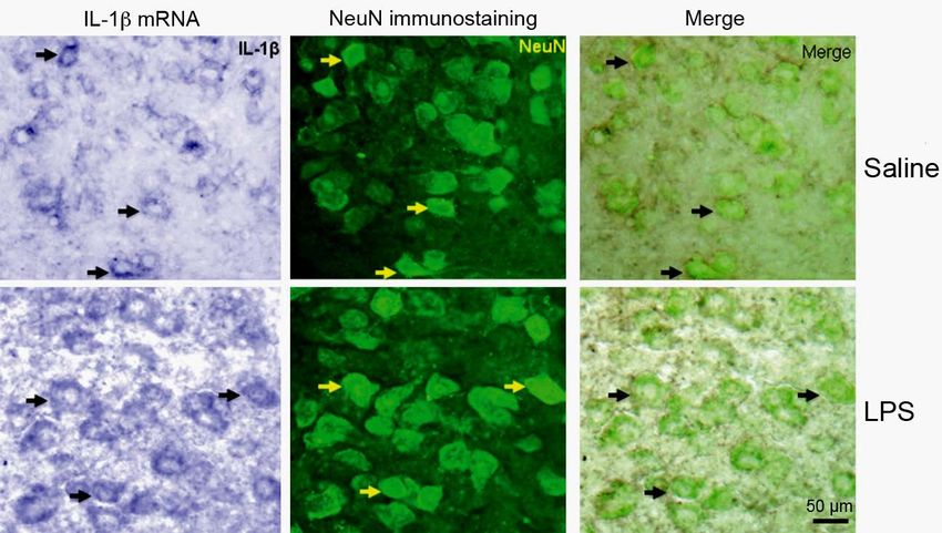
Jafri et al. Resp Physiol Neurobio (2013)IL-1β mRNA expression increased in respiratory areas
Jafri et al. Resp Physiol Neurobio (2013)IL-1β mRNA is expressed in XII motoneurons
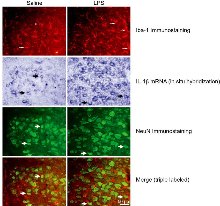
Jafri et al. Resp Physiol Neurobio (2013)Iba-1 (activated microglia) is greater in XII after LPS
Jafri et al. Resp Physiol Neurobio (2013)Microglia appear NOT to express IL-1b
Jafri et al. Resp Physiol Neurobiol, 2013Hypoxia alters IL-1b signaling in the brainstem breathing circuitry
Acute inflammation alters inflammatory drive in the CNS

                       & IX
Changes in nTS neural dynamics after inflammation/lung injury
Getsy et al. Resp Physiol Neurobiol. 2019Changes in nTS neural dynamics after inflammation/lung injury
Getsy et al. Resp Physiol Neurobiol, 2019nTS neurons have smaller sEPSCs after lung injury
Getsy et al. Resp Physiol Neurobiol, 2019Changes in nTS sEPSCs activity after lung injury
Getsy et al. Resp Physiol Neurobiol. 2019nTS evoked EPSCs also show reduced amplitude
Paulina GetsyPGE2 alters breathing pattern in vitro
How do cytokines alter neural activity?
How do cytokines alter neural activity?
How do cytokines alter neural activity?
How do cytokines alter neural activity?

                                          PGE2
When CNS injury occurs, what treatment
options are available and how do we assess
and promote “good,” anti-inflammatory
process while attenuating “bad,” pro-
inflammatory responses?Can we use something besides antibiotics,
corticosteroids, or pharmacological blockade to
reduce/prevent neuro-inflammation in the
CNS?The anti-inflammatory reflex
Tracey KJ, Nature, 2002The Vagus nerve
• The vagus nerve provides extensive afferent & efferent
innervation of the viscera and is a key interface between
CNS circuits and the autonomic control circuitry of the
brainstem.
• The vagus is a mixed autonomic nerve originating in the
medulla oblongata and projects bilaterally along the neck
(bundled with the carotid artery) to the esophagus before
branching to innervate the viscera.
• The anatomy of the vagus and its projections have been
discovered through tract tracing or gross dissection.
• The physiology of the vagus is still an area of active
investigation.The Vagus nerve
NTS = nucleus tractus solitarius
NA = nucleus ambiguus
pBC = preBötzinger Complex
(rhythm generator)Vagus Nerve Stimulation
• Inflammation stimulates the release of pro-inflammatory
cytokines which activate vagal afferents and induce
central neuroinflammation
• Vagal c-fibers are implicated in this inflammatory
upregulation and their first-order synapse is in the
nucleus tractus solitarius (NTS)
• Vagal efferents are implicated in anti-inflammatory
responses via the cholinergic anti-inflammatory pathway
• We have previously shown that vagus nerve stimulation
(VNS) modulates pro-inflammatory cytokine expression
in the central nervous system (CNS) using high
frequency stimulation.
• However, the optimal VNS parameters to reduce
inflammation are not yet known.Vagal nerve stimulation to “knock down” cytokine upregulation
X
Johnson et al. Resp Physiol Neurobiol, 2016FDA-approved clinical uses of VNS
• Treatment of epilepsy. In 1988, the first chronic
implantable stimulator was used to treat drug-resistant
epilepsy.
• VNS has been approved by the FDA since 1997 to treat
partial onset seizures that are drug-resistant.
• Treatment of depression. Chronic or severe depression
affects up to 1.5% of the general population, and many
of these patients obtain little relief from pharmaceutical
treatment.
• Although VNS was not originally developed to treat
depression, the FDA approved VNS for the treatment of
chronic or recurring depression in 2005.Research uses of VNS
• Sepsis. Sepsis is a multibillion dollar health care burden
typically due to systemic bacterial infection and chronic
activation of the pro-inflammatory cytokine cascade.
VNS is being used experimentally to quash runaway
inflammation
• Pain management. The applications of VNS also
extends to disorders associated with chronic or
intermittent bouts of pain such as fibromyalgia and
migraines.
• Cardiovascular disease. VNS must alter cardiovascular
control due to the convergence of inputs in the
autonomic control centers of the brain stem, but for how
long and to what extent is unknown. The descending
cardiac branch of the vagus is key for normal cardiac
function.VNS and cytokines
VNS and cytokines
Methods
Methods
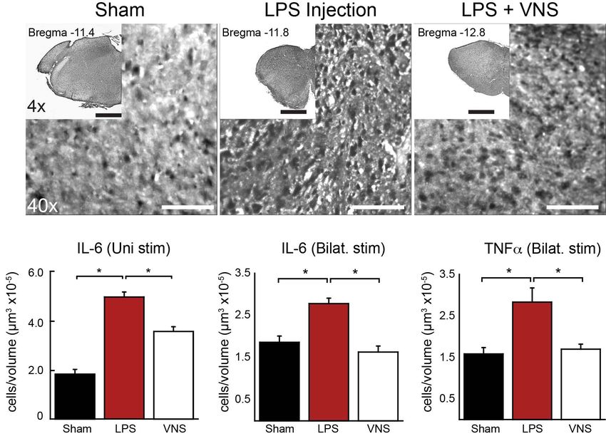
IL-6 and TNFa are reduced after VNS

                              Johnson et al. Resp Physiol Neurobiol, 2016
So if we use “typical” clinical VNS
parameters (current/frequency) we can
reduce cytokine expression.
But, what are the OPTIMAL stimulation
parameters to reduce inflammation?VNS attenuates IL-1b across most frequencies
Cacho et al. submitted to Peds ResearchVNS attenuates TNFa at higher stimulation frequencies
Cacho et al. submitted to Peds ResearchIL-6 is a confusing bugger in response to VNS!
Cacho et al. submitted to Peds ResearchThe alarmin, HMGB1, exhibits a dose-dependent decrease
with VNS
Cacho et al. submitted to Peds ResearchFuture Directions
‣ The likelihood that we will get IRB approval to implant a
vagus nerve stimulator in a preterm infant is vanishingly
small!
‣ Transcutaneous stimulation would allow us to stimulate
non-invasively and attempt to get sufficient current to the
vagus nerve and have an impact on inflammation.
‣ An even more interesting option in the clinic would be the
use of transcutaneous auricular vagus nerve stimulation
(aVNS) which is non-invasive and easy to use in a clinical
setting.Can we modify the method of VNS to use non-invasive stimulators?
Transcutaneous Auricular Vagus Nerve Stimulation (aVNS)

                                         Yap JYY et al. Front Neuroscience, 2020
Transcutaneous
auricular vagus
stimulation
Stavrakos S et al., JACC: Clinical Electrophysiology, 2020aVNS stimulators
Yap JYY et al. Front Neuroscience, 2020aVNS protocols that replicate some of our work….
Sclocco R et al. Brain Stimulation, 2020Summary ‣ Our laboratory has been focused on translational applications of developmental neurophysiology in neonates. ‣ Intratracheal LPS stimulates IL-1b production in the brainstem (nTS, RVLM, and XII) of rodents, activating the COX2 pathway and, ultimately, releasing prostaglandins and other chemokines/cytokines that alter neural network activity. ‣ Bioelectric stimulation may be valuable in controlling acute or chronic inflammation and, using aVNS, may be easily incorporated into current clinical practice.
Thank you for your
attention!

Questions??
References
» 1. Hassan Boskabadi and Maryam Zakerihamidi, “Evaluate the Diagnosis of Neonatal Sepsis by Measuring
  ILs: A Systematic Review,” Pediatrics & Neonatology, October 20, 2017,
  https://doi.org/10.1016/j.pedneo.2017.10.004.
» 2. Lauren Vogel, “Sepsis Kills One Million Newborns a Year: WHO,” CMAJ : Canadian Medical Association
  Journal 189, no. 40 (October 10, 2017): E1272, https://doi.org/10.1503/cmaj.109-5504.
» 3. Celeste M. Torio and Brian J. Moore, “National Inpatient Hospital Costs: The Most Expensive Conditions
  by Payer, 2013: Statistical Brief #204,” in Healthcare Cost and Utilization Project (HCUP) Statistical Briefs
  (Rockville (MD): Agency for Healthcare Research and Quality (US), 2006),
  http://www.ncbi.nlm.nih.gov/books/NBK368492/.
» 4. Kari A. Simonsen et al., “Early-Onset Neonatal Sepsis,” Clinical Microbiology Reviews 27, no. 1 (January
  2014): 21–47, https://doi.org/10.1128/CMR.00031-13.
» 5. Sindhu Sivanandan, Amuchou S. Soraisham, and Kamala Swarnam, “Choice and Duration of Antimicrobial
  Therapy for Neonatal Sepsis and Meningitis,” International Journal of Pediatrics 2011 (2011),
  https://doi.org/10.1155/2011/712150.
» 6. J. Roth and G. E. De Souza, “Fever Induction Pathways: Evidence from Responses to Systemic or Local
  Cytokine Formation,” Brazilian Journal of Medical and Biological Research = Revista Brasileira De
  Pesquisas Medicas E Biologicas 34, no. 3 (March 2001): 301–14.
» 7. P Ng, “Diagnostic Markers of Infection in Neonates,” Archives of Disease in Childhood Fetal and Neonatal
  Edition 89, no. 3 (May 2004): F229–35, https://doi.org/10.1136/adc.2002.023838.
References
» 8. Rhaya L Johnson and Christopher G Wilson, “A Review of Vagus Nerve Stimulation as a Therapeutic
  Intervention,” Journal of Inflammation Research 11 (May 16, 2018): 203–13,
  https://doi.org/10.2147/JIR.S163248.
» 9. Bruno Bonaz, Valérie Sinniger, and Sonia Pellissier, “Anti-inflammatory Properties of the Vagus Nerve:
  Potential Therapeutic Implications of Vagus Nerve Stimulation,” The Journal of Physiology 594, no. 20
  (October 15, 2016): 5781–90, https://doi.org/10.1113/JP271539.
» 10. Robert H. Howland, “Vagus Nerve Stimulation,” Current Behavioral Neuroscience Reports 1, no. 2 (June
  2014): 64–73, https://doi.org/10.1007/s40473-014-0010-5.
» 11. A. O. Hofstetter et al., “The Induced Prostaglandin E2 Pathway Is a Key Regulator of the Respiratory
  Response to Infection and Hypoxia in Neonates,” Proceedings of the National Academy of Sciences 104, no. 23
  (June 5, 2007): 9894–99, https://doi.org/10.1073/pnas.0611468104.
» 12. Frieda A. Koopman et al., “Vagus Nerve Stimulation Inhibits Cytokine Production and Attenuates Disease
  Severity in Rheumatoid Arthritis,” Proceedings of the National Academy of Sciences 113, no. 29 (July 19,
  2016): 8284–89, https://doi.org/10.1073/pnas.1605635113.
» 13. I. Lerman et al., “Noninvasive Transcutaneous Vagus Nerve Stimulation Decreases Whole Blood Culture-
  Derived Cytokines and Chemokines: A Randomized, Blinded, Healthy Control Pilot Trial” 19, no. 3 (April 1,
  2016): 283–91, https://doi.org/10.1111/ner.12398.
References

» 14. Kannan V Balan et al., “Vagal Afferents Modulate Cytokine-Mediated Respiratory Control at the Neonatal
  Medulla Oblongata,” Respiratory Physiology & Neurobiology 178, no. 3 (September 30, 2011): 458–64,
  https://doi.org/10.1016/j.resp.2011.03.003.
» 15. Rhaya L. Johnson et al., “Vagal Nerve Stimulation Attenuates IL-6 and TNFα Expression in Respiratory
  Regions of the Developing Rat Brainstem,” Respiratory Physiology & Neurobiology 229 (April 2, 2016): 1–4,
  https://doi.org/10.1016/j.resp.2016.03.014.
» 16. Yogi A. Patel and Robert J. Butera, “Differential Fiber-Specific Block of Nerve Conduction in Mammalian
  Peripheral Nerves Using Kilohertz Electrical Stimulation,” Journal of Neurophysiology 113, no. 10 (June 1,
  2015): 3923–29, https://doi.org/10.1152/jn.00529.2014.
» 17. Duncan A. Groves and Verity J. Brown, “Vagal Nerve Stimulation: A Review of Its Applications and
  Potential Mechanisms That Mediate Its Clinical Effects,” Neuroscience and Biobehavioral Reviews 29, no. 3
  (May 2005): 493–500, https://doi.org/10.1016/j.neubiorev.2005.01.004.
You can also read