A case of fetal hydrops: prenatal diagnosis and neonatal management - Unime
←
→
Page content transcription
If your browser does not render page correctly, please read the page content below
APMB - Atti della Accademia Peloritana dei Pericolanti                                                                       ISSN 1828-6550
       Classe di Scienze Medico Biologiche
                  Vol. 106(1) 2018
                                                                                            DOI: 10.6092 / 1828-6550 / APMB.106.1.2018.A2

Clinical Case Seminar                                                                                            A2( 1-7)

A case of fetal hydrops: prenatal diagnosis and neonatal
management
Roberta Granese1, Grazia Foti1, Caterina Iozzo1, Alessia Giofrè1, Ilenia
Panasiti2, Gabriella D’Angelo2, Lucia Marseglia2, Eloisa Gitto2
Obstectrics and Gynaecology Unit, Department of Adult and Childhood Human Pathology “Gaetano
1

Barresi”, Messina University Hospital “G . Martino”2 Intensive Care Unit, Department of Adult and
Childhood Human Pathology “Gaetano Barresi”, Messina University Hospital “G . Martino”

Abstract
Hydrops fetalis (HF) is a serious fetal condition defined as an abnormal fluid accumulation in fetal
extravascular compartments and body cavities caused by either immune or non immune conditions.
Immune hydrops is caused by fetal hemolysis mediated by circulating maternal antibodies to fetal red blood
cell antigens. Its most common determinant is rhesus incompatibility. Systemic Lupus Eritematosus (SLE)
is another rare cause of immune fetal hydrops, when the pregnancy is complicated by the presence of a third
degree congenital heart block (CHB). The Neonatal Lupus Syndrome occurs with a prevalence of 2%.
We reported the case of severe fetal hydrops in a 31 weeks pregnant woman affected by mild maternal D
alloimmunization and SLE. Despite fetal hydrops and a mild positive indirect Coombs’ test, the flow-rate
study with the Systolic Peak Velocity (PSV) of the MCA excluded a fetal anemia. At birth, blood gas
showed a condition of severe metabolic and respiratory acidosis (pH 6.43, pO2 9.9 mmHg, pCO2 206
mmHg, Base Excess (BE) -35 mmol/l, HCO3- 2.7 mmol/l) and a mild anemia (Hemoglobin 10.3 g/dl).
ECG revealed a normal sinus rhythm and a CHB was excluded. Despite the critical clinical condition, no
cardiorespiratory or neurological adverse outcomes occurred in the newborn

Keywords: hydrops fetalis, rhesus incompatibility,
Systemic lupus erythematosus

Corresponding Author: roberta.granese@unime.it

Introduction
Hydrops fetalis (HF) is a serious fetal condition defined as an abnormal fluid accumulation in fetal
extravascular compartments and body cavities leading to edema, ascites, pleural and pericardial
effusions, and anasarca (1, 3). It may also be associated with polyhydramnios and placental edema.
Its incidence is reported to be 0.3 to 2.4 per 1,000 live births (4, 5). The etiologies are considered
as immune or non immune hydrops (6).
Immune hydrops is caused by fetal hemolysis mediated by circulating maternal antibodies to fetal
red blood cell antigens (7). Its most common determinant is rhesus incompatibility and it occurs
when antibodies from a Rh-negative mother target and destroy ’foreign’ red blood cells from a Rh-
positive fetus (8). The introduction of postnatal immunoprophylaxis in 1970 reduced the incidence

              APMB - Atti della Accademia Peloritana dei Pericolanti - Classe di Scienze Medico-Biologiche (2018), 106(1):A2(1-7)
                                             DOI: 10.6092 / 1828-6550 / APMB.106.1.2018.A2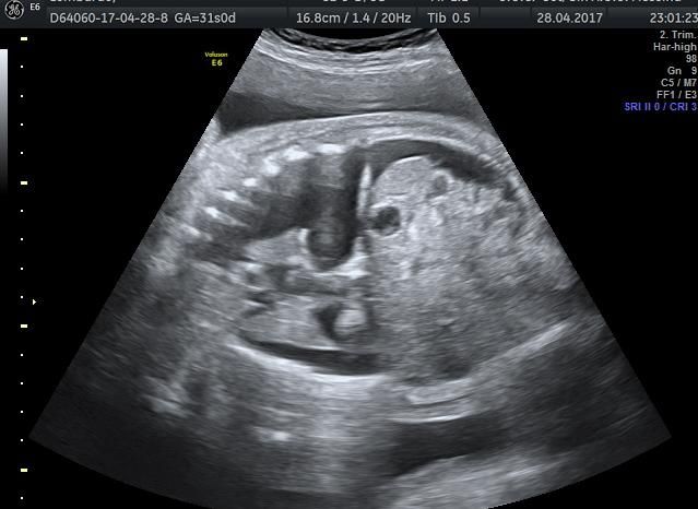
APMB - Atti della Accademia Peloritana dei Pericolanti                                                                       ISSN 1828-6550
       Classe di Scienze Medico Biologiche
                  Vol. 106(1) 2018
                                                                                            DOI: 10.6092 / 1828-6550 / APMB.106.1.2018.A2

of maternal D alloimmunization from 14% to 1-2% (9). Post-partum anti-D immunoglobulin
administered to Rh D-negative women and routine antenatal anti-D prophylaxis have greatly
reduced the incidence of haemolytic disease of the fetus and newborn due to immune anti-D to
0.1% (10) In a Rh-negative mother, maternal anti-D antibodies may cross the placenta and lead to
the immune-mediated destruction of fetal red blood cells. This more rapid destruction of the fetal
red blood cells than normal can lead to anemia and jaundice and, in very severe cases, kernicterus
or even death.
Systemic lupus erythematosus (SLE) is a chronic autoimmune disease that mainly affects women of
reproductive age. Pregnancies in women with SLE resulted in an increase of adverse pregnancy
outcomes (e.g., stillbirth, premature birth, and intrauterine growth restriction) (11). SLE is another rare
cause of immune fetal hydrops, when the pregnancy is complicated by the presence of a third degree
congenital heart block (CHB). The neonatal lupus syndromes occurs with a prevalence of 2%.
Since there was a decline of rhesus iso-immunization by routine screening and prophylaxis (1, 12),
approximately 76-87% of all cases of HF are estimated to be of non-immune origin (6).
Non-immune hydrops can result from a large number of causes including cardiac and non-cardiac
anomaly, syndromes, aneuploidy, congenital infection, twin-to-twin transfusion syndrome,
chrioangioma and other conditions. Moreover, in 15% to 25% of cases, the precise cause may
remain unknown (12,13). Mortality rate is still high, up to 75% (12, 14, 15).
Routine ultrasound examination allows to easily recognize fetal hydrops. A more detailed
examination should be performed in all these cases to understand the etiology and nature of
hydrops, including the location as well as the amount of fluid collections, amniotic fluid index,
placental thickness, fetal echocardiography and Doppler velocimetry (16, 17).
We reported the case of severe immune fetal hydrops in a women at 31 week of pregnancy
complicated by maternal D alloimmunization and SLE.

Case Report
A 36-years-old woman pregnant for the third time, was admitted at 31 week of pregnancy at the
Obstectrics and Gynaecology Unit of the University Hospital “G. Martino” of Messina to perform
a third trimester’s ultrasound examination. A severe hydrops in the fetus was identified for the first
time. Therefore, she was hospitalized.
In her history three miscarriages and two pregnancies were referred. She also suffered of SLE
since 2016. Her blood group was A RhD negative (partner’s group was O RhD positive). She
reported a physiological course of pregnancy except for a threat of abortion in the first trimester. In
this occasion antenatal anti-D prophylaxis were not performed and neither at 28 weeks of
              APMB - Atti della Accademia Peloritana dei Pericolanti - Classe di Scienze Medico-Biologiche (2018), 106(1):A2(1-7)
                                             DOI: 10.6092 / 1828-6550 / APMB.106.1.2018.A2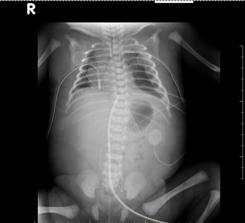
APMB - Atti della Accademia Peloritana dei Pericolanti ISSN 1828-6550
Classe di Scienze Medico Biologiche
Vol. 106(1) 2018
DOI: 10.6092 / 1828-6550 / APMB.106.1.2018.A2
18
pregnancy, as well as current guidelines in pregnancy suggested . In each month during
pregnancy indirect Coombs’ test was reported negative. During this period she was treated with
acetylsalicylic acid, methylprednisolone and hydroxycloroquine.
At the admission blood evaluation showed positive values of antinuclear antibodies (1:2560) and
anti-dsDNA antibodies (1:80). Indirect Coomb’s test was also positive (1: 250).
The ultrasound examination showed a female fetus, with extremely low fetal movements and
regular heartbeats. It was highlighted a polydramnios with a 100 mm of maximum pocket of
amniotic fluid, fetal hydrops with ascites and hydrothorax (Fig. 1 and Fig. 2).
Fig. 1. Fetal edema Fig. 2.Fetal hydrothorax
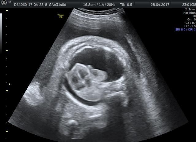
The fetal growth, due to the edema (Fig. 3), was Fig. 3. Fetal hydrops with ascites and hydrothorax
equivalent to 95° centile. The morphological
examination of the fetal anatomy did not detect
any malformation. The echocardiographic exam
showed a slight pericardial effusion, a regular
anatomy of the heart with situs solitus, but in
mesocardia due to the hydrotorax. The flow-rate
study revealed an incontinence of atrioventricular
valves and pulmonary valves.
Doppler velocimetry of the umbilical artery (UA) and middle cerebral artery (MCA) showed a
normal value of pulsatility index (UA: PI 1.07; MCA PI 2.27), and a Systolic Peak Velocity (PSV)
of the Middle Cerebral Artery (MCA) of 45.51 cm/sec (expected 41.93, mOm 1.09), suggesting
for a non-anemic fetus. Given the severity of the clinical picture, the respiratory distress
prophylaxis with betametasone was administered, and a caesarean section was planned.
At birth, the newborn presented severe hydrops and she was cyanotic, apneic and the heart rate
APMB - Atti della Accademia Peloritana dei Pericolanti - Classe di Scienze Medico-Biologiche (2018), 106(1):A2(1-7)
DOI: 10.6092 / 1828-6550 / APMB.106.1.2018.A2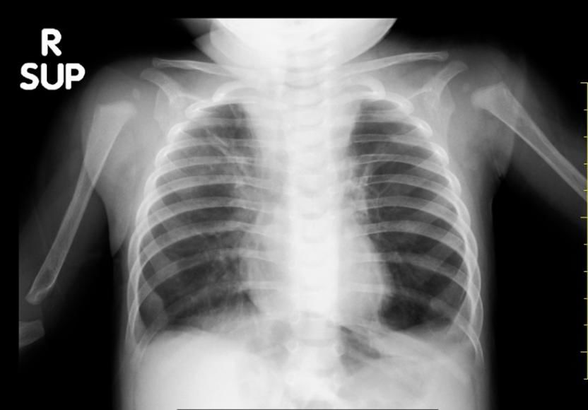
APMB - Atti della Accademia Peloritana dei Pericolanti                                                                        ISSN 1828-6550
       Classe di Scienze Medico Biologiche
                  Vol. 106(1) 2018
                                                                                              DOI: 10.6092 / 1828-6550 / APMB.106.1.2018.A2

was < 100 bpm. According to Newborn Life Support proceedings endotracheal intubation,
surfactant intratracheal instillation and chest compression were performed. Furthermore for the
presence of persistent cardiorespiratory arrest and drug therapy was administered after umbilical
venous catheterization (epinephrine, bolus of 10 ml/kg of normal saline) and, due to a mild anemia
(Hemoglobin 10.3 g/dl), O rhD negative red blood cells were transfused. Moreover bilateral
thoracocentesis was necessary and, after the insertion of chest tubes in the 4th intercostal space of
both chest side, 200 cc of citrine yellow liquid were removed. The Apgar score was 1, 5 and 5, at
1st, 5th and 10th minutes respectively. Nevertheless clinical condition remained extremely critical.
At birth, blood gas showed a condition of severe metabolic and respiratory acidosis (pH 6.43, pO2
9.9 mmHg, pCO2 206 mmHg, Base Excess (BE) -35 mmol/l, HCO3- 2.7 mmol/l). When the
resuscitation maneuvers were completed, control of breathing was first obtained with mechanical
ventilation with SIPPV mode (Synchronized Intermittent Positive Pressure Ventilation) and then
with HFOV mode (High Frequency Oscillation Ventilation). Chest X-ray revealed an increased
cardio-mediastinal shadow, lifting diaphragm and severe soft tissue edema, ascites and
hydrothorax. (Fig. 4).
Fig. 4. Chest X-Ray after few hours from birth.                         Fig. 5. Chest X-Rat at the hospital discharge

After 8 hours from birth blood gas showed a persistently severe metabolic and respiratory acidosis
(pH 7.02, pO2 43.5 mmHg, pCO2 66.5 mmHg, Base Excess (BE) -15mmol/l, HCO3-12.5mmol/l,
Hemoglobin 15.1 g/dl). Due to suspect of pulmonary hypertension caused by the severe pulmonary
hypoplasia a treatment with Nitric Oxide and Fenoldopam were administered successfully. Several
echocardiograms confirmed the moderate-severe pulmonary hypertension but unexpectedly
cardiac pump function was normal. ECG also revealed normal sinus rhythm and a Congenital
Heart Block (CHB) was excluded. The progressive improvement of clinical condition allowed to
remove the chest tubes and Non-invasive ventilation (NCPAP - HHHNC) was started with a great

               APMB - Atti della Accademia Peloritana dei Pericolanti - Classe di Scienze Medico-Biologiche (2018), 106(1):A2(1-7)
                                              DOI: 10.6092 / 1828-6550 / APMB.106.1.2018.A2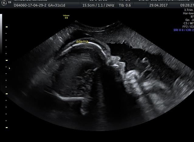
APMB - Atti della Accademia Peloritana dei Pericolanti                                                                       ISSN 1828-6550
       Classe di Scienze Medico Biologiche
                  Vol. 106(1) 2018
                                                                                            DOI: 10.6092 / 1828-6550 / APMB.106.1.2018.A2

response of the infant. (Fig. 5) Neurological evaluation performed by cranial sonography and MRI
scan revealed a normal brain. After a month a healthy baby was discharged.

Discussion
Despite improvements in postnatal care such as advanced mechanical ventilation, surfactant
treatment, and inhaled nitric oxide, hydrops fetalis (HF) is still a serious fetal condition with high
risk of perinatal mortality, morbidity and long-term disability. Some authors report that the number
of fluid collection sites are the strongest antenatal ultrasound risk factor for prediction of poor
outcome (19, 20). Moreover, the risk of fetal death is higher if fetal hydrops is diagnosed at an
earlier gestational age (7).
The severe hydrops we reported could be determined by two maternal risk factors: maternal D
alloimmunization and SLE.
In haemolytic disease of the fetus and newborn (HDFN), fetal hydrops occurs when fetal Hb
deficit exceeds 7 g/dL. It is interesting to observe that in our case the severe hydrops was not
associated with a severe anemia of the newborn. The various mechanisms responsible for hydrops
are hypoalbuminemia secondary to depressed liver function, increased capillary permeability, iron
overload secondary to hemolysis, and increased venous pressures due to poor cardiac function.
Complex genetic factors influence the ability of individuals to respond to red cell antigens. Rh D is
the most effective immunogen and, as little as 0.1 to 1 ml of D positive red cells can stimulate
antibody production (18). Sensitization is believed to have no adverse health effects for the mother
and, the first baby is usually not harmed, as the pregnancy is generally completed by the time that
sensitization has occurred. In addition to feto-maternal hemorrhage at birth, events during
pregnancy may lead to the production of maternal anti-D antibodies and thus sensitization may
also occur during the antenatal period. It has been estimated that approximately half of newborn
infants with hemolytic disease are mildly affected, requiring no treatment. Of the remainder, half
will become hydropic in utero, and half will be born apparently healthy but without treatment may
either die of kernicterus or be left severely disabled (21). In our case, Coombs’ test evaluated
during pregnancy was mild positive and no fetal anemia was suggested to the PVS in the MCA;
these data were confirmed at birth (Hb 10.3 g/dl).
SLE during pregnancy could develop an irreversible 3rd degree heart block in the newborn,
consequently hydrops and high risk of death. Anti–52-kd Ro/SSA and anti-La/SSB antibodies
have been reported to be more strongly associated with CHB than was anti–60-kd Ro/SSA
alone(22). It is considered a pathologic readout of passively acquired autoimmunity consequent to
FcRn-mediated placental transport of antibodies reactive with protein components of the SSA⁄ Ro-
              APMB - Atti della Accademia Peloritana dei Pericolanti - Classe di Scienze Medico-Biologiche (2018), 106(1):A2(1-7)
                                             DOI: 10.6092 / 1828-6550 / APMB.106.1.2018.A2
APMB - Atti della Accademia Peloritana dei Pericolanti ISSN 1828-6550
Classe di Scienze Medico Biologiche
Vol. 106(1) 2018
DOI: 10.6092 / 1828-6550 / APMB.106.1.2018.A2
SSB⁄ La ribonucleoprotein complex (23). It carries a significant mortality (20–30%, primarily fetal
and neonatal) and morbidity (67% of those surviving affected children require permanent pacing
before adulthood) (23). According to the related literature, no difference in mortality, prematurity
or degree of final block or need for pacemaker was founded for fetuses treated with or without
steroids. It was noted that the presence of either pericardial or pleural effusions as well as ascites
and hydrops seemed to improve with the use of steroids(23). Fortunately, in our patience a
Congenital Heart Block (CHB) was excluded and ECG revealed a normal sinus rhythm.
Conclusion
We reported an unusual case of severe hydrops due to RhD alloimmunization, although during
pregnancy hemoglobin value estimated seemed normal and at birth it revealed only a mild anemia.
Nevertheless despite the critical clinical condition at birth, no cardiorespiratory or neurological
adverse outcomes occurred.
Conflicts of Interest: There is no potential conflict of interest, and the authors have nothing to disclose.
This work was not supported by any grant.
References
1. Bellini, C., Hennekam, R., C., (2012) Non-immune hydrops fetalis: a short review of etiology and
pathophysiology. Am J Med Genet A 2012;158A:597-605.
2. Randenberg, A., L., (2010) Nonimmune hydrops fetalis part I: etiology and pathophysiology. Neonatal
Netw;29:281- 95.
3. Favre, R., Dreux, S., Dommergues, M., Dumez, Y., Luton, D., Oury, J., F., (2004) Nonimmune fetal
ascites: a series of 79 cases. Am J Obstet Gynecol, 190:407-12.
4. Liao, C., Wei, J., Li, Q., Li, J., Li, L., Li, D., (2007) Nonimmune hydrops fetalis diagnosed during the
second half of pregnancy in Southern China. Fetal Diagn Ther, 22:302-5.
5. Heinonen, S., Ryynanen, M., Kirkinen, P., (2000) Etiology and outcome of second trimester non-
immunologic fetal hydrops. Acta Obstet Gynecol Scand, 79:15-8.
6. Bellini, C., Hennekam, R.,C., Fulcheri, E., Rutigliani, M., Morcaldi, G., Boccardo, F., (2009) Etiology of
nonimmune hydrops fetalis: a systematic review. Am J Med Genet A, 149A:844e51.
7. Yeom, W., Paik, E.,S., An, J.,J., Oh, S.,Y., Choi, S.,J., Roh, C., R., (2015) Clinicalcharacteristics and
perinatal outcome of fetal hydrops. Obstet Gynecol Sci. Mar;58(2):90-7.
8. Chilcott, J., Lloyd, Jones, M., Wight, J., Forman, K., Wray, J., Beverley, C., (2002) A review of the
clinical effectiveness and cost effectiveness of routine anti-D prophylaxis for pregnant women who are
Rhesus (RhD) negative. London: National Institute of Clinical Excellence. Health Technol Assess, 7(4):iii-
62. Review
9. Basu, (2011) Hemolytic disease of the fetus and newborn: Current trends and perspectives. Asian J
Transfus Sci, 5(1): 3–7.
10. McCauley, C., J., (2017) A review of maternal alloimmunisation to Rh D in Northern Ireland. Transfus
Med., 27(2):132-135.
11. Zhan, Z1, (2017) Fetal outcomes and associated factors of adverse outcomes of pregnancy in southern
Chinese women with systemic lupus erythematosus. PLoS One, 12(4): e0176457.
12. Abrams, M., E., Meredith, K., S., Kinnard, P., Clark, RH., (2007) Hydrops fetalis: a retrospective
review of cases reported to a large national database and identification of risk factors associated with death.
APMB - Atti della Accademia Peloritana dei Pericolanti - Classe di Scienze Medico-Biologiche (2018), 106(1):A2(1-7)
DOI: 10.6092 / 1828-6550 / APMB.106.1.2018.A2APMB - Atti della Accademia Peloritana dei Pericolanti ISSN 1828-6550
Classe di Scienze Medico Biologiche
Vol. 106(1) 2018
DOI: 10.6092 / 1828-6550 / APMB.106.1.2018.A2
Pediatrics, 20:84-9.
13. Randenberg, A., L. Nonimmune hydrops fetalis part II: does etiology influence mortality? (2010)
Neonatal Netw, 29:367-80.
14. Simpson, J., H., McDevitt, H., Young, D., Cameron, A., D. (2006) Severity of non-immune hydrops
fetalis at birth continues to predict survival despite advances in perinatal care. Fetal Diagn Ther, 21:380-2.
15. Moreno, C., A., Kanazawa, T., Barini, R., Nomura, M., L., Andrade, K., C., Gomes, C., P. (2013) Non-
immune hydrops fetalis: a prospective study of 53 cases. Am J Med Genet A, 161A:3078-86.
16. Fukushima, K., Morokuma, S., Fujita, Y., Tsukimori, K., Satoh, S., Ochiail, M., (2011) Short-term and
long-term outcomes of 214 cases of non-immune hydrops fetalis. Early Hum Dev, 87:571-5.
17. Sohan, K., Carroll, S., G., De La Fuente, S., Soothill, P., Kyle, P., (2001) Analysis of outcome in
hydrops fetalis in relation to gestational age at diagnosis, cause and treatment. Acta Obstet Gynecol Scand,
80:726-30.
18. Basu, S., (2011) Hemolytic disease of the fetus and newborn: Current trends and perspectives Asian J
Transfus Sci, 5(1): 3–7.
19. Takci, S., Gharibzadeh, M., Yurdakok, M., Ozyuncu, O., Korkmaz, A., Akcoren, Z., (2014) Etiology
and outcome of hydrops fetalis: report of 62 cases. Pediatr Neonatol, 55(2):108-13.
20. Kim, S., A., Lee, S., M., Hong, J., S., Lee, J., Park, C., W., Kim, B., J., (2014) Ultrasonographic
severity scoring of non-immune hydrops: a predictor of perinatal mortality. J Perinat Med, 43(1):53-9. doi:
10.1515/jpm-2013-0208.
21. Bowman JM. Hemolytic disease of the newborn. VoxSanguinis 1996;70:62–7.
22. Brucato, A., Risk of congenital complete heart block in newborns of mothers with anti-Ro/SSA
antibodies detected by counterimmunoelectrophoresis: a prospective study of 100 women. Arthritis Rheum,
44(8):1832-5.
23. Buyon, J., P., (2009) Autoimmune associated congenital heart block: integration of clinical and research
clues in the management of the maternal ⁄ foetal dyad at risk. J Intern Med, 265(6):653-62.
©2018 by the Author(s); licensee Accademia Peloritana dei Pericolanti (Messina, Italy). This article is an open access
article distributed under the terms and conditions of the Creative Commons Attribution 4.0
International License (https://creativecommons.org/licenses/by/4.0/).
Communicated and received February 20, 2018, revised April 8 2108 , published on line June 15, 2018
APMB - Atti della Accademia Peloritana dei Pericolanti - Classe di Scienze Medico-Biologiche (2018), 106(1):A2(1-7)
DOI: 10.6092 / 1828-6550 / APMB.106.1.2018.A2You can also read