Yaws Disease Caused by - Treponema pallidum subspecies pertenue in Wild Chimpanzee, CDC
←
→
Page content transcription
If your browser does not render page correctly, please read the page content below
DISPATCHES

          Yaws Disease Caused by
       Treponema pallidum subspecies
        pertenue in Wild Chimpanzee,
               Guinea, 2019
        Benjamin Mubemba,1 Emeline Chanove,1 Kerstin Mätz-Rensing, Jan F. Gogarten, Ariane Düx,
   Kevin Merkel, Caroline Röthemeier, Andreas Sachse, Helene Rase, Tatyana Humle, Guillaume Banville,
           Marine Tchoubar, Sébastien Calvignac-Spencer, Christelle Colin, Fabian H. Leendertz

 Yaws-like lesions are widely reported in wild African great         infections with T. pallidum in olive baboons at many
 apes, yet the causative agent has not been confirmed                sites in Tanzania (6,7).
 in affected animals. We describe yaws-like lesions in a                  Both genital and orofacial lesions attributable to
 wild chimpanzee in Guinea for which we demonstrate                  TPE infection have been documented in several NHP
 infection with Treponema pallidum subsp. pertenue. As-              species across sub-Saharan Africa (African green mon-
 sessing the conservation implications of this pathogen              keys [Chlorocebus sabaeus] in Bijilo Forest Park, The
 requires further research.                                          Gambia, and Niokola-Koba National Park, Senegal;
                                                                     sooty mangabeys [Cercocebus atys atys] in Taï Nation-
S   everal monkey species in sub-Saharan Africa
    are infected with Treponema pallidum subspecies
pertenue (TPE) and typically manifest yaws-like le-
                                                                     al Park, Côte d’Ivoire) (1; B. Mubemba et al., unpub.
                                                                     data, https://doi.org/10.1101/848382). Two studies
                                                                     observed that TPE infections remain geographically
sions on the face and distal extremities or syphilis-
                                                                     widespread in Tanzania and affect olive baboons, yel-
like lesions in the anogenital region (1). Reports of
                                                                     low baboons, vervet monkeys (Chlorocebus pygerythrus),
nonhuman primates (NHPs) infected with TPE came
                                                                     and blue monkeys (Cercopithecus mitis), as well as griv-
from West Africa in the 1960s. These studies were
                                                                     et monkeys (Chlorocebus aethiops) from Ethiopia (8,9).
based on seroprevalence studies finding that yellow
                                                                          Symptoms and skeletal deformation have also
baboons (Papio cynocephalus cynocephalus) had a 60%
                                                                     been observed in great apes, specifically gorillas (Go-
seroprevalence rate for treponemal-specific antibod-
                                                                     rilla gorilla) in the Republic of Congo, Gabon, and
ies (2,3). Whole-genome sequencing of the isolate
                                                                     Cameroon (10), as well as in chimpanzees (Pan trog-
collected from these baboons later revealed similari-
                                                                     lodytes) in Cameroon, Uganda, and Côte d’Ivoire, and
ties with TPE causing yaws in humans (3,4). In the
                                                                     are suggestive of TPE infections (10; F.H. Leendertz,
late 1980s in Gombe National Park in Tanzania, ol-
                                                                     pers. comm., 2019 Nov 1), but matching diagnostics
ive baboons (Papio anubis) with genital ulcerations
                                                                     are currently unavailable. The only diagnostic evi-
were found to have yaws-like infections of the skin
                                                                     dence is based on TPE DNA from 2 chimpanzee (P.
(5). Later genetic and serologic studies confirmed
                                                                     troglodytes verus) bones (11) and gorilla feces (9) of
Author affiliations: Copperbelt University, Kitwe, Zambia            unknown individual great apes, so no link between
(B. Mubemba); Robert Koch Institute, Berlin, Germany (B.             diagnostics and clinical signs can be made. We pres-
Mubemba, J.F. Gogarten, A. Düx, K. Merkel, C. Röthemeier,            ent matching clinical and molecular evidence of TPE
A. Sachse, S. Calvignac-Spencer, F.H. Leendertz); University of      infection in a wild great ape.
Agricultural Sciences and Veterinary Medicine, Cluj-Napoca,
Romania (E. Chanove); Chimpanzee Conservation Center,                The Study
Somoria, Faranah, Republic of Guinea (E. Chanove, H. Rase,           We found a cachectic wild adult female chimpanzee
G. Banville, M. Tchoubar, C. Colin); Leibniz Institute for Primate   (Pan troglodytes verus) with severe yaws-like lesions on
Research, Göttingen, Germany (K. Mätz-Rensing); University of        the mouth and lips in a mining concession in Sangare-
Kent, Canterbury, UK (T. Humle)                                      di area, Guinea (Figure 1, panel A). The chimpanzee
DOI: https://doi.org/10.3201/eid2606.191713
                                                                     1
                                                                         These authors contributed equally to this article.

                               Emerging Infectious Diseases • www.cdc.gov/eid • Vol. 26, No. 6, June 2020                     1283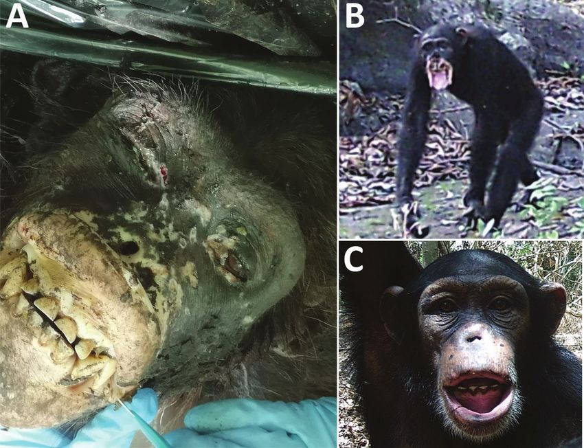
DISPATCHES
Figure 1. Yaws-like lesions
in wild chimpanzees, Guinea.
A) Yaws-like lesions observed
during a necropsy of an adult
female chimpanzee found in the
Sangaredi area, Guinea. B, C)
Camera trap images showing
yaws-like lesions on adult (B) and
juvenile (C) chimpanzees in Haut
Niger National Park, Guinea.
was in visible agony and had to be euthanized; we which is a frequent problem resulting from low num-
performed a necropsy on the body. Gross pathol- bers of bacteria at lesion sites (6).
ogy of the skin revealed a marked depigmentation We extracted DNA from 2 facial lesion biopsies
on hypertrophied edematous lips; crusts and ulcers stored in RNAlater and performed molecular inves-
were present on the head, and much of the nose was tigations (Appendix, https://wwwnc.cdc.gov/EID/
missing. The eyes were shrunken and purulent and article/26/6/19-1713-App1.pdf). High-throughput
surrounded by crusts, and the corneas were opaque. sequencing analysis resulted in a 24-fold average
We preserved samples of lesioned skin in 10% for- coverage of the TPE genome; 98.6% of the genome
malin and RNAlater (Thermo Fisher, https://www. was covered by >1 read and 97.6% by >3 reads.
thermofisher.com). Bayesian Markov chain Monte Carlo analysis of a ge-
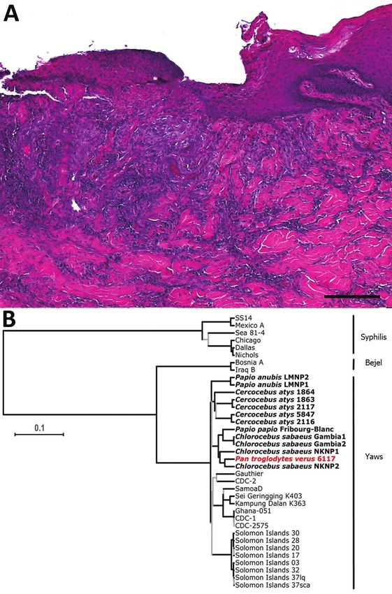
We analyzed formalin-fixed skin samples with nomic alignment comprising this reconstructed TPE
both histological and immunohistochemical meth- genome, all other available TPE and Treponema pal-
ods, as previously described (6). Histopathological lidum subsp. endemicum (TEN, bejel) genomes, and a
features of the skin biopsies were compatible with selection of Treponema pallidum subsp. pallidum (TPA,
treponemal infection (Figure 2, panel A). Skin lesions syphilis) genomes available in Genbank (Appendix
were characterized by irregular epidermal prolifera- Table) revealed that the chimpanzee-derived genome
tion of different extents. The epidermis developed hy- clustered within the well-supported TPE clade, indi-
perkeratosis and hypertrophy of the epidermal rete cating that TPE is responsible for the clinical picture
pegs, which branched and projected deeply into the observed in this particular wild chimpanzee (Figure
corium. Admixed areas with severe superficial ero- 2, panel B). More precisely, this new chimpanzee-
sions or deep ulcerations were visible. A moderate to derived genome belongs to a clade consisting of TPE
severe mixed cell infiltration composed of lympho- strains isolated from NHPs in far western Africa in
cytes and histiocytes was present in the underlying Gambia, Guinea Bissau, Senegal, and Guinea, in
dermal layer. The cellular reaction was most pro- agreement with recent observations that genomic di-
nounced around the dermal blood vessels and hair versity of TPE strains infecting NHPs appears to be
follicles, resulting in superficial and deep perivascu- geographically structured (9; B. Mubemba et al., un-
lar dermatitis. The epidermal surface was covered pub. data, https://doi.org/10.1101/848382). Yaws is
with a dried serosanguineous discharge. Immunohis- principally a skin disease, and it seems likely that the
tochemical analyses failed to visualize treponemes, poor condition of this animal was caused by another
1284 Emerging Infectious Diseases • www.cdc.gov/eid • Vol. 26, No. 6, June 2020Yaws Disease in Wild Chimpanzee, Guinea
unknown but likely traumatic cause, perhaps cou- by camera traps set near the Chimpanzee Conserva-
pled with associated secondary infections, although tion Center in Haut Niger National Park. During 2018–
our field necropsy was not able to identify an alterna- 2019, in 10 different camera trap locations, we observed
tive cause of her cachectic condition. 12 chimpanzees (1 juvenile, 3 subadults, and 8 adults)
To determine whether TPE might affect other with severe lesions. The lesions observed in these im-
chimpanzees in Guinea, we examined videos collected ages closely resembled those of the wild female from
Figure 2. Histopathologic
analysis of yaws-like lesions
in a wild chimpanzee,
Guinea, and phylogenetic
placement of the Treponema
pallidum subspecies pertenue
strain. A) Histopathologic
evidence suggestive of a
treponemal infection. Shown
here is superficial ulcerative
pyogranulomatous dermatitis
including formation of a mixed
inflammatory cell infiltration,
predominantly neutrophil
granulocytes. Deeper dermal
layers show the formation of
a perivascular lymphocytic
inflammatory cell infiltrate, focal
folliculitis, and perifolliculitis.
Skin areas adjacent to ulcerated
parts show irregular epidermal
hyperplasia, consistent with
treponemal infections. The
ulcerated areas were covered
by a serocellular crust. Scale
bar indicates 200 µm. B)
Maximum clade credibility
tree of T. pallidum strain
genomes. Red indicates the
chimpanzee genome generated
in this study. All simian-infecting
strains are shown in bold with
labels showing the species of
nonhuman primate, and the
diseases caused by each type
of bacteria are shown at right.
Branches supported by posterior
probabilitiesDISPATCHES
the Sangaredi region described in this article, includ- References
ing shrunken eyes, deformation of the face, absence of 1. Knauf S, Gogarten JF, Schuenemann VJ, Nys HM De, Düx A,
Strouhal M, et al. Nonhuman primates across sub-Saharan
the nose, and hypertrophied and depigmented lips (in Africa are infected with the yaws bacterium Treponema
1 case, the lips were completely missing; Figure 1, pan- pallidum subsp. pertenue. Emerg Microbes Infect. 2018;7:1–4.
els B, C). Molecular investigations of the pathogen(s) https://doi.org/10.1038/s41426-018-0156-4
causing these infections is clearly warranted, perhaps 2. Fribourg-Blanc A, Mollaret HH, Niel G. Serologic and
microscopic confirmation of treponemosis in Guinea baboons
through noninvasive screening of TPE in feces, bones, [in French]. Bull Soc Pathol Exot Filiales. 1966;59:54–9.
or primate-associated flies (9,12). 3. Fribourg-Blanc A, Mollaret HH. Natural treponematosis of
the African primate. Primates Med. 1969;3:113–21.
Conclusions 4. Zobaníková M, Strouhal M, Mikalová L, Cejková D,
Ambrožová L, Pospíšilová P, et al. Whole genome sequence
This study links yaws-like pathology to the actual of the Treponema Fribourg-Blanc: unspecified simian isolate
detection of TPE in a wild chimpanzee, providing is highly similar to the yaws subspecies. PLoS Negl Trop Dis.
evidence that at least part of the suggestive lesions 2013;7:e2172. https://doi.org/10.1371/journal.pntd.0002172
often observed in wild great apes are caused by this 5. Wallis J, Lee DR. Primate conservation: the prevention of
disease transmission. Int J Primatol. 1999;20:803–26.
pathogen. These data join a growing body of evi- https://doi.org/10.1023/A:1020879700286
dence demonstrating that many NHP species across 6. Knauf S, Batamuzi EK, Mlengeya T, Kilewo M, Lejora IAV,
sub-Saharan Africa are infected with TPE (1,9). This Nordhoff M, et al. Treponema infection associated with
finding could potentially be problematic for the ongo- genital ulceration in wild baboons. Vet Pathol. 2012;49:292–
303. https://doi.org/10.1177/0300985811402839
ing campaign to eradicate TPE globally by 2030 (13), 7. Harper KN, Fyumagwa RD, Hoare R, Wambura PN,
although, clearly, data from TPE-infected humans Coppenhaver DH, Sapolsky RM, et al. Treponema pallidum
in this region are needed to determine whether zoo- infection in the wild baboons of East Africa: distribution
notic transmission of this pathogen occurs. Given the and genetic characterization of the strains responsible.
PLoS One. 2012;7:e50882. https://doi.org/10.1371/
severity of lesions, it is evident that individual ani- journal.pone.0050882
mal fitness is affected. The impact of this disease on 8. Chuma IS, Batamuzi EK, Collins DA, Fyumagwa RD,
NHP populations is unknown but could be assessed Hallmaier-Wacker LK, Kazwala RR, et al. Widespread
through long-term monitoring. Treponema pallidum infection in nonhuman primates,
Tanzania. Emerg Infect Dis. 2018;24:1002–9. https://doi.org/
10.3201/eid2406.180037
Acknowledgments 9. Chuma IS, Roos C, Atickem A, Bohm T, Anthony Collins D,
For their collaboration and help, we thank the Office Grillová L, et al. Strain diversity of Treponema pallidum subsp.
Guinéen des Parcs et Resérves and Haut Niger National pertenue suggests rare interspecies transmission in African
nonhuman primates. Sci Rep. 2019;9:14243. https://doi.org/
Park Authorities. We are also grateful to the Convention 10.1038/s41598-019-50779-9
on International Trade in Endangered Species, Germany 10. Knauf S, Liu H, Harper KN. Treponemal infection in non
and CITES, Guinea for facilitating the import of samples human primates as possible reservoir for human yaws.
from Guinea to Germany. Emerg Infect Dis. 2012;19:2058–60. https://doi.org/10.3201/
eid1912.130863
The Tusk Trust funded the camera trap project and the 11. Gogarten JF, Düx A, Schuenemann VJ, Nowak K, Boesch C,
Wittig RM, et al. Tools for opening new chapters in the book
German Research Foundation Great Ape Health project
of Treponema pallidum evolutionary history. Clin Microbiol
no. LE1813/14-1 funded the laboratory investigations. Infect. 2016;22:916–21. https://doi.org/10.1016/
B.M. was supported through the Robert Koch Institute’s j.cmi.2016.07.027
PhD program, Berlin, Germany. 12. Gogarten JF, Düx A, Mubemba B, Pléh K, Hoffmann C,
Mielke A, et al. Tropical rainforest flies carrying pathogens
form stable associations with social nonhuman primates.
About the Author Mol Ecol. 2019;28:4242–58. https://doi.org/10.1111/
mec.15145
Dr. Mubemba is a PhD student with the Epidemiology 13. Dyson L, Mooring EQ, Holmes A, Tildesley MJ, Marks M.
of Highly Pathogenic Organisms research group at the Insights from quantitative and mathematical modelling on
Robert Koch Institute, Berlin, Germany. Dr. Chanove is the the proposed 2030 goals for Yaws. Gates Open
Res. 2019;3:1576. https://doi.org/10.12688/
veterinarian in charge at the Chimpanzee Conservation gatesopenres.13078.1
Center, Somoria, Faranah, Republic of Guinea. Both Dr.
Mubemba and Dr. Chanove are veterinarians interested in Address for correspondence: Fabian H. Leendertz, Robert Koch
infectious diseases of wildlife with a focus on wild Institute, Epidemiology of Highly Pathogenic Microorganisms,
nonhuman primates. Seestr. 10, Berlin 13353, Germany; email: LeendertzF@rki.de
1286 Emerging Infectious Diseases • www.cdc.gov/eid • Vol. 26, No. 6, June 2020You can also read


