Maximum intensity projection aids in diagnosing acute appendicitis and mobile caecum: A case report and literature review
←
→
Page content transcription
If your browser does not render page correctly, please read the page content below
SA Journal of Radiology
ISSN: (Online) 2078-6778, (Print) 1027-202X
                                                            Page 1 of 4     Case Report

 Maximum intensity projection aids in diagnosing acute
  appendicitis and mobile caecum: A case report and
                   literature review

 Authors:                              Appendicitis is a common childhood condition requiring surgical intervention and delayed
 Kakia A.F. Namugenyi1                 diagnosis can have serious consequences. This report describes the case of a child who
 Ferdinand M. Oompie2
 Kasandji F. Kabambi1                  presented with an acute abdomen and intestinal obstruction. Multidetector (MD) CT
                                       demonstrated a left-sided caecum and an inflamed appendix with a faecolith. Maximum
 Affiliations:                         intensity projection (MIP) post-processing was key in identifying the appendicular artery and
 1
  Department of Surgery,               determine the diagnosis. At surgery, however, a mobile caecum and the appendix were
 Faculty of Health Sciences,
 Walter Sisulu University,
                                       positioned on the right side.
 Mthatha, South Africa
                                       Keywords: maximum intensity projection; multi detector computed tomography; childhood;
                                       acute appendicitis; mobile caecum.
 2
  Department of Radiology,
 Nelson Mandela Academic
 Hospital, Mthatha,
 South Africa                         Introduction
 Corresponding author:                Acute appendicitis is a common condition in childhood, but a left-sided appendicitis related to a
 Kakia Namugenyi,                     mobile caecum is rare.1 Appendicitis is usually diagnosed on the basis of clinical presentation and
 faithkakia@gmail.com                 laboratory results; however, an atypical presentation may pose a diagnostic dilemma. The
                                      position of the appendix varies considerably depending on the position of the caecum.2 In
 Dates:
 Received: 18 Mar. 2021               individuals with situs inversus, midgut malrotation or an unusually long appendix (more than
 Accepted: 11 May 2021                6.8 cm in children), the appendix may be found on the left side of the abdomen.1,3,4 There are a few
 Published: 28 July 2021              case reports of CT scan findings demonstrating a perforated appendix in association with caecal
                                      redundancy.2,5
 How to cite this article:
 Namugenyi KAF, Oompie FM,
 Kabambi KF. Maximum
 intensity projection aids in
                                      Patient presentation
 diagnosing acute appendicitis        A 12-year-old boy presented with generalised abdominal pain, abdominal distension and
 and mobile caecum: A case            vomiting for 1 week. There was history of intermittent abdominal pain with constipation, which
 report and literature review.
                                      was managed at a nearby primary healthcare unit. There were no known comorbidities and no
 S Afr J Rad. 2021;25(1),
 a2153. https://doi.                  history of previous surgery.
 org/10.4102/sajr.v25i1.2153
                                      On clinical examination, the child was ill looking and febrile (38 °C) but fully conscious. The abdomen
 Copyright:
                                      was distended with generalised tenderness and guarding. Biochemistry was normal except for a
 © 2021. The Authors.
 Licensee: AOSIS. This work           raised white cell count of 15 500 mm3 (normal range: 5000 mm3 – 10 000 mm3).
 is licensed under the
 Creative Commons                     Abdominal ultrasound demonstrated interloop free fluid and tenderness on probe compression. The
 Attribution License.                 appendix could not be identified because of overlying bowel gas in the distended bowel loops. Post-
                                      contrast-enhanced abdominal CT scan showed distended small bowel loops with multiple pelvic
                                      collections. The caecum was left sided (Figure 1a–c) with a 50 mm diameter collection in the left iliac
                                      fossa associated with a 7-mm long dense sausage-shaped structure, presumed to be the appendix
                                      with an appendicolith (Figure 2a and b). Differential diagnosis included a left-sided appendix with a
                                      faecolith, a foreign body complicated by perforated small bowel, infected mesenteric or duplication
                                      cyst and a Meckel’s diverticulitis with a stone. The radiologist sought to follow the appendicular
                                      artery, a branch of the ileocolic artery from the superior mesenteric artery (SMA) using maximum
                                      intensity projection (MIP) post-processing on a Philips IntelliSpace workstation (Philips Healthcare
                                      Netherlands B.V. Veenpluis 8.0, the Netherlands). The distal SMA was seen coursing towards the left
                                      in the region where the high-density sausage-shaped structure was seen (Figure 3). Interloop and
     Read online:                     right paracolic gutter fluid was present. A radiological diagnosis of a ruptured left-sided acute
                    Scan this QR      appendicitis probably due to a mobile caecum or an unusually long appendix was made.
                    code with your
                    smart phone or
                    mobile device     Differentials remained as given here. There were no features of midgut malrotation or situs
                    to read online.
                                      inversus. The referring surgeons were informed and an emergency laparotomy was performed.

                                                 http://www.sajr.org.za     Open Access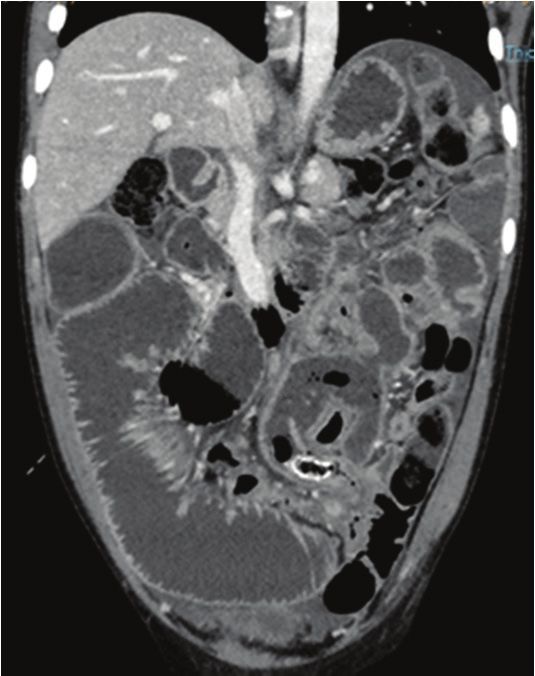
Page 2 of 4         Case Report

                                                 a                                                     b                                                     c

FIGURE 1: (a-c) (cephalad to caudad): A 12-year-old boy presented with abdominal distension, vomiting and a raised white cell count. Axial CT revealed
the abnormal caecal position (blue star) in the left lower quadrant with an anterior cephalad thickened appendix containing an appendicolith (arrow) and adjacent
fat stranding.

                                                                                          a                                                                 b

FIGURE 2: (a and b) Axial and coronal CT images demonstrating appendiceal wall thickening and enhancement with a faecolith (blue arrows) anterior and
cephalad to the caecum (star). The right iliac fossa was unremarkable with fluid in the paracolic gutter (red arrow). Note the liver (red circle) is in the normal
anatomic position.

The surgeons performed a midline incision extending from
epigastrium to the suprapubic region. Multiple dilated
                                                                                     Discussion
friable small bowel loops were found. Accidental iatrogenic                          A mobile caecum is a congenital abnormality with a defective
                                                                                     right colonic mesenteric attachment at the lateral peritoneum
rupture of small bowel was primarily repaired. The dilated
                                                                                     because of agenesis of the caecal mesocolon.6 The estimated
loops were decompressed and the caecum was found on a
                                                                                     prevalence of a mobile caecum and ascending colon is
free mesentery on the right side.
                                                                                     10% – 20%.2,7,8 During embryological development, a mobile
                                                                                     caecum arises from failure of fusion of the colonic mesentery
The appendix was inflamed with an appendicolith at the
                                                                                     with the posterolateral peritoneum. This results in a free
base and a ruptured gangrenous tip. Appendectomy was                                 mesentery of the caecum and occasionally the right colon,
performed and the specimen was sent for histology. Interloop                         allowing them to freely move to any part of the abdominal
collections were drained followed by peritoneal lavage.                              cavity. Occasionally, the caecum may rotate causing volvulus
There were no adhesions. The patient was discharged after                            but most of the time it remains in the normal anatomic position.7
four days later, following an uneventful recovery.
                                                                                     Mobile caecum and right colon are commonly present
Histopathology reported a 7 cm × 3 cm appendix with an                               in children as mobile caecal syndrome.9 Patients with
occluded lumen and a faecolith.                                                      this syndrome may present with chronic intermittent

                                                     http://www.sajr.org.za          Open Access
Page 3 of 4 Case Report
The differential of a left-sided acute appendicitis may not be
promptly established in the emergency setting and is often
delayed because of atypical clinical signs.1 Delayed diagnosis
may result in complications such as perforation, abscess
formation, peritonitis, sepsis, bowel obstruction, infertility
and death.13 Chest and abdominal radiographs may be
helpful in excluding situs inversus with identification of
the heart and gastric bubble in the correct anatomical
position. Midgut malrotation may need contrasted upper
gastrointestinal studies or cross-sectional imaging. Graded
compression ultrasound plays a vital role in the diagnosis of
acute appendicitis, especially in children. However, in the
presence of bowel obstruction, this modality may be limited
by bowel gas. Multidetector CT is the gold standard for
imaging acute appendicitis.14 By following the intestinal
segments sequentially from the stomach to the anus or vice
versa, one is able to demonstrate the location of the caecum
and ascending colon.
Multidetector CT also has the capability to assess the
vasculature.5,7 One limitation of MDCT in acute appendicitis
is the overlapping range in maximal appendiceal diameter
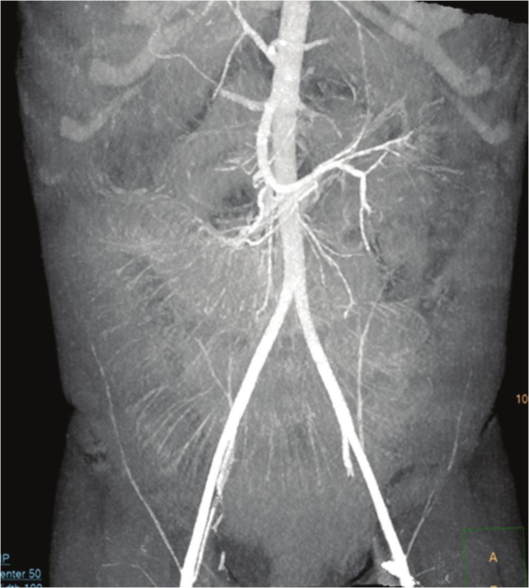
FIGURE 3: Coronal reconstructed maximum intensity projection image between inflamed and non-inflamed appendices. The
demonstrating the superior mesenteric artery (blue arrow) and its branches
including the ileocolic (black arrow) from which the appendicular artery arises. presence of fat stranding, abscesses and an appendicolith are
The vessels are seen coursing to the left, corresponding to the position of the valuable ancillary findings in confirming the diagnosis. In
caecum and appendicolith. This is in contrast to the normal course towards the
right lower quadrant. children with minimal abdominal fat, pericaecal fat
stranding may not be present, limiting confidence in a
abdominal pain, distension, flatulence, dyspareunia, caecal definitive diagnosis. The current case demonstrated a left-
volvulus and partial or complete bowel obstruction.7 sided caecum and faecolith associated with an abscess in
the left iliac fossa (see Figure 1a–c). The presence of
Left-sided acute appendicitis may be difficult to multiple dilated bowel loops and collapsed large bowel
differentiate from an infected duplication, mesenteric or with interloop collections provided challenges in tracing
urachal cyst or a Meckel’s diverticulitis.10 Meckel’s the bowel. The MIP was very useful in helping to visualise
diverticulum is the most common congenital anomaly of the course of the appendicular vessels. The SMA was
the gastrointestinal tract, present in 2% of the population. followed to the ileocolic and the appendicular artery
It is usually asymptomatic, but 4% – 40% of people with (Figure 3). These vessels normally course towards the right
Meckel’s diverticulum may experience complications of iliac fossa. However, in our patient they deviated to the left
diverticulitis, haemorrhage, intussusception, small-bowel where an abscess and the appendicolith were seen. This,
obstruction, stone formation or neoplasm. A urachal cyst combined with the left-sided caecum improved confidence
may become infected and present with abdominal pain, in diagnosing appendicitis.
fever, nausea, vomiting, dysuria, voiding difficulty,
epididymitis and orchitis. Maximum intensity projection software is available on
most radiology workstations in South Africa. The MIP
Mesenteric cysts have an incidence of less than 1:100 000 provides improved display of vascular maps, improving
cases in the general population. They may be located in the visualisation of most segments of the vessel including the
mesentery, peritoneum or retroperitoneum. Most are intraparenchymal branches.15 Source images are used to
benign but there is a 3% incidence of malignancy.11 Patients display the maximum intensities in the voxels with
with symptomatic mesenteric cysts present with abdominal selection of optimum slab thickness depending on the
pain and distension with cysts rarely becoming infected. orientation of the vessels and the density of the adjacent
Duplication cysts of the gastrointestinal system are rare and structures. As MIP is not a 3D volume-rendered application,
often associated with vertebral defects, anal atresia, one of the pitfalls is the false interpretation of the
cardiac defects, tracheo-esophageal fistula, renal anomalies relationship of the vessels and adjacent structures in the
and limb abnormalities (VACTERL anomalies), especially presence of high-intensity structures.15,16,17 This necessitates
imperforate anus and hemivertebrae. If symptomatic, the user to adjust the display parameters to include the
they present with abdominal distension, gastrointestinal degree of opacification and slab thickness that correctly
obstruction or obstipation and caecal duplications, resulting depicts the vasculature. The MIP requires substantial editing
in intussusception.12 in most cases to clearly depict the anatomical display of the
http://www.sajr.org.za Open AccessPage 4 of 4 Case Report
vessels. The quality of the final product may vary according Data availability
to the experience of the user and the vendor.15
Original images, consent form and any required information
are available on request from the corresponding author
Conclusion (K.A.F.N.).
This case report highlights MIP as a diagnostic problem-
solving tool in the atypical presentation of a mobile caecum. Disclaimer
Familiarity with this post-processing software is useful for
The views and opinions expressed in this article are those of
advanced imaging application in clinical practice. This case
the authors and not an official position of the institution.
report also increases awareness of the mobile caecum as an
anatomic variant, which may result in an atypical presentation
of acute appendicitis. References
1. Akbulut S, Ulku A, Senol A, Tas M, Yagmur Y. Left-sided appendicitis: Review of 95
Acknowledgements published cases and a case report. World J Gastroenterol. 2010;16(44):5598.
https://doi.org/10.3748/wjg.v16.i44.5598
2. Rogers RL, Harford FJ. Mobile cecum syndrome. Dis Colon Rectum. 1984;27(6):
The authors would like to acknowledge C. Naidoo and 399–402. https://doi.org/10.1007/BF02553011
P. Kobo who diagnosed the patient at the Radiology 3. Yang CY, Liu HY, Lin HL, Lin JN. Left-sided acute appendicitis: A pitfall in the
emergency department. J Emerg Med. 2012;43(6):980–982. https://doi.
Department and L.T. Mtshabe and S. Mduna who operated org/10.1016/j.jemermed.2010.11.056
on the patient (Division of General Surgery). They are also 4. Alzaraa A, Chaudhry S. An unusually long appendix in a child: A case report. Cases
J. 2009;2(1):7398. https://doi.org/10.4076/1757-1626-2-7398
grateful to Z. Njumba and A. Ndabankulu who followed
5. Yazawa K, Azuma Y, Kurokawa T, Yoshioka Y, Tsurita G, Shinozaki M. Abdominal CT-
up with the patient for consent. aided diagnosis of acute appendicitis in the presence of mobile cecum: A case report.
Int J Surg Case Rep. 2018;42:258–260. https://doi.org/10.1016/j.ijscr.2017.12.035
6. Garude K, Rao S. Mobile cecum: An incidental finding. Indian J Surg.
Competing interests 2013;75(4):265–267. https://doi.org/10.1007/s12262-012-0529-1
7. Lee YJ, Lee YA, Liu TJ, Chang TH. Mobile cecum syndrome: A report of two cases.
The authors declare that they have no financial or personal Zhonghua yi xue za zhi = (Taipei). Chin Med J; Free China ed. 1996;57(5):380.
relationships that may have inappropriately influenced them 8. Ingelfinger FJ. Intermittent volvulus of the mobile cecum. Arch Surg.
1942;45(1):156–163. https://doi.org/10.1001/archsurg.1942.01220010159012
in writing this article.
9. Printen KJ. Mobile cecal syndrome in the adult. Am Surg. 1976;42(3):204–205.
10. Wong CH, Trinh TM, Robbins AN, Rowen SJ, Cohen AJ. Diagnosis of appendicitis:
Authors’ contributions Imaging findings in patients with atypical clinical features. Am J Roentgenol.
1993;161(6):1199–1203. https://doi.org/10.2214/ajr.161.6.8249725
K.A.F.N. drafted and wrote the article. F.M.O. and K.F.K. 11. Kurtz RJ, Heimann TM, Holt JA, Beck AR. Mesenteric and retroperitoneal cysts. Ann
Surg. 1986;203(1):109. https://doi.org/10.1097/00000658-198601000-00017
critically revised the article with important conceptual and
12. Blickman JG, Rieu PH, Buonomo C, Hoogeveen YL, Boetes C. Colonic duplications:
editorial input. Clinical presentation and radiologic features of five cases. Eur J Radiol.
2006;59(1):14–19. https://doi.org/10.1016/j.ejrad.2006.03.012
13. Sivit CJ, Applegate KE. Imaging of acute appendicitis in children. Semin Ultrasound
Ethical considerations CT MRI. 2003;24(2):74–82. https://doi.org/10.1016/S0887-2171(03)90003-5
14. Toprak H, Bilgin M, Atay M, Kocakoc E. Diagnosis of appendicitis in patients with
Ethical approval to conduct the study was obtained from the abnormal position of the appendix due to mobile caecum. Case Rep Surg.
2012;2012:921382. https://doi.org/10.1155/2012/921382
Walter Sisulu University’s Ethics Committee (protocol
15. Fishman EK, Ney DR, Heath DG, Corl FM, Horton KM, Johnson PT. Volume rendering
number 002/2021). versus maximum intensity projection in CT angiography: What works best, when, and
why. Radiographics. 2006;26(3):905–922. https://doi.org/10.1148/rg.263055186
16. Rubin GD, Dake MD, Napel S, et al. Spiral CT of renal artery stenosis: Comparison
Funding information of three-dimensional rendering techniques. Radiology. 1994;190(1):181–189.
https://doi.org/10.1148/radiology.190.1.8259402
This work received no specific grant from any funding 17. Dalrymple NC, Prasad SR, Freckleton MW, Chintapalli KN. Introduction to the
language of three-dimensional imaging with multidetector CT. Radiographics.
agency in the public, commercial or not-for-profit sectors. 2005;25(5):1409–1428. https://doi.org/10.1148/rg.255055044
http://www.sajr.org.za Open AccessYou can also read