Successful re-entry using the outback elite catheter via retrograde popliteal access with IVUS guidance for femoropopliteal occlusion: a case ...
←
→
Page content transcription
If your browser does not render page correctly, please read the page content below
Hayakawa et al. CVIR Endovascular      (2020) 3:63
https://doi.org/10.1186/s42155-020-00156-9
                                                                                                                                   CVIR Endovascular

 CASE REPORT                                                                                                                                        Open Access

Successful re-entry using the outback® elite
catheter via retrograde popliteal access
with IVUS guidance for femoropopliteal
occlusion: a case report
Naoki Hayakawa1* , Satoshi Kodera2, Masataka Arakawa1 and Junji Kanda1

  Abstract
  Background: There are still cases that are difficult to treat for femoropopliteal chronic total occlusion (CTO). The
  Outback® Elite catheter is effective re-entry device to treat such kind of difficult cases, however, it might be difficult
  to use the Outback® Elite catheter antegradely in cases with severely calcified lesions. In this case, we performed
  EVT using the Outback Elite® catheter via the retrograde popliteal approach.
  Case presentation: We report a case of a 77-year-old male with end-stage renal disease who presented with pain and
  cyanosis of his left foot. Control angiography showed total occlusion from the middle of the left superficial femoral artery to
  the proximal portion of the popliteal artery. The CTO lesion was severely calcified, which prevented the
  antegrade advancement of any guidewire. Retrograde popliteal puncture was performed with the patient in
  the supine position. After intentional retrograde subintimal wiring, the Outback® Elite catheter was advanced
  via the retrograde approach after the identification of a suitable re-entry site using intravascular ultrasound.
  After wire crossing, one nitinol stent was deployed and sufficient antegrade flow was achieved without any
  complications.
  Conclusions: Using Outback® Elite from retrograde should be considered in cases where antegrade
  advancement fails and bidirectional wiring cannot pass through the CTO lesion.
  Keywords: Endovascular therapy, Outback, Retrograde approach, Chronic total occlusion

Background                                                                             high success rate with the Outback® Elite catheter, they
The success rate of endovascular treatment (EVT) for                                   also reported that severe calcification caused many cases
chronic total occlusion (CTO) of the superficial femoral                               of treatment failure (Kitrou et al. 2015). Similarly, Shin
artery (SFA) has improved due to the development of re-                                et al. reported that substantial calcification at the proposed
entry devices and CTO crossing devices and the retro-                                  re-entry site is a strong predictor of recanalization failure
grade approach (Schneider 2017; Soga et al. 2018; Schmidt                              (Shin et al. 2011).
et al. 2012). However, certain lesions remain challenging                                Herein, we report a case in which SFA CTO with se-
to treat. One re-entry device that is simple to use and ef-                            vere calcification was successfully recanalized via the use
fective is the Outback® Elite catheter (Cordis, Florida,                               of intravascular ultrasound (IVUS) to identify a portion
USA). However, although Kitrou et al. reported a very                                  with relatively little calcification at which retrograde re-
                                                                                       entry with the Outback® Elite catheter was possible. To
* Correspondence: haya.naoki1981@gmail.com                                             the best of our knowledge, this is the first report of the
1
 Department of Cardiovascular Medicine, Asahi General Hospital, Asahi                  successful use of the Outback® Elite catheter via the
General Hospital, I-1326 Asahi, Chiba 289-2511, Japan
Full list of author information is available at the end of the article                 retrograde approach under IVUS guidance.

                                       © The Author(s). 2020 Open Access This article is licensed under a Creative Commons Attribution 4.0 International License,
                                       which permits use, sharing, adaptation, distribution and reproduction in any medium or format, as long as you give
                                       appropriate credit to the original author(s) and the source, provide a link to the Creative Commons licence, and indicate if
                                       changes were made. The images or other third party material in this article are included in the article's Creative Commons
                                       licence, unless indicated otherwise in a credit line to the material. If material is not included in the article's Creative Commons
                                       licence and your intended use is not permitted by statutory regulation or exceeds the permitted use, you will need to obtain
                                       permission directly from the copyright holder. To view a copy of this licence, visit http://creativecommons.org/licenses/by/4.0/.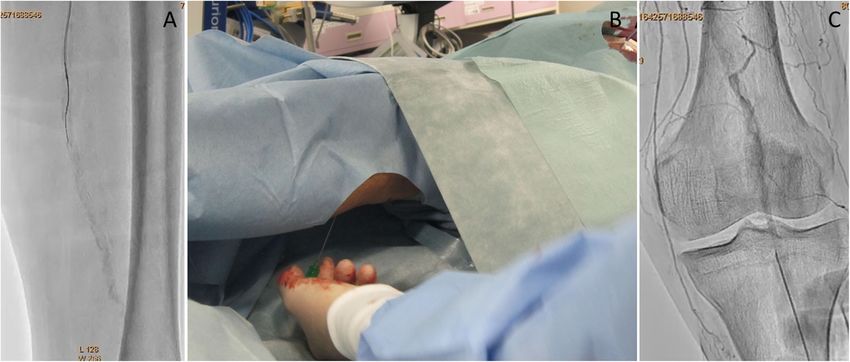
Hayakawa et al. CVIR Endovascular         (2020) 3:63                                                                                       Page 2 of 6

Case report                                                                   to advance a 0.014-in. Jupiter T45® guidewire with a 45 g
A 77-year-old man with progressive pain at rest and cyan-                     tapered wire tip (Boston Scientific) inside the CTO, but
osis of his left lower limb was referred to our department                    its progress was hindered by severe calcification and it
for revascularization. At presentation, the patient had                       could not be advanced beyond the distal SFA (Fig. 2a). A
end-stage renal disease, diabetic nephropathy, hyperten-                      CROSSER® 14S microcatheter (Bard, Tempe, AZ) with a
sion, dyslipidemia, peripheral artery disease, and chronic                    small balloon was also unable to pass through the lesion,
heart failure due to severe coronary artery disease. The                      and a 0.035-in. knuckle-shaped wire was unable to
ankle-brachial index was 0.48 on the right side and 0.35                      proceed at all. Retrograde popliteal puncture was then
on the left. A vascular surgeon treated the left common                       performed with the patient in the supine position (Fig.
femoral artery occlusion via endoatherectomy. After con-                      2b, c). The middle of the popliteal artery (P2 segment)
sultation with the Department of Vascular Surgery, EVT                        was punctured with a micropuncture kit (Cook, Tokyo,
was selected as the treatment method because there was                        Japan) under angiographic guidance. After successful
no graftable vein and bypass was likely to be difficult due                   puncture, a 0.014-in. Cruise® guidewire (Asahi Intec) was
to the poor quality of the distal run-off vessels.                            advanced into the popliteal artery, and a 2.6-F Corsair
   A Parent Plus60® guiding sheath (Medikit, Tokyo,                           Armet® microcatheter (Asahi Intec) was introduced to
Japan) was inserted into the left common femoral artery                       support the guidewire using a sheathless technique. A
via the ipsilateral antegrade approach. Control angiog-                       0.014-in. Jupiter MAX® guidewire with a 100 g tip load
raphy showed severe stenosis of the proximal SFA and                          (Boston Scientific) was introduced via the retrograde ap-
total occlusion with severe calcification from the middle                     proach. However, the severe calcification prevented it
of the SFA to the proximal popliteal artery (Fig. 1a, b).                     from advancing to the true lumen. Thus, we exchanged
Furthermore, the popliteal artery was severely stenosed,                      Corsair Armet® microcatheter to 6-Fr sheath. And the
and the below-the knee vessels were totally occluded                          guidewire was replaced by a 0.035-in. Radifocus wire,
(Fig. 1c, d). A 0.014-in. Jupiter FC® guidewire (Boston                       which was successfully advanced into the CTO lesion by
Scientific, Tokyo, Japan) was initially advanced to the                       knuckle wire technique (Fig. 3a). IVUS showed that the
site of the CTO, and the proximal stenotic lesion was di-                     retrograde wire was in the subintimal space and that the
lated using a 4.0 × 15 mm Peripheral Cutting Balloon®                         vessel walls were hardened by severe calcification, sug-
(Boston Scientific). A 2.6-F Corsair Armet® microcath-                        gesting that the CTO lesion would be extremely difficult
eter (Asahi Intec, Aichi, Japan) and Guidezilla2 PV®                          to negotiate with a guidewire or the controlled antegrade
guide extension catheter (Boston Scientific) were then                        and retrograde subintimal tracking (CART) technique.
inserted to achieve stronger backup force. We managed                         An attempt to pass a hard guidewire through the lesion

 Fig. 1 Control angiography. a Digital subtraction angiography showing severe stenosis with severe calcification in the left proximal superficial
 femoral artery. b Digital angiography of the middle to distal part of the left superficial femoral artery showing chronic total occlusion with severe
 calcification. c: Digital subtraction angiography of the distal superficial femoral artery to the proximal popliteal artery showing severe tandem
 stenosis with severe calcification. d Digital subtraction angiography of the below-the-knee lesions showing the total occlusion of three vessels

Hayakawa et al. CVIR Endovascular          (2020) 3:63                                                                                           Page 3 of 6

 Fig. 2 a Antegrade wiring with heavy weight 0.014-in. tapered wire. Severe calcification prevents the advancement of antegrade wire into the lesion. b
 Retrograde popliteal puncture with the patient in the supine position. c: Puncture of the middle part of the popliteal artery under angiographic guidance

via the retrograde approach under IVUS guidance via                             expansion and sufficient antegrade flow (Fig. 4c, d, e, f).
the antegrade approach was unsuccessful. Therefore, it                          There were no major dissections and/or vessel perfor-
was decided that re-entry would be attempted using an                           ation. The patient’s symptoms resolved immediately
Outback® Elite catheter via the retrograde approach. The                        after the procedure, and there were no complications.
retrograde wire route was then dilated using a 3.0 × 40                         The pain at rest was markedly improved, but mild pain
mm Bellona® balloon (Medicos Hirata, Osaka, Japan) to                           at rest remained. The ankle-brachial index improved to
enable the advancement of the Outback® Elite catheter                           1.4 and the pain at rest was completely resolved after
(Fig. 3b). The Outback® Elite catheter was advanced to                          the performance of additional EVT 1 month later for the
the proximal subintimal space adjacent to the recon-                            below-the-knee lesions.
structed area of the proximal true lumen where there
were relatively few calcified parts seen on antegrade                           Discussion
IVUS (Fig. 3c, d, e, f). Two orthogonal angiographic                            The treatment of TASC C and D femoropopliteal occlu-
views were obtained to determine the best direction for                         sion has become feasible with the development of various
the puncture (Fig. 3g, h). IVUS was inserted via the ante-                      EVT techniques and devices (Kitrou et al. 2015; Shin et al.
grade approach, and the position was adjusted so that                           2011; Urasawa et al. 2014; Kawasaki et al. 2008; Tan et al.
the Outback® Elite catheter needle entered the true                             2017; Bolia et al. 1990); for example, CTO can be crossed
lumen in which the IVUS transducer was located. A 22G                           via the intentional subintimal approach using the loop
re-entry cannula was inserted into the proximal true                            wire technique (Bolia et al. 1990). A high procedural suc-
lumen in the middle of the SFA. A 0.014-in. Chevalier                           cess rate is achieved with re-entry devices such as the
Universal® guidewire (Cordis, Florida, USA) was success-                        Outback® Elite catheter (Schneider 2017). However, it is
fully advanced into the true lumen and into the                                 difficult to use the antegrade subintimal approach in cases
antegrade guiding sheath (Fig. 3i, j). After wire                               with severely calcified lesions. In such cases, it is often dif-
externalization, the Outback® Elite catheter was removed                        ficult to perform re-entry even via the retrograde ap-
and the lesion was dilated using a 4.0 × 220 mm Coyote®                         proach. Bypass surgery is a possible solution in such
balloon (Boston Scientific). Next, a 5.0 × 220 mm Coy-                          situations, as it is technically simple and achieves good
ote® balloon (Boston Scientific) was dilated over a 10-                         long-term patency. However, in the present case, bypass
min period to achieve intravascular hemostasis of the                           was likely to be difficult due to the poor distal run-off ves-
popliteal puncture site (Fig. 4a). After confirmation of                        sels and the absence of graftable veins.
good hemostasis, a 6.0 × 150 mm INNOVA® stent (Bos-                                In the present case, the severe calcification of the le-
ton Scientific) was deployed in the SFA lesion (Fig. 4b).                       sion made it difficult to obtain re-entry using standard
Post-dilatation of the whole SFA lesion was performed                           bidirectional wiring. This difficult situation was over-
using a 6.0 × 150 mm SHIDEN HP® balloon (Kaneka,                                come with the use of the Outback® Elite catheter via the
Tokyo, Japan). Final angiography showed appropriate                             retrograde approach. Although re-entry devices such as
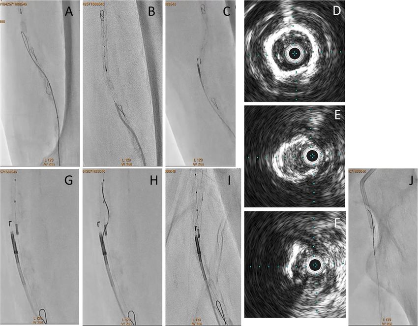
Hayakawa et al. CVIR Endovascular           (2020) 3:63                                                                                            Page 4 of 6

 Fig. 3 a: Advancement of knuckle-shaped 0.035-in. Radifocus wire. b: Both wires are closed. Antegrade intravascular ultrasound showing that the
 antegrade wire is in the intraplaque space and the retrograde wire is in the subintimal space. The retrograde route is dilated with a 3.0-mm balloon to
 enable the advancement of the Outback® Elite catheter (Cordis, Florida, USA). c: The Outback® Elite catheter is advanced retrogradely under intravascular
 ultrasound guidance from the antegrade direction. d: The IVUS findings of proximal SFA showed 360 degree heavy calcification. e: The IVUS findings
 showed 180 to 270 degree calcification. f: The IVUS findings from retrograde showed the retrograde IVUS catheter was in the subintimal space, and true
 lumen was relatively few calcified parts. This was the place where we tried out re-entry from retrograde using Outback® Elite catheter. g: Use of the
 Outback® Elite catheter. Adjustment of the L marker. h: Adjustment of the T marker. i: Successful re-entry. j: Advancement of the retrograde wire into the
 antegrade guiding sheath

the Outback® Elite catheter were originally used via the                         subintimally, as in the present case. In such cases, it is
antegrade approach, Kim et al. (2013) reported success-                          difficult to negotiate both wires, even with a bidirec-
ful retrograde re-entry using the Outback LTD catheter                           tional approach. In this situation, procedural success
for an aortoiliac lesion, and Patrone and Stehno (2019)                          may only be possible by using a re-entry device to enter
reported the retrograde insertion of the Outback® reen-                          the proximal true lumen via the retrograde approach.
try device via a tibial artery for infrainguinal recanaliza-                     When a re-entry device is applied via the contralateral
tion. However, to the best of our knowledge, there have                          approach, an acute aortic bifurcation angle is reportedly
been no reported cases in which the Outback® Elite cath-                         a predictor of procedural failure (Shin et al. 2011). In
eter has been used via the retrograde approach to treat                          such cases, the retrograde popliteal approach may be
femoropopliteal lesions. We usually consider the use of                          effective, as it uses the ipsilateral approach.
re-entry devices when the antegrade wire enters the                                Many CTO cases with re-entry failure are unsuccessful
subintimal space and cannot be returned to the distal                            due to the presence of marked calcification (Schneider
true lumen. However, there are some severe cases in                              2017). In the present case, IVUS via the antegrade ap-
which the antegrade wire cannot be advanced even                                 proach was very helpful in identifying potential re-entry
Hayakawa et al. CVIR Endovascular           (2020) 3:63                                                                                            Page 5 of 6

 Fig. 4 a: Dilation of a long balloon from the distal superficial femoral artery to the popliteal artery with hemostasis of the retrograde puncture site.
 b: Deployment of a bare nitinol stent at the site of chronic total occlusion. c: Digital subtraction angiography of the proximal superficial femoral artery.
 d: Digital subtraction angiography of the middle to distal superficial femoral artery. e: Digital subtraction angiography of the proximal popliteal artery.
 e: Digital subtraction angiography of the bellow the ankle artery

points for the Outback® Elite catheter via the retrograde                        2017). In contrast, some studies have reported the suc-
approach. Although angiography suggested that the prox-                          cess of the subintimal approach (Ishihara et al. 2016). In
imal true lumen was markedly calcified over the entire                           the present case, although the IVUS had passed through
length of the obstruction, IVUS was used to identify a por-                      most of the CTO via the subintimal route, a sufficient
tion with relatively mild calcification. The IVUS catheter                       minimal stent area was obtained by placing the stent
was also a good landmark for the Outback® Elite catheter                         after sufficient pre-dilatation and firm post-dilatation.
from the retrograde approach. Re-entry devices such as                           The SUPERA® stent (Abbott Vascular, USA) is report-
the Pioneer catheter use IVUS guidance to identify the                           edly useful for subintimal recanalization (Palena et al.
puncture point; however, with the Outback® Elite catheter,                       2017), but was not yet available in our institute at that
the puncture point is usually determined based on the LT                         time; this stent may be considered for use in the future.
marker and the angiography findings (Scheinert et al.                              The technique described in the present case has some
2005). Such use of the Outback® Elite catheter via the                           limitations. The approach site is limited because a sheath
retrograde approach under antegrade IVUS guidance to                             of 6Fr or more must be inserted from the retrograde ap-
identify an appropriate puncture point may increase the                          proach. Thus, the distal puncture site must be carefully
success rate of the Outback® Elite catheter in complex                           examined. In addition, balloon dilatation may be neces-
cases. The efficacy of several image-guided CTO crossing                         sary to retrieve the Outback Elite® catheter from the
devices has been reported (Cawich et al. 2014; Jacobs et al.                     retrograde direction, and care must be taken to avoid
2006), but the use of such devices was not approved when                         complications such as vascular perforation. Previous
the present patient was treate.                                                  studies have suggested that re-entry devices including
  For femoropopliteal CTO, the likelihood of restenosis                          the Outback Elite® catheter may be unsuccessful in se-
in the remote phase increases in tandem with the length                          verely calcified lesions (Kitrou et al. 2015; Shin et al.
passed through by the subintimal route (Mori et al.                              2011). The present patient achieved good short-term
Hayakawa et al. CVIR Endovascular               (2020) 3:63                                                                                                    Page 6 of 6

outcomes; however, the long-term outcomes remain un-                                       implantation for femoropopliteal chronic total occlusion. J Endovasc Ther 23:
clear. Further follow-up is needed to assess the long-                                     889–895
                                                                                      Jacobs DL, Motaganahalli RL, Cox DE et al (2006) True lumen re-entry devices
term outcomes.                                                                             facilitate subintimal angioplasty and stenting of total chronic occlusions: initial
                                                                                           report. J Vasc Surg 43:1291–1296. https://doi.org/10.1016/j.jvs.2006.02.051
Conclusions                                                                           Kawasaki D, Tsujino T, Fujii K et al (2008) Novel use of ultrasound guidance for
                                                                                           recanalization of iliac, femoral, and popliteal arteries. Catheter Cardiovasc
We successfully performed EVT using the Outback Elite®                                     Interv 71:727–733
catheter via the retrograde popliteal approach with IVUS                              Kim TH, Ahn JH, Kim DH (2013) A successful retrograde re-entry at aorta using
guidance for severely calcified femoropopliteal CTO. This                                  the outback LTD catheter for a bilateral common iliac artery occlusion.
                                                                                           Catheter Cardiovasc Interv 81:E250–E254. https://doi.org/10.1002/ccd.24506
technique should be considered in cases where EVT is un-                              Kitrou P, Parthipun A, Diamantopoulos A et al (2015) Targeted true lumen re-
successful via the antegrade approach and the lesion can-                                  entry with the Outback catheter: accuracy, success, and complications in 100
not be passed even using bidirectional wiring.                                             peripheral chronic total occlusions and systematic review of the literature. J
                                                                                           Endovasc Ther 22:538–545
Abbreviations                                                                         Mori S, Hirano K, Ito Y et al (2017) Initial and 3-year results after subintimal versus
CTO: Chronic total occlusion; EVT: Endovascular therapy; SFA: Superficial                  intraluminal approach for long femoropopliteal occlusion treated with a self-
femoral artery; IVUS: Intravascular ultrasound; ABI: Ankle-brachial index                  expandable nitinol stent. J Atheroscler Thromb 24:477–486
                                                                                      Palena LM, Diaz-Sandoval LJ, Sultato E et al (2017) Feasibility and 1-year
                                                                                           outcomes of subintimal revascularization with supera® stenting of long
Acknowledgments
                                                                                           femoropopliteal occlusions in critical limb ischemia: the "Supersub" study.
We thank Kelly Zammit, BVSc, from Edanz Editing (www.edanzediting.com/
                                                                                           Catheter Cardiovasc Interv 89:910–920. https://doi.org/10.1002/ccd.26863
ac), for editing a draft of this manuscript.
                                                                                      Patrone L, Stehno O (2019) Retrograde insertion of the outback reentry device
                                                                                           from a tibial artery for complex infrainguinal recanalization. CVIR Endovasc 2:
Conflict of interest
                                                                                           47. https://doi.org/10.1186/s42155-019-0088-7
The authors declare that there is no conflict of interest regarding the
                                                                                      Scheinert D, Bräunlich S, Scheinert S et al (2005) Initial clinical experience with an
publication of this article.
                                                                                           IVUS-guided transmembrane puncture device to facilitate recanalization of
                                                                                           total femoral artery occlusions. EuroIntervention 1:115–119
Authors’ contributions
                                                                                      Schmidt A, Bausback Y, Piorkowski M et al (2012) Retrograde recanalization
NH, MA: performed the procedure and performed pre and post procedure
                                                                                           technique for use after failed antegrade angioplasty for chronic superficial
follow-up. SK: drafted the manuscript and revised it critically for important in-
                                                                                           femoral artery occlusion. J Endovasc Ther 19:23–29
tellectual content. JK: gave final approval for the submitted manuscript. The
                                                                                      Schneider PA (2017) Evolution and current use of technology for superficial
authors read and approved the final manuscript.
                                                                                           femoral and popliteal artery interventions for claudication. J Vasc Surg 66:
                                                                                           916–923
Funding
                                                                                      Shin SH, Baril D, Rhee R et al (2011) Limitations of the outback LTD re-entry
No specific grants from any funding agency in the public, commercial, or
                                                                                           device in femoropopliteal chronic total occlusions. J Vasc Surg 53:1260–1264
not-for-profit sectors were received for this study.
                                                                                      Soga Y, Nakamura M, Hirose K et al (2018) Primary use of the TruePath crossing
                                                                                           device for infrainguinal chronic total occlusions with intravascular ultrasound
Availability of data and materials
                                                                                           evaluation. J Endovasc Ther 25:592–598
The datasets used and/or analysed during the current study are available
                                                                                      Tan M, Urasawa K, Koshida R et al (2017) Anterolateral popliteal puncture
from the corresponding author on reasonable request.
                                                                                           technique: a novel retrograde approach for chronic femoropopliteal
                                                                                           occlusions. J Endovasc Ther 24:525–530
Ethics approval and consent to participate
                                                                                      Urasawa K, Sato K, Koshida R et al (2014) Trans-collateral angioplasty for the
All procedures were performed in accordance with the ethical standards of
                                                                                           treatment of long chronic total occlusions of superficial femoral arteries: a
the institutional and/or national research committee and with the 1964
                                                                                           novel wiring technique. J Cardiovasc Surg 55:395–400
Declaration of Helsinki and its later amendments or comparable ethical
standards.
                                                                                      Publisher’s Note
Consent for publication                                                               Springer Nature remains neutral with regard to jurisdictional claims in
Written informed consent was obtained from the patient described in the               published maps and institutional affiliations.
case report.

Competing interests
The authors declare that they have no competing interests.

Author details
1
 Department of Cardiovascular Medicine, Asahi General Hospital, Asahi
General Hospital, I-1326 Asahi, Chiba 289-2511, Japan. 2Department of
Cardiovascular Medicine, University of Tokyo Hospital, Tokyo, Japan.

Received: 31 July 2020 Accepted: 2 September 2020

References
Bolia A, Miles KA, Brennan J (1990) Percutaneous transluminal angioplasty of
     occlusion of the femoral and popliteal arteries subintimal dissection.
     Cardiovasc Intervent Radiol 13:357–363
Cawich I, Marmagkiolis K, Cilingiroglu M (2014) Ocelot catheter for the treatment
     of long SFA occlusion. Catheter Cardiovasc Interv 83:144–147. https://doi.org/
     10.1002/ccd.25187
Ishihara T, Takahara M, Iida O et al (2016) Comparable 2-year restenosis rates
     following subintimal approach and intraluminal drug-eluting stent
You can also read