Hemodynamic Monitoring: Pulse Oximetry - ASATT
←
→
Page content transcription
If your browser does not render page correctly, please read the page content below
SCIENCE AND TECHNOLOGY

Hemodynamic
Monitoring:
          Pulse
          Oximetry

          MICHAEL A. CRAIG
          SUTV MEELY
          OKLAHOMA CITY COMMUNITY COLLEGE

          The Standards for Basic Anesthetic Monitoring were set and
          approved by the American Society of Anesthesiologists House of
          Delegates on October 21st, 1986. Pulse Oximetry is referenced
          in ASA Standard II, sub-section 2.2.2 and standard IV, sub-section
          4.2.3 (ASA, 2015). The ASA guidelines state that during all
          anesthetics, the patient’s oxygenation, ventilation, circulation,
          and temperature shall be continually evaluated, whilst ensuring
          the adequacy of a patient’s circulatory system throughout all
          anesthetics (ASA, 2015). The ASA also denotes that: (“continual”
          is defined as “repeated regularly and frequently in steady rapid
          succession” whereas “continuous” means “prolonged without any
          interruption at any time.”).

                                                                         MORE

      Sensor Summer 2020
    The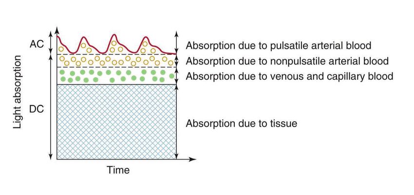
SCIENCE AND TECHNOLOGY
CONTINUING FROM PREVIOUS PAGE

History                                                            the need for a zero calibration in a bloodless sample (Van
                                                                   Meter et. Al, 2017). Aoyagi chose different wavelengths of
Johann Heinrich Lambert’s book “Photometria sive de                light than had been previously used, using 630 nm (red) and
mensura et gradibus luminis, colorum et umbrae”, published         900 nm (infrared) instead of using 805 nm, an isosbestic
in 1760, formulated the law which states that absorbance           point; a wavelength at which the absorption of light by a
of a material sample is directly proportional to its thickness     mixed solution remains constant as the equilibrium between
(path length) (Columbia University Archive, 2016). Almost          the components in the solution changes (UCDavis, 2019),
100 years later, in 1852 a man by the name of August Beer;         for hemoglobin, which is a point of equal absorption by
a German physicist, chemist, and Professor of Mathematics          oxyhemoglobin and deoxyhemoglobin (Severinghaus, 2007).
at University of Bonn, added that “the absorbance is               Nihon Kohden Corporation produced the first commercial
proportional to the concentrations of the attenuating species      pulse oximeter, the OLV-5100, and applied for a patent
in the material sample” (Blood in the case of pulse oximetry).     to the Japanese Patent Office on March 29, 1974, but not
Together, these two scientists developed the Lambert-Beer          elsewhere in the world (Aoyagi, 2003).
Law, which describes the disruption in amplitude
of the wavelength of light, in relation to the thickness
of the material in which it is traveling through (Van Meter
et al, 2017). The Lambert-Beer Law is the foundational                These are key events throughout
idea of oximetry.
                                                                       history that have pushed pulse
In 1860, two professors at the University of Heidelberg in             oximetry to where it currently
Germany, Gustav Kirchoff, and Robert Bunsen (inventor of
the Bunsen burner) established the technique of analytical
                                                                         is and have helped serve as
spectroscopy. The discovery of spectroscopy aided Felix                 an establishment for the ASA
Hoppe-Seyler; a German physiologist and chemist in the
discovery of the oxygen carrying material in blood called
                                                                            Monitoring Standards.
Hemoglobin thereafter, in 1864. He defined hemoglobin
as two parts, the heme dark-red, iron-containing, non-
protein part, and the globin, the colorless protein part.          Although the probe was very sensitive to motion, it showed
He then applied absorption spectroscopy to hemoglobin,             that the principle of pulse oximetry was accurate. Based on
based on the principle that substances are colored because         Aoyagi’s foundation, several groups within the United States
they absorb and reflect certain wavelengths of light. He           began to develop their own versions of pulse oximeters (Van
demonstrated that if light passed through a solution of            Meter et al, 2017). Improvements in diode technology led
oxygenated hemoglobin; at that time, 540nm and 560nm               to several American companies to enter the field of pulse
wavelengths would be absorbed (twin-peak absorption                oximetry. In 1980, Biox Technology, an American medical
pattern) (Hazelwood, 2001).                                        technology company headquartered in Denver, Colorado
                                                                   marketed their first pulse oximeter in the United States
It was not until more than 100 years later, in Tokyo, Japan
                                                                   (USPTO, 1983).
that the term “pulse” had been studied in coordination to
the field of oximetry, by a young Japanese bio-engineer by         These are key events throughout history that have pushed
the name of Takuo Aoyagi who worked at the Nihon Kohden            pulse oximetry to where it currently is and have helped serve
Corporation, a Tokyo-based manufacturer that developed             as an establishment for the ASA Monitoring Standards.
and distributed medical equipment. At the time, Aoyagi was
researching the measurement of cardiac output through              Principles
dye dilution. An ear oximeter, designed previously by Earl         The World Health Organization defines pulse oximeters
Wood in the United States in 1949, was used during the             as medical devices that monitor the level of oxygen in a
research. Aoyagi was troubled by interference from pulsatile       patient's blood and alert the health-care worker if oxygen
variations in the light signal, encountering difficulty because    levels drop below safe levels, allowing rapid intervention
of the constant artifact created by these pulsations. After this   (WHO, 2019). Practitioners can quickly recognize changes
finding, he concluded that the change in arterial blood flow       in blood oxygen saturation due to the changes in audible
could be utilized to measure the oxygen saturation without         pitches and cadence.
                                                                                                                         MORE
                                                 The
                                                       Sensor Summer 2020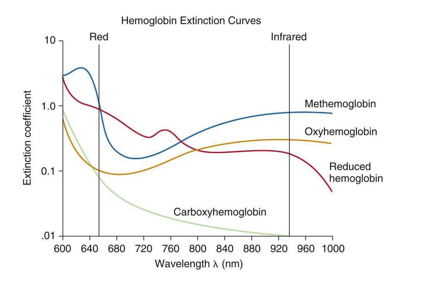
SCIENCE AND TECHNOLOGY
CONTINUING FROM PREVIOUS PAGE
The pulse oximeter is often the very first monitor placed on through the tissue for the photo-detector to receive. After
the patient upon arrival to the operating room (Guimaraes the signal is received, the photo-detector relays that signal
et al, 2019). This noninvasive method is used to measure to a computer that utilizes an algorithm, which are company
oxygenation, ventilation and circulation by determining specific and proprietary, to transmit the data to the monitor.
the oxygen levels within the arterial blood. The oxygen The probes can either be disposable or reusable and are
levels are determined by measuring hemoglobin saturation available in different sizes. In operating room type setting
(SpO2) via red and infrared light transmission through tissue. it is more common to utilize disposable probes in order to
Hemoglobin is a protein that is found in red blood cells prevent any potential nosocomial infections. Proper size
(RBCs) and can either contain oxygen (oxyhemoglobin) or selection is important because it ensures that accurate
not contain oxygen (deoxyhemoglobin). Oxyhemoglobin and values are recorded. For example, if the size if too large then
deoxyhemoglobin absorb light differently: oxyhemoglobin light from the diode can be overcompensated and not reach
absorbs more infrared light than red light and the photocell without passing through the tissue, which can
deoxyhemoglobin absorbs more red light than infrared. The result in a false high SpO2 reading. It is important for the
oxyhemoglobin has significantly lower absorption of the 660 photocell to be aligned with the LED so readings can be
nm wavelength than deoxyhemoglobin, while at 940 nm the recorded accurately.
oxyhemoglobin absorption is slightly higher. This difference
The next component is the cable. The cable connects the
is used for the measurement of the amount of oxygen in a
probe to the oximeter console and it is important that there
patient's blood by the pulse oximeter.
is a complete connection between the two components or
While SpO2 is used by the anesthetist to continuously else the monitor will not have an accurate reading or even
monitor the oxygen delivered to metabolically active tissues, a reading at all. The values are displayed on the console for
it is not a direct measurement of the oxygen content of the operator to read and monitor. Once the console receives
blood. SpO2 serves as a surrogate measurement of oxygen the signal from the probe transducer via the cable then it
saturation of hemoglobin in arterial blood (SaO2) (Guimaraes is displayed in pulsatile waveform and oxygen saturation
et al, 2019). is displayed in a percentage with the strength of the probe
signal.
Equipment
There are three constituents that comprise a pulse oximeter:
probe transducer, cable and monitor. Each of these works in
conjunction with one another to provide an accurate reading
of the patient’s oxygen saturation levels.
Pulse Pitch
The pitch of the pulse oximeter sound correlates with
the oxygen saturation. The lower the pitch, the lower the
saturation will be. There are some pitfalls with this system
and one of the main distractions tends to be the OR
environment (Lichtor, 2014). The OR environment tends to
be quite loud with respect to the staff and the music that the
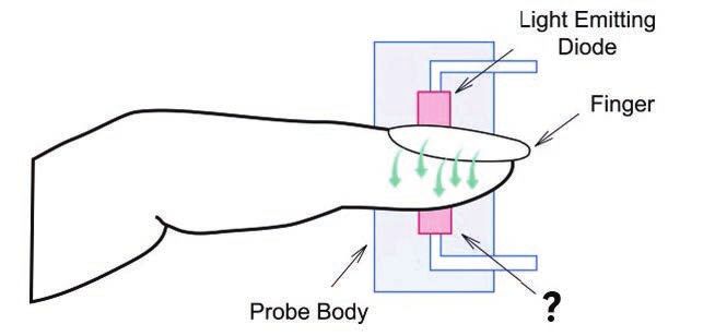
The element that comes in direct contact with the patient surgeon has requested. A loud environment is by no means
is the probe transducer. The pulse oximeter contains a red conducive to utilizing a monitor that has a sound that is
(650nm) diode, an infrared (940-1000nm) diode, and a designed to help you readily identify SpO2 saturation when
photoreceptor. The light-emitting diode (LED) is part of the you are performing multiple tasks that directly affect the care
probe that emits light at a specific wavelength and sends it of a patient undergoing a surgical intervention.
MORE
The
Sensor Summer 2020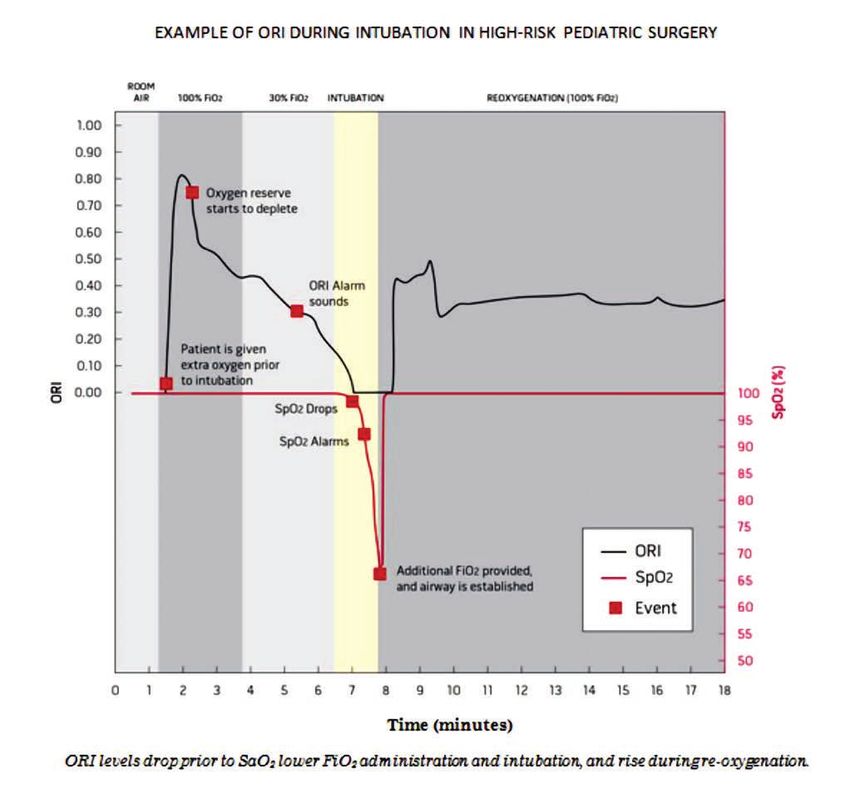
SCIENCE AND TECHNOLOGY
CONTINUING FROM PREVIOUS PAGE
The department of anesthesia at Vanderbilt University             administered. This allows some ease for those patients who
assessed whether training to make use of combined visual          fear the idea of surgery and may have some concern with
and auditory cues might improve resident physicians’              needles and pain. The measurement of oxygen saturation
ability to detect frequency changes due to oxygen                 is important throughout surgery because providers must be
saturation. The results were just as lackluster as one may        alerted when there is a drop of saturation due to anesthetics
imagine. It was concluded that both environmental noise           or other factors. It is the most readily available because it
and attentional load impaired response time to detect             is easy and fast to place on the patient and it also provides
changes in tones representative of decreasing oxygen              a variety of sizes and different probes for a variety of site
saturation. Environmental noise also impaired accuracy of         applications.
tone determination. The utilization of perceptual training
improved the residents’ ability to detect changes in oxygen
saturation determined by auditory pitch changes. Perceptual
training also improved their response time in a noisy and
attention-demanding environment like that of an operating
room (Lichtor, 2014).

Measurement Method
There are two types of methods that are used to collect
data from the pulse oximeter: transmission and reflection.
The most common and readily used method for measuring
saturation is transmission pulse oximetry. With this method,
the light source is transmitted through tissue to the detector
that lies directly on the opposite side. There are situations
where it is beneficial and even crucial to utilize transmission
and reflective probes in conjunction with one another. In
cardiac and vascular surgery in particular, practitioners seem
to be adopting the use of cerebral oximeters (reflective)
in order to get a more accurate reading of SpO2. Wax et al
referenced a study in their research that stated “one study
suggested that they may be more reliable than finger probes in
patients with poor peripheral perfusion or low cardiac index”
(Wax et al, 2009).

Common sites for transmission probes are the fingertip,
toe, nose and earlobe because it provides a direct line with
the light source and the photodetector, in contrast of the
cheek or forehead sites. Unlike transmission, reflection pulse
oximetry relies on backscattering; therefore, producing a
weaker impulse. With reflection, the LED and photocell are
on the same plane. There are ways that can maximize the
signal such as heating the site being measured and applying
pressure.                                                         Limitations
                                                                  A limitation of the pulse oximeter that is often overlooked
Advantages                                                        is the inability to detect hyperoxemia. There is growing
Pulse oximetry possesses qualities that make it advantageous      evidence that the administration of oxygen in concentrations
from other monitors. These qualities include being noninvasive,   that produce hyperoxemia is associated with cellular
serving as a continuous monitor and being the most readily        injury (Vanderveen et al, 2006). More recent evidence also
available. Being noninvasive, pulse oximetry is considered        indicates that resuscitation of premature neonates with a
a routine monitor and can be placed before anesthesia is          high fraction of inspired oxygen (FiO2) is associated with

                                                                                                                          MORE
                                                 The
                                                      Sensor Summer 2020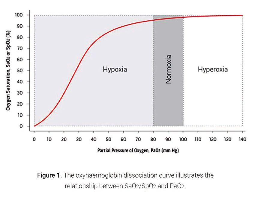
SCIENCE AND TECHNOLOGY

greater mortality and worse outcomes (Rabi et al, 2007).
The inability of the pulse oximeter to detect hyperoxemia is
profound and worth noting.
                                                                References
Perfusion greatly affects the quality of information provided   A technique to improve detection of pulse oximetry pitch perception
by the pulse oximeter. If a patient does not have adequate      given the background noise of an operating room. (2018, December
                                                                31). Retrieved from https://aa2day.org/2014/06/technique/
perfusion to their extremities it is impossible to get an
                                                                Ahrens; Kimberley, Basham (1993). Essentials of Oxygenation:
accurate SpO2 reading. However, severity of poor perfusion
                                                                Implication for Clinical Practice. Jones & Bartlett Learning. p. 194.
should be noted. A recent study published in the 2018           ISBN 978-0867203325
edition of Anesthesiology, tested four different brands and
                                                                Aoyagi, T. (2003). Pulse oximetry: Its invention, theory, and future.
discovered a confidence (p-value), in most cases of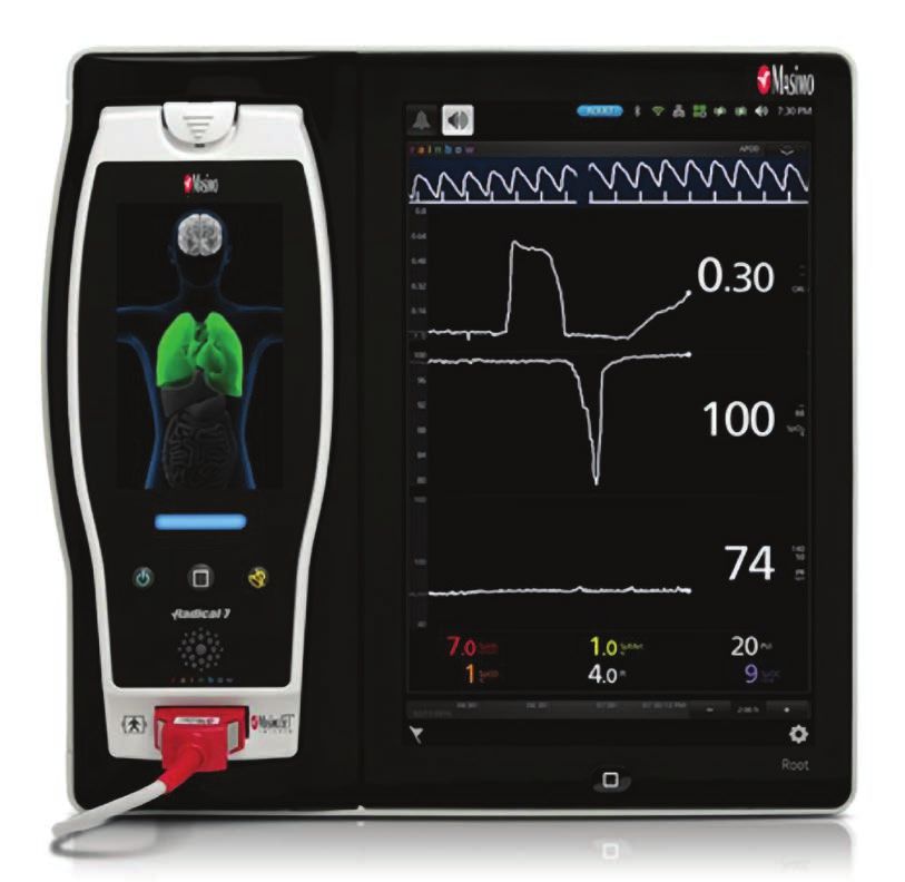
SCIENCE AND TECHNOLOGY
                                The
                                      Sensor Summer 2020
Continuing Education Quiz
                        To test your knowledge on this issue’s article, provide correct answers to the following
                                    questions on the form below. Follow the instructions carefully.

                                                                         6. Pulse oximetry has a fairly significant
                                                                         (often overlooked) disadvantage.
                                                                         What is this disadvantage?
                                                                         a) The inability to spontaneously ventilate
                                                                         b) The inability to detect hyperoxemia
                                                                         c) The Lack of wattage in the diode
                                                                         d) The inability to detect respirations

1. The image below shows the placement of a pulse                        7. Pulse Oximetry is a non-invasive method
oximetry probe on a patient’s finger. What part of                       of monitoring used to measure?
the pulse oximetry probe is indicated by the “?”?                        (Select all that apply)
a) L.E.D.			                c) Photodetector                             a) Hyperoxemia		                 d) Blood loss
b) Reflector		              d) Pulsatile flow sensor                     b) Oxygenation		                 e) Ventilation
2. This principle describes the disruption in                            c) Circulation		                 f) Hemoglobin Levels

amplitude of the wavelength of light, in relation to                     8. The article referenced a wavelength of light
the thickness of the material in which it is traveling                   from the diode that measures oxyhemoglobin;
through.                                                                 what was the wavelength and color of the
a) Boyle’s Law		            c) The Venturi Effect
                                                                         spectrum? (Select two)
b) Lambert-Beer Law         d) Poiseuille's Law                          a) 560 nm		                      e) Indigo
                                                                         b) 740 nm		                      f) Red
3. All of the choices below are components of a
                                                                         c) 940 nm			                     g) Infrared
typical pulse oximeter; which component is not                           d) 660 nm		                      h) Ethyl Violet
typical?
a) Transducer		             c) Monitor                                   9. The article references two principles that are
b) Cable			                 d) Piezoelectric Wafer                       used to collect data from the pulse oximeter, what
4. The article referenced a wavelength of light                          are these two principles? (Select two)
                                                                         a) Transmission		                c) Reflection
from the diode that measures deoxyhemoglobin;                            b) Absorption		                  d) Oxidation
what was the wavelength and color of the
                                                                         10. The pitch of the pulse oximeter sound
spectrum? (Select two)
a) 560 nm         c) 940 nm		         e) Indigo        g) Infrared       correlates with the SpO2 of the patient. The_______
b) 740 nm         d) 660 nm           f) Red	         h) Ethyl Violet   the pitch, the lower the oxygen _______ will be.
5. In pulse oximetry, the probe may be placed in                         (Fill in the blanks by selecting two)
                                                                         a) Lower			                      c) Saturation
different locations on the patient in order to obtain                    b) Higher			                     d) Detection
a reading. Which location is NOT utilized for pulse
oximetry?
a) Earlobe			               c) Toes
b) Forehead		               d) Fingertips

    To apply for Continuing Education/
                                                                                                   1: A B C D                6: A B C D
    Contact Hours:                                                        The answers to
                                                                          the Summer 2020          2: A B C D                7: A B C D E F
    1) Provide all the information requested on this form.                Science and Technology
                                                                          Continuing Education     3: A B C D                8: A B C D E F G H
    2) Provide correct answers to this issue’s quiz in this box > > >     Quiz are:                4: A B C D E F G H        9: A B C D
    3) Mail this form along with $10.00 Member $20 Non-Member             (circle answers)         5: A B C D               10: A B C D
      (check or money order, payable to ASATT) to:
      "ASATT", 7044 S 13th St, Oak Creek, WI 53154                                                                               Quiz 1 of 2
    Name: 								 ASATT Number:
    Street Address: 							 Phone Number:
    City: 								 State: 		                                                                              Zip:

    Signature: 								 Date:
                     SUBMISSIONS FOR THIS ISSUE’S QUIZ EXPIRE DECEMBER 31, 2021.
                ACHIEVE 80% IN THIS QUIZ TO EARN ONE (1) CONTINUING EDUCATION CREDIT.
You can also read