If Not Otitis Externa Then What? - Clinical - Journal of Urgent Care ...
←
→
Page content transcription
If your browser does not render page correctly, please read the page content below
CME: This peer-reviewed article is offered for AMA PRA Category 1 Credit.™
  Clinical                                                   See CME Quiz Questions on page 9.

If Not Otitis Externa…Then What?
Urgent message: Complaints of ear pain in children are among the most common pre-
sentations in the urgent care setting. While acute otitis media and acute otitis externa
are high on the list of possible causes, it is essential that the urgent care provider be
prepared to differentiate these from other possible etiologies.

SADIA ANSARI, MD; TIMOTHY MARTIN, MD; and ELIZABETH FLASCH, MD

Citation: Ansari S, Martin T, Flasch E. If not otitis
externa…then what ? J Urgent Care Med. 2021;15 (5):
11-15.

Introduction

E
   ar pain is one of the most common presentations in
   urgent care, especially among pediatric patients. Fur-
   ther, acute otitis media (AOM) is the most common
condition for which antibacterial agents are prescribed
for children in the United States. There were 634 clini-
cian visits per 100 children during 2005-2006. It is
imperative that clinicians differentiate AOM from new
onset of otorrhea not due to acute otitis externa (AOE),
the most common diagnoses made by clinicians with
regional variations based on age and geography.1
   The majority of AOE-related visits occur during the
summer months (June through August); visits occur

                                                                                                                                                                      ©AdobeStock.com
most commonly in the South and least commonly in
the West.2-4
   Rosenfeld, et al noted that data from ambulatory care
centers suggest there are about 2.4 million visits for AOE,
affecting 1 in 123 persons in the United States. Just less                     AOE and AOM. In addition, coordination with a sub-
than half of all visits for AOE were for children 5 to 14                      specialist is often necessary.
years of age. Direct costs are estimated at half a billion                       Here, we offer three illustrative cases involving chil-
dollars annually, and ambulatory care providers spent                          dren who presented to Children’s Wisconsin Urgent
about 600,000 hours treating AOE.2-4                                           Care facilities with chief complaints of “ear infection”
   Clearly, urgent care providers must be able to distin-                      or “ear pain.”
guish AOE and AOM from other causes of otalgia, otor-
rhea, and inflammation of the external auditory canal.                         Case 1
“Ear infections” that do not present as AOE or AOM                             A 3-year-old female presents with a complaint of “left
make for a difficult case in an urgent care setting. In                        ear infection” and facial swelling. Symptoms started 36
such cases, the treatment and management differ from                           hours prior to presentation. While cleaning her hair,

Author affiliations: Sadia Ansar, MD, Department of Urgent Care, Division of Primary Care, Children’s Wisconsin. Timothy Martin, MD, Department of Oto-
laryngology, Children’s Wisconsin; Medical College of Wisconsin. Elizabeth Flasch, MSN, APNP, PNP, Department of Urgent Care, Division of Primary Care,
Children’s Wisconsin; Marquette University College of Nursing. The authors have no relevant financial relationships with any commercial interests.

w w w. j u c m . c o m                                               JUCM T h e J o u r n a l o f U r g e n t C a r e M e d i c i n e | Fe b r u a r y 2 0 2 1   11
I F N O T O T I T I S E X T E R N A … T H E N W H AT ?

 Figure 1.                                                                               Decision-making/diagnosis
                                                                                          Patient was diagnosed with an infected pre-auricular
                                                                                           pit with clearance of the previous AOM

                                                                                         Treatment
                                                                                         The patient was discharged home on high-dose amox-
                                                                                         icillin clavulanate with outpatient referral to otolaryn-
                                                                                         gology for evaluation and possible excision of the
                                                                                         preauricular pit. Unfortunately, as you will read, this was
                                                                                         likely the incorrect choice for this patient.

                                                                                         Follow-up
                                                                                         The ENT nurses phoned the family the next day to set
                                                                                         up an appointment for follow-up. Due to tactile fever
                                                                                         and increasing ear pain, as relayed by the parents, the
                                                                                         child was referred to the emergency department, where
                                                                                         incision-and-drainage (I&D) was performed under seda-
                                                                                         tion by ENT. Bacterial culture was ordered and sent. The
                                                                                         patient was discharged from ED with oral clindamycin
                                                                                         with outpatient ENT follow-up to be scheduled in 7-10
                                                                                         days. Culture was positive for Serratia marcescens, Aggre-
parent noticed a swelling on outer left ear. The child had                               gatibacter, and coagulase negative Staphylococcus (CONS).
been diagnosed with right AOM 1 month prior and was                                      The oral amoxicillin clavulanate was recommended to
treated with amoxicillin. Her right ear was not reassessed                               be stopped by the ENT specialist.
after completion of treatment. She had a past medical                                       Three days later, the family contacted ENT via tele-
history significant for atopic dermatitis and shellfish                                  phone and reported bloody drainage and “new white
allergy which were under good control with low-dose                                      patches” on the ear. The patient was evaluated in the out-
topical steroids as needed. Parent denied fever but stated                               patient ENT clinic, then admitted immediately after for
presence of upper respiratory symptoms.                                                  surgical drainage of the pre-auricular pit. After drainage
                                                                                         of the pit, she was discharged on clindamycin PO (30
Exam – Case 1                                                                            mg/kg/day dosed three times daily) for an additional 7
Vitals                                                                                   days. Per parent report, this course was completed.
 Afebrile                                                                                  The patient returned to the ENT clinic with continued
 Within normal limits for age                                                           drainage 3 weeks later. She was started on 10 days of oral
                                                                                         ciprofloxacin (40 mg/kg/day dosed twice daily). Due to
Physical exam findings                                                                   recurrent infections, the decision was made to excise the
 Left pinna with moderate-to-significant swelling and                                   pre-auricular pit. She had surgery 2 months postpresen-
  a visualized preauricular pit                                                          tation, with successful resection of the pit.
 On palpation, mild to moderate tenderness and mild
  fluctuance                                                                             Discussion
                                                                                         Pre-auricular sinuses are distinct from first branchial cleft
Ear canal                                                                                anomalies and derive from ectodermal inclusions
 No drainage with dry membranes                                                         formed during development of the external ear. They
 Tympanic membranes were clear bilaterally                                              are quite common in pediatric patients and may present
                                                                                         in 1% of Caucasians, 5% of African-Americans, and 10%
Nares                                                                                    of Asians. Infants born with pre-auricular pits should
 Clear rhinorrhea bilaterally                                                           have formal audiologic evaluation.
 Remainder of the exam was benign                                                          The pre-auricular sinuses may be the first indication
 Prior to examination, the mother was unaware of the                                    of branchio-oto-renal (BOR) syndrome—one of the
  definition of a pre-auricular pit                                                      most common hereditary causes of hearing loss. BOR is

12   JUCM T h e J o u r n a l o f U r g e n t C a r e M e d i c i n e | Fe b r u a r y 2 0 2 1                                        w w w. j u c m . c o m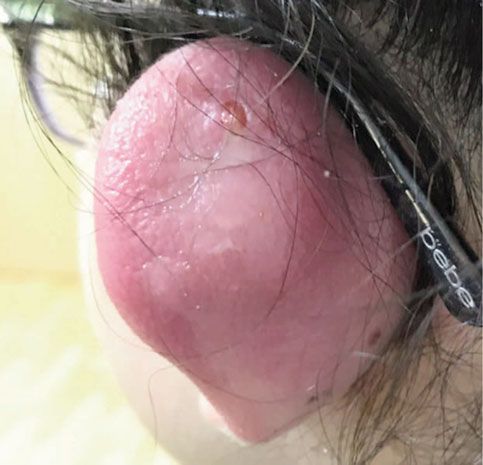
I F N O T O T I T I S E X T E R N A … T H E N W H AT ?
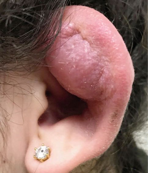
Figure 2. Figure 3.
lage pierced 17 days prior to urgent care visit. Seen at
another hospital 10 days prior with erythema and puru-
lent drainage from the piercing site. She was started on
high-dose amoxicillin clavulanate 875-125 mg twice
an autosomal dominant syndrome characterized by daily. Seen again at same hospital, 6 days prior to the
preauricular pits, branchial cysts or tracts, malformed visit with worsening symptoms. The patient was referred
ears, and renal anomalies, including renal dysplasia and to a general surgeon’s clinic immediately following her
bifid renal pelvises. They may present as cysts and fre- urgent care visit. There, I&D was performed and she was
quently become infected to form abscesses.5-10 continued on amoxicillin clavulanate 875-125 mg twice
First-line treatment of abscess is I&D with oral antibi- daily. Instructions to follow up in 1 week were provided.
otics. Following resolution of inflammation, the sinus She continued to have worsening swelling, erythema,
should be excised to prevent recurrence. Pits are most and purulent thick drainage and thus presented to our
commonly infected with Staphylococcus species. Edema, urgent care.
erythema, fluid drainage, and pain are common signs
infection. Exam – Case 2
For this case presentation, Serratia species is likely Vitals
from an exogenous environmental source—water, soil, Heart rate 64
plants, animals, insects. Serratia species are generally sus- Respiratory rate 16
ceptible to fluoroquinolones, aminoglycosides, trime- Temperature 36.8° C
thoprim-sulfamethoxazole, Zosyn (piperacillin and Weight 108.2 kg
tazoabactam), Timentin (ticarcillin and clavulanate),
fourth-generation cephalosporins, macrolides, tetracy- General
cline, nitrofurantoin, and colistin. CONS was presumed Afebrile
to be a contaminant. Mild distress with level of pain reported
Patient tearful with examiner
CASE 2
A 16-year-old female with an insignificant past medical Ear
history presents with chief complaint of ear pain, On inspection of left pinna: auricular erythema with
swelling, and “ear infection” for the last 10 days. Carti- significant swelling
w w w. j u c m . c o m JUCM T h e J o u r n a l o f U r g e n t C a r e M e d i c i n e | Fe b r u a r y 2 0 2 1 13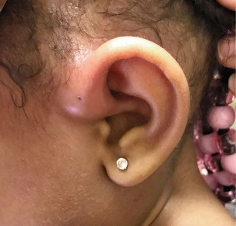
I F N O T O T I T I S E X T E R N A … T H E N W H AT ?

 Figure 4.                                                                                 Figure 5.

 On palpation, moderate to severe tenderness with                                       are associated with poor healing and more serious infec-
  fluctuance noted                                                                       tion due to the avascular nature of auricular cartilage.
                                                                                         Piercings are usually carried out by nonauthorized or
Decision-making/diagnosis                                                                untrained professionals with no consensus on asepsis
 Cellulitis of left ear with abscess, perichondritis of left                            techniques. The risk of developing infection is higher in
  ear                                                                                    the ear cartilage than in the lobe.11-17
 Digital images taken via Haiku into EPIC
 ENT phone consult done during urgent care visit. ENT                                   Pathogens
  recommended admission with intravenous anti-                                           The most common pathogen is Pseudomonas aeruginosa,
  biotics                                                                                followed by Staphylococcus aureus.17 Symptoms usually
 After admission, she was started on IV ciprofloxacin                                   develop between 3 days and 4 weeks after the ear pierc-
  (400 mg every 12 hours) with plans for I&D in the                                      ing and include pain, erythema, edema, and abscess for-
  operating room                                                                         mation. Diagnosis is clinical; wound culture with
                                                                                         antibiogram must be performed. Fluoroquinolones are
Follow-up                                                                                the treatment of choice since they show antipseudo-
After successful I&D and short hospital stay (72 hours),                                 monal activity in addition to anti staphylococcal effect.
the patient was discharged on 10 days of oral cipro-                                     Once an abscess develops, surgical I&D is often neces-
floxacin (500 mg every 12 hours). An aerobic and anaer-                                  sary. Good cosmetic preservation of the cartilage is dif-
obic culture was sent and grew few Pseudomonas                                           ficult to maintain.11-17
aeruginosa which were susceptible to ciprofloxacin (pan-
susceptible). She followed up in ENT clinic 2 weeks later.                               Case 3
During that visit, her ear was noted to be healing well.                                 An 8-year-old female presented to urgent care with a 12-
Three months after the initial urgent care visit, the ear                                hour history of ear “redness.” She complained that her
was healed with a residual defect in the cartilage.                                      ear felt “thick” and warm to the touch. She had minimal
                                                                                         pain. She denied drainage, difficulty hearing, fever, URI,
Discussion                                                                               or trauma. She had been swimming quite a bit in the
Auricular abscess/perichondritis complicating helical ear                                days leading up to the visit. There was no known injury
piercing is a frightening complication of the traumatized                                or trauma to the ear. Past medical history and surgical
ear that can lead to a residual deformity. These piercings                               history were negative.

14   JUCM T h e J o u r n a l o f U r g e n t C a r e M e d i c i n e | Fe b r u a r y 2 0 2 1                                      w w w. j u c m . c o m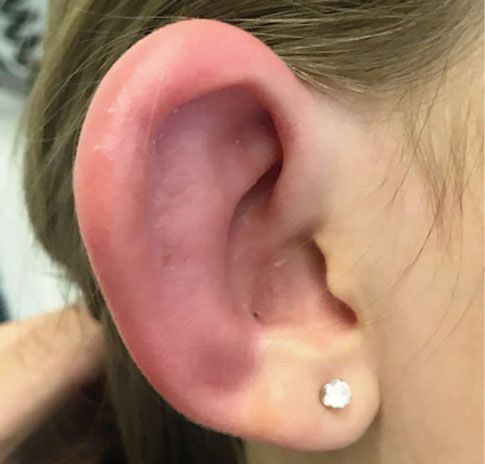
I F N O T O T I T I S E X T E R N A … T H E N W H AT ?

Exam – Case 3                                                     “The fact that there is a broad differential
Vitals
 Heart rate 80                                                        for outer ear infections requires the
 Respiratory rate 20                                             clinician to look beyond acute otitis media
 Temperature 37.7° C
 Weight 28.1 kg
                                                                     and acute otitis externa when a child
                                                                     presents with a chief complaint of ear
Ear                                                                   pain. Taking a detailed history and
 Significant and well-demarcated erythema
 No pain to mastoid process or the entire helix                      conducting a thorough examination
                                                                          can aid in assessing for other
Decision-making/diagnosis
 Digital images taken via Haiku into EPIC
                                                                              potential diagnoses.”
 ENT consult via telephone during urgent care visit
 The patient was admitted for IV antibiotics. The ENT           likely to include Pseudomonas and Staphylococcus species.
  resident initially started nafcillin for Staphylococcus        Antibiotic resistance rates are high. Intravenous antibi-
  coverage; this was later changed to ceftazidime                otics, hospitalization, surgical I&D, and culture may be
 Diagnosis of perichondritis thought to be caused by            necessary. Oral antibiotics may require double coverage.
  a previous injury/laceration to the ear that had gotten        Amoxicillin is used often and is the incorrect choice.
  infected                                                       Consultation with ENT is often necessary to preserve
 Although the patient denied injury/trauma, the likely          the form and function of the ear. n
  cause of the perichondritis was some injury with
                                                                 References
  infection from her recent swimming                             1. Lieberthan A, Carroll A, Chonmaitree T, et al. The diagnosis and management of acute
                                                                 otitis media. Pediatrics. 2013;131(3):e964-e999.
                                                                 2. Rosenfeld RM, Schwartz SR, Cannon CR, et al. Clinical practice guideline: acute otitis
Follow-up                                                        externa. Otolaryngology Head Neck Surgery. 2014;150:S1.
The patient was discharged home after 1 day of IV                3. Rosenfeld RM, Singer M, Wasserman JM, Stinnett SS. Systematic review of topical
                                                                 antimicrobial therapy for acute otitis externa. Otolaryngol Head Neck Surg. 2006;134(4
antibiotics. She was switched to 10 days of ciprofloxacin        Suppl):S24.
(500 mg twice daily for 10 days) and clindamycin (300            4. Rosenfeld RM, Brown L, Cannon CR, et al. Clinical practice guideline: acute otitis
                                                                 externa. Otolaryngol Head Neck Surg. 2006;134:S4.
mg every 8 hours for 10 days).                                   5. Bellini C, Piaggio G, Massocco D, et al. Branchio-oto-renal syndrome: a report on nine
                                                                 family groups. Am J Kidney Dis. 2001;37:505-509.
                                                                 6. Ehlers Klug T, Holm N, Greve T, Ovesen T. Perichondritis of the auricle: bacterial findings
Discussion                                                       and clinical evaluation of different antibiotic regimens. Eur Arch Otorhinolaryngol.
Perichondritis is an infection of the pinna. Pseudomonas         2019;276:2199-2203
                                                                 7. Huang XY, Tay GS, Wansaicheong GK, Low WK. Preauricular sinus: clinical course and
and Staphylococcus species are the most common                   associations. Arch Otolaryngol Head Neck Surg. 2007;133:65-68
pathogens6,13 (Staphylococcus species being the major            8. Kugelman A, Tubi A, Bader D, et al. Pre-auricular tags and pits in the newborn: the role
                                                                 of renal ultrasonography. J Pediatr. 2002;141:388-391.
pathogen in non-abscess perichondritis6). Pseudomonas            9. Roth DA, Hildesheimer M, Bardenstein S, et al. Preauricular skin tags and ear pits are
is widespread in nature and thrives on most surfaces and         associated with permanent hearing impairment in newborns. Pediatrics. 2008;122:e884-
                                                                 890.
is known to cause otitis externa, keratitis, hot tub folli-      10. Scheinfeld NS, Silverberg NB, Weinberg JM, Nozad V. The preauricular sinus: a review
culitis, postoperative abscesses, and burn infections.           of its clinical presentation, treatment, and associations. Pediatr Dermatol. 2004;21:191-
                                                                 196
Double antibiotic therapy is recommended with a peni-            11. Keene WE, Markum AC, Samadpour M. Outbreak of Pseudomonas aeruginosa infec-
cillin and fluroquinolone.6,13                                   tions caused by commercial piercing of upper ear cartilage. JAMA. 2004;291:981-985.
                                                                 12. Lee TC, Gold WL. Necrotizing Pseudomonas chondritis after piercing of the upper ear.
                                                                 CMAJ. 2011;183:819-821.
Conclusion                                                       13. Mitchell S, Ditta K, Minhas S, Dezso A. Pinna Abscesses: Can we manage them better?
                                                                 A case series and review of the literature. Eur Arch Otorhinolaryngol. 2004;272(11):3163-
There is a broad differential for outer ear infections.          3167.
Patients presenting with a chief complaint of “ear infec-        14. Rodriguez J, Thone N, Duque J, Branes R. Infected transcartilaginous ear piercings. A
                                                                 case report and review of the literature. Revista de Ciencias Medicas 2019;44(2):23-25.
tion” or “ear pain” automatically prompt the clinician           15. Rowshan HH, Keith K, Baur D, Skidmore P. Pseudomonas aeruginosa infection of the
to consider AOM and AOE. However, a detailed history             auricular cartilage caused by “high ear piercing”: a case report and review of the literature.
                                                                 J Oral Maxillofac Surg. 2008;66:543.
and thorough examination can aid in assessing for pre-           16. Sosin M, Weissler JM, Pulcrano M, Rodriguez ED. Transcartilaginous ear piercing and
auricular sinus infection, infected piercing site, or peri-      infectious complications: a systematic review and critical analysis of outcomes. Laryngo-
                                                                 scope. 2015;125:1827-1834.
chondritis on the differential diagnosis list.                   17. Stewart G, Thorp A, Brown L. Perichondritis—a complication of high ear piercing. Pedi-
   In these conditions, implicating pathogens are more           atr Emerg Care. 2006;22(12):804-806.

w w w. j u c m . c o m                                 JUCM T h e J o u r n a l o f U r g e n t C a r e M e d i c i n e | Fe b r u a r y 2 0 2 1           15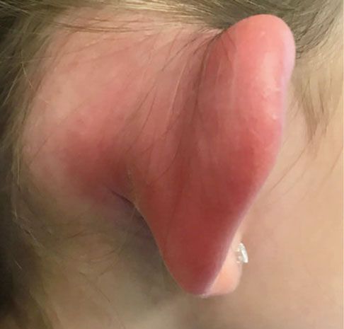
You can also read