Primary orbital ganglioneuroblastoma: A case report - De Gruyter
←
→
Page content transcription
If your browser does not render page correctly, please read the page content below
Open Medicine 2021; 16: 1076–1081
Case Report
Ruixin Ma#, Yujiao Wang#, Weimin He*
Primary orbital ganglioneuroblastoma:
A case report
https://doi.org/10.1515/med-2021-0230 Keywords: primary ganglioneuroblastoma, orbit, adrenal
received September 30, 2020; accepted December 19, 2020 gland
Abstract
Background ‒ Ganglioneuroblastoma (GNB) is a neo-
plasm that arises from the primitive cells of the sympa-
thetic nervous system during childhood. The current case 1 Introduction
is very unique because of the initial primary tumor mani-
festation in the orbit and an adrenal tumor being observed Ganglioneuroblastoma (GNB) is a neuroblastic tumor ori-
later during follow-up. ginating from the primordial neural crest cells in the sym-
Case presentation ‒ A 2-year-old girl presented to the pathetic nervous system and is frequently observed in the
Ophthalmology Department of our hospital complaining pediatric population [1]. GNB, first reported by Wahl and
of swelling of the left upper eyelid for approximately one Craig, is a histological subgroup of neuroblastomas and
month. Orbital computed tomography (CT) revealed a left has an intermediate risk of malignancy compared with
orbital mass with bone destruction. Thoracic and abdo- ganglioneuromas and neuroblastomas [2,3]. The clinical
minal CT indicated no abnormalities. The mass was surgi- behavior of GNB is unpredictable as the tumor may
cally resected, and histopathological analysis confirmed regress spontaneously, mature into ganglioneuroma, or
it as GNB. During follow-up, abdominal CT detected an metastasize rapidly as observed in neuroblastoma. The
adrenal tumor with internal calcification, a calcified nodule common sites of GNB are the adrenal gland (35%) and
on the left side of the abdominal aorta, and mesenteric retroperitoneum (30%), whereas metastases usually occur
lymph nodes. Accordingly, primary orbital GNB and meta- in the bone marrow (70.5%) and the bone (30.9%) [4]. To
static adrenal GNB were the possible considerations. We the best of our knowledge, there are four case reports on
removed the adrenal tumor, and the patient underwent the orbital GNB [5–8]. However, primary orbital GNB has
chemotherapy. However, the patient died 18 months after never been reported previously. Herein, we described a
the ophthalmic surgery. rare case wherein a primary GNB was detected in the orbit
Conclusion ‒ Primary orbital GNB in children is easily and a metastatic adrenal tumor was found later during
misdiagnosed because of its rare occurrence and atypical follow-up.
clinical findings. Imaging methods combined with histo-
pathological examination contribute to the detection and
diagnosis of primary and metastatic GNBs. Thus, timely
surgery combined with adjuvant chemotherapy and long- 2 Case presentation
term follow-up is essential for controlling the metastasis
of GNB and improving the survival rate of patients. A 2-year-old girl was admitted to the Ophthalmology
Department of our hospital with a swelling of the left
upper eyelid for approximately one month. No redness
# These authors contributed equally to this work. in the eye or other abnormal symptoms were observed.
There were no pregnancy or childbirth-related abnorm-
alities reported by the patient’s mother. Also, no familial
* Corresponding author: Weimin He, Department of Ophthalmology, anamnesis of cancer existed.
Ophthalmic Laboratory, West China Hospital, Sichuan University,
During the initial clinical assessment, the patient had
Chengdu 610041, Sichuan, China, e-mail: hewm888@hotmail.com
Ruixin Ma, Yujiao Wang: Department of Ophthalmology, Ophthalmic
normal growth and good nutritional status and had no
Laboratory, West China Hospital, Sichuan University, Chengdu abdominal symptoms such as abdominal pain and abdo-
610041, Sichuan, China minal swelling. Thoracic and abdominal CT revealed no
Open Access. © 2021 Ruixin Ma et al., published by De Gruyter. This work is licensed under the Creative Commons Attribution 4.0
International License.
Primary orbital ganglioneuroblastoma 1077
Figure 1: The ocular appearance shows swelling of the lateral
superior part and the lower eyelid of the left eye.
detectable abdominal masses. On ophthalmic examina-
tion, a mass on the lateral superior orbital rim of the left
eye, with congestion and edema of the superotemporal Figure 3: The gross picture shows two masses measuring approxi-
mately 2.5 cm × 2 cm × 1.5 cm and 2.5 cm × 1.5 cm × 1 cm in size.
conjunctiva and swelling of the lower eyelid, was detected
(Figure 1). Orbital CT showed a soft tissue mass on the
lateral superior orbital rim of the left eye with bone (Figure 4). After surgery, although the doctor suggested
destruction in the lateral wall of the left orbit (Figure 2). that the patient undergo further chemotherapy or radio-
To determine the composition of the left orbital mass, therapy, the patient’s parents refused. During one month
the patient underwent a resection surgery under general follow-up period after surgery, abdominal CT indicated a
anesthesia. During the surgery, we found that the mass 1.5 cm × 2.2 cm × 2.7 cm mass with calcification in the left
was growing close to the lateral and superior wall of the adrenal gland, a calcified nodule on the left side of the
orbit and showed a cord-like shape, dark red color, and an abdominal aorta, and several mesenteric lymph nodes
irregular surface. It adhered extensively to the surround- (Figure 5). Subsequently, the previously observed ocular
ing tissues, with adjacent bones widely destroyed. The symptoms recurred in the operated eye two months after
mass was removed as much as possible. On a general surgery. The patient was scheduled for regular chemotherapy
observation, the two tumor masses measured approxi- including arsenicals combined with cyclophosphamide,
mately 2.5 cm × 2 cm × 1.5 cm and 2.5 cm × 1.5 cm × 1 cm adriamycin, and vincristine. However, at five months after
(Figure 3). Postoperative histopathological analysis revealed chemotherapy, the patient was not doing well, and frontal
neuroblasts in different differentiation stages arranged in bone and rib destruction was detected on a bone scinti-
nests and mature gangliocytes, with fibrous connective graphy. The adrenal tumor was removed by surgery and
tissue septa. Immunohistochemical staining results were was histologically confirmed to be GNB. Postoperative
positive for CD56, neuron-specific enolase (NSE), and synap- adjuvant chemotherapy was initiated. Unfortunately, at
tophysin (Syn), partly positive for neuron-specific nuclear eight months after the adrenal surgery, the patient died
protein (NeuN) and Ki-67 (2–5%), and negative for leukocyte of bone metastasis. Informed consent was obtained from
common antigen (LCA), confirming a diagnosis of GNB the patient’s parents for publishing the study.
Figure 2: The radiological findings of the 2-year-old girl with an orbital mass. (a) Orbital axial CT showing the soft tissue mass on the lateral
orbital rim of the left eye; (b) orbital coronal CT in the bone window showing the soft tissue mass on the lateral superior orbital rim of the left
eye, with bone destruction in the lateral wall of the left orbit.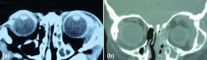
1078 Ruixin Ma et al.
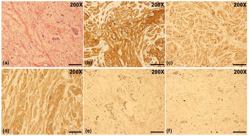
Figure 4: Histopathological examination of the tumor. (a) Hematoxylin-eosin (H&E) staining results of the tumor showed neuroblasts at
different degrees of differentiation arranged in nests and mature gangliocytes with fibrous connective tissue septa (H&E, original mag-
nification, 200×); (b) the number of CD56-positive tumor cells was 175.7 ± 6.5 per 3 high power field (HPF) (CD56, original magnification,
200×); (c) immunohistochemical staining results were positive for NSE and the number of these tumor cells was 163.3 ± 5.6 per 3 HPF (NSE,
original magnification, 200×); (d) the number of Syn-positive tumor cells was 172.1 ± 25.2 per 3 HPF (Syn, original magnification, 200×);
(e) the number of NeuN-positive cells was 122.7 ± 11.6 per 3 HPF (NeuN, original magnification, 200×); (f) the proportion of Ki-67-positive
cells was 2–5% per HPF (Ki-67, original magnification, 200×). Scale bar = 200 μm. ImageJ 1.50b with cell counter plugin (https://imagej.nih.
gov/ij/) was used for cell counting following the online guide.
3 Discussion tissues [13]. However, because of the atypical clinical
characteristics and imaging features of CT, histopatho-
Neuroblastic tumors are the most common childhood logical examination plays a significant role in the defini-
solid extracranial tumors, and GNB constitutes 20% of tive diagnosis of neuroblastoma subtypes, in differential
these tumors [9]. Clinical symptoms of patients with diagnosis, and in the determination of cellular matura-
orbital GNB include Horner’s syndrome and periorbital tion degree. These tumors vary in their relative propor-
bony lesions that destroy the palpebral vessels draining tion of neuroblasts and Schwann cells. Undifferentiated
the periorbital soft tissue [10,11]. Thus, patients pre- neoplasms are composed almost entirely of immature and
senting with periorbital ecchymosis and edema called undifferentiated neuroblasts. Differentiated cells include
“raccoon eyes” account for 30% of all orbital GNB neuroblasts differentiating into ganglion cells and mature
patients [12]. However, “raccoon eyes” is a nonspecific ganglion cells. Malignant neuroblastoma shows less than
symptom. Skull base fractures, Kaposi’s sarcoma, mul- 50% differentiated elements, with poor stroma and thin
tiple myeloma, and amyloidosis can also present “rac- fibrovascular septa. Homer Wright rosette is a typical char-
coon eyes.” Thus, imaging and pathological differential acteristic of a neuroblastoma, with circular or ovoid columns
diagnosis are indispensable for confirming GNB. Imaging of tumor cells arranged around a central core of the neuropil.
examinations, such as CT, magnetic resonance imaging However, it is not always present. Benign ganglioneuroma
(MRI), and ultrasonography, can be used to find neuro- comprises 100% differentiated cells, with a dominant stroma
blastic tumors. On plain CT, a tumor usually shows an characterized by extensive growth of Schwann cells. GNB is
irregular soft tissue mass, unclear edge, no obvious cap- intermediate, with more than 50% differentiated cells, as
sule, proneness to cystic change, and necrosis, accompa- was also observed in this case [2,14,15]. GNB can secrete
nied by calcification and invasion to the surrounding catecholamines, such as the vanillylmandelic acid (VMA)
Primary orbital ganglioneuroblastoma 1079
Figure 5: Abdominal CT findings. (a) A plain scan showing a left adrenal tumor of approximately 1.5 cm × 2.2 cm × 2.7 cm size (black arrow);
(b) a plain scan showing a calcified nodule on the left side of the abdominal aorta (black arrow); (c) an enhanced scan revealing left adrenal
tumor with increased calcification inside (black arrow); (d) an enhanced scan showing the mesenteric lymph nodes (black arrow).
and homovanillic acid (HVA). Furthermore, the VMA-to- the left orbital mass with bone destruction was found on
HVA ratio is often used as an indicator of tumor maturity orbital and abdominal CT at the initial visits. Abdominal
[16]. Moreover, NSE, CD56, Syn, and chromogranin A (CgA) CT conducted during the postoperative follow-up revealed
are the frequently used neuronal markers and have high the adrenal tumor, which was pathologically confirmed as
specificity; their positive rates are indicative of the severity GNB. Thus, the primary orbital GNB and metastatic adrenal
of neuroblastomas [17,18]. Furthermore, it is the increased GNB were considered.
amplification and expression of MYCN, a proto-oncogene, Surgical resection is the mainstay treatment for GNBs, and
and it always predicts poor prognosis [19,20]. In the pre- chemotherapy is recommended for high-risk patients [21,22].
sented case, the immunohistochemical results for NSE, In this case, the primary orbital tumor was timely resected, but
CD56, and Syn were positive and those for NeuN were partly postoperative chemotherapy was delayed, which may have
positive, favoring the diagnosis of GNB. been responsible for tumor recurrence and metastasis.
In 60–70% of cases, metastases are present with pri- To the best of our knowledge, this is the first reported
mary lesions at the initial diagnosis [9]. Two cases have case of primary orbital GNB and metastatic adrenal GNB.
been previously reported wherein GNB originated from The four previous case studies associated with orbital
the adrenal gland and metastasized to the orbit, and in GNB have been reported abroad, and we could not find
both these cases, the primary and metastatic sites were similar domestic studies on literature review. Two of
diagnosed simultaneously [7,8]. However, in our case, only these studies reported cases with both adrenal GNB and
Table 1: Comparison of published cases of orbital ganglioneuroblastoma
Author Year Age Organ involvement Treatment Outcome
Kim et al. 2011 2 years Skull, orbit, adrenal gland, Chemotherapy, surgery, bone marrow Survived, no
bone marrow transplantation recurrence
Johnson and 2003 10 months Adrenal gland, orbit, skull Chemotherapy Survived (>7 years)
Toledano
Dhermy et al. 1985 Not reported
Salas and Esparza 1963 Not reported
1080 Ruixin Ma et al.
orbital GNB. One study reported the case of a 10-month- Australian children: a population-based study (1983–2015).
old girl with metastatic orbital GNB and primary adrenal J Paediat Child Health 2020;56(7):1046–52.
GNB. She underwent chemotherapy and survived after [2] Shimada H, Ambros IM, Dehner LP, Hata JI, Joshi VV, Roald B,
et al. The international neuroblastoma pathology classification
7 years of follow-up. Another study reported a 2-year-old
(the Shimada system). Cancer. 1999;86(2):364–72.
girl with primary adrenal GNB and metastatic lesions including [3] Wahl HR, Craig PE. Report of a case showing a distinct gang-
the calva, orbit, and bone marrow. She underwent neoadjuvant lioneuroma, a neroblastoma and a cystic calcifying ganglio-
chemotherapy, surgery, and bone marrow transplantation and neuroblastoma. Am J Pathol. 1938;14:797–808.
survived without recurrence (Table 1). [4] Emre Ş, Zcan R, Bakır AC, Kuruğoğlu S, Omunoğlu N, Şen HS,
et al. Adrenal masses in children: imaging, surgical treatment
and outcome. Asian J Surg. 2020;43(1):207–12.
[5] Salas M, Esparza H. A case of ganglioneuroblastoma with
metastasis to the orbital bones. Boletin Med del Hospital
4 Conclusion Infant de Mexico. 1963;20:343–60.
[6] Dhermy P, Sekkat A, Moussaoui M, Bellakhdar N, Haye C,
Charlot JC. Ganglioneuroblastoma of the orbit. J Francais
Our report highlights the importance of early diagnosis
d’ophtalmol. 1985;8(2):139–46.
and intervention for primary orbital GNB in children with
[7] Johnson TE, Toledano SR. Ganglioneuroblastoma metastatic to
an orbital bony lesion, especially in cases with a primary the orbit. Ophthalmic Plastic Reconst Surg. 2003;19(4):330–3.
orbital tumor with “raccoon eyes.” Imaging methods com- [8] Kim SD, Jung TY, Jung S, Baek HJ. Neo-adjuvant chemotherapy
bined with histopathological examination can contribute followed by surgery for extensive calvarial metastases of a
to the accurate diagnosis of the primary and metastatic neuroblastoma. J Korean Neurosurg Soc. 2011;49(1):68–70.
[9] Ioana BT, Samașca G, Aldea C, Lupan I, Farcau D, Makovicky P.
lesions, particularly in the adrenal gland. Furthermore,
Ganglioneuroblastoma in children. Neurological Sci.
timely surgery combined with adjuvant chemotherapy 2019;40(9):1985–9.
and long-term follow-up is essential for reducing the [10] Lu D, Liu J, Chen Y, Chen F, Yang H. Primary cervical gang-
recurrence rate and metastasis of GNB and for improving lioneuroblastoma: a case report. Med (US).
the survival rate of the patients. 2018;97(12):e0090.
[11] John MM. Recent advances in neuroblastoma research. N Engl J
Med. 2010;362:2202–11.
Acknowledgments: The authors are grateful to the patient
[12] Alvi S, Karadaghy O, Manalang M, Weatherly R. Clinical man-
and her family for signing informed consent for publication. ifestations of neuroblastoma with head and neck involvement
in children. Int J Pediatric Otorhinolaryngol. 2017;97:157–62.
Funding information: This study was supported by a grant [13] Zhang X, Li C, Xu C, Hao X, Yu X, Li Q. Correlation of CT signs
from the 1.3.5 Project for disciplines of excellence-Clinical with lymphatic metastasis and pathology of neuroblastoma in
children. Oncol Lett. 2018;16(2):2439–43.
Research incubation Project, West China Hospital, Sichuan
[14] Peuchmaur M, D’Amore EG, Joshi VV, Hata JI, Roald B,
University (2018HXFH024) and Sichuan provincial science Dehner LP, et al. Revision of the international neuroblastoma
and technology program (2018SZ0128). pathology classification: confirmation of favorable and unfa-
vorable prognostic subsets in Ganglioneuroblastoma,
Author contributions: Ma RX gathered medical records of Nodular. Cancer. 2003;98(10):2274–81.
the patient; Ma RX and Wang YJ wrote the paper; Wang YJ [15] Joshi VV, Cantor AB, Altshuler G, Larkin EW, Neill JS, Shuster JJ,
et al. Age-linked prognostic categorization based on a new
and He WM revised the paper; Ma RX and Wang YJ con-
histologic grading system of neuroblastomas. A clinicopatho-
tributed equally to this work. logic study of 211 cases from the pediatric oncology group.
Cancer. 1992;69(8):2197–211.
Conflict of interest: None of the authors reports conflicts [16] Lonergan GJ, Schwab CM, Suarez ES, Carlson CL.
of interest in this work. Neuroblastoma, ganglioneuroblastoma, and ganglioneuroma:
radiologic-pathologic correlation. Radiographics.
2002;22(4):911–34.
Data availability statement: All data generated or analysed [17] Park SJ, Park CJ, Kim S, Jang S, Chi HS, Kim MJ, et al. Detection
during this study are included in this published article. of bone marrow metastases of neuroblastoma with immuno-
histochemical staining of CD56, chromogranin A, and synapto-
physin. Appl Immunohistochem Mol Morphol.
2010;18(4):348–52.
[18] Lam AK. Update on adrenal tumours in 2017 World Health
References Organization (WHO) of endocrine tumours. Endocr Pathol.
2017;28(3):213–27.
[1] Youlden DR, Jones BC, Cundy TP, Karpelowsky J, Aitken JF, [19] He WG, Yan Y, Tang W, Cai R, Ren G. Clinical and biological
McBride CA. Incidence and outcomes of neuroblastoma in features of neuroblastic tumors: a comparison ofPrimary orbital ganglioneuroblastoma 1081
neuroblastoma and ganglioneuroblastoma. Oncotarget. [21] Decarolis B, Simon T, Krug B, Leuschner I, Vokuhl C, Kaatsch P,
2017;8(23):37730–9. et al. Treatment and outcome of Ganglioneuroma and
[20] Vo KT, Matthay KK, Neuhaus J, London WB, Hero B, Ambros PF, Ganglioneuroblastoma intermixed. BMC Cancer. 2016;16(1):1–11.
et al. Clinical, biologic, and prognostic differences on the [22] Yang T, Huang Y, Xu T, Tan T, Yang J, Pan J, et al. Surgical
basis of primary tumor site in neuroblastoma: a report from management and outcomes of ganglioneuroma and ganglio-
the international neuroblastoma risk group project. neuroblastoma-intermixed. Pediatric Surg Int.
J Clin Oncol. 2014;32(28):3169–76. 2017;33(9):955–9.You can also read


