Manual for the use of mating-based Split ubiquitin system "mbSUS" - version B Petr Obrdlik December 2004
←
→
Page content transcription
If your browser does not render page correctly, please read the page content below
Manual for the use of
mating-based Split ubiquitin system
            "mbSUS"

                                   version B

                                 Petr Obrdlik
                               December 2004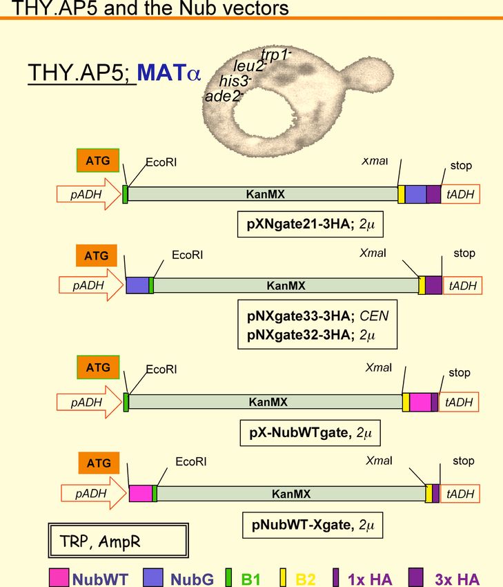
1     INTRODUCTION AND GENERAL INFORMATION                                3

1.1     ABBREVIATIONS                                                      4
1.2     MBSUS PACKAGE CONTENTS                                             5
1.3     REFERENCES AND CITATION                                            6
1.4     THE MBSUS IN BRIEF                                                 7
1.4.1    GENERAL INFORMATION                                               7
1.4.2    PNXGATE33-3HA VERSUS PNXGATE32-3HA                                7
1.4.3    CONTROL CONSTRUCTS (C=CUB, N=NUBG)                                8
1.4.4    NUBG-X OR X-NUBG FUSIONS?                                         8
1.4.5    QUANTITATIVE b-GALACTOSIDASE ASSAYS                               8
1.4.6    THE SENSITIVITY OF THE METHODS FOR DETECTION OF INTERACTION:      8
1.4.7    THE "POOLING" STRATEGY                                            9
1.5     CHECK-LIST BEFORE YOU START MBSUS EXPERIMENTS                     10

2     PROTOCOLS                                                           11

2.1     IN VIVO CLONING INTO MBSUS VECTORS                                12
2.2     MBSUS TESTS PROTOCOL                                              13
2.3     VERIFYING DETECTED INTERACTIONS                                   15
2.4     LIAC TRANSFORMATION OF YEAST                                      16
2.5     X-GAL ASSAYS                                                      19
2.6     "LAZY BONES" METHOD FOR RAPID RELEASE OF PLASMID DNA FROM YEAST   20

3     FIGURES                                                             21

4     MAPS OF VECTORS AND CONTROL CONSTRUCTS                              29

                                                                           2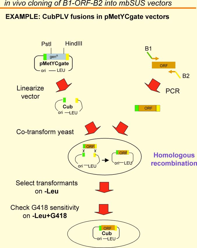
1   Introduction and general information

                                           3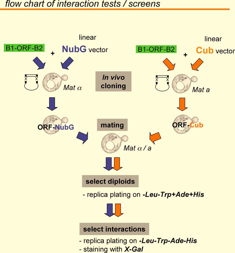
1.1 Abbreviations

X = prey peptide/ORF
Y = bait peptide/ORF
N = NubG
C = Cub
PLV = protA-LexA-VP16 peptide
HA tag = hemagglutinin epitope tag

Ade = adenine
His = histidine
Trp = tryptophan
Leu = leucine
Ura = uracil
Met = methionine

X-Gal = 5-bromo-4-chloro-3-indol-b-D-galactosidase
ONPG = o-nitrophenylgalactopyranoside

SC = synthetic complete medium
SD = synthetic dextrose / minimal medium
SSDNA = carrier DNA from salmon sperm

                                                     4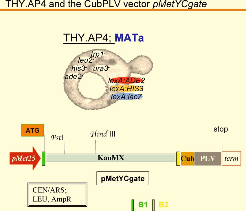
1.2 mbSUS package contents

You have received the package with mating-based Split ubiquitin system " mbSUS". This
package contains:
      yeast strains THY.AP4 and THY.AP5
      vectors (resuspended in dH2O, ca. 50 ng/ul)
               1     pXNgate21-3HA
               2     pNXgate33-3HA
               3     pX-NubWTgate
               4     pNubWT-Xgate
               5     pMetYCgate
              12     pNXgate32-3HA
      control constructs (resuspended in dH2O, ca. 50 ng/ul)
               6     KAT1-Cub (Arabidopsis K+ channel, Acc. At5g46240)
               7     KAT1-N
              11     N-KAT1-3HA(33)
              13     N-KAT1-3HA(32)

Restriction analysis:
The maps of the vectors and control constructs are shown in the section "Maps of vectors and
control constructs". The sequences of the vectors1-5 and 12 in genebank format are in the
"Appendix".

General information, protocols, maps and sequences of the vectors are
included in this manual. For further information please check Obrdlik et
al., 2004 and Ludewig et al., 2003.

                                                                                          5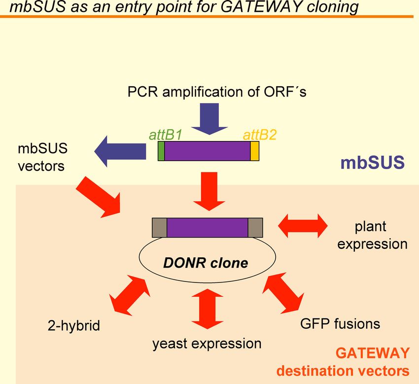
1.3 References and citation

When citing the system, the vectors pXNgate21-3HA, pMetYCgate, pX-NubWTgate,
pNubWT-Xgate and pNXgate32-3HA, and the control constructs please refer to the
publication Obrdlik et al., 2004, PNAS 101; 12242-12247.

When citing the vector pNXgate33-3HA, please refer to C. Cappellaro and E. Boles,
University of Frankfurt, unpublished.

Differences to the vectors published in Obrdlik et al., 2004

The expression cassette of the vectors pXNgate21-3HA, pNXgate33-3HA and pNXgate32-
3HA is based on the corresponding vectors pXNgate and pNXgate in Obrdlik et al. (2004).
The additional feature is the triple HA tag, which allows the construction of ORF-NubG-
3HA and NubG-ORF-3HA fusions. The triple HA tag is readily detectable with anti-HA
antibodies.

The difference between pNXgate33-3HA and pNXgate32-3HA: pNXgate33-3HA is a low-
copy CEN plasmid, which mediates significantly lower expression of NubG-ORF fusions
when compared to pNXgate32-3HA. To read more about the differences between
pNXgate32-3HA and pNXgate33-3HA please see Chapter 1.4.2 of the Manual.

Please note that the control construct KAT1-N has not a triple but single HA-tag peptide
fused to the C-terminus!

                                                                                      6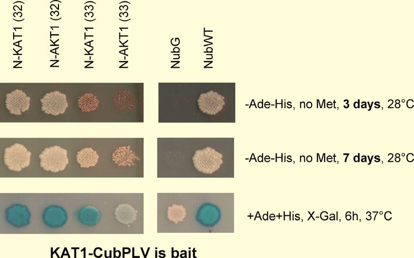
1.4 The mbSUS in brief

1.4.1 General information
-The vectors are designed for recombinational in vivo cloning in yeast: you need only one
PCR-product of the gene of interest, which you can insert in any of the Nub or Cub vectors
("In vivo cloning into mbSUS vectors" and Fig. 4 and 5).
- Big plus: the linkers in our system contain attB1 and attB2 sites, which are compatible with
  GATEWAY cloning (Invitrogen). Thus you can use either the PCR products or the resulting
  Split-ubiquitin constructs for further cloning into any of the GATEWAY destination vectors
  (Fig. 6). Nevertheless, keep in mind that the mbSUS vectors themselves are N O T
  DESTINATION VECTORS!
-Expression of CubPLV fusions can be optimized since it is under the control of MET25
  promoter. We routinely test interactions on different methionine concentrations.
-We use two different yeast strains: THY.AP5 is Mat  mating type strain and is designed for
  the transformation with Nub fusions, the second strain THY.AP4 [Mat a] is designed for
  transformation with CubPLV fusions. Therefore we can test the interactions by mating
  approach in diploid cells. (see "mbSUS tests protocol" and Fig. 5)
-There are three reporter constructs in our yeast strains: lexA-HIS3, lexA-Ade2 and lexA-lacZ.
  Thus we can select the interacting partners via growth (on -Ade-His media), by white color
  on non-selective media (+Ade+His) or via ß-galactosidase assays (Fig. 3)
-NubG vectors possess a 3 x HA tag at their C-termini, which is detectable in most of the Nub
  fusions tested. This is useful if you want to show that the Nub fusions are indeed expressed.
- There are also vectors for construction NubWT-X and X-NubWT fusions. Especially X-
  NubWT fusions can be useful since the affinity of NubG for Cub in X-NubG is often very
  low and hardly shows any interaction (see also below).
- The empty NubWT-X gate vector (i.e. soluble NubWT peptide) should also be used as a
  positive control.

1.4.2 pNXgate33-3HA versus pNXgate32-3HA
pNXgate33-3HA and pNXgate32-3HA vectors differ by their copy number per cell:
(i) The pNXgate33-3HA is a low-copy CEN plasmid, which mediates significantly lower
expression of NubG-ORF fusions when compared to pNXgate32-3HA. Therefore the use of
pNXgate33-3HA results in higher signal/background ratio in growth assays on solid media
(Fig. 7) when compared to pNXgate32-3HA, however, the expression of NubG-ORF fusions
may be too low to allow quantitative analysis of the interactions (see below). Moreover, the
low expression levels of NubG-ORF fusions may not allow the detection of weak interactions
(false negatives)!
(ii) The pNXgate32-3HA is a high-copy 2µ plasmid, which mediates higher expression of
NubG-ORF fusions when compared to pNXgate33-3HA. It may - but does not have to - result
in low signal/background ratio in growth assays (false positives). If you observe such
background problems you can perform quantitative -galactosidase assays (measuring lacZ
activity, Obrdlik et al., 2004), which can distinguish between false and true interactions.
Alternatively try to adjust a proper methionine concentration in the medium.
Our recommendation:

                                                                                             7
Start with the pNXgate33-3HA vector and detect the interactions via selective growth on solid
media without His, Ade, Trp, Leu and Ura. Then select the protein pairs of interest, fuse them
into the pNXgate32-3HA vector and quantify them via quantitative -galactosidase assays.
If you have problems to detect the expected interactions (false negatives) with pNXgate33-
3HA vector on solid His-,Ade-,Trp-,Leu-,Ura- media, use the pNXgate32-3HA vector. If you
then face the problem of low signal/background ratio (false positives) try to quantify the
interactions via quantitative -galactosidase assays.
1.4.3 Control constructs (C=Cub, N=NubG)
The system works very well for KAT1 interactions: we use the homomerization of KAT1 as a
positive control in our assays. Hence the KAT1-C, KAT1-N, N-KAT1-3HA(33) and N-
KAT1-3HA(32) are included as positive controls. KAT1-N contains a single HA tag at the C-
terminus.
1.4.4 NubG-X or X-NubG fusions?
It is crucial for mbSUS function that the CubPLV and the Nub peptides are both located on
the cytosolic face of membranes. We strongly recommend you to check the topology of the
proteins of interest before you start the experiments. The topology of plant membrane proteins
can be checked via ARAMEMNON database (http://aramemnon.botanik.uni-koeln.de/).
X-NubG:
In general, Cub has higher affinity to Nub fused to the N-termini of prey (Nub-X fusions) than
to Nub fused to C-termini of prey (X-Nub fusions). When testing interactions with a set of X-
NubG fusions (e.g. KAT1-Cub with X-NubG fusions) the signal/background ratio is usually
very high. However you may not be able to detect "weak" interactions.
NubG-X:
When using NubG-X fusions it is important to test that your CubPLV fusions do not show
background activities in growth assays with different NubG-X fusions (false positives)! It is
crucial to use several different NubG-X fusions as negative controls! An empty
pNXgate33-3HA and/or pNXgate32-3HA vector (soluble NubG peptide) is not enough. This
is especially important if you decide to use pNXgate32-3HA. The pNXgate33-3HA produces
less background but the expression of NubG-ORF fusions may be too low to allow
quantitative analysis of the interactions (chapter 1.4.2, Fig. 7).
1.4.5 Quantitative -galactosidase assays
To obtain direct comparison of two or more interactions you can perform quantitative -
galactosidase assays by measuring lacZ activity, (Obrdlik et al., 2004). This may on one
hand help to distinguish between false and true interactions, on the other hand the
quantification is very useful when performing interaction domain mapping.
1.4.6 The sensitivity of the methods for detection of interaction
The growth assays via selection on media -Ade-His-Trp-Leu-Ura is the most sensitive one.
The detection of interactions via lacZ-activity by X-Gal assays and liquid ONPG assays is not
as sensitive but liquid assays are useful for quantitative analysis. The detection of interactions
by monitoring the white (interaction) and red (no interaction) color of diploid cells (Fig. 3) is
the least sensitive and can be used only in addition to the other detection methods.
81.4.7 The "pooling" strategy
For in vivo cloning of the PCR product combined with direct interaction tests use pools of 5-
10 independent clones as described in the protocol for screening "mbSUS tests protocol". In
this way it is possible to suppress the effects of non-functional mutants. Such mutations can
be caused by the RT-PCR as well as by the recombination events during in vivo cloning (rare
but possible mutations in the vicinity of homologous regions).
91.5 Check-list before you start mbSUS experiments
1) In order to check CubPLV fusions, use the empty pNXgate33-3HA or pNXgate32-3HA,
and pNubWT-Xgate vectors as negative and positive controls, respectively.
2) It is not sufficient to use the empty pNXgate33-3HA or pNXgate32-3HA vector as a
negative control. Use also NubG fusions of several membrane proteins as negative controls
together with the membrane protein of your interest!
3) If you want to construct X-Nub fusions, consider also creating X-NubWT fusions. X-
NubWT fusions can provide specific results if you analyze the interactions via quantitative
lacZ assays (but do not forget negative controls from 2!).
4) For the production of B1-ORF-B2 inserts by PCR ("In vivo cloning into mbSUS vectors"
and Fig. 4) it is better to start from a sequenced DNA template. Using this you will reduce the
risk of mistakes produced during PCR.
102 Protocols
This section contains protocols adapted for mbSUS. General protocols and the
preparation of general media and chemicals should be performed as described in
"Methods in Yeast Genetics" (Adams et al., Cold Spring Harbor Lab. Press) and in
"Current Protocols in Molecular Biology" (Ausubel et al., John Wiley & Sons Inc.).
112.1 In vivo cloning into mbSUS vectors
The principle of the in vivo cloning is described in Obrdlik et al., 2004 and in Fig. 4 of this manual.
For in vivo cloning into mbSUS vectors the ORFs have to be flanked by B1 and B2 linkers via PCR. In parallel
the vector pMetYCgate and the Nub vectors have to be restricted with PstI/HindIII and with EcoRI/SmaI,
respectively.
Both the B1-ORF-B2 PCR product and the appropriate linear vector are used to co-transform either THY.AP4 or
THY.AP5 yeast (Fig. 4 and 5). Homologous recombination between B1 and B2 sequences of the B1-ORF-B2
and of the linear vector produces circular vector harboring the ORF. Transformants are selected on -Leu
(CubPLV fusions) or on -Trp (Nub fusions).
1): B1 and B2 linker sequences of the vectors
Linker B1
attB1
aca agt ttg tac aaa aaa gca ggc tct cca acc acc atg
T S L Y K K A G S P T T Met
Linker B2
attB2 BamHI
tac cca gct ttc ttg tac aaa gtg gtt ggt ggt ggc gga tcc ggt gga ggt gga tca
Y P A F L Y K V V G G G G S G G G G S
The underlined B1 and B2 nucleotid sequences are crucial for GATEWAY cloning and may not be changed according to
codon usage rules (even if the corresponding amino acid is the same)! (Invitrogen; Hartley et al. (2000) Genome Research
10: 1788; Walhout et al. (2000) Science 287:116).
2): Primer design for in vivo cloning into mbSUS vectors
B1
forward primer
open reading frame (ORF)
5` 3`
3` 5`
B2
reverse primer
B1-forward primer (5´strand)
B1-linker start-ORF
aca agt ttg tac aaa aaa gca ggc tct cca acc acc atg xxx-5´strand cDNA
B2-reverse primer (3´strand)
B2-linker
tcc gcc acc acc aac cac ttt gta caa gaa agc tgg gta xxx-3´strand cDNA w/o stop!
3): Enzymes for the cleavage of the mbSUS vectors for in vivo cloning
pXNgate3HA, pX-NubWTgate: EcoRI & Sma I
pNXgate33-3HA, pNXgate32-3HA, pNubWT-Xgate: EcoRI & Sma I
pMetYCgate: Pst I & Hind III
122.2 MbSUS tests protocol
See also Fig. 3, 4 and 5
Recombinational in vivo cloning:
1. Cut the vectors with restriction enzymes as described in "In vivo cloning into mbSUS
vectors". Purify the fragments via agarose gel.
2. Make a B1-ORF-B2 insert by PCR using primers described in "In vivo cloning into
mbSUS vectors". Purify the PCR fragments via affinity columns.
3. Co-transform a linear vector and the B1-ORF-B2 insert into either THY.AP4 (Cubs)
or THY.AP5 (Nubs) as described in "LiAc Transformation". Do not forget to
transform the linear vector alone (negative control!). Plate on appropriate media
without Leu (Cub vectors in THY.AP4) or without Trp (Nub vectors in THY.AP5).
Mating:
4. Collect 5-10 independent clones and mix them in 0.1 ml dH2O. Use 20-100 µl of this
suspension to inoculate 5ml of the appropriate SC media without G418 and 5 ml with
G418 (starting cultures should be only slightly turbid).
5. Grow cells over night to the stationary phase (stock culture). Cells carrying vectors
with inserts should not grow on G418.
6. Concentrate 1ml of the cultures by centrifugation in a final volume of 200µl YPD.
200 µl are enough for 13 crossings. For higher number of crossings use a higher
volume of the culture (e.g. for 26 crossings concentrate 2ml culture in 400 µl YPD).
Mating and replica plating is more efficient if the final suspension is not too thin!
7. Mix 15 µl of the appropriate mating types for each cross. You can use microtiter plates
for large number of interactions.
8. Drop 4 µl of the mixed suspensions on a YPD-plate (this should give a patch of about
7mm). It takes a while for the liquid to be absorbed. For that reason plates should be
pre-dried under a hood.
9. Mate the cells for 6-8h at 28°C.
10. Replica plating on SC/+Ade+His; make minimum 2 replicas of each YPD plate
11. Selection of diploid cells (2-3 days incubation at 28°C).
Interaction growth tests:
12. Replica plating of the cells on 4 different plates (synthetic minimal medium )
a) + 0 mM methionine
b) + 75 µM methionine
c) + 150 µM methionine
d) + 400 µM methionine
13. In addition to replica plating it is recommended to streak out the colonies on synthetic
minimal medium with different methionine concentrations to check for growth
properties!
14. After two days at 30°C start to record the growth.
X-gal tests:
15. Replica plating of the cells on different plates
(synthetic minimal medium +ADE, +HIS, )
a) + 0 mM methionine
b) + 150 µM methionine
1316. After 2-3 days test the -galactosidase activity by "X-Gal Overlay assay".
Synthetic minimal medium (SD)
2% w/v Glucose
0,17% w/v yeast nitrogen base without ammonium sulfate and amino acids
0,5% ammonium sulfate
pH adjusted to 6-6,3 with NaOH
Synthetic complete medium (SC)
like SD medium but add to 1L medium
1,5g of "AHTLUM"-drop out (-Ade, -His, -Leu, -Trp, -Ura, -Met)
add appropriate chemicals for auxotrophy selection (see below)
AHTLUM minus drop-out
standard protocol for drop-out, but without Adenine, Histidine, Leucine, Tryptophan,
Uracil, Methionine
Chemicals for auxotrophy selection
all dissolved in dH2O and sterilized by filtering
chemical stock conc. vol (ml) stock storage
(g/100ml) for 1L medium
A adenine sulfate 0.2 10 RT
U uracil 0.2 10 RT
T L-tryptophan 1 2 4°C
L L-leucine 1 10 4°C
H L-histidine HCl 1 2 4°C
Met L-methionine 1 2 4°C
all these can be added to the media before autoclaving !!!
142.3 Verifying detected interactions
The interactions identified in the directed mbSUS tests with the in-vivo cloned ORFs (Fig. 5
and "mbSUS tests protocol") have to be verified.
1) isolate the plasmid DNA form yeast ("Lazy-bones method for rapid release of plasmid
DNA from yeast")
2) Amplify the plasmid DNA in E.coli.
3) Isolate the plasmid DNA from E.coli and verify the expression cassette by restriction
analysis and by sequencing
4) Use the verified plasmid DNA instead of B1-ORF-B2 and linear vectors for mbSUS
interaction tests as described "mbSUS tests protocol".
152.4 LiAc transformation of yeast
(modified protocol of Gietz & Schiestl, 1995)
Work all the time under strictly sterile conditions!
Making competent cells
1. Incubate one colony of THY.AP5 and THY.AP4, each at 28°C for ca. 24 h (shaking in 5 ml
YPAD)
2. Early in the morning: Inoculate 100 ml pre-warmed YPAD with the pre-culture to an OD600
0.08-0,1.
3. Incubate at 28°C and 180-200 rpm till OD600 0.5-0,6.
Important: Minimal incubation time = time necessary for 2-3 duplications!
4. Place cells in sterile tubes (FALCON), centrifuge at 2.500 x g for 5 min (20°C).
5. Remove the medium, re-suspend pellets in 5 ml sterile ddH2O each and re-centrifuge again
as described above.
6. Remove ddH2O, re-suspend each pellet in 2,5 ml LiAc/TE, pool the suspensions together
and mix carefully.
7. Centrifuge (5 min; 2.500 x g, 20°C) and remove the supernatant.
8. Re-suspend the pellet carefully in 0.5-0.8 ml LiAc/TE, let the suspension incubate for
30min at RT (competent cells!). If you use the cells later than 30 min after this step keep them
at 4°C.
Transformation
before starting: boil the SSDNA for 3 min and chill it on ice immediately
9. For each transformation add in the following order:
-20 µl carrier-SSDNA (5 or 10 mg/ml)
-20 µl DNA-mix: = 1x TE buffer and DNA
either: plasmid alone (0,1 - 10 µg)
or: linear plasmid (100 ng) plus insert (>100 ng), (molecular ratio of vector
vs insert at least 1:4)
! mix well !
- 4,5 µl 1M LiAc
! mix well !
-50 µl competent cells, !mix well!
-300 µl PEG/LiAc mix (prepare it fresh each time!!!) and mix well!!!
13. Incubate shaking for 20 min at 30°C (thermo-mixer)
1614. Heat-shock in 42°C-waterbath for 20 min (keep the time!!!)
15. Centrifuge at 6000 - 8000 rpm for 1 min and carefully remove the supernatant with a
micropipette
16. Resuspend the pellet carefully in 100µl sterile dH2O (or sterile 1x TE buffer) with the
pipette.
17. Streak the transformation on appropriate selective media (The media on petri dishes
should be dry!).
For transformation of THY.AP4 with Cub constructs: SC/+AHTU
For transformation of THY.AP5 with Nub constructs: SC/+AHL
!!! For some purposes the transformation efficiency may be too high and it will be difficult to
isolate single colonies from the plates! In that case resuspend the cells (step 16) in 1ml of
sterile dH2O and streak out 100µl on the plate
18. Grow for 2-4 days at 28°C (30°C) on selective media.
Working solutions:
LiAc/TE
1 ml 10 x TE
1 ml 1M LiAc
8 ml ddH2O
PEG/LiAc mix:
0,5 ml 10 x TE
0,5 ml 1M LiAc
4,0 ml 50% PEG
Stock solutions:
1M LiAc:
LiAc in ddH2O, pH not adjusted!, sterilize by filtering
10 x TE:
100 mM Tris HCl,
10 mM EDTA, pH 7,5; adjust with NaOH
50% PEG 3350 (Roth ) or PEG 4000 (Fluka)
sterilize by autoclaving
5mg/ml salmon sperm DNA (SSDNA), in 1xTE
make aliquots and keep in -20°C
2 x 100 ml sterile ddH2O
17chemicals for auxotrophy selection:
all solutions are in ddH2O and sterilized by filtering:
amino acid stock conc. vol (ml) stock storage
(g/100ml) for 1L medium
A adenine sulfate 0.2 10 RT
U uracil 0.2 10 RT
T L-tryptophan 1 2 4°C
L L-leucine 1 10 4°C
H L-histidine HCl 1 2 4°C
Met L-methionine 1 2 4°C
all these can be added to the media before autoclaving !!!
Media:
YPAD:
add 10 ml of sterile adenine stock to YPD media
AHTLUM minus drop-out:
standard protocol for drop-out, but without Adenine, Histidine, Leucine, Tryptophan,
Uracil, Methionine
SC- (synthetic complete medium):
for 1L:
1,7 g yeast nitrogen base w/o amino acids and ammonium sulfate
5,0 g (NH4)2 SO4
20 g glucose
1,5 g of AHTLUM minus drop-out
20 g Oxoid Agar; adjust pH at 6,0-6,3 with NaOH
Add the appropiate amount of auxotrophy selection chemicals. Leu, Trp, Met, Ade,
His and Ura can be added to the medium before autoclaving.
SD- (synthetic dextrose minimal medium):
the same protocol as SC-medium, but without the AHTLUM minus drop-out
182.5 X-Gal assays
Overlay assay
1): Dissolve 0.25g agarose in 50ml Z-buffer (pH 7.2) in the microwave. Do not overheat, otherwise
precipitation may occur! 50ml is enough for 3-4 petri dishes.
2): Cool down the solution to 50°C and add 1ml of 10% SDS and 1ml of X-Gal solution (100mg/ml in
DMF). The final concentration of X-Gal is 2mg/ml.
3): Pour the solution carefully over the plate. Incubate at 37°C for 5min to several hours.
Solutions
Z-buffer
Na2HPO4 x 2H2O 10,68g/L (60mM) or: Na2HPO4 x 7 H2O; 16.1 g/L (60 mM)
NaH2PO4 x H2O 5.5 g/L
KCl 0.75 g/L
MgSO4 x 7H2O 0.246 g/L
adjust to pH 7.0 and autolave
100mg/ml X-Gal, in DMF,
store at -20°C in dark
10% SDS
Filter assay
1): Cut nitorcellulose filter under sterile conditions and place it (use sterile forceps) on a selective
media plate.
2): Streak the clones on the filter and incubate the petri dish at 30 °C for 2-4 days.
3): Remove the filter carefully from the plate and place it for 5-10 sec. in liquid N2. Take it out and let
it thaw at RT for 1 min.
4): Repeat the step 3).
5): Place the filter with colonies facing up on Whatman paper soaked with 2ml of freshly prepared Z-
buffer/X-Gal solution (in a petri dish). Incubate closed petri dish in the dark for 30 min to ON (RT or
30°C)
Solutions:
Z-buffer
described above
X-Gal solution
80 mg/ml in DMF
each time freshly prepared: Z-buffer/X-Gal solution
100 ml Z-buffer
0.27 ml -mercaptoethanol
1.67 ml X-Gal solution (final X-Gal conc. ca. 1.4 mg/ml)
192.6 "Lazy bones" method for rapid release of plasmid DNA
from yeast
(Adapted from Kaiser et al., Methods in yeast Genetics, CSHL Press, 1994)
1: Incubate one yeast colony at 28°C in 5ml YPD (ON) or in selective medium
(1-2 days)
2: Pellet 1.5ml of ON yeast cell culture at 5000xg for 5 min. Discard the supernatant.
3: Add 0.2ml of the DNA release solution and resuspend the pellet.
4: Add 0.2ml phenol:chloroform:isoamylalcohol (25:24:1) and 0.3ml of acid washed glas
beads.
5: Vortex at high speed for 5-10 min.
6: Centrifuge for 5 min at RT and full speed (Eppendorf centrifuge).
7: Transfer 120µl of the aqueous layer (the upper layer) to a new tube. Precipitate the
DNA with 1xVol isopropanol and wash with 1xVol 70% EtOH. Resuspend the pellet in
30µl dH2O.
8: To transform chemo-competent E.coli use 5-10µl of the DNA. For electro-competent
cells use 1µl of the DNA.
Solutions
DNA release solution:
2% Triton X-100
1% SDS
100mM NaCl
10mM Tris HCl (8.0)
1mM Na-EDTA
! do not autoclave !
Acid washed beads:
Use 250-500 micron beads (0,7-1,0mm should also work), work under a fume hood !!!
Wash 100ml glass beads in 200ml conc. HCl (stir them carefully from time to time) and
let them stand ON in a fume hood.
Discard the HCl and wash the beads several times with dH2O, to the pH >5.0
(if the pH is still below pH 3.0 after several washing steps, make the last wash with
20mM Na2CO3, pH 7.0).
Dry the beads in a glass beaker at 70°C ON and transfer them into a bottle.
203 Figures
21Fig. 1: Reporter yeast strain THY.AP4 and expression cassette of the vector
pMetYCgate. pMetYCgate is suited for "Y-CubPLV” fusions of bait peptides “Y”. B1-
KanMX-B2 is identical with the B1-KanMX-B2 cassettes of the Nub vectors (Fig. 2).
Promoter is red, term marks the terminator, the linkers B1 and B2 are green and yellow,
respectively. Marked restriction sites are used to produce linear vectors for in vivo cloning.
„ATG“ and „stop“ mark the start and the stop codon in the expression cassette. AmpR refers to
ampicillin resistance cassette. CEN/ARS refers to a low-copy yeast origin of replication. For
more details on the pMetYCgate sequence see the appendix.
22Fig. 2: Yeast strain THY.AP5 and the expression cassettes of the Nub vectors. Nub
vectors are suited for ”X-Nub” or “Nub-X” fusions of prey peptides “X”. All B1-KanMX-B2
cassettes are identical (see also Fig. 1). pADH is the ADH1 promoter, tADH marks the ADH1
terminator, NubG, NubWT, HA tag and the linkers B1 and B2 are shown below. Marked
restriction sites are used to produce linear vectors for in vivo cloning. „ATG“ and „stop“ mark
the start and the stop codons in the expression cassettes. AmpR refers to ampicillin resistance
cassette. 2µ refers to a high-copy, CEN to a low-copy yeast origin of replication. All vectors
carry TRP1 and AmpR selection markers. For more details on the vector sequences see the
appendix.
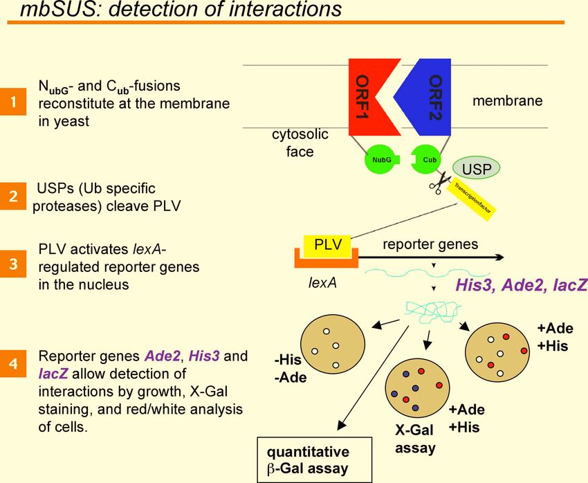
23Fig. 3: Detection of interactions with mbSUS. To enable interaction-dependent cleavage of
the PLV peptide, NubG and Cub have to be on the cytosolic face of the membrane. The
cleaved PLV transcription factor diffuses into the nucleus, binds lexA-regulated promoters
and activates the reporter genes. Activation of the reporter genes Ade2 and His3 allows
selection of interactions via growth. In addition, lacZ allows detection of interactions by X-
Gal staining and Ade2 enables red/white selection of interacting proteins (white = interaction,
red = no interaction). LacZ activity can also be measured with quantitative Gal assays.
24Fig. 4: The principle of in vivo cloning into mbSUS vectors. The figure shows the
construction of CubPLV fusions. Nub fusions are constructed in similar way using linear
pXNgate21-3HA, pNXgate33-3HA, pNXgate32-3HA, pX-NubWTgate or pNubWT-Xgate
vectors.
25Fig. 5: Flow chart of mbSUS interaction tests. Mat strain is THY.AP5, Mat a strain is
THY.AP4. Leu, Trp, Ade and His is leucine, tryptophan, adenine and histidine.
26Fig. 6: mbSUS as an entry point for versatile analysis of membrane proteins. B1-ORF-
B2 PCR products as well as the mbSUS fusions can be used as entry points to subclone the
inserts into different GATEWAY destination vectors. DONR marks the pDONR vectors of
GATEWAY (Invitrogen). Please note that the mbSUS vectors are NOT DESTINATION
VECTORS and thus are not suitable for direct cloning via GATEWAY!
27Fig. 7: The “strength” of NubG-ORF interactions depends on the applied pNXgate
vectors. The K+ channels KAT1 and AKT1 from Arabidopsis were cloned into the vectors
pNXgate32 and pNXgate33 and the resulting NubG-ORF fusions were tested against
KAT1-CubPLV fusion peptide as bait. The interactions were visualized via growth
selection after 3 and 7 days incubation, and via X-Gal assays. Soluble NubG (an empty
pNXgate33) and NubWT (an empty pNubWT-Xgate) were used as negative and positive
control, respectively.
284 Maps of vectors and control constructs
29pXNgate21-3HA, 8194 bp
(TRP1, AmpR, 2µ)
pNXgate33-3HA, 6682 bp
(TRP1, AmpR, CEN)
30pX-NubWTgate, 8120 bp
(TRP1, AmpR, 2µ)
pNubWT-Xgate, 8118 bp
(TRP1, AmpR, 2µ)
31pMetYCgate, 10033 bp
(LEU2, AmpR, CEN/ARS)
pNXgate32-3HA, 8177 bp
(TRP1, AmpR, 2µ)
32KAT1-C, 10580 bp
(LEU2, AmpR, CEN/ARS)
KAT1-N, 8667 bp
(TRP1, AmpR, 2µ)
33N-KAT1-3HA(33), 7229 bp
(TRP1, AmpR, CEN)
N-KAT1-3HA(32), 8724 bp
(TRP1, AmpR, 2µ)
34You can also read