Diagnostic studies for common invasive and infectious pathologies of the Orenburg region
←
→
Page content transcription
If your browser does not render page correctly, please read the page content below
E3S Web of Conferences 282, 03020 (2021)                                     https://doi.org/10.1051/e3sconf/202128203020
EFSC2021

         Diagnostic studies for common invasive and
         infectious pathologies of rabbits in the
         Orenburg region
         Z.Kh. Terentyeva1,*, R.Sh. Taiguzin1, O.A. Matveev1, Kh.Kh. Shakhbiyev2, and
         A.L. Kryazhev3
         1Orenburg  FSBEI HE Orenburg SAU, Orenburg, Russia
         2Chechen  State University, Grozny, Russia
         3Federal State Budgetary Educational Institution of Higher Education Vologda State Dairy

         Academy named after N.V. Vereshchagin, Vologda, Russia.

                       Abstract. Rabbits, like other animal species, are susceptible to infection
                       with invasive and infectious diseases. Both ecto- and endoparasites are
                       registered in them, including helminths, insects on the body, acarus
                       scabies, protozoa. Rabbits are characterized by a high degree of precocity,
                       high fecundity and the breadth of use of the products obtained from them.
                       As a product of rabbit breeding, first, meat is in demand as a dietary
                       product. Part of the products from rabbits - hair, wool-molt is obtained
                       during the life of the animal, and meat, skins, other products - after
                       slaughter. Skins and rabbit hair are used for the manufacture of fur
                       products, as well as in the felt and knitting industry. In addition, biologists,
                       doctors, veterinarians use rabbits as laboratory animals in scientific
                       research, for testing cosmetics, as well as in laboratories and bio factories
                       for drug testing. To preserve the population of these species, it is important
                       to prevent infection with various pathologies inherent in rabbits, including
                       invasive diseases.

         1 Introduction
             Rabbits, in comparison with farm animals, are distinguished by their precocity, high
         fecundity, and the breadth of use of the products obtained from them. The products of
         rabbit breeding as a food product primarily include meat, which is in demand as a dietary
         product. Such products from rabbits as - hair, wool-molt are obtained during the life of the
         animal, and meat, skins, and other products - after slaughter. Skins and rabbit hair are used
         for the manufacture of fur products, as well as for the manufacture of felt and knitwear. In
         addition, biologists, doctors, and veterinarians use rabbits as laboratory animals, as well as
         in scientific research, for testing cosmetics, and in laboratories and bio factories for drug
         testing [1, 2].

         *
             Corresponding author: zoy19570501@mail.ru

   © The Authors, published by EDP Sciences. This is an open access article distributed under the terms of the Creative
   Commons Attribution License 4.0 (http://creativecommons.org/licenses/by/4.0/).
E3S Web of Conferences 282, 03020 (2021)                             https://doi.org/10.1051/e3sconf/202128203020
EFSC2021

             The quality and quantity of rabbit breeding products depends on the breed and
         hereditary characteristics of animals, on the impact of various environmental factors, on the
         economic use of this type of animal and on the type of animal maintenance, as well as on
         various pathologies, including infestations inherent in this type of animal [3, 5, 6, 10, 13,
         14]. In Russia, in 1927, the breeding of rabbits was mainly amateur in nature, during this
         period, several thousand high-value breeds were imported to the country such as: Belgian
         Hare, chinchilla (small), white giant, Viennese white, Viennese blue, champagne, Angora
         ram, Havanna. In 1929-1931, large rabbit farms were established in different parts of the
         country for breeding pedigree animals. At that time, the main task of the rabbit breeding
         revival was the reproduction of breeding rabbits by crossing with purebred animals and
         thereby improving the quality of local mongrel animals [4, 7, 9, 14].
             During this period, factories for processing of rabbit skins were built in the country.
         Thus in 1934-1935, the volume of rabbit skin products in the fur processing industry was
         35%. The range of products from rabbit raw materials was diverse and high-quality skins
         were produced, both in natural form and imitated for more valuable furs (beaver, leopard,
         cat, sable), which were used to make hats, women's coats, collars, jackets. From skins
         unsuitable for the fur industry, leather was made for the manufacture of haberdashery
         products, light shoes. Felt boots, hats, berets, and other items were made from rabbit hair,
         and high-quality berets, sweaters, children's suits, shawls, scarves, stoles, gloves, [8,9] were
         made from the hair of Angora rabbits.
             Currently, several rabbit farms are engaged in rabbit breeding in Russia, including: LLC
         "Agricultural enterprise "Kroliks", in the Leningrad region, Rabbit breeding farm "Ushastiy
         Dvorik" in Novoorsk (Orenburg region), CJSC APCC "Roshchinsky", in Tyumen, the
         Company "Agrocomplex" in the vil. Vyselki (Krasnodar Krai), LLC "Agricultural
         Enterprise "Kroliks", Volosovo, city of Argun, the Chechen Republic, and some others.
             The purpose of our research was to identify the most common invasive and infectious
         pathologies of rabbits and to clarify the dynamics of prevalence in farm and individual
         households of owners in the territory of Orenburg region.
             The tasks of diagnostic studies were to determine the prevalence of invasive pathologies
         in rabbits on the basis of helminthologic, pathoanatomic studies and veterinary and sanitary
         assessment of slaughter products.

         2 Materials and Methods
         The material on the study of the species composition of pathogens of rabbit infestations in
         individual and rabbit farms was collected by us for several years. As a result of the
         conducted studies (helminth coprological studies, post-mortem examination, veterinary and
         sanitary assessment of slaughter products) in such farms as: individual farm Evdokimova
         M.I. (Ivanovka), Govorova K.N. (Kushkuli), Egorova Yu.A. (vil. Vostochny), Petrova
         Yu.P. (vil. Rostoshi), Kalantaryan G.G. (Nezhinka), subsidiary farm at the fire station (vil.
         Chebenki) and others, we studied rabbits of different age groups, in different seasons of the
         year, with different types of housing.

         3 Results and Discussion
         During helminth coprological studies, autopsy of rabbit corpses, veterinary and sanitary
         assessment of slaughter products and carcass research, we registered such invasive diseases
         as: eimeriosis (coccidiosis of the liver and intestinal forms), cysticercosis pisiformis,
         passalurosis, dicroceliosis, fasciolosis, psoroptosis. Studying the material from rabbits in

                                                        2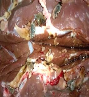
E3S Web of Conferences 282, 03020 (2021)                             https://doi.org/10.1051/e3sconf/202128203020
EFSC2021

         different individual and subsidiary farms, significant differences in the composition of the
         parasite fauna were noted. The dominant position in the prevalence of parasite species was
         occupied by pathogens belonging to the genus Eimeria. We have identified the following
         species:
             1. E. intestinalis (Cheissin, 1948)
             2. E. perforans (Leuckart, 1879)
             3. E. magna (Perard, 1925)
             4. E. stiedae (Lindemann, 1865)
             5. E. media (Kessel, 1929)
             During helminthologic studies in individual farms of the owners, monoinvasions were
         most often recorded in rabbits. In eimeriosis, the most common type of eimeria was E.
         perforans. This type of parasite in different degrees of intensity has been recorded and
         established in different age groups of rabbits. In descending order of the protozoa
         prevalence, the following were found, respectively: E. magna, E. intestinalis, and E. media.
         Other Eimeria species were very rare. In rabbits up to 2 months of age, Eimeria of E.
         perforans species dominated, with EI-22.0-24.0%, and in 3-month-old rabbits - E.
         perforans and E. Intestinalis, respectively, 10.0-13.0%. Similar types of pathogens were
         also found in rabbits up to 6 months of age (EI-6.7%). In adult rabbits, all the listed types of
         protozoa pathogens were found, but with lower rates of invasion (EI-1.2-1.7%), and in
         males, the degree of invasion and species composition was low, even in comparison with
         females (0.2-4.0%). In the subsidiary farms of individual owners, 5 types of pathogens of
         eimeriosis were also identified: E. stiedae, E. intestinalis, E. magna, E. perforans, and E.
         media. Of these, Eimeria stiedae was the most widely distributed species.
             The most unfavorable among all age groups of animals in terms of the degree of
         infection with eimeriosis were rabbits of 1-3 months of age. In animals of this age group,
         the intensity of invasion, depending on the farm in which they were raised, was in the range
         of 10.0±6.0 - 60.6±5.5 ind. of eimerium oocyst in the field of vision of the microscope A
         high degree of eimeria oocysts (25.0±7.0-50±9.0 ind.) was observed in rabbits up to one
         month of age. The first isolation of eimeria oocysts in young animals of this group was
         observed at the age of 21 days. With an increase in the age of rabbits, including at 6 months
         of age, the intensity of eimeriotic invasion was low. At the same time, the intensity of the
         invasion was single oocysts.
             In subsidiary farms, when analyzing the age dynamics of eimeriotic invasion among
         doe-rabbits and male rabbits, it was also found that females were most intensely invaded in
         comparison with adult male rabbits, which also had a low degree of invasion. The degree of
         infection of animals with eimeria in different farms differed significantly (EI - 2.0-26.0%).
         The study of the seasonal dynamics of rabbit eimeriosis was carried out on the example of
         several farms with different animal housing: cage and floor in a heated capital premises.
             The results of studies with cellular content showed that the peak of invasion occurred in
         the autumn-winter period, and the most invaded at this time were rabbits from 1 to 3
         months. A low degree of invasion was observed in rabbits up to 1 month (5.0±2.0 -7.0±3.1
         ind.) and rabbits of 5-6 months of age and adult livestock. In the autumn period, eimeriotic
         invasion was recorded in the range of 60.6±5.5 ind. of oocyst eimerium. In the autumn-
         winter period, an increase in the invasion intensity was observed in rabbits of all age
         groups. The high degree of invasion in the autumn-winter period is due to the increased
         humidity in the premises, which favorably affected the sporulation and development of
         oocysts. In the spring period, when the heat comes, the eimeriotic invasion was reduced due
         to the ventilation of the premises. In this season, the least infestation was observed in
         rabbits up to 30 days of age (10.2 ±1.6 ind.) and in animals 1-2 months (15.0+4.0 ind.).

                                                        3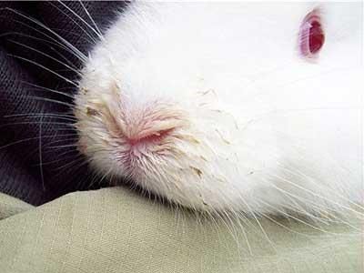
E3S Web of Conferences 282, 03020 (2021)                             https://doi.org/10.1051/e3sconf/202128203020
EFSC2021

             The study of the seasonal dynamics of eimeriosis with floor housing in capital-built
         premises showed that the indicators of prevalence dynamics in all periods of the year
         changed slightly and were approximately at the same level in each age group. Thus, in
         rabbits at the age of 30 days, according to the seasons of the year, the EI was 5.2±2.0% - in
         winter, and 3.0±2.75% - in summer. In rabbits of 3 months of age - from 20.3±2.0% to
         25.2±1.8%, similar changes in indicators were observed in animals of older age groups. In
         contrast to rabbits kept in cages, with the dynamics of infestations of animals located in
         heated rooms, the peak of infestation was observed from autumn to spring in rabbits at the
         age of up to 1-2 months.
             During the veterinary and sanitary assessment of slaughter products with rabbit
         eimeriosis (coccidiosis), the affected organs (liver, intestines) are destroyed or disposed of,
         the depleted carcass is rejected, the carcasses of the first and second categories are used
         without restrictions.
             When dissecting rabbits that died from eimeriosis, the body was depleted, and
         destructive changes were noted in the liver (Fig. 1). The liver is enlarged, dark in color on
         the surface of the organ and in the parenchyma of which there are also rounded whitish-
         yellow overlays filled with the contents.

         Fig. 1. Liver damage by Eimeria.
         In the digestive tract: the intestinal mucosa is pale and yellowish, microscopic examination
         revealed the presence of clusters of oocysts (Fig. 2). On the mucosa, puffiness and redness
         were detected, as well as detachment of the epithelial layer of the intestine, covered with
         curd-like overlays with an admixture of blood and the presence of yellowish-white foci.

         Fig. 2. Eimeria oocysts (high degree of invasion).

                                                              4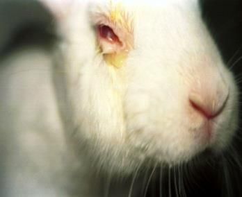
E3S Web of Conferences 282, 03020 (2021) https://doi.org/10.1051/e3sconf/202128203020
EFSC2021
The trematode - Fasciola hepatica causes fasciolosis in rabbits. When eating
grass or hay from meadows and pastures affected by fasciolosis, rabbits become
infected with fasciolosis. In the region of the Southern Urals, fasciolosis pathogens
were found only in rabbits imported from the Stavropol Krai. During the
coprological examination, single eggs of fascioles were identified in the field of
visions of the microscope (Fig. 3)
Fig. 3. Fasciola egg.
Fig. 4. Liver affected by fascioles.
If the liver is affected by fascioles, it is disposed of (Fig. 4), and the carcass is used
without restriction, if there are no degenerative changes in the organs and tissues. If after
the rabbit slaughter yellowish color remained in the carcass for 48 hours, it is also disposed
of. When autopsies are performed on corpses, the conjunctiva is pale, the hair is matted, the
mucous membranes are inflamed, and yellowish. The anus area is stained with fecal masses
with bloody clots, there are signs of tympanitis. With a chronic course, the hair is brittle,
easily falls out. There are edemas in the submandibular space, chest. The liver is slightly
enlarged on autopsy.
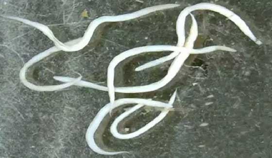
In case of passalurosis of rabbits path. Passalurus ambiguus (Fig. 5) is localized in the
large intestine
5E3S Web of Conferences 282, 03020 (2021) https://doi.org/10.1051/e3sconf/202128203020
EFSC2021
Fig.5. Passalurus ambiguus. Fig. 6. Eggs of Passalurus ambiguus.
Passalurosis was registered mainly in animals with floor housing. During the
helminthologic examination in the field of vision of the microscope, an average degree of
invasion was noted (II-25-35 ind., EI-2.0-4.0%) (Fig.6). The carcasses are atrophied, the
mesenteric lymph nodes are enlarged, hyperemic, and swollen. In case of degenerative
changes, the carcass is disposed of. On autopsy, serous-catarrhal and catarrhal-necrotic
inflammation of the intestine was noted in rabbits. The mucosa of the caecum, rectum, and
anus were hyperemic, edematous, with punctate hemorrhages. On autopsy of the large
intestine, dozens of helminth specimens were found. There are traumatic injuries around the
vulva and anus, due to scratching
In cysticercosis pisiformis (larval stage - Cysticercus pisiformis). An accurate
diagnosis is established on the autopsy of corpses or at the veterinary and sanitary
examination of rabbit carcasses after slaughter. Cysticercus were found on the serous
membranes of the internal organs in the form of grapes (Fig. 7).
Fig. 7. Cysticercus on the serous membranes of internal organs.
At the same time, the liver was enlarged, dark cherry-colored, with hemorrhages in the
parenchyma and hyperplasia of the bile ducts, serous-fibrinous peritonitis was noted.
Carcasses and organs of rabbits with severe exhaustion and lesion of the mesenterium,
omentum with thin-necked cysticercus are disposed of. In cysticercosis taenuicollis, the
affected organs are rejected, the carcasses are used without restriction (Fig. 7).
The psoroptosis pathogens in rabbits is the mite Psoroptos cuniculi (Fig. 8), mainly
adult rabbits are susceptible to this disease. The maximum degree of damage to rabbits is
6E3S Web of Conferences 282, 03020 (2021) https://doi.org/10.1051/e3sconf/202128203020
EFSC2021
noted in the winter and early spring, due to a decrease in the body resistance. Inflammation
of the external auditory canal and the inner surface of the auricle, which are covered with
thick crusts, are developed in animals (Fig. 9). The pathology is accompanied by itching,
the affected areas are infected with microflora, followed by the development of a purulent
process and the accumulation of purulent-bloody mass, with a fetid smell. Sick rabbits lose
their orientation. Rabbits are exhausted, they have seizures. Psoroptosis was recorded in
rabbits aged over 7-8 months.
Fig. 8. Psoroptosis in a rabbit Figure 9 - Psoroptes cuniculi.
Infectious rhinitis was recorded from infectious pathologies of rabbits. Infectious
rhinitis is caused by conditionally pathogenic microflora (Pasteurella, staphylococci,
Pseudomonas aeruginosa, etc.) and all age groups of animals are susceptible to it. Animals
have a reduced appetite, they sneeze, mucosal or purulent leak from the nose, light green
discharge, crusts form in the nasal passages, there is difficulty and increased breathing. An
increase in nose and swelling of the mucous membrane, while heavy breathing is noted.
With this disease, rabbits die in 30-40 days. Of the 30 rabbits studied, 2 of them were
infected with the infection (Fig. 10). Pathoanatomic changes. The nasal mucosa is
hyperemic and edematous, covered with mucopurulent or purulent secretions. The mucous
membrane of the trachea is swollen, hyperemic, with hemorrhages, the blood vessels of the
trachea are blood-filled. The bronchial mucosa is also hyperemic, there is a foamy exudate
in the lumen. Catarrhal, hemorrhagic, purulent-fibrinous or croup inflammation was noted
with lung damage. There are hemorrhages on the surface of the lungs. The lower lobes of
the lungs are swollen, and there are encapsulated abscesses in the tissues.
7E3S Web of Conferences 282, 03020 (2021) https://doi.org/10.1051/e3sconf/202128203020
EFSC2021
Fig. 10. Clinical manifestation of infectious rhinitis.
The diagnosis of infectious rhinitis was established on the basis of clinical and
pathoanatomical studies, as well as using the results of a bacteriological study in the
regional laboratory, where cultures of microorganisms from the lungs, heart, and blood
were isolated with the establishment of pathogenic properties of pathogens on laboratory
animals - white mice and rabbits. After confirmation of the viral etiology of the disease, a
retrospective diagnosis was performed, examining the paired blood sera of rabbits in the
reaction of delayed hemagglutination. At the same time, infectious rhinitis of rabbits was
differentiated from rhinitis of non-infectious origin, which is characterized by the release
of serous or serous-mucosal exudate from the nasal passages. In pasteurellosis, caseous
lymphadenitis, aspergillosis, they are distinguished by the specific nature of the exudate for
these pathologies and the results of laboratory tests.
Infectious keratoconjunctivitis is caused by different types of staphylococci. In this
pathology, the cornea is affected, with turbidity and the appearance of ulcers, acquiring a
white or yellowish-green color, conjunctivitis occurs with the release of purulent exudate,
photophobia occurs, the epithelium is rough, with purulent or purulent-fibrinous exudate
and ulcers, leading to an abscess, resulting in a thorn with vision loss. Animals lose weight
and often followed by the death after 10-12 days from the onset of the disease. Of the 30
studied rabbits of different age groups, 5 of them were infected. Rabbits of different age
groups were susceptible to the disease. The diagnosis was set on the basis of characteristic
clinical signs and confirmed by bacteriological examination of the material in the
laboratory from sick rabbits (Fig. 11). Pathoanatomical changes are similar to the data of
the clinical picture of the disease.
8E3S Web of Conferences 282, 03020 (2021) https://doi.org/10.1051/e3sconf/202128203020
EFSC2021
Fig. 11. Clinical manifestation of infectious keratoconjunctivitis.
4 Conclusions
Thus, when conducting diagnostic helminthological studies, during autopsy of corpses and
veterinary and sanitary assessment of rabbit slaughter products and the study of carcasses,
we obtained the following results:
1. In rabbits in the farms of the Orenburg region, during autopsy and coprological
studies, such invasive diseases as eimeriosis (coccidiosis of the liver and intestinal forms),
passalurosis were registered, in which the average indicators of II and EI were,
respectively, from several units in fascioliasis, to several thousand individuals in eimeriosis;
2. During the veterinary and sanitary assessment of carcasses in different age groups of
animals, cysticercosis pisiformis was detected with a degree of invasion of 25-27 ind.,
fasciolosis with an intensity of invasion - 15-30 ind.;
3. Such pathologies as: passalurosis, dicroceliosis, fascioliasis was less common, mainly
in animals acquired in other regions, with a low degree of invasion;
4. Psoroptosis affected animals at the age of 3-4 months, with EI, on average, - 3-6%;
5. The dominant invasion was eimeriosis, caused by several types of eimeria, in most
cases, the intestinal form was recorded in rabbits;
6. Infectious keratoconjunctivitis and infectious rhinitis were more common among
infectious pathologies. The intensity of the prevalence of these infections was at a low
level, in comparison with invasive pathologies.
References
1. E.S. Volkova, V.N. Baimatov, Methods of scientific research in veterinary medicine
183 (M., KolosS, 2010)
2. V.V. Gorokhov, J. Veterinary medicine, 8, 3-5 (2002)
3. A.A. Dubnitsky, Proc. of the cent. of sc.-res. labor. fur farming, 6, 335-342 (1950)
4. A.A. Dubnitsky, Passalurosis. Diseases of rabbits, 184-190 (M. Kolos, 1974)
5. E.I. Kabanova, Diagnosis, clinical and ophthalmic characteristics and treatment of
postoperative dry keratoconjunctivitis in small domestic animals, Veterinary surgery,
Moscow (2021)
9E3S Web of Conferences 282, 03020 (2021) https://doi.org/10.1051/e3sconf/202128203020
EFSC2021
6. B.E. Ketema, Eimeriosis of rabbits in different housing systems and improvement of
control and prevention measures, Parasitology, Moscow (2002)
7. S.V. Leontyuk, A.A. Dubnitsky, et al. Diseases of rabbits, 235 (M., Kolos, 1974)
8. I.S. Minina, A.I. Mayorov, All about rabbits, Album, 210 (M., Kolos, 1985)
9. A.I. Rakhmanov, Home animal farm. Housing and breeding of rabbits and fur-bearing
animals in the small garden plots, 256 (M., Aquarium LTD., 2000)
10. Z.Kh. Terentyeva, Russian Parasitological Journal, 4, 342-346 (2011)
11. Z.Kh. Terentyeva, Proceedings of the Orenburg State Agrarian University, 3 (41), 257-
260 (2013)
12. Z.Kh. Terentyeva, E.P. Pyatachkova, Theory and practice of combating parasitic
diseases, 17, 467-469 (2016)
13. L.I. Ulyikhina, Handbook of the rabbit breeder from A to Z, 256 (M., Aquarium-Print,
2009)
14. A.V. Uspensky, Veterinary consultant, 17, 10 (2003)
15. Yu.N. Shilyaeva, Eimeriosis of rabbits in the Republic of Tatarstan: Epizootology,
control measures, Parasitology, Moscow (2004)
16. D.A. Sharafutdinov, Complex treatment of rabbits with experimental conjunctival
keratitis, 45-47 (Notes of the KSAVM n.a. Bauman. Kazan, 2013)
17. A.A. Shevchenko, Diseases of rabbits, 224 (M., Aquarium-Print, 2009)
18. Katleen Hermans, Ilse Moeremans. E. cuniculiand other diseases in a geriatric rabbit.
DVM Case Report as part of the Master`s Dissertation Ghent University faculty of
veterinary medicine Academic year 2015, 234-238 (2016)
10You can also read