Large zooming range adaptive microscope employing tunable objective and eyepiece - Nature
←
→
Page content transcription
If your browser does not render page correctly, please read the page content below
www.nature.com/scientificreports
OPEN Large zooming range adaptive
microscope employing tunable
objective and eyepiece
Feng‑Lin Kuang1, Rong‑Ying Yuan2, Qiong‑Hua Wang2* & Lei Li1*
The conventional microscope has discrete magnification and slow response time in zoom process,
which is difficult to capture the dynamic activity of the live specimen. We demonstrate an adaptive
microscope employing a tunable objective and a tunable eyepiece with large zooming range. The
tunable objective consists of three glass lenses and four electrowetting liquid lenses. The tunable
eyepiece consists of an achromatic eyepiece and an electrowetting liquid lens. The focal point between
the objective and the eyepiece is designed to be tunable, which are controlled by voltages. Thus, the
tuning range is relatively large. We fabricate the adaptive microscope and observe the specimen. In
the experiment, the magnification of the microscope changes continuously from ~ 59.1 × to ~ 159.2 × ,
and the largest numerical aperture is ~ 0.212. The tunable eyepiece can release the back focal
length of the tunable objective, which increases the zoom range of the microscope. No mechanical
movement is required and the aberrations can be corrected over a wide wavelength range. Thus, the
proposed adaptive microscope has a potential application in biological research and clinical medical
examination.
Microscopes play an important role in scientific research and production, such as physiological applications1,
biomedical engineering2 and m icrofabrication3. For targets of different sizes, microscopes need different magni-
fications. Besides, to observe large area of cells and a zoom-in area with high resolution, the higher requirements
of real-time observation and continuous zoom change on microscopes e merges4. The conventional microscopes
can change magnifications by manually converting objectives. But its magnifications are not continuous and the
conversion operation introduces sample vibration. One of the solutions for continuous zoom change is to use the
mechanical or optical compensation system driven by mechanical m ovement5,6. However, such compensation
systems are bulky and sample vibration is still an issue due to the mechanical movement. Besides the slow zoom
speed affects the real-time observation. Fortunately, the liquid lenses have changed the traditional lens s ystems7.
Due to lightweight, low power consumption and fast response speed, liquid lenses have important applications in
imaging8–10, display11, and c ommunication12. Moreover, liquid lenses are wildly used in microscopy as focusing
component for axial s canning13–16, increasing the depth-of-field17,18 and a utofocusing19,20. However, they cannot
achieve continuous zoom change. For example, a five-dimensional microscopy using liquid lens is proposed
and able to scan volumes rapidly and reproducibly21. This microscopy has fast scan speed however it cannot
zoom continuously. A adaptive microscope objective using liquid lens is p roposed22. The magnification of the
adaptive microscope objective tune from ~ 7.8 × to ~ 13.2 × , but its zoom range is very limited due to fixed back
focal length (BFL). Therefore, it is still urgent to study microscopes with large zoom range and fast zoom speed.
We demonstrate an adaptive microscope with tunable objective and tunable eyepiece. The tunable objective
consists of three glass lenses and four electrowetting liquid lenses. The tunable eyepiece consists of an achro-
matic eyepiece and an electrowetting liquid lens. The focus of both objective and eyepiece can be adjusted by
applying voltage. Different from our previous work22, the position of image plane between the tunable objective
and eyepiece is flexible, which largely increases the tuning range. We fabricate the adaptive microscope and set
up the experiment to observe a resolution chart and a specimen. The magnification of the adaptive microscope
changes continuously from ~ 59.1 × to ~ 159.2 × and the largest numerical aperture (NA) is ~ 0.212. The response
time is ~ 50 ms. Without introducing mechanical moving parts while zooming, the aberrations can be corrected.
The adaptive microscope is suitable for real-time observations that require fast continuous zoom changes.
1
School of Electronics and Information Engineering, Sichuan University, Chengdu 610065, China. 2School of
Instrumentation and Optoelectronic Engineering, Beihang University, Beijing 100191, China. *email: qionghua@
buaa.edu.cn; leili@scu.edu.cn
Scientific Reports | (2020) 10:14644 | https://doi.org/10.1038/s41598-020-71507-8 1
Vol.:(0123456789)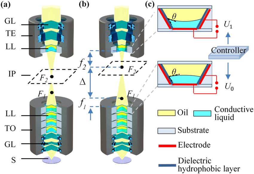
www.nature.com/scientificreports/
Figure 1. Configuration of the adaptive microscope. (a) Initial magnification. GL glass lens, TE tunable
eyepiece, LL liquid lens, IP image plane, TO tunable objective, S sample. (b) Zoom in. Focal length f1 and f2
become shorter, and optical spacing becomes longer. (c) Liquid lens with different applied voltages, of which
U0 is lower and U1 is higher. θ is the contact angle of the liquid–liquid interface.
Structure and theoretical analysis
A simplified configuration of the proposed adaptive microscope is shown in Fig. 1a. The microscope consists of
a tunable objective and a tunable eyepiece, and both are composed of liquid lenses and glass lenses. The dashed
box in Fig. 1a, b represent the intermediate image plane of the tunable objective. F1 is the focus of the tunable
objective and F2 is the focus of the tunable eyepiece. F2 and the intermediate image plane always overlap.
The design of continuous zoom change is shown in Fig. 1b, where f1 is the focal length of the tunable objec-
tive, and f2 is the focal length of the tunable eyepiece. is the optical spacing between the focus of the tunable
objective and the tunable eyepiece. The focuses (F1 and F2) are tuned between the tunable objective and tunable
eyepiece. Thus, the focal length f1 and f2 are changed, which results in the magnification change of the proposed
microscope. The magnification of the microscope can be expressed as:
250�
Ŵ=− , (1)
f1 f2
where 250 represent the distinct vision of 250 mm. When the focal length f1 becomes shorter, the magnification
of the tunable objective increases, and the imaging plane becomes closer to the tunable eyepiece. To make the
focus F2 coincide with the intermediate image plane, the focal length f2 becomes shorter, which increases its
magnification and the optical spacing . Thus, the zoom range of the microscope is increased.
Figure 1c shows the structural changes of the electrowetting liquid lens when different voltages are applied.
Each liquid lens consists of an oil and a conductive liquid, and can change its optical power by electrowetting
effect. θ is the contact angle of the liquid–liquid interface. The relationship between the contact angle θ and the
applied voltage U can be described by Young-Lippmann e quation7:
εU 2
cos θ = cos θ0 + , (2)
2dγ
where θ0 is the initial contact angle, γ is the surface tension between the conductive liquid and the dielectric
hydrophobic layer, ε is the dielectric constant and d is the thickness of the dielectric hydrophobic layer.
The zoom range of the proposed adaptive microscope increases in two ways. (1) For the tunable objective,
the BFL can be adjusted within a certain range instead of fixed, which means the aberrations correction capabil-
ity of the liquid lenses is released. As a result, the zoom ability of the objective is increased. (2) For the tunable
eyepiece, to make the focus F2 coincide with the intermediate image plane, the focal length f2 changes with the
tunable objective. This change increases the magnification of the eyepiece and the optical spacing , increasing
Scientific Reports | (2020) 10:14644 | https://doi.org/10.1038/s41598-020-71507-8 2
Vol:.(1234567890)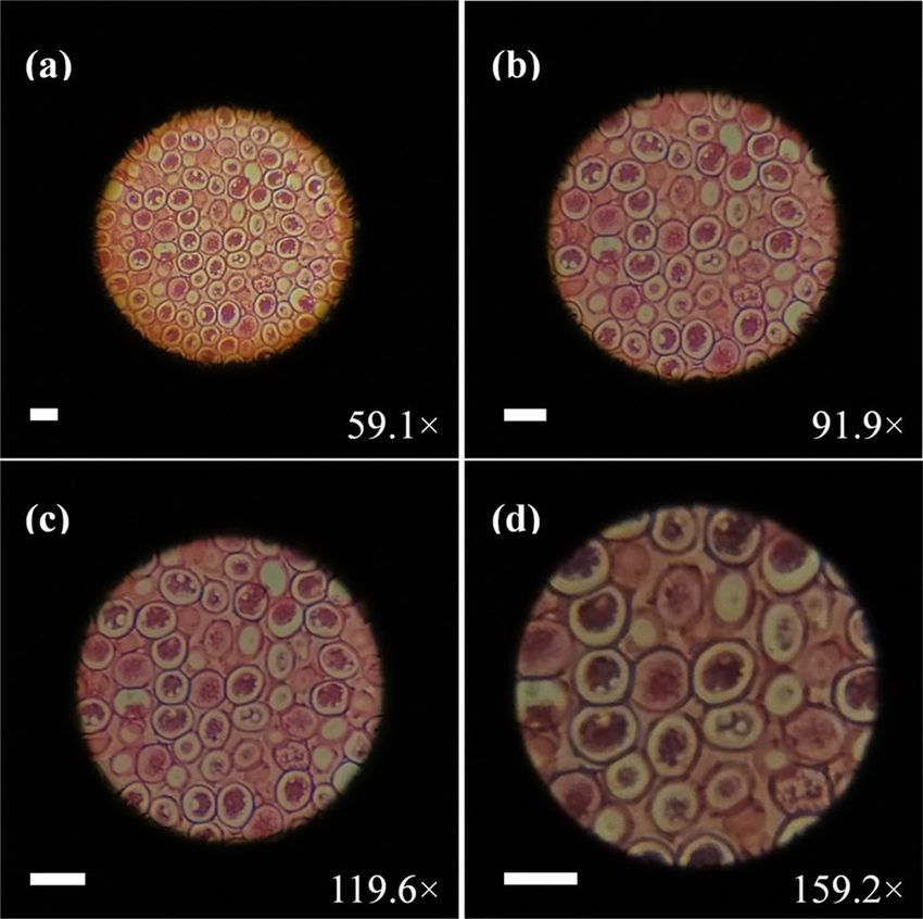
www.nature.com/scientificreports/

                            Figure 2.  Adaptive microscope and its components. (a) The adaptive microscope. (b) Tunable eyepiece. (c)
                            Tunable objective. (d) The controller.

                             Material               Oil             Conductive liquid        N-SK16        ZF10          QK1
                             Refractive index        1.500           1.388                    1.620         1.689         1.470
                             Abbe number            34.63           58.13                    60.32         31.18         66.87

                            Table 1.  Refractive index and Abbe number of the materials used in the tunable objective.

                            the zoom range of the microscope. Because the tunable objective plays a decisive role in magnification and image
                            quality, four liquid lenses are used. Two liquid lenses are used to increase the zoom ability, and the other two
                            liquid lenses are used to adjust the position of the image plane and correct aberrations. Using the commercial
                            software Zemax, we constructed a merit function to optimize the radii of four liquid lenses in tunable objective.
                            Then we get the radius solutions that meets the image quality and aberration correction requirements. The radii
                            are converted into applied voltages to achieve continuous zoom change. Since BFL changes while zooming,
                            we adjust the focus of the tunable eyepiece to the intermediate image plane to get the clear image. The tunable
                            eyepiece uses only one liquid lens, because the movement of the BFL is limited, and one liquid lens is enough
                            to adjust its focus to the image plane.

                            Fabrication
                            We developed an adaptive microscope shown in Fig. 2a. The microscope consists of a tunable eyepiece, a tun-
                            able objective, a controller, a lens cone and a light source. We fabricated a tunable eyepiece as shown in Fig. 2b.
                            The eyepiece consists of a 10 × achromatic eyepiece and an electrowetting liquid lens. Its size is approximately
                            36 × 36 × 32 (mm).
                               The fabricated tunable objective is shown in Fig. 2c. Its size is approximately 25 × 25 × 61 (mm), and the focal
                            length ranges from 11.2 to 4.8 mm. For the tunable objective, four electrowetting liquid lenses and three glass
                            lenses are used. The clear aperture of the electrowetting liquid lenses is ~ 4 mm and its main materials are oil and
                            conductive liquid. The diameter of three glass lenses are ~ 6 mm and its material are N-SK16, ZF10 and QK1,
                            respectively. The refractive index and Abbe number of the oil, the conductive liquid and three kinds of glass are
                            shown in Table 1. The controller shown in Fig. 2d is based on ARM 32-bit Cortex M3 CPU (STM32F103ZET6). It
                            provides multiple sets of specific voltages for the adaptive microscope. Each set of voltage includes 5 independent
                            voltage signals. These voltages are applied to the liquid lenses in the tunable objective and the tunable eyepiece,
                            which, according to Eq. (2), results in the change of the focal length of the liquid lenses. Below the target is a
                            LED surface light source (DM-906823) with an adjustable diaphragm.

                            Simulation and experiments
                            We simulated the proposed adaptive microscope in Zemax within the zoom range of ~ 60 × to ~ 160 × . A paraxial
                            surface was used to simulate the lens of the human eye. The MTF obtained by ray tracing is shown in Fig. 3. The
                            MTF at 60 × , 110 × , and 160 × are given by three wavelengths at 0.486μm, 0.587μm, and 0.656μm, respectively.
                            The black line represents the MTF at diffraction-limited resolution (limit). Axis, T and S represent the zero,
                            tangential and sagittal field of view, respectively. The simulation shows that proposed adaptive microscope can
                            achieve near-diffraction limit resolution at different magnifications and different wavelengths.

Scientific Reports |   (2020) 10:14644 |                  https://doi.org/10.1038/s41598-020-71507-8                                          3

                                                                                                                                         Vol.:(0123456789)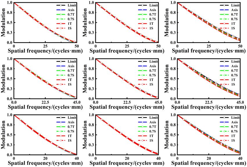
www.nature.com/scientificreports/
Figure 3. MTF of the proposed microscope. The columns from left to right represent magnifications of 60 × ,
110 × , and 160 × , respectively. The lines from top to bottom represent wavelengths of 0.486μm, 0.587 μm and
0.656μm, respectively.
Magnification 60 × 80 × 100 × 120 × 140 × 160 ×
NA 0.157 0.169 0.178 0.188 0.196 0.212
Table 2. Detailed optical parameters of the adaptive microscope.
We measured the focal length of the electrowetting liquid lens at different applied voltages. The negative
focal length varies from ~ − 50 mm to infinity, and the positive focal length varies from ~ 23 mm to infinity. The
corresponding negative radius of the liquid–liquid interface varies from ~ − 5.6 mm to infinity and the positive
radius varies from ~ 2.7 mm to infinity. Magnification and the NA of the microscope are given in Table 2. The
smallest and largest NA are ~ 0.157 and ~ 0.212.
The proposed microscope can be used directly for visual observation. To evaluate the imaging quality of the
proposed adaptive microscope, we set up an imaging experiment by using a camera of a smartphone to simulate
the human eyes, as shown in Fig. 4a. The experiment setup consists of a tunable objective, a tunable eyepiece, a
smartphone and a USAF 1951 chart which is used as the resolution target, shown in Fig. 4b. The resolution of
the camera (S5KGM1 from Samsung) is 1.6 microns. The focus of the camera is set to a fixed value during the
experiment.
For each magnification, firstly, we used the commercial software Zemax to optimize the radii of four elec-
trowetting liquid lenses in the tunable objective. Then the radii were picked up and converted into voltages. When
we applied those voltages to the electrowetting liquid lenses, the focal length and BFL of the tunable objective
changed. According to the BFL of the tunable objective, we changed the voltage applied to the electrowetting
liquid lens in the tunable eyepiece. The focus of the tunable eyepiece was adjusted to the intermediate image plane
to obtain clear image. All these voltages were stored in the controller by groups. When we switched between
different groups, the microscope can achieve optical zoom.
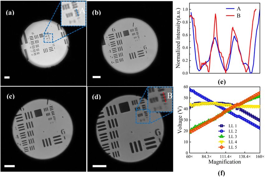
The captured results are shown in Fig. 5a–d. To obtain the normalized intensity of the resolution target,
Fig. 5a–d are converted into grayscale images. The magnification of Fig. 5a is ~ 59.1 × , the entire sixth and seventh
group, and part of the fourth and fifth group of resolution target can be seen. When the magnification increases,
the resolution target is gradually magnified and the visible parts of the fourth and fifth group are becoming
Scientific Reports | (2020) 10:14644 | https://doi.org/10.1038/s41598-020-71507-8 4
Vol:.(1234567890)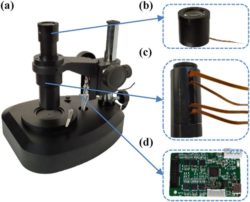
www.nature.com/scientificreports/

                            Figure 4.  Microscope setup. (a) A smartphone camera is used to simulate the human eye. (b) Imaging
                            experimental setup. The microscope used directly for visual observation. A USAF 1951 chart is used as
                            resolution target.

                            Figure 5.  Captured images of the resolution target. (a) Zoom 59.1 × . (b) Zoom 91.9 × . (c) Zoom 119.6 × .
                            (d) Zoom 159.2 × . The scale bars in those pictures are 50 μm. (e) The blue line (A) and red line (B) which
                            represent the normalized intensity distribution of the third element of the seventh group at 59.1 × and 159.2 × ,
                            respectively. (f) Magnification of the microscope versus voltages applied to the liquid lenses. LL liquid lens. The
                            liquid lenses in the tunable objective are numbered sequentially from the object side to image side as liquid
                            lenses 1 to 4. Liquid lens 5 represents the liquid lens in the tunable eyepiece.

Scientific Reports |   (2020) 10:14644 |               https://doi.org/10.1038/s41598-020-71507-8                                                 5

                                                                                                                                         Vol.:(0123456789)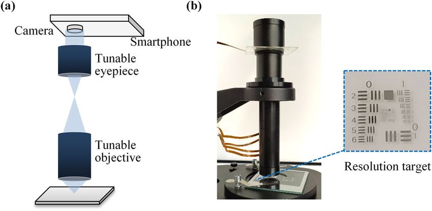
www.nature.com/scientificreports/

                                      Figure 6.  Captured images of the female ascaris sec. When the applied voltage is changed, the cell is
                                      continuously enlarged (see Visualization 1). (a) Zoom 59.1 × . (b) Zoom 91.9 × . (c) Zoom 119.6 × . (d) Zoom
                                      159.2 × . The scale bars in those pictures are 50 μm.

                                      smaller. When the magnification is larger than ~ 119.6 × , as shown in Fig. 5c, only the sixth and seventh group
                                      can be seen. The largest magnification is ~ 159.2 × , the captured image is shown in Fig. 5d. In Fig. 5d, the third
                                      element of seventh group is clear. From Fig. 5a–d, the visual field becomes larger and the field of view becomes
                                      smaller because the focal length of the tunable eyepiece becomes shorter and its magnification becomes larger.
                                      The magnification can be linear theoretically. However, the step size of the voltages makes the changes of the
                                      magnification in some cases are non-linear. The approximate magnification step size is about 0.1 × . The mid-
                                      dle two magnifications show that the microscope can achieve continuous zoom change within a certain range,
                                      instead of discrete magnifications such as 10 × , 20 × , 40 × , etc. The color difference between the pictures may be
                                      caused by the exposure difference of the images and the NA difference during zooming.
                                          Figure 5e shows the normalized intensity distribution of the third element of the seventh group, where the
                                      blue line (A) represents the intensity at Zoom 59.1 × . The modulation of the lines in the center is about 0.19. The
                                      red line (B) represents the intensity at Zoom 159.2 × , and the modulation of the lines in the center is about 0.34.
                                      The results show that the resolution has been improved by zooming. Figure 5f shows the magnification of the
                                      adaptive microscope versus the voltage applied to the liquid lenses in the tunable objective and tunable eyepiece.
                                      When the magnification changes, two liquid lenses in the objective work as the zoom part, and the other two
                                      liquid lenses are used to adjust the position of the image plane and to correct aberrations. The liquid lens in the
                                      eyepiece is mainly responsible for adjusting the focal length of the tunable eyepiece.
                                          To verify biological applications of the proposed adaptive microscope, we used a section specimen of female
                                      ascaris as the target. The result is shown in Fig. 6. Figure 6 remains unchanged, which makes its color different
                                      from that in Fig. 5a–d. From Fig. 6a–d, as we continuously zoom in, the cells of the female ascaris are gradually
                                      enlarged and the image remains sharp at each magnification. A video of the different magnification (see Visu-
                                      alization 1) shows that the zooming process is continuous and fast. The response time is measured to be ~ 50 ms.
                                      As the focal length of the eyepiece becomes shorter, the visual field becomes larger and the field of view becomes
                                      smaller, as we observed in Fig. 5. The lower brightness of the picture in Fig. 6d is due to the lack of light intensity.
                                      As the magnification increases, the light intensity needs to be increased to maintain the same brightness of the
                                      image, which is similar to the traditional microscope.
                                          We note that there are small vibrations in Visualization 1. There are two reasons for that: (1) Since the optical
                                      power of the liquid lenses are controlled by voltages, the vibrations of the voltages are the main reason for the
                                      small vibrations. (2) The vibrations of the external environment cause the stage to move up and down, which
                                      will also cause the image vibrations.
                                          Since the aperture of the liquid lens in the eyepiece is ~ 4 mm, the field of view of the microscope relatively
                                      small. We believe that using a liquid lens with a larger diameter can get a larger filed. However, the microscope
                                      will be complicate to operate and zoom speed will be slower, which will affect the real-time observation. The
                                      zoom range of our proposed microscope is ~ 59.1 × to ~ 159.2 × . Increasing the optical power range of single
                                      electrowetting liquid lens, or using more liquid lenses will increase the zoom range of the microscope. However,
                                      more liquid lenses will make the microscope more complicated and will increase the costs. In this microscope,
                                      working distance is fixed which means no moving parts are required. However, achieving a larger zoom range
                                      at a fixed working distance will be a challenge, because the requirement of optical power range is relatively large.

          Scientific Reports |   (2020) 10:14644 |                https://doi.org/10.1038/s41598-020-71507-8                                                6

Vol:.(1234567890)
www.nature.com/scientificreports/
With the development of the liquid lenses, more and more research will be put into the microscopes to achieve
larger zoom range. Changing working distance without moving part may address these issues in the future.
Conclusion
In this paper, we present an adaptive microscope consisting of a tunable objective and a tunable eyepiece. The
tunable objective consists of three glass lenses and four electrowetting lenses. The tunable eyepiece consists
of an achromatic eyepiece and an electrowetting lens. The focus of the objective and the eyepiece are tunable
by changing the applied voltages. Because the focus of the eyepiece always coincides with the intermediate
image plane during zooming, the BFL of the tunable objective is released, which increases the zoom range of
the microscope. Without introducing mechanical moving parts, the magnification of the microscope changes
from ~ 59.1 × to ~ 159.2 × and its response time is ~ 50 ms. Observation of resolution targets and specimens shows
that our microscope has the ability of real-time observation with continuous zoom change. In specific application,
such as tumor metastasis. It is necessary to observe a relatively large field of view, while being able to magnify
the area of interest and to observe the morphology of the cell. Our adaptive microscope is suitable for such
applications. Besides, the proposed adaptive microscope has potential applications in physiological research
and manufacturing.
Received: 23 April 2020; Accepted: 17 August 2020
References
1. Glaser, A. K. et al. Light-sheet microscopy for slide-free non-destructive pathology of large clinical specimens. Nat. Biomed. Eng.
1, 0084 (2017).
2. Tanaka, N. et al. Whole-tissue biopsy phenotyping of three-dimensional tumours reveals patterns of cancer heterogeneity. Nat.
Biomed. Eng. 1, 796–806 (2017).
3. Choudhary, A., Naskar, A. & Paul, S. An investigation on application of nano-fluids in high speed grinding of sintered alumina.
J. Manuf. Process 35, 624–633 (2018).
4. Ji, N., Freeman, J. & Smith, S. L. Technologies for imaging neural activity in large volumes. Nat. Neurosci. 19, 1154–1164 (2016).
5. Smith, W.J. Modern Lens Design. (McGraw-Hill, 2005).
6. Huang, Y., Chen, X., Zhang, H., Huang, S. & Lin, F. Design of a high-performance digital slit-lamp microscope with five-switched
zoom. Appl. Sci. Basel 10, 2757–2769 (2020).
7. Berge, B. & Peseux, J. Variable focal lens controlled by an external voltage: An application of electrowetting. Eur. Phys. J. E 3,
159–163 (2000).
8. Kuiper, S. & Hendriks, B. H. W. Variable-focus liquid lens for miniature cameras. Appl. Phys. Lett. 85, 1128–1130 (2004).
9. Li, L., Wang, D., Liu, C. & Wang, Q. H. Ultrathin zoom telescopic objective. Opt. Express 24, 18674–18684 (2016).
10. Yang, A., Cao, J., Zhang, F. H., Cheng, Y. & Hao, Q. Ultrathin tunable lens based on boundary tension effect. Sensors-Basel 19, 4018
(2019).
11. Lee, Y. H., Peng, F. L. & Wu, S. T. Fast-response switchable lens for 3D and wearable displays. Opt. Express 24, 1668–1675 (2016).
12. Moosavi, S. A. et al. Improvement of coupling efficiency in free-space optical communication with a multi-actuator adaptive lens.
Opt. Lett. 44, 606–609 (2019).
13. Supekar, O. D. et al. Two-photon laser scanning microscopy with electrowetting-based prism scanning. Biomed. Opt. Express 8,
5412–5426 (2017).
14. Lan, G. P., Mauger, T. F. & Li, G. Q. Design of high-performance adaptive objective lens with large optical depth scanning range
for ultrabroad near infrared microscopic imaging. Biomed. Opt. Express 6, 3362–3377 (2015).
15. Murali, S., Thompson, K. P. & Rolland, J. P. Three-dimensional adaptive microscopy using embedded liquid lens. Opt. Lett. 34,
145–147 (2009).
16. Qu, Y. F. & Hu, Y. B. Analysis of axial scanning range and magnification variation in wide-field microscope for measurement using
an electrically tunable lens. Microsc. Res. Tech. 82, 101–113 (2019).
17. Llavador, A., Scrofani, G., Saavedra, G. & Martinez-Corral, M. Large depth-of-field integral microscopy by use of a liquid lens.
Sensors-Basel 18, 3383 (2018).
18. Kim, W., Lee, C., Kim, C. & Kim, D. S. Dual-mode reconfigurable focusing using the interface of aqueous and dielectric liquids.
Lab Chip 17, 4031–4039 (2017).
19. Chang, Z., Xiaoming, H. & Ya, Z. An induced fluorescence detecting system with autofocus electrically tunable len. Int. J. Eng.
Technol. (Singapore) 8, 297–300 (2016).
20. Wang, Z. J. et al. Compact multi-band fluorescent microscope with an electrically tunable lens for autofocusing. Biomed. Opt.
Express 6, 4353–4364 (2015).
21. Tehrani, K. F. et al. Five-dimensional two-photon volumetric microscopy of in-vivo dynamic activities using liquid lens remote
focusing. Biomed. Opt. Express 10, 3591–3604 (2019).
22. Li, L., Wang, D., Liu, C. & Wang, Q. H. Zoom microscope objective using electrowetting lenses. Opt. Express 24, 2931–2940 (2016).
23. https://www.sipmv.com/products/microscope/parts/light/dm-9608/
Acknowledgements
This work is supported by the National Natural Science Foundation of China under Grant Nos. 61927809 and
61975139.
Author contributions
F.-L.K., R.-Y.Y., Q.-H.W. and L.L. wrote the main text, and Q.-H.W. prepared Figures 1–6. All authors reviewed
the manuscript.
Competing interests
The authors declare no competing interests.
Scientific Reports | (2020) 10:14644 | https://doi.org/10.1038/s41598-020-71507-8 7
Vol.:(0123456789)www.nature.com/scientificreports/
Additional information
Supplementary information is available for this paper at https://doi.org/10.1038/s41598-020-71507-8.
Correspondence and requests for materials should be addressed to Q.-H.W. or L.L.
Reprints and permissions information is available at www.nature.com/reprints.
Publisher’s note Springer Nature remains neutral with regard to jurisdictional claims in published maps and
institutional affiliations.
Open Access This article is licensed under a Creative Commons Attribution 4.0 International
License, which permits use, sharing, adaptation, distribution and reproduction in any medium or
format, as long as you give appropriate credit to the original author(s) and the source, provide a link to the
Creative Commons licence, and indicate if changes were made. The images or other third party material in this
article are included in the article’s Creative Commons licence, unless indicated otherwise in a credit line to the
material. If material is not included in the article’s Creative Commons licence and your intended use is not
permitted by statutory regulation or exceeds the permitted use, you will need to obtain permission directly from
the copyright holder. To view a copy of this licence, visit http://creativecommons.org/licenses/by/4.0/.
© The Author(s) 2020
Scientific Reports | (2020) 10:14644 | https://doi.org/10.1038/s41598-020-71507-8 8
Vol:.(1234567890)You can also read