Diagnostic Dilemma of Recurrent Pulmonary Embolism - Mdpi
←
→
Page content transcription
If your browser does not render page correctly, please read the page content below
diagnostics
Case Report
Diagnostic Dilemma of Recurrent
Pulmonary Embolism
Alexandra Dadarlat-Pop 1,2 , Irina Burian 1,2 , Laura Cadis 1 , Raluca Tomoaia 2, * and
Alexandru Oprea 1,3
 1    Cardiology Department, Heart Institute Niculae Stăncioiu, 19-21 Mot, ilor street, 400001 Cluj-Napoca,
      Romania; dadarlat.alexandra@yahoo.ro (A.D.-P.); irinaburian@yahoo.com (I.B.); laura30@yahoo.com (L.C.);
      alexandru_oprea2002@yahoo.com (A.O.)
 2    Department of Cardiology, Iuliu Haţieganu University of Medicine and Pharmacy, 8 Victor Babes street,
      400012 Cluj-Napoca, Romania
 3    Cardiovascular Surgery Department, Heart Institute Niculae Stăncioiu, 19-21 Mot, ilor street,
      400001 Cluj-Napoca, Romania
 *    Correspondence: raluca.tomaia@gmail.com

 Received: 24 January 2020; Accepted: 9 February 2020; Published: 11 February 2020

 Abstract: Popliteal venous aneurysms are rare vascular disorders associated with a high risk
 of pulmonary embolism. We present the case of a 56-year-old woman hospitalized for a third
 episode of unprovoked pulmonary embolism. Venous ultrasonography identified a popliteal
 aneurysm, repeatedly missed by two-point compression venous ultrasonography, which was
 eventually confirmed by a magnetic resonance examination. Because of its highly symptomatic nature
 despite optimal anticoagulant treatment, the decision was made to undergo surgery, consisting of
 aneurysmectomy followed by patch angioplasty. The goal of this paper is to report a rare case of
 popliteal venous aneurysm and its treatment strategies and postoperative evolution.

 Keywords: popliteal venous aneurysm; recurrent pulmonary embolism; therapy

1. Introduction
     Popliteal venous aneurysms (PVAs) are rare pathologic vascular disorders. They may have
various aetiologies including congenital, trauma, varicose veins, localized degenerative changes,
and inflammation. Moreover, they usually remain silent until a thromboembolic event is developed.
Given the rarity and potential fatal complications of this condition, without an established consensus
of its management we report a case of primary popliteal venous aneurysm and discuss the
therapeutic approach.

2. Patient Information and Physical Examination
     A 56-year-old female patient presented to the emergency room with a recent syncope followed by
dyspnoea of acute onset. The medical history was significant for a left Baker’s cyst, Hashimoto
thyroiditis, and recurrent episodes of unprovoked pulmonary embolism (2017, 2018). During
previous hospitalizations, Doppler venous ultrasonography showed no signs of deep vein thrombosis.
Furthermore, no predisposing factors were identified, and the patient underwent screening tests
for malignancy and blood tests for acquired or inherited coagulation disorders, all of which had
been negative.
     Since the hospital discharge after the first diagnosis of pulmonary embolism, the patient was
commenced on long-term treatment with direct oral anticoagulant which she has been following up
until the current hospital admission.

Diagnostics 2020, 10, 96; doi:10.3390/diagnostics10020096                    www.mdpi.com/journal/diagnostics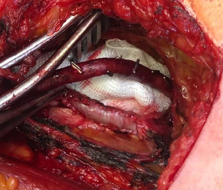
Diagnostics 2020, 10, 96                                                                                      2 of 7
Diagnostics 2020, 10, 96                                                                                        2 of 7
3. Clinical Examination
      Physical
 3. Clinical     examination revealed a hemodynamically stable patient with an oxygen saturation of
              Examination
96% without additional oxygen, blood pressure of 110/70 mmHg, heart rate of 76 bpm, respiratory
      Physical
frequency    (RF)examination
                  of 18 r.p.m.,revealed a hemodynamically
                                and a temperature of 36.8 °C. stable
                                                               Heart patient
                                                                     sounds with
                                                                              werean   oxygen
                                                                                    regular,     saturation
                                                                                               and           of
                                                                                                    no cardiac
 96%  without    additional  oxygen, blood pressure  of 110/70  mmHg,    heart  rate of 76  bpm,
murmurs were detected. Lung sounds were normal, with no dry or moist rales. Examination of the     respiratory
 frequency   (RF) of was
                     18 r.p.m., and a temperature of 36.8 ◦ C. Heart sounds were regular, and no cardiac
lower   extremities       unremarkable—no    palpable  masses  and no signs of deep vein thrombosis were
 murmurs were
identified.        detected.
              By using    the Lung sounds were
                              recommended       normal, with
                                             pulmonary          no dry(PE)
                                                          embolism      or moist  rales. Examination
                                                                            prediction                   of the
                                                                                           rules, the revised
Geneva and the Wells score, the patient was classified in the moderate-probability category, were
 lower  extremities  was   unremarkable—no  palpable  masses   and  no signs of  deep  vein  thrombosis   thus
 identified.  By using the  recommended   pulmonary   embolism   (PE)  prediction  rules, the revised
having a 20–30% risk of PE. The pulmonary embolism severity score (PESI) was 56, corresponding to      Geneva
aand  the Wells score, the patient was classified in the moderate-probability category, thus having a
   low-risk.
 20–30% risk of PE. The pulmonary embolism severity score (PESI) was 56, corresponding to a low-risk.
4. Diagnostic Assessment
4. Diagnostic Assessment
     Blood work showed elevated plasma D-dimer levels (2125 ng/mL) and normal cardiac
      Blood work showed elevated plasma D-dimer levels (2125 ng/mL) and normal cardiac biomarkers.
biomarkers.
      ECG showed signs of RV strain, with inverted T waves in leads V1–V4, and incomplete right
     ECG showed signs of RV strain, with inverted T waves in leads V1–V4, and incomplete right
bundle-branch block.
bundle-branch block.
      Echocardiogram revealed no signs of RV pressure overload or dysfunction.
     Echocardiogram revealed no signs of RV pressure overload or dysfunction.
      A computed tomographic pulmonary angiography was performed that revealed filling defects
     A computed tomographic pulmonary angiography was performed that revealed filling defects
located in the segmental arterial branches of the right inferior lobe, confirming the diagnosis of
located in the segmental arterial branches of the right inferior lobe, confirming the diagnosis of
pulmonary embolism.
pulmonary embolism.
      Two-point compression ultrasonography showed no signs of deep vein thrombosis. However, a
     Two-point compression ultrasonography showed no signs of deep vein thrombosis. However,
complete ultrasonography of the lower extremity venous system in real-time B-mode revealed a 6 × 5
a complete ultrasonography of the lower extremity venous system in real-time B-mode revealed a 6
× 4 cm anechoic mass located in the cranial extremity of the left popliteal fossa, contiguous to the
× 5 × 4 cm anechoic mass located in the cranial extremity of the left popliteal fossa, contiguous to the
popliteal vein, with sluggish, swirling blood flow, but no signs of thrombosis (Figure 1).
popliteal vein, with sluggish, swirling blood flow, but no signs of thrombosis (Figure 1).

                Proximal left
      Figure 1. Proximal left popliteal
                              popliteal vein
                                        vein transverse
                                             transverse axis
                                                        axis view
                                                             view demonstrates
                                                                  demonstrates aa simple
                                                                                  simple saccular
                                                                                         saccular dilatation,
                                                                                                  dilatation,
                                                               PVA’s dimensions.
      with partial thrombosis. The dotted lines represent the PVA’s  dimensions.

      Doppler spectral
      Doppler   spectral analysis
                           analysis revealed
                                     revealed lowlow velocity
                                                      velocity blood
                                                                 blood flow flow with
                                                                                  with normal
                                                                                         normal phasic
                                                                                                  phasic variation
                                                                                                           variation
corresponding    to a venous  waveform.
corresponding to a venous waveform.
      The ultrasonography
      The ultrasonography also
                             also identified
                                   identifiedthe
                                               theBaker’s
                                                    Baker’scyst
                                                              cystasasaafluid-filled
                                                                          fluid-filled“speech
                                                                                        “speechbubble”
                                                                                                 bubble” structure at
                                                                                                           structure
the  postero-medial   knee  with a  “neck”  at its deepest   extent.
at the postero-medial knee with a “neck” at its deepest extent.
      Knee MRI
      Knee  MRI waswas performed
                        performedtotobetter
                                        betterdescribe
                                                  describethetheextension
                                                                  extensionand  and   anatomy
                                                                                    anatomy   of of
                                                                                                  thethe  aneurysm.
                                                                                                      aneurysm.    It
It described
described  an an   aneurysmal
               aneurysmal        dilatation
                             dilatation      of the
                                         of the       distal
                                                  distal       popliteal
                                                          popliteal   veinvein    measuring
                                                                              measuring         19 ×× 31
                                                                                           64 × 64        × 31 with
                                                                                                      19 mm,    mm,
with turbulent
turbulent  bloodblood   flow which
                  flow which         exerted
                               exerted  a mass a mass
                                                  effecteffect
                                                         on theonadjacent
                                                                  the adjacent     muscle
                                                                              muscle  groupsgroups  (Figure
                                                                                               (Figure 2). 2).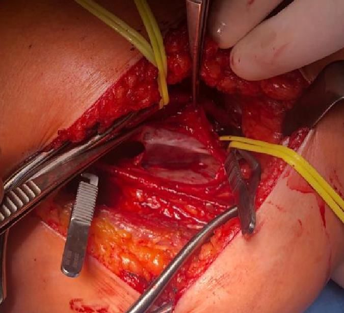
Diagnostics 2020, 10,
Diagnostics 2020, 10, 96
                      96                                                                                     33 of
                                                                                                                of 7
Diagnostics 2020, 10, 96                                                                                     3 of 7

      Figure 2.
              2. Magnetic resonance
                           resonance (MRI) of   the knee,
                                                    knee, longitudinal axis
                                                                       axis view, showing
                                                                                  showing the anatomy
                                                                                              anatomy and
      Figure 2. Magnetic
      Figure     Magnetic resonance (MRI)
                                       (MRI) of
                                             of the
                                                the knee, longitudinal
                                                          longitudinal axis view,
                                                                            view, showing the
                                                                                          the anatomy and
                                                                                                      and
      location of
      location of the popliteal
                      popliteal vein aneurysm.
                                     aneurysm.
      location of the
                  the popliteal vein
                                vein aneurysm.
5. Treatment
5. Treatment
     After a Heart Team
                   Team session,
                          session, and
                                    and in
                                        in accordance
                                           accordance to
                                                      to the
                                                         the patient’s
                                                             patient’s wishes, the decision was made to
     After a Heart Team session, and in accordance to the patient’s wishes, the decision was made to
proceed
proceed with
         with surgical
              surgical repair
                       repair of
                              of the
                                  the aneurysm.
                                      aneurysm.
proceed with surgical repair of the aneurysm.
Surgery
Surgery
Surgery
    Access
     Access to
             to the
                the popliteal
                    popliteal fossa
                              fossa was
                                    was obtained
                                         obtained via
                                                  via aa posterior
                                                         posterior approach.
                                                                    approach.
     Access to the popliteal
    Intraoperative            fossa  was obtained  via aofposterior approach.
     Intraoperative findings confirmed the presence of 6/4 saccular aneurysm
                     findings  confirmed  the presence     6/4 saccular aneurysm located
                                                                                 located along
                                                                                         along the
                                                                                               the distal
                                                                                                   distal
     Intraoperative
popliteal vein       findings
               (Figure  3).    confirmed the presence of 6/4 saccular aneurysm located along the distal
popliteal vein (Figure 3).
popliteal vein (Figure 3).

      Figure 3.
      Figure 3. Intraoperative
                Intraoperative view
                               view after
                                    after the
                                          the isolation,
                                              isolation, incision and drainage
                                                         incision and          of the
                                                                      drainage of the popliteal vein aneurysm.
                                                                                      popliteal vein aneurysm.
      Figure 3. Intraoperative view after the isolation, incision and drainage of the popliteal vein aneurysm.
     The    popliteal vein and the aneurysm         were isolated,    followed    by longitudinal incision and
     The    popliteal vein and the aneurysm         were isolated,    followed    by longitudinal incision and
drainage    of the aneurysm, which revealed         no thrombotic     material.   Next, the surgical procedure
drainage    of the aneurysm, which revealed         no thrombotic     material.   Next, the surgical procedure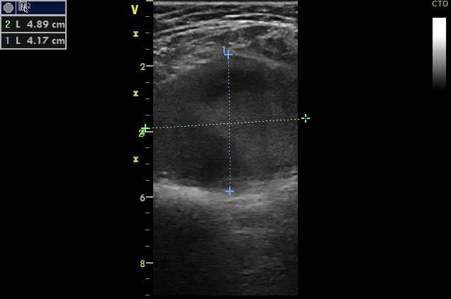
Diagnostics 2020, 10, 96 4 of 7
Diagnostics 2020, 10, 96 4 of 7
The popliteal vein and the aneurysm were isolated, followed by longitudinal incision and drainage
of the aneurysm,
consisted which aneurysmectomy
of tangential revealed no thrombotic material.
followed by patchNext, the surgical
angioplasty procedure
with consisted of
bovine pericardium
tangential
(Figure 4). aneurysmectomy followed by patch angioplasty with bovine pericardium (Figure 4).
Figure
Figure 4.
4. Tangential and patch
Tangential aneurysmectomy of the PVA and patch angioplasty
angioplasty with
with bovine
bovine pericardium.
pericardium.
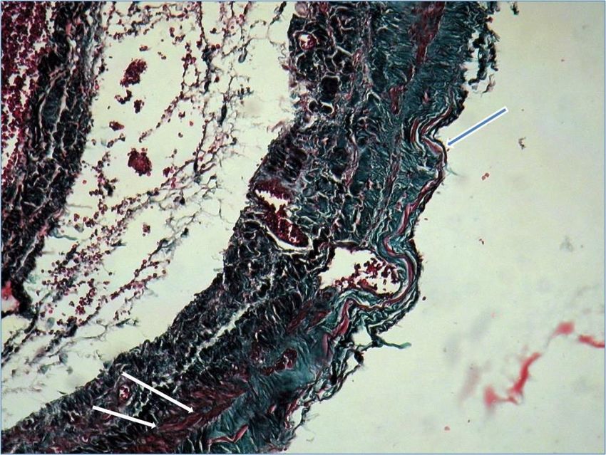
Microscopic examination
Microscopic examination with tricrom masson
with tricrom masson staining
staining protocol
protocol showed
showed continuous
continuous thinned
thinned
venous layer with fragmentation of elastin fibres (blue arrow) and few smooth muscle fibers (white
venous layer with fragmentation of elastin fibres (blue arrow) and few smooth muscle fibers (white
arrow) confirming
arrow) confirming the
the diagnosis
diagnosis of
of popliteal
popliteal venous
venous aneurysm
aneurysm (Figure
(Figure 5).
5).
Figure 5.
Figure Microscopy confirming
5. Microscopy confirming the
the diagnosis
diagnosis of
of popliteal
popliteal venous
venous aneurysm.
aneurysm.
Early post-surgery, the patient developed a hematoma that required surgical haemostasis and
Early post-surgery, the patient developed a hematoma that required surgical haemostasis and
drainage. Otherwise, the recovery was uneventful. Post-discharge medication consisted of oral
drainage. Otherwise, the recovery was uneventful. Post-discharge medication consisted of oral
anticoagulant and aspirin.
anticoagulant and aspirin.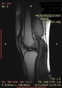
Diagnostics 2020, 10, 96                                                                           5 of 7

6. Follow-up
     The patient remained symptom-free at 2 months after the surgery. The antiplatelet treatment was
discontinued after 3 months. After consulting with the patient, the decision has been made to maintain
the anticoagulant treatment.

7. Discussion
      Venous aneurysms are rare findings, having different aetiologies and locations throughout
the body [1,2]. They are described in various anatomical locations as head and neck, thoracic,
intraabdominal, or extremities, some of them being associated with congenital malformations such as
Klippel-Trenaunay syndrome [1]. As for arterial aneurysms, a distinction is made between fusiform
and saccular aneurysms, primary and secondary venous aneurysm, respectively. While the aetiology
of primary venous aneurysms remains unclear, venous hypertension, external compression, or direct
trauma are the most frequent causes of secondary venous aneurysms. Primary popliteal venous
aneurysms (PVA) are the most common deep venous aneurysms of the lower extremity. They were
first described by Dahl et al. in 1976 [2]. They present several histological changes, such as decreased
smooth muscle cells in the media and a fibrous intima [1]. The major complications of these aneurysms
are thromboembolic events. Unfortunately, based on the existing data, between 24% and 51% of
patients with popliteal venous aneurysm have pulmonary embolism (PE) as the presenting symptom,
which may be a life-threatening condition [2]. Actually, large aneurysms are associated with a 70–80%
chance of PE formation despite efficient anticoagulation [2]. PE clinical signs and symptoms are
non-specific, including dyspnoea, chest pain, haemoptysis, or syncope [3]. When syncope is a symptom
of PE, it is associated with a higher risk of sudden death [4]. On the other hand, patients with deep
venous thrombosis may frequently develop silent recurrent PE. Other clinical presentations may
be related to chronic venous disease or local mass effect. However, the risk of rupture is low [5].
Approximately two-thirds of PVAs are saccular and the majority are located in the left lower limb
because of the compression of the left common iliac vein by the right common iliac artery [6].
      Also, a higher prevalence in women was reported. A possible explanation could be the
oestrogen-related effects on angiogenesis [5].

7.1. Diagnosis
      Ultrasound, CT, or magnetic resonance (MR) imaging are used as non-invasive diagnostic tools for
this condition. We believe that the tool of choice remains the color duplex ultrasound, which besides
describing the morphology of the aneurysm can predict the risk of thrombus formation by detecting
the turbulent flow in the aneurysmal segments. But, when an intervention is warranted, computed
tomography venography or magnetic resonance venography may be required for preoperative planning.
Diagnosing PVAs venous thromboembolic complications should follow the guideline recommended
diagnostic workup [3]. In our case, using the guideline proposed algorithm for diagnosing PE, by
combining the pre-test prediction rules—the Wells score or the modified Geneva score with D-Dimers
determination correctly lead to the indication of performing imaging testing, which confirmed PE.

7.2. Surgical Management
     Currently, there are no criteria with regard to the venous size in defining venous aneurysms.
Isolated dilatation of one and a half, two or three times more than the native vein have been described
in the existing literature [1,2,4]. Moreover, no consensus regarding the management of primary venous
aneurysms exists.
     Even though there is controversial data regarding the therapeutic management of PVAs, expectant
monitoring, medical management with anticoagulation, and surgical repair are the possible therapeutic
plans [1]. Anticoagulation may reduce the risk of thromboembolism but may theoretically worsen the
outcome in the event of aneurysmal rupture [5]. The natural history of asymptomatic PVAs remains
Diagnostics 2020, 10, 96 6 of 7
unknown, therefore the ideal treatment strategy is controversial. But, asymptomatic saccular or large
fusiform PVAs are associated with potentially life-threatening embolic events, therefore surgical repair
is generally advised. There are studies showing that a popliteal venous aneurysm with turbulent flow
detected by duplex ultrasound and a diameter of more than 20 mm has a firm indication for surgery
because of the unpredictable risk of PE [5]. Also, studies suggest surgery as the method of choice in
patients with recurrent thrombosis or pulmonary embolism after anticoagulation or having a high risk
of venous thrombosis [5]. Nasr et al. showed in a study that anticoagulation therapy alone fails in 43%
of cases in preventing thromboembolic events in patients with primary venous aneurysms [7]. As in
our case, patients may develop recurrent pulmonary emboli despite anticoagulation therapy when a
nonoperative management strategy is employed.
The operative technique must be selected on a case-by-case basis. Posterior access is the most
common, followed by resection of the aneurysm sac and lateral venorrhaphy to reconstruct the vein.
Primary venorrhaphy can be done if the aneurysm wall appears to be of good integrity. In our case,
intraoperative findings of a severely diseased venous wall dictated the use of a patch. The procedure
usually has few complications, including transient common peroneal nerve palsy and hematoma
formation, as in our case [8].
Whereas endovascular techniques have become a mainstay of vascular surgery, they currently
have no defined role in the treatment of venous aneurysms [2].
7.3. Postoperative Management
There are cases of recurrent popliteal venous aneurysms after surgical patchplasty that require
surgical reintervention. The role and duration of anticoagulation in postoperative asymptomatic
patients is unclear. Several reports suggest oral anticoagulation between 3–12 months, the use of
compression stocking, or lifelong aspirin therapy [1].
The small number of existing reported cases make definitive recommendations hardly to be made.
Even though current guidelines for the diagnosis and management of acute pulmonary embolism [3]
do not mention venous aneurysms of the extremities as major transient factors for pulmonary embolism
we strongly believe that it is very important for physicians to suspect PVAs until it is otherwise
ruled-out. In this context, we hope that our case adds evidence to the body of knowledge regarding
primary popliteal venous aneurysms.
Consent for Publication: The patient provided her written informed consent for the publication of the case report.
Funding: This research received no external funding.
Conflicts of Interest: The authors declare no conflict of interest.
References
1. Yang, G.K.; Hsiang, Y.N. Primary Popliteal Vein Aneurysm. Clin. Surg. 2018, 3, 2076.
2. Teter, K.A.; Maldonado, T.M.; Adelman, M.A. A systematic review of venous aneurysms by anatomic location.
J. Vasc. Surg. Venous Lymphat. Disord. 2018, 6, 408–413. [CrossRef] [PubMed]
3. Konstantinides, S.V.; Meyer, G.; Becattini, C.; Bueno, H.; Geersing, G.J.; Harjola, V.P.; Huisman, M.V.;
Humbert, M.; Jennings, C.S.; Jiménez, D.; et al. 2019 ESC Guidelines for the diagnosis and management
of acute pulmonary embolism developed in collaboration with the European Respiratory Society (ERS).
Eur. Heart J. 2020, 41, 543–603. [PubMed]
4. Prandoni, P.; Lensing, A.W.; Prins, M.H.; Ciammaichella, M.; Perlati, M.; Mumoli, N.; Bucherini, E.; Visonà, A.;
Bova, C.; Imberti, D.; et al. Prevalence of Pulmonary Embolism Among Patients Hospitalized for Syncope.
N. Engl. J. Med. 2016, 375, 1524–1531. [CrossRef] [PubMed]
5. Noppeney, T.; Kopp, R.; Pfister, K.; Schierling, W.; Noppeney, J.; Cucuruz, B. Treatment of popliteal vein
aneurysms. J. Vasc. Surg. Venous Lymphat. Disord. 2019. [CrossRef] [PubMed]Diagnostics 2020, 10, 96 7 of 7
6. Miyamotto, M.; de Lorenzo Costa, M.; Granella, V.H.; Angelo, B.Z.; de Andrade, D.C.; Raymundo, C.L.;
Moreira, R.C.R. Popliteal vein aneurysm: report of two cases. J. Vasc. Bras. 2018, 17, 170–173. [CrossRef]
[PubMed]
7. Nasr, W.; Babbitt, R.; Eslami, M.H. Popliteal vein aneurysm: A case report and review of literature.
Vasc. Endovascular Surg. 2007, 41, 551–555. [CrossRef] [PubMed]
8. Zhao, S.; Wang, X.; Sheng, H.; Huang, W.; Zhu, Y. Our experience of symptomatic and asymptomatic popliteal
venous aneurysm. J. Vasc. Surg. Cases Innov. Tech. 2017, 4, 1–4. [CrossRef] [PubMed]
© 2020 by the authors. Licensee MDPI, Basel, Switzerland. This article is an open access
article distributed under the terms and conditions of the Creative Commons Attribution
(CC BY) license (http://creativecommons.org/licenses/by/4.0/).You can also read