HHS Public Access Author manuscript Histopathology. Author manuscript; available in PMC 2016 September 19 - IUPUI ...
←
→
Page content transcription
If your browser does not render page correctly, please read the page content below
HHS Public Access
                                          Author manuscript
                                          Histopathology. Author manuscript; available in PMC 2016 September 19.
Author Manuscript

                              Published in final edited form as:
                               Histopathology. 2016 January ; 68(1): 5–21. doi:10.1111/his.12876.

                              Phyllodes tumours of the breast: a consensus review
                              Benjamin Y Tan, Geza Acs1, Sophia K Apple2, Sunil Badve3, Ira J Bleiweiss4, Edi Brogi5,
                              José P Calvo6, David J Dabbs7, Ian O Ellis8, Vincenzo Eusebi9, Gelareh Farshid10, Stephen
                              B Fox11, Shu Ichihara12, Sunil R Lakhani13, Emad A Rakha8, Jorge S Reis-Filho5, Andrea L
                              Richardson14, Aysegul Sahin15, Fernando C Schmitt16, Stuart J Schnitt17, Kalliopi P
                              Siziopikou18, Fernando A Soares19, Gary M Tse20, Anne Vincent-Salomon21, and Puay
                              Hoon Tan
Author Manuscript

                              Department of Pathology, Singapore General Hospital, Singapore
                              1Women’s      Pathology Consultants, Ruffolo Hooper & Associates, Tampa, FL, USA
                              2Department      of Pathology, UCLA Medical Center, Santa Monica, CA, USA
                              3Departments    of Pathology and Internal Medicine, Clarian Pathology Laboratory of Indiana
                              University, Indianapolis, IN, USA
                              4Department      of Pathology, Icahn School of Medicine at Mount Sinai, New York, NY, USA
                              5Department      of Pathology, Memorial Sloan Kettering Cancer Center, New York, NY, USA
                              6Servicio   de Anatomía Patológica, Hospital Universitario Ramón y Cajal, Madrid, Spain
                              7University   of Pittsburgh Medical Center, Pittsburgh, PA, USA
Author Manuscript

                              8Department of Histopathology, Nottingham City Hospital NHS Trust, Nottingham University,
                              Nottingham, UK
                              9Sezione    Anatomia e Istologia Patologica, ‘M. Malpighi’ Università di Bologna, Bologna, Italy
                              10BreastScreen SA, Discipline of Medicine, Adelaide University and Directorate of Surgical
                              Pathology, SA Pathology, Adelaide, South Australia
                              11Pathology   Department, Peter MacCallum Cancer Centre, St Andrews Place, East Melbourne,
                              Vic., Australia
                              12Department      of Pathology, Nagoya Medical Center, Nagoya, Japan
                              13School of Medicine and Pathology Queensland, The Royal Brisbane & Women’s Hospital,
Author Manuscript

                              University of Queensland Centre for Clinical Research, Brisbane, Qld, Australia
                              14Department      of Pathology, Brigham and Women’s Hospital and Harvard Medical School, Boston,
                              MA, USA
                              15Departmentof Pathology, Division of Pathology/Laboratory Medicine, University of Texas MD
                              Anderson Cancer Center, Houston, TX, USA

                              Address for correspondence: Dr Puay Hoon Tan, Department of Pathology, Singapore General Hospital, Diagnostics Tower, Level 7,
                              Academia, 20 College Road 169856, Singapore. tan.puay.hoon@sgh.com.sg.
                              Conflicts of interest
                              The authors declare no conflicts of interest with respect to the authorship, research and/or publication of this article

                    ___________________________________________________________________

                    This is the author's manuscript of the article published in final edited form as:
                    Tan, B. Y., Acs, G., Apple, S. K., Badve, S., Bleiweiss, I. J., Brogi, E., ... & Farshid, G. (2016). Phyllodes tumours of the breast: a
                    consensus review. Histopathology, 68(1), 5-21. https://doi.org/10.1111/his.12876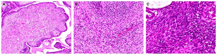
Tan et al.                                                                                                      Page 2

                        16Laboratoire      national de santé, Luxembourg city, Luxembourg
Author Manuscript

                        17Beth      Israel Deaconess Medical Center and Harvard Medical School, Boston, MA, USA
                        18Northwestern University Feinberg School of Medicine, Robert H. Lurie Comprehensive Cancer
                        Center, Chicago, IL, USA
                        19Department        of Anatomic Pathology, A. C. Camargo Cancer Centre, São Paulo, Brazil
                        20Department   of Anatomical and Cellular Pathology, Prince of Wales Hospital, The Chinese
                        University of Hong Kong, Shatin, Hong Kong
                        21Pôle      Pathologie-Génétique-Immunologie, Institut Curie, Paris, France

                        Abstract
                                 Phyllodes tumours constitute an uncommon but complex group of mammary fibroepithelial
Author Manuscript

                                 lesions. Accurate and reproducible grading of these tumours has long been challenging, owing to
                                 the need to assess multiple stratified histological parameters, which may be weighted differently
                                 by individual pathologists. Distinction of benign phyllodes tumours from cellular fibroadenomas is
                                 fraught with difficulty, due to overlapping microscopic features. Similarly, separation of the
                                 malignant phyllodes tumour from spindle cell metaplastic carcinoma and primary breast sarcoma
                                 can be problematic. Phyllodes tumours are treated by surgical excision. However, there is no
                                 consensus on the definition of an appropriate surgical margin to ensure completeness of excision
                                 and reduction of recurrence risk. Interpretive subjectivity, overlapping histological diagnostic
                                 criteria, suboptimal correlation between histological classification and clinical behaviour and the
                                 lack of robust molecular predictors of outcome make further investigation of the pathogenesis of
                                 these fascinating tumours a matter of active research. This review consolidates the current
                                 understanding of their pathobiology and clinical behaviour, and includes proposals for a rational
Author Manuscript

                                 approach to the classification and management of phyllodes tumours.

                        Keywords
                                 classification; fibroadenoma; malignant; metastasis; phyllodes

                        Introduction
                                           Phyllodes tumours of the breast constitute an uncommon but fascinating group of
                                           fibroepithelial neoplasms that have a morphological resemblance to the intracanalicular
                                           fibroadenoma at the benign end of the spectrum, but with increased stromal cellularity and
                                           leaf-like architecture. Phyllodes tumours are classified into benign, borderline and malignant
Author Manuscript

                                           grade categories on the basis of a constellation of histological parameters, i.e. the degree of
                                           stromal cellularity and atypia, mitotic count, stromal overgrowth, and the nature of their
                                           tumour borders.1 As each microscopic parameter has two to three tiers of stratification, there
                                           are significant challenges in accurate and reproducible categorization.

                                           Apart from grading difficulties, the benign phyllodes tumour shows overlapping features
                                           with cellular fibroadenoma, whereas, at the other end of the spectrum, the malignant
                                           phyllodes tumour may be mistaken for primary breast sarcoma or spindle cell metaplastic

                                             Histopathology. Author manuscript; available in PMC 2016 September 19.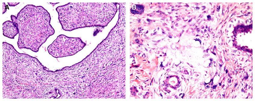
Tan et al. Page 3
carcinoma. The distinction between benign phyllodes tumour and cellular fibroadenoma is
Author Manuscript
especially problematic on core biopsies. Currently, cellular fibroepithelial lesions diagnosed
on core biopsy may be subjected to complete removal through either vacuum-assisted or
open excision. Surgical excision is usually the preferred procedure, as it allows negative
margins to be obtained in the event that the final diagnosis is a phyllodes tumour. What
constitutes a sufficient margin for phyllodes tumours is yet another unresolved dilemma.
Debate regarding the relationship between fibroadenoma, a common benign neoplasm, and
phyllodes tumour, a rare tumour with uncertain behaviour, continues. Fibroadenoma-like
areas are not infrequently encountered in phyllodes tumours, although the frequency of such
an observation is not known.
In this review, we provide a collective stance on these issues that can serve as a practical
guide for pathological reporting and understanding of phyllodes tumours.
Author Manuscript
Grading of phyllodes tumours
The criteria for diagnosis and grading of phyllodes tumours are summarized in the
recommendations of the World Health Organization (WHO) classification of tumours of the
breast.1 Briefly, phyllodes tumours are diagnosed when the fibroepithelial architecture
shows an exaggerated intracanalicular pattern with leaf-like fronds protruding into cystically
dilated spaces accompanied by stromal hypercellularity. A benign phyllodes tumour shows
mildly increased stromal cellularity as compared with a fibroadenoma, and has minimal
nuclear atypia, pushing borders, and mitoses of ≤4/10 high-power fields (HPFs). Stromal
overgrowth (defined as the presence of stroma without epithelium in at least one low-power
field as observed with a × 4 microscope objective) is not present. The key feature
distinguishing a benign phyllodes tumour from a fibroadenoma with an exaggerated
Author Manuscript
intracanalicular growth pattern is the presence of increased stromal cellularity. In the
absence of well-developed stromal fronds, the presence of elongated, branching and cleft-
like ducts meandering through the cellular stroma, giving a staghorn appearance, may be a
histological clue to the diagnosis of a phyllodes tumour.
At the other end of the spectrum, a malignant phyllodes tumour shows marked stromal
cellularity and atypia, has permeative margins, and has mitotic activity of at least 10/10
HPFs. Stromal overgrowth is usually easily identified. Phyllodes tumours with intermediate
features are assigned to the borderline category. Previous grading schemes have assessed
similar histological parameters, including that described by Azzopardi in 1979, which
incorporated the nature of the tumour edge, stromal overgrowth, mitotic activity, and cellular
atypia.2
Author Manuscript
It is important to acknowledge that there are no objective criteria for separating minimal/
mild from moderate and marked degrees of stromal hypercellularity and atypia, and this may
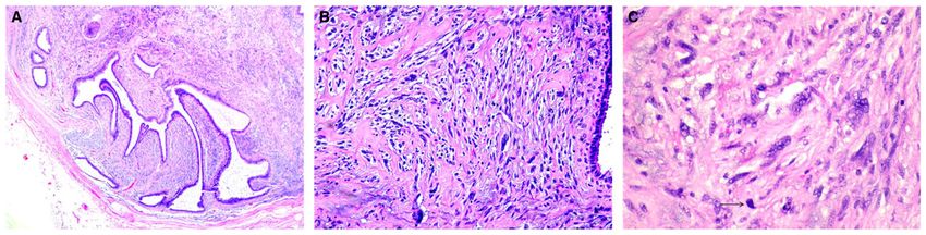
confound grading attempts. A practical guide for assessing stromal cellularity is to centre on
the most cellular zones of the lesion, with mild hypercellularity characterized by a slight
increase in stromal cells as compared with normal perilobular stroma, with evenly spaced
nuclei that are not touching or overlapping. Marked stromal cellularity shows confluent
Histopathology. Author manuscript; available in PMC 2016 September 19.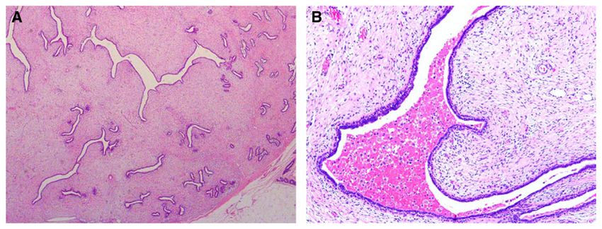
Tan et al.                                                                                              Page 4

                                  areas of densely overlapping nuclei, whereas moderate stromal cellularity has findings that
Author Manuscript

                                  are intermediate, with some overlapping stromal nuclei (Figure 1). Mild stromal atypia
                                  shows nuclei with little variation in size, with smooth nuclear contours. Moderate atypia
                                  shows some variation in nuclear size, with wrinkled nuclear membranes, to an extent
                                  exceeding that in mild atypia but less than that in marked atypia. Marked atypia shows
                                  marked variation in nuclear size, coarse chromatin, and irregular nuclear membranes with
                                  discernible nucleoli (Figure 2).3

                                  The perceived clinical relevance of grading phyllodes tumours is to predict clinical
                                  behaviour: benign tumours have the potential to locally recur; borderline tumours have the
                                  potential to recur locally, and have a very low risk of metastasis; and malignant tumours
                                  have the highest risk of metastatic behaviour, which may eventually prove fatal. However, it
                                  is accepted that adverse events are, in general, rare for all forms of phyllodes tumours when
                                  they are subjected to complete local excision.
Author Manuscript

                                  Although the guidelines may appear straightforward, their application can be fraught with
                                  ambiguity. Furthermore, how the subdivisions for each microscopic parameter interact to
                                  constitute the final grade is subjective. It is also not uncommon for phyllodes tumours to
                                  show intratumoral heterogeneity, and harbour features that typify benign lesions in some
                                  areas, and characteristics of borderline and malignant lesions in other foci. For instance, a
                                  phyllodes tumour with marked stromal atypia and brisk mitotic activity, but without
                                  permeative margins or stromal overgrowth, may be considered by some pathologists to be
                                  borderline, whereas others may regard the tumour as malignant, owing to different
                                  weighting of the relevance of each feature, with prioritization of stromal atypia (Figure 3). A
                                  practical approach is to grade a phyllodes tumour as malignant when it shows all of the
                                  histological changes of malignancy, and as borderline when not all malignant characteristics
Author Manuscript

                                  are present. The presence of a malignant heterologous element such as liposarcoma,
                                  chondrosarcoma or osteosarcoma relegates the tumour into the malignant category
                                  regardless of whether other histological parameters (stromal hypercellularity, atypia, mitotic
                                  rate, overgrowth, and nature of tumour borders) show changes characteristic of malignant
                                  phyllodes tumours. In an effort to comprehend which microscopic parameters are more
                                  influential in determining the clinical behaviour of phyllodes tumours, a study of 605 cases
                                  concluded that stromal atypia, mitoses, overgrowth and surgical margins (AMOS criteria)4
                                  were of independent significance in predicting behaviour, with surgical margin status being
                                  the most important. A nomogram was developed by the use of a mathematical formula that
                                  could be applied to counsel patients about their individual risk for recurrence.4

                                  Despite the host of biological markers studied in phyllodes tumours, many with an
                                  association with grade,5–13 their use in defining grade and potential clinical behaviour in
Author Manuscript

                                  specific cases remains limited.

                        Biological behaviour and metastatic potential of phyllodes tumours
                                  Recurrence rates in a large Asian series of phyllodes tumours were 10.9%, 14.4% and 29.6%
                                  for benign, borderline and malignant phyllodes tumours, respectively. 4 In another review of
                                  33 cases from Germany, recurrence rates were reported to be 8%, 20% and 50% for benign,

                                    Histopathology. Author manuscript; available in PMC 2016 September 19.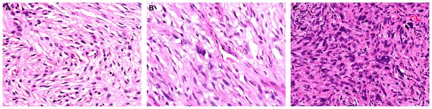
Tan et al.                                                                                            Page 5

                                 borderline and malignant tumours, respectively,1 with distant metastases being encountered
Author Manuscript

                                 in 9% of patients with malignant tumours. Overall, recurrence rates in the literature are 10–
                                 17%, 14–25% and 23–30% for benign, borderline and malignant phyllodes tumours,
                                 respectively.1 Interestingly, in a clinicopathological analysis by Karim et al., there was a
                                 suggestion that Asian patients experienced a higher recurrence rate than those of non-Asian
                                 ethnicity.14

                                 Grade progression during local recurrence of phyllodes tumours can occur. There have been
                                 several suggestions regarding why this happens, including a lack of representative sampling
                                 of the initial tumour, tumour heterogeneity with the presence of stromal subclones,15 and
                                 loss of stromal–epithelial interdependency. 16

                                 In a study of 335 phyllodes tumours, it was noted that metastases and death from phyllodes
                                 tumours were consistently preceded by a primary malignant diagnosis,17 suggesting that a
Author Manuscript

                                 key aim should be to recognize the malignant category, in order for effective therapy to be
                                 given at the outset. Metastases in phyllodes tumours invariably indicate a dismal prognosis,
                                 with ensuing death.4,18 Distant metastases occur mostly to the lung and skeleton, but almost
                                 all organ sites have been affected (Figure 4). Histologically, metastases comprise malignant
                                 stromal elements without accompanying epithelium.17,19,20 Two exceptional cases of
                                 metastatic phyllodes tumours harbouring an epithelial component have been described as
                                 case reports. One represented inclusion of lung alveolar tissue within the metastatic tumour
                                 stroma, as confirmed by immunoreactivity of the epithelial component for antisurfactant
                                 apoproteins.21 The other case showed floridly hyperplastic, adenosis-like epithelium,
                                 rimmed by actin and calponin-positive myoepithelial cells, within both the primary and
                                 metastatic tumours, which showed liposarcomatous differentiation.22 The metastatic
                                 epithelial component duplicated the immunoreactivity for oestrogen receptor (ER),
Author Manuscript

                                 progesterone receptor and gross cystic disease fluid protein 15 seen in the epithelium of the
                                 primary tumour.

                                 How often do phyllodes tumours metastasize, and do benign tumours ever do so? Table 1
                                 shows metastatic rates according to phyllodes tumour grades that have been described by
                                 various authors.4,23–35 The singular documentations of metastatic disease following a
                                 diagnosis of benign phyllodes tumour were by Abdalla et al. and Chaney et al., where distant
                                 metastases were reported to occur in 3.2%, 11.1% and 28.6% of benign (1/31), borderline
                                 (3/27) and malignant (6/21) tumours,26 and in 1.7%, 0% and 26.7% of benign (1/59),
                                 borderline (0/12) and malignant (8/30) tumours,23 respectively. However, pathological
                                 details of these unusual cases of metastatic benign phyllodes tumour were not provided.

                                 It may be reasonably inferred that metastatic disease is a vanishingly rare occurrence in
Author Manuscript

                                 benign phyllodes tumours, with the qualification that all tumours should be adequately
                                 sampled to account for intratumoral heterogeneity. Conversely, metastatic behaviour is an
                                 established risk for malignant phyllodes tumours, albeit still uncommon, and pathological
                                 diagnosis should focus on accurately identifying this group of tumours.

                                   Histopathology. Author manuscript; available in PMC 2016 September 19.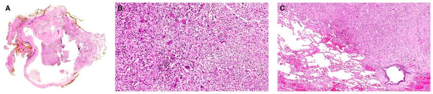
Tan et al.                                                                                               Page 6

                        Relationship between fibroadenoma and phyllodes tumour
Author Manuscript

                                  Phyllodes tumours are generally regarded as de-novo lesions derived from periductal and
                                  specialized lobular stroma. The initiation of tumorigenesis may hinge on epithelial–stromal
                                  interactions. However, the histological overlap between fibroadenoma and phyllodes tumour
                                  has long raised the question of pathogenetic kinship. Table 2 shows the studies that have
                                  explored this relationship and their salient findings.36–50

                                  Interestingly, a mother and daughter pair with benign phyllodes tumours was also described,
                                  raising the possibility of hereditary linkage,43 and a TP53 founder mutation was discovered
                                  in phyllodes tumours from Brazil.51 A study from France discovered chromosome
                                  imbalances in 55%, 91% and 100% of benign, borderline and malignant phyllodes tumours,
                                  respectively, with 1q gains being associated with borderline and malignant grades. It was
                                  suggested that phyllodes tumours could be divided into two genetically distinct classes, with
Author Manuscript

                                  benign tumours in one group and borderline/malignant phyllodes in the other.52

                                  More recently, highly recurrent mediator complex subunit 12 (MED12) somatic mutations in
                                  exon 2 were discovered in 59% of 98 fibroadenomas studied, 44 with most mutations
                                  occurring in codon 44. The same mutation in MED12 is a common genetic anomaly in
                                  uterine smooth muscle tumours.53,54 Laser capture microdissection established that MED12
                                  mutations were present in stromal but not in epithelial cells of fibroadenomas. A subsequent
                                  study by Ng et al. found that MED12 mutations were also prevalent in phyllodes tumours,
                                  with 65.1% of benign, 65.6% of borderline and 42.8% of malignant phyllodes tumours,
                                  respectively, showing mutations. 50 The overall rate of MED12 mutations was strikingly
                                  similar in phyllodes tumours (62.5%) and fibroadenomas (59%), with a comparable rate of
                                  mutations in codon 44 of MED12 supporting a close molecular relationship.44,50 Other
Author Manuscript

                                  studies have confirmed the high prevalence of MED12 mutations in fibroadenomas and
                                  phyllodes tumours.45–49 Using targeted next-generation sequencing, Cani et al. found that
                                  malignant phyllodes tumours harboured additional genetic aberrations in tumour suppressor
                                  genes, consistent with their aggressive biological behaviour.45 Of particular prognostic
                                  import is the finding by Ng et al. that tumours with MED12 mutations were significantly
                                  associated with longer disease-free survival, possibly related to hormonal dependence.50

                                  Although evidence for the direct evolution of phyllodes tumours from fibroadenomas
                                  remains limited, with very recent confirmation of linear progression in some cases,55 it is
                                  clear that these fibroepithelial lesions possess molecular similarities, in addition to their
                                  striking morphological resemblance in some cases.

                        Distinguishing cellular fibroadenoma from benign phyllodes tumour
Author Manuscript

                                  In a study of 21 cellular fibroepithelial lesions evaluated by 10 specialist breast pathologists
                                  using the WHO criteria, only for two cases was uniform agreement achieved with regard to
                                  whether the lesion represented a cellular fibroadenoma or phyllodes tumour. It is noted,
                                  however, that these cases were highly selected from a consultation series, and problematic
                                  lesions were therefore over-represented.56 When cellular fibroadenomas and benign
                                  phyllodes tumours were combined and compared with borderline and malignant phyllodes

                                    Histopathology. Author manuscript; available in PMC 2016 September 19.
Tan et al. Page 7
tumours as another group, the level of agreement improved considerably, with complete
Author Manuscript
concordance in 53% of cases. These findings testify to the challenges of separating cellular
fibroadenomas from benign phyllodes at one end of the spectrum, and also highlight the
difficulty of achieving consensus in grading phyllodes tumours (Figure 5). It is important to
note that fibroadenomas in the paediatric age group tend to have increased stromal
cellularity, which should not be overinterpreted. 57 Of 68 paediatric breast fibroepithelial
lesions analysed in a recent study, 16 cases showed mitotic activity. These included 15
fibroadenomas of simple, cellular and juvenile types, 11 of which showed 1–2 mitoses/10
HPF, and the remaining five fibroepithelial lesions showed mitotic activity ranging from 3 to
5/10 HPF.57 In another study, by Ross et al., of breast fibroepithelial tumours in adolescent
females aged up to 18 years, the mean stromal mitotic counts/10 HPF for 11 benign
phyllodes tumours, five ‘usual’ fibroadenomas, 12 juvenile fibroadenomas and three
‘variant’ juvenile fibroadenomas (with pericanalicular stromal expansion) were 3 (range: 1–
7), 1 (range: 0–2), 2 (range: 0–4) and 3 (range: 0–7), respectively.58 Faiz et al. also found
Author Manuscript
stromal mitoses in all fibroadenoma subtypes (cellular, classic, and juvenile) among 119
paediatric cases with up to 5 mitoses/10 HPF being observed in two cases.59 Therefore,
apart from potentially increased stromal cellularity in paediatric fibroepithelial tumours,
mitotic activity may also be encountered, up to 7 mitoses/10 HPF.57–59 A cautious and
measured approach is therefore needed when cellular and mitotically active paediatric
fibroepithelial lesions are evaluated. A diagnosis of phyllodes tumour should be based on the
finding of well-developed stromal fronds accompanied by increased stromal hypercellularity.
Numerous studies have attempted to analyse the histology of phyllodes tumours.60–64 A
study by Choi et al. found a concordance rate of only 60% between core needle biopsies and
excision specimens, with larger tumour size being significantly correlated with discordant
biopsy results.65 Assessment of clinicoradiological tumour attributes such as size and
Author Manuscript
radiographic density may contribute to clinical decision-making. 66,67 Notwithstanding that,
histomorphological assessment of the excised lesion remains the practical gold standard in
diagnosis and grading,60,68 with the presence of leafy architecture and increased stromal
cellularity typically being used as the discriminants between cellular fibroadenoma and
benign phyllodes tumour. Fibroadenomas that contain stromal multinucleated giant cells can
also be mistaken for phyllodes tumours,69 and there may be a potential role for Ki67
proliferation activity as an adjunctive aid.3
The question of whether there is always a need to precisely delineate a benign phyllodes
tumour from a cellular fibroadenoma arises. The answer is important, as many surgeons
would perform a second surgical procedure to achieve negative margins for a benign
phyllodes tumour initially enucleated without margin clearance. The WHO Working Group
Author Manuscript
has proposed that the term ‘benign fibroepithelial neoplasm’ be employed in equivocal
cases,70 in order to avoid overtreatment. This term, however, should be used sparingly, as it
does not represent a new diagnostic category.
There has been both direct and indirect evidence that benign phyllodes tumours may be
treated less aggressively. In a study of 37 women with locally recurrent phyllodes tumours, it
was concluded that an expectant approach may be acceptable for initially diagnosed benign
and borderline tumours, with complete resection being achieved during any subsequent
Histopathology. Author manuscript; available in PMC 2016 September 19.Tan et al. Page 8
recurrent episode.27 Although most surgeons would be uncomfortable with not re-excising a
Author Manuscript
borderline phyllodes tumour with positive margins, it would be reasonable to assume a
‘watchful waiting’ strategy for benign lesions. The rate of recurrence for fibroadenomas
after ultrasound-guided vacuum assisted percutaneous excision is listed as 15%,71 whereas
Organ et al. described a recurrence rate of 17% for surgically excised fibroadenomas.72
However, as acknowledged by the authors, determining whether the recurrence was a ‘true
recurrence’ of the same tumour or another primary tumour was difficult, if not impossible,
owing to the retrospective nature of the study, particularly if the same breast was involved. In
a previous article, also by Organ,73 it was stated that ‘recurrences’ of fibroadenomas were
undoubtedly serial presentations of multicentric lesions. This contrasts with a 10.9%
recurrence rate of benign phyllodes tumours in one series,4 occurring typically at the site of
previous surgery. A very low recurrence rate of 3.4% was reported in benign phyllodes
tumours in a study by Korean investigators, with all recurrent cases remaining benign,74
Author Manuscript
without any association with surgical margin status. Teo et al., in a retrospective review of
44 Asian cases, found no cases of local recurrence in benign tumours treated with simple
excision (enucleation), regardless of margin status, after a mean follow-up of 47.6 months.75
Hence, a benign phyllodes tumour diagnosed after representative sampling of an excision
specimen may be conservatively handled even when positive margins are encountered.
Conversely, malignant phyllodes tumours are associated with a recurrence rate of 29.6%4,
with metastases and death being observed in 22%,1 underscoring the need to recognize this
subset of aggressive phyllodes tumours for complete surgical eradication.
Distinguishing malignant phyllodes tumour from primary breast sarcoma
and spindle cell metaplastic breast carcinoma
Author Manuscript
At the other end of the histological spectrum, a high-grade spindle cell neoplasm of the
breast invokes different diagnostic considerations, namely malignant phyllodes tumour with
sarcomatous overgrowth, spindle cell metaplastic breast carcinoma, and primary or
secondary breast sarcoma.
The architectural hallmark of leaf-like fronds surmounted by benign glandular epithelium
serves to delineate phyllodes tumour from its mimics. In some malignant phyllodes tumours,
however, the stromal overgrowth may be so prominent that epithelial elements can be
difficult to find, requiring extensive sampling with many sections for their identification. The
stroma of a malignant phyllodes tumour may, on occasion, show heterologous sarcomatous
differentiation, most frequently liposarcoma, but also including myosarcoma, angiosarcoma,
chondrosarcoma, and osteosarcoma (Figure 6). A spindle cell metaplastic breast carcinoma
Author Manuscript
contains varying proportions of a malignant epithelial component, which may be of
squamous, glandular or adenosquamous type. Metaplastic carcinomas can also be entirely
devoid of frank epithelial elements, or additionally show heterologous mesenchymal
differentiation, although liposarcomatous elements are hardly ever seen. The presence of
ductal carcinoma in situ adjacent to a malignant mammary spindle cell tumour greatly
favours a diagnosis of metaplastic carcinoma. Primary breast sarcomas, which are distinctly
uncommon,76 and sarcomas metastatic to the breast, which are exceptionally rare, have no
Histopathology. Author manuscript; available in PMC 2016 September 19.Tan et al. Page 9
distinguishing histological features of either phyllodes tumour or metaplastic breast
Author Manuscript
carcinoma, and can have histological attributes common to sarcomas at any site. A history of
previous or metastatic sarcoma, imaging and clinical correlation may be helpful. Table 3
summarises the features of these three entities.
On limited samples such as needle core biopsies, accurate diagnosis of high-grade malignant
mammary spindle cell lesions can prove exceedingly challenging, especially when an
epithelial element is elusive. The demonstration of diffuse cytokeratin or p63
immunoreactivity in the malignant spindle cells supports a diagnosis of metaplastic
carcinoma,77,78 although interpretation must be tempered in cases of focal keratin or p63
expression, as such reactivities have been described in stromal cells of phyllodes
tumours.79,80 The utility of p40 in a similar diagnostic setting remains under investigation;
so far, it has been found to be more specific but less sensitive than p63.80–82 However, like
p63, p40 may stain stromal cells of phyllodes tumours in some cases. CD34 reactivity,
Author Manuscript
which is well described in the stromal cells of phyllodes tumours, has been reported to be
inversely related to adverse histological features;6,8,13,83–86 this assumes importance if
CD34 were to be considered for diagnostic utility in differentiating high-grade spindle cell
lesions of the breast, as malignant phyllodes tumours are less likely to express CD34. Other
markers, including bcl-286,87 which is more frequently expressed in phyllodes tumours,
CD117,83,88,89 which shows increased expression in higher-grade phyllodes tumours, and
sarcoma-specific molecular cytogenetic alterations are possible diagnostic adjuncts.
Although routinely employed in the diagnosis of fibromatosis that may occur in the breast,
aberrant nuclear expression of β-catenin has been reported in the stroma of phyllodes
tumours, as well as in metaplastic carcinomas.90–94 The use of β-catenin as a solitary marker
in the assessment of mammary spindle cell lesions must be cautioned against.
Author Manuscript
Adequate sampling, entailing at least one section per centimetre of maximal tumour size,
with additional sampling of grossly heterogeneous areas and meticulous morphological
assessment, remains the keystone of diagnosis, buttressed by clinical, radiological and
immunohistochemical correlation.
Epithelial–stromal interactions in phyllodes tumours
In 1992, Sawhney et al. observed that stromal mitotic activity tended to occur close to the
epithelial compartment in fibroepithelial lesions, and hypothesized that stromal growth in
these tumours depended, in part, on the epithelial component. It was suggested that
increasing malignancy correlated with loss of stromal dependency on the epithelium.95
Since then, there have been a number of studies supporting this view. Sawyer et al.
Author Manuscript
demonstrated allelic imbalances in chromosomes 3p and 1q in both epithelial and stromal
elements of phyllodes tumours.96 These authors also noted that stromal proliferation in
benign phyllodes tumours was dependent on abnormalities in the Wnt pathway consequent
to Wnt5a expression in the epithelial component, with malignant progression being linked to
independence from the latter.91 E-cadherin is a known epithelial differentiation marker that
is affected by the Wnt signalling pathway, and its expression in the epithelium of phyllodes
tumours was correlated with recurrence and shorter tumour-specific survival.97 Feakins et al.
Histopathology. Author manuscript; available in PMC 2016 September 19.Tan et al. Page 10
found epithelial platelet-derived growth factor (PDGF)/stromal PDGF receptor-β
copositivity that correlated with disease-related death in 43% of phyllodes tumours.98 Clonal
Author Manuscript
abnormalities have been detected in both epithelial and stromal components of phyllodes
tumours in studies by Dietrich et al.99 and Kuijper et al.39 Additional work on biomarker
expression in epithelial and stromal elements of phyllodes tumours lends further credence to
their interactions.16,97,100–102 For example, the level of CXCR4, an epithelial–stromal
interaction-related molecule, was found to be increased in the stromal component of higher-
grade phyllodes tumours.10
Hormone receptors
Table 4 summarizes findings from studies that have evaluated the expression of hormone
receptors in phyllodes tumours,103–109 with one report documenting HER2/c-erbB2
reactivity as well.106 ERα expression was confined to the epithelial compartment, without
Author Manuscript
any stromal positivity, with one study demonstrating an inverse correlation with grade.106
ERβ expression, on the other hand, has been observed in stromal cells of phyllodes
tumours.108,109 The practical significance of these observations is unclear. Although the
current data indicate a limited role for hormones in phyllodes tumours, the recently
discovered MED12 mutations in these tumours may lead to a resurgence of interest, as the
MED12 abnormality is linked to aberrantly activated oestrogen signalling.44,50
Surgical margins
The mainstay of phyllodes tumour management has traditionally consisted of surgical
excision with wide tumour-free margins, generally defined by some authors as at least 10
mm.31 However, more recent data suggest that not all phyllodes tumours require excision
with such wide margins.32,110,111 In cases of large tumours, this may render breast
Author Manuscript
conservation impracticable.112,113 Table 5 summarizes pertinent findings from studies that
have addressed surgical margins in phyllodes tumours.23–25,28,32,110,111,114–120
Drawing a parallel from an opinion advanced by Wood regarding the issue of surgical
margins in invasive breast cancer, there appears to be a dearth of data supporting a precise
width of tumour-free tissue that is significantly associated with reduced tumour
recurrence.121 Although an increasing amount of normal tissue confers greater confidence in
the adequacy of excision (with ensuing diminishing cosmetic results), a single layer of cells
between the tumour and the surgical plane constitutes, in theory, a clear margin.121 This
does not take into account myriad factors that may undermine the accuracy of representation
in any slide, including tumour irregularity, multifocality, ink seepage, sampling and technical
sectioning issues, among others. Onkendi et al., in a study of 67 borderline and malignant
Author Manuscript
phyllodes tumours from the Mayo Clinic, found that the extent of surgical excision had no
impact on disease-free survival.110 An analysis of 164 cases by Jang et al. revealed no
significant local control advantage conferred by wide (at least 10 mm) margins over
narrower margins.32 Lin et al., in a single-institution series of 33 cases, found no relationship
between width of surgical margin and disease recurrence.111
Histopathology. Author manuscript; available in PMC 2016 September 19.Tan et al. Page 11
Notwithstanding the above, many institutions elect to offer additional surgical treatment for
Author Manuscript
close margins. Mangi et al., in a study of 40 cases from the Massachusetts General Hospital,
found that post-excision recurrences were confined to cases with positive margins, or
margins ofTan et al. Page 12
1. Grading of phyllodes tumours should aim to achieve accuracy and
Author Manuscript
consistency at the benign and malignant ends of the spectrum.
2. Definitive distinction between cellular fibroadenomas and benign
phyllodes tumours may not be crucial, in light of similar reported
recurrence rates. The term benign fibroepithelial lesion/neoplasm may be
recommended for cases where clear diagnostic distinction cannot be made,
although this should be used sparingly.
3. Malignant phyllodes tumours are diagnosed when there are marked
stromal hypercellularity, atypia, increased mitoses of ≥10/10 HPFs,
permeative tumour borders, and stromal overgrowth. The presence of a
malignant heterologous component places the tumour into the malignant
category regardless of other histological features.
Author Manuscript
4. A conservative approach can be accorded to benign phyllodes tumours that
have been initially enucleated without margins.
5. Excision with negative margins should be achieved for recurrent and
malignant phyllodes tumours. Most would recommend that borderline
tumours should also be completely excised. Although the literature often
refers to a margin width of at least 10 mm, a robust evidence base to
support this approach is lacking. Therefore, an ideal margin width remains
to be determined, and may need to be considered in relation to factors such
as tumour size and cosmesis.
6. From a diagnostic and management perspective, it is important to
accurately recognize malignant phyllodes tumours, which should be
Author Manuscript
surgically eradicated and effectively treated at diagnosis, as these tumours
have a well-established but relatively infrequent risk of metastasis and
death.
7. The role of adjuvant radiation therapy in borderline and malignant
tumours remains to be defined. Routine axillary dissection is not
recommended.
References
1. Lakhani, SR.; Ellis, IO.; Schnitt, SJ.; Tan, PH.; van de Vijver, MJ., editors. World Health
Organization Classification of Tumours of the Breast. Lyon: IARC Press; 2012.
2. Azzopardi, J. Problems in Breast Pathology. London: WB Saunders; 1979. p. 346-365.
3. Jara-Lazaro AR, Akhilesh M, Thike AA, Lui PC-W, Tse GM-K, Tan PH. Predictors of phyllodes
Author Manuscript
tumours on core biopsy specimens of fibroepithelial neoplasms. Histopathology. 2010; 57:220–232.
[PubMed: 20716164]
4. Tan PH, Thike AA, Tan WJ, et al. Predicting clinical behaviour of breast phyllodes tumours: a
nomogram based on histological criteria and surgical margins. J Clin Pathol. 2012; 65:69–76.
[PubMed: 22049216]
5. Tsang JYS, Ni Y-B, Ng EK, et al. MicroRNAs are differentially deregulated in mammary malignant
phyllodes tumour. Histopathology. 2015; 67:294–305. [PubMed: 25585495]
Histopathology. Author manuscript; available in PMC 2016 September 19.Tan et al. Page 13
6. Vilela MHT, de Almeida FM, de Paula GM, et al. Utility of Ki- 67, CD10, CD34, p53, CD117, and
mast cell content in the differential diagnosis of cellular fibroadenomas and in the classification of
Author Manuscript
phyllodes tumors of the breast. Int J Surg Pathol. 2014; 22:485–491. [PubMed: 24492332]
7. Tan WJ, Thike AA, Bay BH, Tan PH. Immunohistochemical expression of homeoproteins Six1 and
Pax3 in breast phyllodes tumours correlates with histological grade and clinical outcome.
Histopathology. 2014; 64:807–817. [PubMed: 24438019]
8. Ho SK, Thike AA, Cheok PY, Tse GM-K, Tan PH. Phyllodes tumours of the breast: the role of
CD34, vascular endothelial growth factor and β-catenin in histological grading and clinical
outcome. Histopathology. 2013; 63:393–406. [PubMed: 23772632]
9. Kim G-E, Kim J-H, Lee KH, et al. Stromal matrix metalloproteinase-14 expression correlates with
the grade and biological behavior of mammary phyllodes tumors. Appl Immunohistochem Mol
Morphol. 2012; 20:298–303. [PubMed: 22505012]
10. Kwon JE, Jung W-H, Koo JS. Molecules involved in epithelial– mesenchymal transition and
epithelial–stromal interaction in phyllodes tumors: implications for histologic grade and prognosis.
Tumour Biol. 2012; 33:787–798. [PubMed: 22203494]
11. Ang MK, Ooi AS, Thike AA, et al. Molecular classification of breast phyllodes tumors: validation
Author Manuscript
of the histologic grading scheme and insights into malignant progression. Breast Cancer Res Treat.
2011; 129:319–329. [PubMed: 20945089]
12. Tsai W-C, Jin J-S, Yu J-C, Sheu L-F. CD10, actin, and vimentin expression in breast phyllodes
tumors correlates with tumor grades of the WHO grading system. Int J Surg Pathol. 2006; 14:127–
131. [PubMed: 16703173]
13. Chen CM, Chen CJ, Chang CL, Shyu JS, Hsieh HF, Harn HJ. CD34, CD117, and actin expression
in phyllodes tumor of the breast. J Surg Res. 2000; 94:84–91. [PubMed: 11104647]
14. Karim RZ, Gerega SK, Yang YH, et al. Phyllodes tumours of the breast: a clinicopathological
analysis of 65 cases from a single institution. Breast. 2009; 18:165–170. [PubMed: 19329316]
15. Karim RZ, O’Toole SA, Scolyer RA, et al. Recent insights into the molecular pathogenesis of
mammary phyllodes tumours. J Clin Pathol. 2013; 66:496–505. [PubMed: 23404800]
16. Karim RZ, Scolyer RA, Tse GM, Tan P-H, Putti TC, Lee CS. Pathogenic mechanisms in the
initiation and progression of mammary phyllodes tumours. Pathology. 2009; 41:105–117.
[PubMed: 19152185]
Author Manuscript
17. Tan P-H, Jayabaskar T, Chuah K-L, et al. Phyllodes tumors of the breast: the role of pathologic
parameters. Am J Clin Pathol. 2005; 123:529–540. [PubMed: 15743740]
18. Goh CHR, Lim YP, Su JW, et al. Cardiopulmonary thromboembolism of epithelioid angiosarcoma
arising from malignant phyllodes tumour of the breast. J Clin Pathol. 2014; 67:450–454. [PubMed:
24399035]
19. Tsubochi H, Sato N, Kaimori M, Imai T. Osteosarcomatous differentiation in lung metastases from
a malignant phyllodes tumour of the breast. J Clin Pathol. 2004; 57:432–434. [PubMed:
15047752]
20. Fernandez BB, Hernanzez FJ, Spindler W. Metastatic cystosarcoma phyllodes: a light and electron
microscopic study. Cancer. 1976; 37:1737–1746. [PubMed: 177175]
21. West TL, Weiland LH, Clagett OT. Cystosarcoma phyllodes. Ann Surg. 1971; 173:8.
22. Kracht J, Sapino A, Bussolati G. Malignant phyllodes tumor of breast with lung metastases
mimicking the primary. Am J Surg Pathol. 1998; 22:1284–1290. [PubMed: 9777991]
23. Chaney AW, Pollack A, McNeese MD, et al. Primary treatment of cystosarcoma phyllodes of the
breast. Cancer. 2000; 89:1502–1511. [PubMed: 11013364]
Author Manuscript
24. Chen W-H, Cheng S-P, Tzen C-Y, et al. Surgical treatment of phyllodes tumors of the breast:
retrospective review of 172 cases. J Surg Oncol. 2005; 91:185–194. [PubMed: 16118768]
25. Sotheran W, Domjan J, Jeffrey M, Wise MH, Perry PM. Phyllodes tumours of the breast—a
retrospective study from 1982–2000 of 50 cases in Portsmouth. Ann R Coll Surg Engl. 2005;
87:339–344. [PubMed: 16176692]
26. Abdalla HM, Sakr MA. Predictive factors of local recurrence and survival following primary
surgical treatment of phyllodes tumors of the breast. J Egypt Natl Cancer Inst. 2006; 18:125–133.
27. Tan EY, Tan PH, Hoon TP, et al. Recurrent phyllodes tumours of the breast: pathological features
and clinical implications. ANZ J Surg. 2006; 76:476–480. [PubMed: 16768772]
Histopathology. Author manuscript; available in PMC 2016 September 19.Tan et al. Page 14
28. Cheng S-P, Chang Y-C, Liu T-P, Lee J-J, Tzen C-Y, Liu C-L. Phyllodes tumor of the breast: the
challenge persists. World J Surg. 2006; 30:1414–1421. [PubMed: 16865317]
Author Manuscript
29. Belkacémi Y, Bousquet G, Marsiglia H, et al. Phyllodes tumor of the breast. Int J Radiat Oncol
Biol Phys. 2008; 70:492– 500. [PubMed: 17931796]
30. Lenhard MS, Kahlert S, Himsl I, Ditsch N, Untch M, Bauerfeind I. Phyllodes tumour of the breast:
clinical follow-up of 33 cases of this rare disease. Eur J Obstet Gynecol Reprod Biol. 2008;
138:217–221. [PubMed: 17868973]
31. Guillot E, Couturaud B, Reyal F, et al. Management of phyllodes breast tumors. Breast J. 2011;
17:129–137. [PubMed: 21251125]
32. Jang JH, Choi M-Y, Lee SK, et al. Clinicopathologic risk factors for the local recurrence of
phyllodes tumors of the breast. Ann Surg Oncol. 2012; 19:2612–2617. [PubMed: 22476816]
33. Sawalhi S, Al-Shatti M. Phyllodes tumor of the breast: a retrospective study of the impact of
histopathological factors in local recurrence and distant metastasis. Ann Saudi Med. 2013; 33:162–
168. [PubMed: 23563006]
34. Wang H, Wang X, Wang C-F. Comparison of clinical characteristics between benign borderline
and malignant phyllodes tumors of the breast. Asian Pac J Cancer Prev. 2014; 15:10791–10795.
Author Manuscript
[PubMed: 25605178]
35. Bumpers HL, Tadros T, Gabram-Mendola S, et al. Phyllodes tumors in African American women.
Am J Surg. 2015; 210:74–79. [PubMed: 25873163]
36. Noguchi S, Motomura K, Inaji H, Imaoka S, Koyama H. Clonal analysis of fibroadenoma and
phyllodes tumor of the breast. Cancer Res. 1993; 53:4071–4074. [PubMed: 8395336]
37. Noguchi S, Yokouchi H, Aihara T, et al. Progression of fibroadenoma to phyllodes tumor
demonstrated by clonal analysis. Cancer. 1995; 76:1779–1785. [PubMed: 8625047]
38. Kasami M, Vnencak-Jones CL, Manning S, Dupont WD, Jensen RA, Page DL. Monoclonality in
fibroadenomas with complex histology and phyllodal features. Breast Cancer Res Treat. 1998;
50:185–191. [PubMed: 9822223]
39. Kuijper A, Buerger H, Simon R, et al. Analysis of the progression of fibroepithelial tumours of the
breast by PCR-based clonality assay. J Pathol. 2002; 197:575–581. [PubMed: 12210075]
40. Wang ZC, Buraimoh A, Iglehart JD, Richardson AL. Genomewide analysis for loss of
heterozygosity in primary and recurrent phyllodes tumor and fibroadenoma of breast using single
Author Manuscript
nucleotide polymorphism arrays. Breast Cancer Res Treat. 2006; 97:301–309. [PubMed:
16791486]
41. Hodges KB, Abdul-Karim FW, Wang M, et al. Evidence for transformation of fibroadenoma of the
breast to malignant phyllodes tumor. Appl Immunohistochem Mol Morphol. 2009; 17:345–350.
[PubMed: 19276971]
42. Abe M, Miyata S, Nishimura S, et al. Malignant transformation of breast fibroadenoma to
malignant phyllodes tumor: long-term outcome of 36 malignant phyllodes tumors. Breast Cancer.
2011; 18:268–272. [PubMed: 22121516]
43. Foucar CE, Hardy A, Siziopikou KP, et al. A mother and daughter with phyllodes tumors of the
breast. Clin Breast Cancer. 2012; 12:373–377. [PubMed: 22981939]
44. Lim WK, Ong CK, Tan J, et al. Exome sequencing identifies highly recurrent MED12 somatic
mutations in breast fibroadenoma. Nat Genet. 2014; 46:877–880. [PubMed: 25038752]
45. Cani AK, Hovelson DH, McDaniel AS, et al. Next-gen sequencing exposes frequent MED12
mutations and actionable therapeutic targets in phyllodes tumors. Mol Cancer Res. 2015; 13:613–
619. [PubMed: 25593300]
Author Manuscript
46. Yoshida M, Sekine S, Ogawa R, et al. Frequent MED12 mutations in phyllodes tumours of the
breast. Br J Cancer. 2015; 112:1703–1708. [PubMed: 25839987]
47. Piscuoglio S, Murray M, Fusco N, et al. MED12 somatic mutations in fibroadenomas and
phyllodes tumours of the breast. Histopathology. 2015; 67:529–537. [PubMed: 25688711]
48. Nagasawa S, Maeda I, Fukuda T, et al. MED12 exon 2 mutations in phyllodes tumors of the breast.
Cancer Med. 2015; 7:1117–1121. [PubMed: 25865354]
49. Pfarr N, Kriegsmann M, Sinn P, et al. Distribution of MED12 mutations in fibroadenomas and
phyllodes tumors of the breast—implications for tumor biology and pathological diagnosis. Genes
Chromosom Cancer. 2015; 54:444–452. [PubMed: 25931199]
Histopathology. Author manuscript; available in PMC 2016 September 19.Tan et al. Page 15
50. Ng CCY, Tan J, Ong CK, et al. MED12 is frequently mutated in breast phyllodes tumours: a study
of 112 cases. J Clin Pathol. 2015; 68:685–691. [PubMed: 26018969]
Author Manuscript
51. Giacomazzi J, Koehler-Santos P, Palmero EI, et al. A TP53 founder mutation, p. R337H, is
associated with phyllodes breast tumors in Brazil. Virchows Arch. 2013; 463:17–22. [PubMed:
23794094]
52. Laé M, Vincent-Salomon A, Savignoni A, et al. Phyllodes tumors of the breast segregate in two
groups according to genetic criteria. Mod Pathol. 2007; 20:435–444. [PubMed: 17334353]
53. Croce S, Chibon F. MED12 and uterine smooth muscle oncogenesis: state of the art and
perspectives. Eur J Cancer. 2015; 51:1603–1610. [PubMed: 26037152]
54. Je EM, Kim MR, Min KO, Yoo NJ, Lee SH. Mutational analysis of MED12 exon 2 in uterine
leiomyoma and other common tumors. Int J Cancer. 2012; 131:E1044–E1047. [PubMed:
22532225]
55. Tan J, Ong CK, Lim WK, et al. Genomic landscapes of breast fibroepithelial tumours. Nat Genet.
2015 Oct 5. (epub).
56. Lawton TJ, Acs G, Argani P, et al. Interobserver variability by pathologists in the distinction
between cellular fibroadenomas and phyllodes tumors. Int J Surg Pathol. 2014; 22:695–698.
Author Manuscript
[PubMed: 25161205]
57. Tay TKY, Chang KTE, Thike AA, Tan PH. Paediatric fibroepithelial lesions revisited: pathological
insights. J Clin Pathol. 2015; 68:633–641. [PubMed: 25998513]
58. Ross DS, Giri DD, Akram MM, Catalano J, Van Zee KJ, Brogi E. Fibroepithelial lesions in the
breast of adolescent females: a clinicopathological profile of 35 cases. Mod Pathol. 2012; 25(Suppl
2):64a.
59. Faiz S, Tudor V, Yasim G-P, Badve S. Fibroadenomatous lesions in pediatric age group. Mod
Pathol. 2013; 26(Suppl 2):39A.
60. Giri D. Recurrent challenges in the evaluation of fibroepithelial lesions. Arch Pathol Lab Med.
2009; 133:713–721. [PubMed: 19415945]
61. Tsang AKH, Chan SK, Lam CCF, et al. Phyllodes tumours of the breast—differentiating features
in core needle biopsy. Histopathology. 2011; 59:600–608. [PubMed: 21916949]
62. Lee AHS, Hodi Z, Ellis IO, Elston CW. Histological features useful in the distinction of phyllodes
tumour and fibroadenoma on needle core biopsy of the breast. Histopathology. 2007; 51:336–344.
Author Manuscript
[PubMed: 17727475]
63. Aiyer HM, Jain M, Thomas S, Logani KB. Diagnostic stromal histomorphology in fibroepithelial
breast lesions: a fresh perspective. Indian J Pathol Microbiol. 2000; 43:5–12. [PubMed: 12583412]
64. Yasir S, Gamez R, Jenkins S, Visscher DW, Nassar A. Significant histologic features differentiating
cellular fibroadenoma from phyllodes tumor on core needle biopsy specimens. Am J Clin Pathol.
2014; 142:362–369. [PubMed: 25125627]
65. Choi J, Koo JS. Comparative study of histological features between core needle biopsy and
surgical excision in phyllodes tumor. Pathol Int. 2012; 62:120–126. [PubMed: 22243782]
66. Gould DJ, Salmans JA, Lassinger BK, et al. Factors associated with phyllodes tumor of the breast
after core needle biopsy identifies fibroepithelial neoplasm. J Surg Res. 2012; 178:299–303.
[PubMed: 22524977]
67. Resetkova E, Khazai L, Albarracin CT, Arribas E. Clinical and radiologic data and core needle
biopsy findings should dictate management of cellular fibroepithelial tumors of the breast. Breast
J. 2010; 16:573–580. [PubMed: 21070433]
68. Yang X, Kandil D, Cosar EF, Khan A. Fibroepithelial tumors of the breast: pathologic and
Author Manuscript
immunohistochemical features and molecular mechanisms. Arch Pathol Lab Med. 2014; 138:25–
36. [PubMed: 24377809]
69. Heneghan HM, Martin ST, Casey M, Tobbia I, Benani F, Barry KM. A diagnostic dilemma in
breast pathology—benign fibroadenoma with multinucleated stromal giant cells. Diagn Pathol.
2008; 3:33. [PubMed: 18673528]
70. Tan PH, Ellis IO. Myoepithelial and epithelial–myoepithelial, mesenchymal and fibroepithelial
breast lesions: updates from the WHO Classification of Tumours of the Breast 2012. J Clin Pathol.
2013; 66:465–470. [PubMed: 23533258]
Histopathology. Author manuscript; available in PMC 2016 September 19.Tan et al. Page 16
71. Grady I, Gorsuch H, Wilburn-Bailey S. Long-term outcome of benign fibroadenomas treated by
ultrasound-guided percutaneous excision. Breast J. 2008; 14:275–278. [PubMed: 18397185]
Author Manuscript
72. Organ CH, Organ BC. Fibroadenoma of the female breast: a critical clinical assessment. J Natl
Med Assoc. 1983; 75:701–704. [PubMed: 6887274]
73. Nigro DM, Organ CH. Fibroadenoma of the female breast. Some epidemiologic surprises. Postgrad
Med. 1976; 59:113–117. [PubMed: 177964]
74. Kim S, Kim J-Y, Kim DH, Jung WH, Koo JS. Analysis of phyllodes tumor recurrence according to
the histologic grade. Breast Cancer Res Treat. 2013; 141:353–363. [PubMed: 24062207]
75. Teo JY, Cheong CS-J, Wong CY. Low local recurrence rates in young Asian patients with
phyllodes tumours: less is more. ANZ J Surg. 2012; 82:325–328. [PubMed: 22507352]
76. Rakha EA, Tan PH, Shaaban A, et al. Do primary mammary osteosarcoma and chondrosarcoma
exist? A review of a large multi-institutional series of malignant matrix-producing breast tumours.
Breast. 2013; 22:13–18. [PubMed: 23084962]
77. Koker MM, Kleer CG. p63 expression in breast cancer: a highly sensitive and specific marker of
metaplastic carcinoma. Am J Surg Pathol. 2004; 28:1506–1512. [PubMed: 15489655]
78. Tse GM, Tan P-H, Chaiwun B, et al. p63 is useful in the diagnosis of mammary metaplastic
Author Manuscript
carcinomas. Pathology. 2006; 38:16–20. [PubMed: 16484002]
79. Chia Y, Thike AA, Cheok PY, Yong-Zheng Chong L, Man-Kit Tse G, Tan PH. Stromal keratin
expression in phyllodes tumours of the breast: a comparison with other spindle cell breast lesions.
J Clin Pathol. 2012; 65:339–347. [PubMed: 22259180]
80. Cimino-Mathews A, Sharma R, Illei PB, Vang R, Argani P. A subset of malignant phyllodes
tumors express p63 and p40: a diagnostic pitfall in breast core needle biopsies. Am J Surg Pathol.
2014; 38:1689–1696. [PubMed: 25046342]
81. Kim SK, Jung WH, Koo JS. p40 (ΔNp63) expression in breast disease and its correlation with p63
immunohistochemistry. Int J Clin Exp Pathol. 2014; 7:1032–1041. [PubMed: 24696720]
82. D’Alfonso TM, Ross DS, Liu Y-F, Shin SJ. Expression of p40 and laminin 332 in metaplastic
spindle cell carcinoma of the breast compared with other malignant spindle cell tumours. J Clin
Pathol. 2015; 68:516–521. [PubMed: 25795733]
83. Noronha Y, Raza A, Hutchins B, et al. CD34, CD117, and Ki- 67 expression in phyllodes tumor of
the breast: an immunohistochemical study of 33 cases. Int J Surg Pathol. 2011; 19:152–158.
Author Manuscript
[PubMed: 21087983]
84. Moore T, Lee AH. Expression of CD34 and bcl-2 in phyllodes tumours, fibroadenomas and spindle
cell lesions of the breast. Histopathology. 2001; 38:62–67. [PubMed: 11135048]
85. Cîmpean AM, Raica M, Nariţa D. Diagnostic significance of the immunoexpression of CD34 and
smooth muscle cell actin in benign and malignant tumors of the breast. Rom J Morphol Embryol.
2005; 46:123–129. [PubMed: 16286998]
86. Lee AHS. Recent developments in the histological diagnosis of spindle cell carcinoma,
fibromatosis and phyllodes tumour of the breast. Histopathology. 2008; 52:45–57. [PubMed:
18171416]
87. Dunne B, Lee AHS, Pinder SE, Bell JA, Ellis IO. An immunohistochemical study of metaplastic
spindle cell carcinoma, phyllodes tumor and fibromatosis of the breast. Hum Pathol. 2003;
34:1009–1015. [PubMed: 14608534]
88. Esposito NN, Mohan D, Brufsky A, Lin Y, Kapali M, Dabbs DJ. Phyllodes tumor: a
clinicopathologic and immunohistochemical study of 30 cases. Arch Pathol Lab Med. 2006;
130:1516–1521. [PubMed: 17090194]
Author Manuscript
89. Tan P-H, Jayabaskar T, Yip G, et al. p53 and c-kit (CD117) protein expression as prognostic
indicators in breast phyllodes tumors: a tissue microarray study. Mod Pathol. 2005; 18:1527–1534.
[PubMed: 16258510]
90. Lacroix-Triki M, Geyer FC, Lambros MB, et al. β-catenin/Wnt signalling pathway in fibromatosis,
metaplastic carcinomas and phyllodes tumours of the breast. Mod Pathol. 2010; 23:1438–1448.
[PubMed: 20693983]
91. Sawyer EJ, Hanby AM, Rowan AJ, et al. The Wnt pathway, epithelial–stromal interactions, and
malignant progression in phyllodes tumours. J Pathol. 2002; 196:437–444. [PubMed: 11920740]
Histopathology. Author manuscript; available in PMC 2016 September 19.Tan et al. Page 17
92. Sawyer EJ, Hanby AM, Poulsom R, et al. Beta-catenin abnormalities and associated insulin-like
growth factor overexpression are important in phyllodes tumours and fibroadenomas of the breast.
Author Manuscript
J Pathol. 2003; 200:627–632. [PubMed: 12898599]
93. Tsang JYS, Mendoza P, Lam CCF, et al. Involvement of α- and β-catenins and E-cadherin in the
development of mammary phyllodes tumours. Histopathology. 2012; 61:667–674. [PubMed:
22571452]
94. Hayes MJ, Thomas D, Emmons A, Giordano TJ, Kleer CG. Genetic changes of Wnt pathway
genes are common events in metaplastic carcinomas of the breast. Clin Cancer Res. 2008;
14:4038–4044. [PubMed: 18593979]
95. Sawhney N, Garrahan N, Douglas-Jones AG, Williams ED. Epithelial–stromal interactions in
tumors. A morphologic study of fibroepithelial tumors of the breast. Cancer. 1992; 70:2115–2120.
[PubMed: 1327488]
96. Sawyer EJ, Hanby AM, Ellis P, et al. Molecular analysis of phyllodes tumors reveals distinct
changes in the epithelial and stromal components. Am J Pathol. 2000; 156:1093– 1098. [PubMed:
10702425]
97. Tsang JYS, Mendoza P, Putti TC, et al. E-cadherin expression in the epithelial components of
Author Manuscript
mammary phyllodes tumors. Hum Pathol. 2012; 43:2117–2123. [PubMed: 22820000]
98. Feakins RM, Wells CA, Young KA, Sheaff MT. Platelet-derived growth factor expression in
phyllodes tumors and fibroadenomas of the breast. Hum Pathol. 2000; 31:1214–1222. [PubMed:
11070114]
99. Dietrich CU, Pandis N, Rizou H, et al. Cytogenetic findings in phyllodes tumors of the breast:
karyotypic complexity differentiates between malignant and benign tumors. Hum Pathol. 1997;
28:1379–1382. [PubMed: 9416694]
100. Dacic S, Kounelis S, Kouri E, Jones MW. Immunohistochemical profile of cystosarcoma
phyllodes of the breast: a study of 23 cases. Breast J. 2002; 8:376–381. [PubMed: 12390361]
101. Karim RZ, Gerega SK, Yang YH, et al. Proteins from the Wnt pathway are involved in the
pathogenesis and progression of mammary phyllodes tumours. J Clin Pathol. 2009; 62:1016–
1020. [PubMed: 19861560]
102. Logullo AF, Nonogaki S, Do Socorro Maciel M, Mourão-Neto M, Soares FA. Stromal and
epithelial cells react differentially to c-kit in fibroepithelial tumors of the breast. Mol Med Rep.
2008; 1:857–861. [PubMed: 21479497]
Author Manuscript
103. Rao BR, Meyer JS, Fry CG. Most cystosarcoma phyllodes and fibroadenomas have progesterone
receptor but lack estrogen receptor: stromal localization of progesterone receptor. Cancer. 1981;
47:2016–2021. [PubMed: 6261932]
104. Mechtersheimer G, Krüger KH, Born IA, Möller P. Antigenic profile of mammary fibroadenoma
and cystosarcoma phyllodes. A study using antibodies to estrogen- and progesterone receptors
and to a panel of cell surface molecules. Pathol Res Pract. 1990; 186:427–438. [PubMed:
2174150]
105. Singh Y, Hatano T, Uemura Y, et al. Immunohistochemical profile of phyllodes tumors of the
breast. Oncol Rep. 1996; 3:677–681. [PubMed: 21594434]
106. Shpitz B, Bomstein Y, Sternberg A, et al. Immunoreactivity of p53, Ki-67, and c-erbB-2 in
phyllodes tumors of the breast in correlation with clinical and morphologic features. J Surg
Oncol. 2002; 79:86–92. [PubMed: 11815995]
107. Tse GMK, Lee CS, Kung FYL, et al. Hormonal receptors expression in epithelial cells of
mammary phyllodes tumors correlates with pathologic grade of the tumor: a multicenter study of
Author Manuscript
143 cases. Am J Clin Pathol. 2002; 118:522–526. [PubMed: 12375638]
108. Sapino A, Bosco M, Cassoni P, et al. Estrogen receptor-beta is expressed in stromal cells of
fibroadenoma and phyllodes tumors of the breast. Mod Pathol. 2006; 19:599–606. [PubMed:
16554735]
109. Kim Y-H, Kim G-E, Lee JS, et al. Hormone receptors expression in phyllodes tumors of the
breast. Anal Quant Cytol Histol. 2012; 34:41–48. [PubMed: 22590818]
110. Onkendi EO, Jimenez RE, Spears GM, Harmsen WS, Ballman KV, Hieken TJ. Surgical treatment
of borderline and malignant phyllodes tumors: the effect of the extent of resection and tumor
characteristics on patient outcome. Ann Surg Oncol. 2014; 21:3304–3309. [PubMed: 25034817]
Histopathology. Author manuscript; available in PMC 2016 September 19.You can also read