An Update on Molecular Genetic Aberrations in Spitz Melanocytic Proliferations: Correlation with Morphological Features and Biological Behavior
←
→
Page content transcription
If your browser does not render page correctly, please read the page content below
Clinical Science
Narrative Review
Acta Medica Academica 2021;50(x):xx-xx
DOI: 10.5644/ama2006-124.XX
An Update on Molecular Genetic Aberrations in Spitz Melanocytic Proliferations:
Correlation with Morphological Features and Biological Behavior
Daja Šekoranja, Jože Pižem, Boštjan Luzar
Institute of Pathology, Faculty of Medicine, University of Ljubljana, Korytkova 2, 1000 Ljubljana, Slovenia
Correspondence: bostjan.luzar@mf.uni-lj.si; Tel.: + 386 1 543 7130; Fax.: + 386 1 543 7101
Received: 14 February 2021; Accepted: 15 March 2021
Abstract
The aim of the paper is to give an update on molecular genetic aberrations in Spitz melanocytic proliferations with special em-
phasis on their correlation with morphological features and biological behavior. The Spitz group of melanocytic proliferations
is defined by a combination of distinctive morphological features and driver molecular genetic events. Morphologically, these
neoplasms are characterized by large, oval, polygonal, or spindled melanocytes with abundant eosinophilic cytoplasm, vesicular
nuclei with prominent nucleoli, often in association with epidermal hyperplasia. Molecular aberrations in Spitz melanocytic
proliferations can be divided into two main groups, according to the driver genetic change: 1) 11p amplification/HRAS muta-
tion, present in about 20% of cases, and 2) kinase fusions, present in about 50%, further subdivided into tyrosine kinase fusions
(ALK, ROS1, NTRK1, NTRK3, MET, RET) or serine-threonine kinase fusions (MAP3K8, BRAF). Driver genetic aberrations
can be detected along the whole biological spectrum of Spitz melanocytic proliferations, and are mutually exclusive. Although
driver genetic aberrations enable proliferation of melanocytes, additional genetic events (often biallelic inactivation of CDKN2A
and TERT promoter mutations) are necessary for the development of overt Spitz malignancy. Conclusions. Recent studies have
demonstrated that certain driver genetic aberrations are more often associated with the benign spectrum of Spitz melanocytic
proliferations and indolent biological behavior (11p amplification/HRAS mutation, tyrosine kinase fusions). In contrast, some
driver aberrations are more frequent in the atypical/malignant spectrum of Spitz melanocytic proliferations with a potential
for aggressive biological behavior (serine-threonine kinase fusions). In addition, certain driver aberrations are often associated
with distinctive morphological features. However, none of the morphological features is entirely specific for any of these driver
genetic aberrations. Immunohistochemistry for ALK, ROS1, and pan-TRK can be used for screening purposes to detect cor-
responding fusion proteins.
Key Words: Spitz Melanocytic Proliferations Kinase Fusions 11p Amplification/HRAS Mutation Morphology Clinical Be-
havior.
Introduction hyperplasia (1). They encompass the whole bio-
logical spectrum of melanocytic proliferations, in-
According to the most recent 4th WHO Classifica- cluding Spitz nevi, atypical Spitz tumors, and Spitz
tion of Skin Tumours, the Spitz group of melano- melanomas, also referred to as malignant Spitz
cytic proliferations is defined by a combination of tumors (1). Based on the underlying genetic ab-
morphological characteristics and driver molecu- errations, Spitz melanocytic proliferations can be
lar genetic abnormalities (1). Spitz melanocytic divided into four distinct groups: 1) 11p amplified/
proliferations are characterized morphologically HRAS mutated proliferations, 2) proliferations
by a proliferation of large, oval, polygonal epithe- with tyrosine kinase fusions (ALK, ROS1, NTRK1,
lioid or spindled melanocytes with abundant eo- NTRK3, MET, RET), 3) proliferations with serine/
sinophilic cytoplasm, vesicular nuclei with promi- threonine kinase fusions (MAP3K8, BRAF) and
nent nucleoli, often in association with epidermal 4) proliferations with Spitz morphology but lack-
Copyright © 2021 by the Academy of Sciences and Arts of Bosnia and Herzegovina.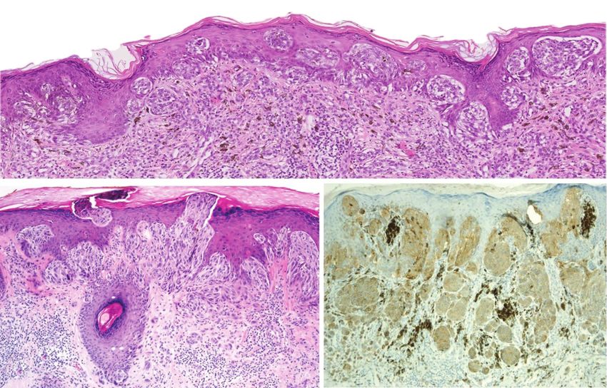
Daja Šekoranja et al: Spitz Melanocytic Proliferations – An Update

ing an 11p/HRAS mutation, kinase fusions, BRAF,                    section, a table is presented with short summary of
NRAS, GNAQ, GNA11 mutations and other driver                       key data for each particular Spitz group of melano-
genetic changes characteristic of other defined                    cytic proliferations).
melanocytic subgroups (1). The last group is at
present poorly defined and will not be discussed
further in this review. Importantly, driver genetic                11p Amplified and/or HRAS Mutated Spitz
events are mutually exclusive in a particular Spitz                Melanocytic Proliferations
melanocytic proliferation and are by themselves                    The first recurrent genetic alterations discovered
insufficient for the development of overt malig-                   in Spitz melanocytic proliferations were 11p am-
nancy, generally characterized by the development                  plification and HRAS mutation. They were also the
of distant metastases and aggressive biological be-                first genetic alterations associated with a specific
havior (2, 3).                                                     morphologic phenotype in this group of melano-
    As has been demonstrated by recent studies,                    cytic neoplasms (12, 13). Both 11p amplification
such a combined morphological/genetic classifica-                  and HRAS mutation can appear exclusively or si-
tion better correlates with the biological behavior                multaneously in a single lesion and are most com-
of different groups of Spitz melanocytic prolifera-                monly associated with a desmoplastic Spitz nevus
tions. The vast majority of Spitz melanomas re-                    morphology (13, 14). HRAS (Harvey rat sarcoma
sulting in distant metastatic spread harbor serine/                viral oncogene homolog) resides on 11p chromo-
threonine kinase fusions (4-11). Furthermore,                      some arm (11p15.5) and belongs to the Ras family
since several biological drugs are available to treat              of oncogenes, encoding a GTPase that is involved
melanocytic proliferations with aggressive clinical                in cellular signaling (the MAP kinase-signaling
behavior, the characterization of particular driver                pathway) (15, 16). Mutations in HRAS lead to con-
genetic events and additional genetic abnormali-                   stitutive activation of an altered protein that im-
ties is becoming increasingly important.                           pacts the expression of various transcription fac-
    Herein, we review recent advances in the mo-                   tors involved in cell cycle progression, thus stimu-
lecular genetics of Spitz melanocytic proliferations.              lating cell growth and differentiation (16). HRAS
Special emphasis is given to the correlation of mo-                mutations have also been detected in urothelial
lecular genetic aberrations with morphological                     and squamous cell carcinomas, adenocarcinomas
features and with the biological behavior of Spitz                 of various origins, leukemias, and myelodysplastic
melanocytic proliferations. Immunohistochemis-                     syndromes (17-19). HRAS mutations in Spitz me-
try can be a reliable surrogate tool for certain mo-               lanocytic proliferations are most commonly mis-
lecular abnormalities to molecular genetic testing,                sense mutations involving codons 61 and 13, with
as discussed further in this review (for practical                 the three most commonly reported mutations
purposes, the list of antibodies reflecting possible               being Q61L (2, 6, 14, 20), Q61R (14, 20-23), and
kinase fusions the authors are using routinely is                  G13R (6, 21, 24-28).
summarized in the Table 1. Also, at the end of each                    The prototypic 11p amplified/HRAS mutated
                                                                   Spitz nevus is a symmetrical, predominantly der-
Table 1. The List of Antibodies Reflecting Possible Kinase         mal, relatively hypocellular proliferation com-
Fusions Used Routinely by the Authors of the Current               posed of large, epithelioid and spindled melano-
Review                                                             cytes with desmoplastic stromal reaction (i.e.,
 Antigen   Clone       Manufacturer              Dilution          thickened collagen fibers between single neo-
 ALK       5A4         Leica Biosystems,         1:10              plastic cells) and an infiltrative base (Figure 1) (2,
                       Wetzlar, Germany                            13, 14). Melanocytes have abundant eosinophilic
 ROS1      SP384       Ventana, Roche, Tucson,   RTU (Ready        or amphophilic cytoplasm, vesicular nuclei, and
                       USA                       To Use)           mild to moderate nuclear pleomorphism (2, 13,
 panTRK    EPR17341    Abcam, Cambridge, UK      1:50              14). The proliferation rate is usually low, although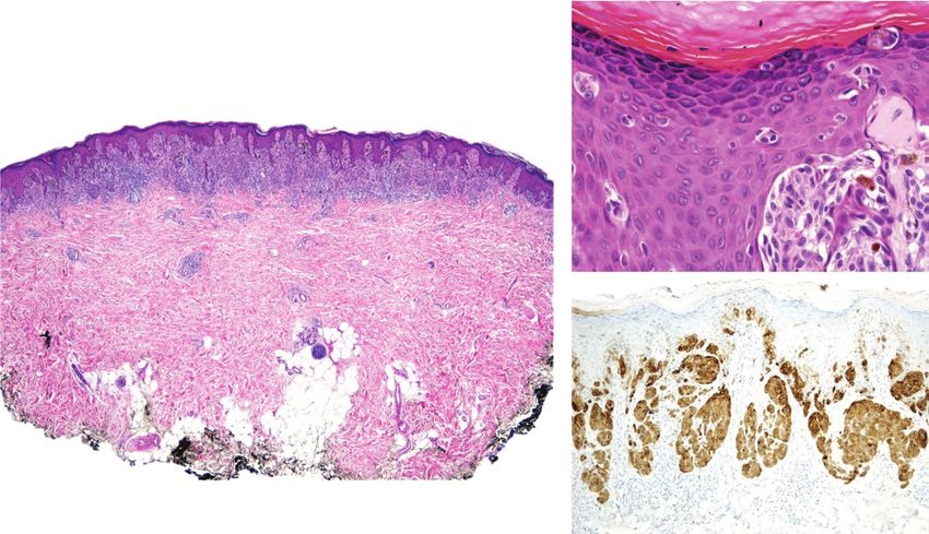
Acta Medica Academica 2021;50(x):xx-xx

A                                                             B

Figure 1. Desmoplastic Spitz nevus. A. Low power magnification reveals a fairly symmetrical intradermal proliferation with
infiltrative growth pattern and a desmoplastic stromal reaction. B. Dense collagenous stroma containing epithelioid mela-
nocytes with abundant eosinophilic cytoplasm with darkly stained nuclei and prominent nucleoli is typical for the entity.

isolated deep mitoses and, in rare instances, mul-            reaction) in a case of a 50-year-old female, and a
tiple mitoses may be present and are not associated           three-codon deletion in MAP2K1 (p.102_104del),
with malignancy (2, 13, 14, 29). Atypical mitoses             a hemizygous mutation in ARID1A, homozygous
are generally absent.                                         deletion of CDKN2A and NOTCH2 amplifica-
    In addition to solitary lesions with typical des-         tion in a case of a 75-year-old female (23). Un-
moplastic Spitz nevus morphology, other melano-               fortunately, clinical follow-up was not available
cytic proliferations with HRAS mutations and/or               in either of the two cases (23). Lazova et al. also
11p gain have also been described, including agmi-            reported two melanomas with HRAS mutations in
nated Spitz nevi with or without associated nevus             their series (6). One case was of a 73-year-old male
spilus (25-28), recurrent Spitz nevi (30), melano-            diagnosed with Spitz melanoma that harbored an
cytic nevi with deep penetrating nevus-like mor-              HRAS Q61L mutation, which developed metasta-
phology (21), pseudogranulomatous Spitz nevi                  ses two years later, but he was still alive at a 4-year
(31) and a combination of syringocystadenoma                  follow-up (6). The second case was a 60-year-old
papilliferum, tubular adenoma and a Spitz nevus               male diagnosed with conventional melanoma that
(24). Moreover, the desmoplastic Spitz nevus phe-
notype is not restricted to HRAS mutated lesions              Table 2. Spitz Melanocytic Proliferations with 11p
since rare Spitz nevi harboring ROS1, ALK or even             Amplification/HRAS Mutation
a BRAF fusion exhibiting a desmoplastic Spitz ne-
                                                                  Morphological         • Symmetrical, predominantly
vus morphology have been described (2, 32).                       features                intradermal, infiltrative base
    Even though the vast majority of Spitz prolif-                                      • Epithelioid and spindled melanocytes
erations with HRAS mutations are essentially as-                                          – Large
                                                                                          – Abundant cytoplasm
sociated with benign clinical behavior (14, 29, 33),                                      – Mild to moderate pleomorphism
there are occasional reports of HRAS mutated Spitz                                        – Mitoses rare, can be deep
                                                                                          – Low proliferation rate
melanomas (6, 23). Recently, Raghavan et al. pub-
                                                                                        • Desmoplastic stromal reaction
lished a series of Spitz melanomas, two of which
                                                                  Biological behavior   Generally benign
harbored HRAS hotspot mutations (23). Both
                                                                  Confirmatory test     Next generation sequencing
lesions were associated with additional genetic                                         Fluorescence in situ hybridization (for
aberrations, namely a loss of chromosome 9 (ac-                                         11p amplification)
companied by negative p16 immunohistochemical                                           Comparative genomic hybridization (for
                                                                                        11p amplification)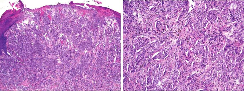
Daja Šekoranja et al: Spitz Melanocytic Proliferations – An Update

harbored an HRAS G13R mutation, who devel-                  Spitz melanocytic proliferations with ALK fu-
oped metastases two years later, and died at the        sions have the largest average diameter among all
age of 63 (6).                                          Spitz melanocytic proliferations (43). The vast ma-
                                                        jority of Spitz proliferations with ALK fusion are
                                                        polypoid/dome and/or wedge-shaped solitary le-
Spitz Melanocytic Proliferations with                   sions with a bulbous and/or infiltrative base. They
Tyrosine Kinase Fusions                                 are composed of plexiform and intersecting fas-
                                                        cicles of fairly large, fusiform/spindle cell or mixed
ALK Fusions                                             spindle and epithelioid cell melanocytes with
                                                        amphophilic cytoplasm and vesicular nuclei with
The anaplastic lymphoma kinase (ALK) gene re-           prominent nucleoli (Figure 2) (2, 23, 43-46, 49, 50,
sides on chromosome 2p23 and encodes a tyrosine         54, 55, 58). Nuclear pleomorphism is usually mild
kinase receptor, a transmembrane protein that be-       and occasionally moderate. Melanocytes may ap-
longs to the insulin receptor family (34). Genetic      pear discohesive with clefts or small vesicle-like
alterations of the ALK gene include point muta-         spaces in between (23, 50, 55, 59). Ulceration may
tions, gene fusions, ALK locus amplification, alter-    be present, as may be dermal (even deep) mito-
native transcription, and small deletions (35) and      ses and perineural invasion, but Kamino bodies
influence cell proliferation and survival via consti-   are rare (23, 43, 45, 47, 50, 58). Melanin pigment
tutive activation of the RAS-ERK, JAK3-STAT3,           is typically lacking or presents in limited amounts
and PI3K-AKT-mTOR pathways (36-38). Fusions             in the cytoplasm of melanocytes. Focal mucin
involving the ALK gene have been discovered in          deposits have been described (45). Proliferations
diverse cutaneous neoplasms, including primary          with abundant myxoid areas with ALK+/SOX10+/
cutaneous anaplastic large cell lymphoma (39),          MelanA- spindle cells underneath the superficial
epithelioid fibrous histiocytoma (40), acral mela-      nevoid or Spitzoid component have been termed
nomas (41, 42), and Spitz melanocytic prolifera-        melanocytic myxoid spindle cell tumors with ALK
tions (2, 43-50).                                       rearrangement (MMySTAR) (56). They have been
    In Spitz melanocytic proliferations, various        shown to harbor ALK fusions with different fu-
different fusion partners have been identified, in-     sion partners, namely FBXO28, NPAS2, PPFIBP1,
cluding TPM3 (2, 11, 45, 51-54), DCTN1 (2, 23,          and TPM3 (56). Interestingly, a single example of a
45, 50, 54), MLPH (9, 44, 45, 55), KANK1 (9, 45),       desmoplastic Spitz nevus harboring a TPM3-ALK
CLIP1 (50), DDX3Y (9), EEF2 (45), GTF3C2 (50),          fusion has also been reported (2).
MYO5A (45), NPM1 (47), PPFIBP1 (9), SPTAN1                  ALK immunohistochemistry is a reliable sur-
(9) and TPR (50), in descending order of frequen-       rogate marker for molecular genetic techniques
cy. Lesions from the whole biological spectrum          in cases with diffuse and strong immunopositiv-
ranging from Spitz nevi, atypical Spitz tumors          ity in most melanocytes (Figure 2c) (2, 43-46, 50).
to Spitz melanomas have been distributed fairly         In contrast, weak and focal or heterogeneous ALK
equally among different fusion partners (2, 9, 11,      staining has been demonstrated in non-Spitz me-
23, 44, 45, 47, 50-52, 54-56). Although different fu-   lanocytic proliferations with ALK overexpression
sions in Spitz melanocytic proliferations are gener-    due to other molecular mechanisms (e.g., alterna-
ally believed to be mutually exclusive with BRAF        tive transcription initiation that leads to the ex-
mutations, a few examples (two Spitz melanomas,         pression of a novel ALK isoform ALKATI (60, 61)
one atypical Spitz tumor, and an acral melanoma)        or in cases with chromosome 2p23 gain (62)), in
with concurrent ALK fusion and a BRAF mutation          rare cases of cellular blue nevi and in a single case
have been reported in the literature (41, 54, 57).      of deep penetrating nevus (63).
Such a combination of ALK fusion and BRAF mu-               Exceptionally rare examples of Spitz melano-
tation is, nevertheless, exceptionally rare.            cytic proliferations with ALK fusion have har-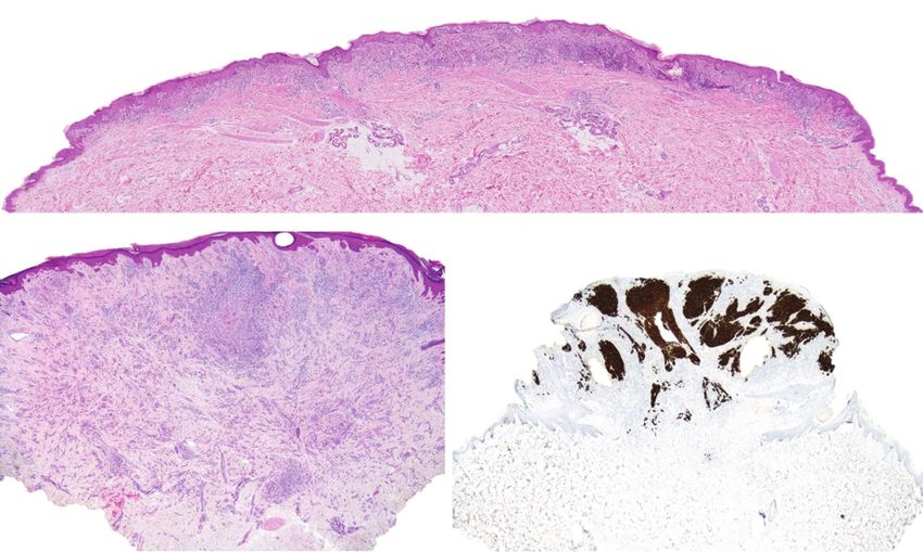
Acta Medica Academica 2021;50(x):xx-xx

   A

   B                                                              C

Figure 2. Spitz nevus with ALK fusion. A. A large diameter of the proliferation is a characteristic feature of ALK-fused mela-
nocytic proliferations. B. A plexiform growth pattern with deep extension of melanocytes is also frequently observed. C.
ALK immunohistochemistry.

Table 3. Spitz Melanocytic Proliferations with ALK Fusions       case (45), which had no effect on the biological be-
 Morphological         • Symmetrical, polypoid/dome and/or
                                                                 havior of the proliferation. Only two cases of Spitz
 features                wedge-shaped                            melanocytic proliferations with ALK fusion and
                       • Large diameter                          with deposits in lymph nodes have been described,
                       • Plexiform growth pattern
                       • Epithelioid and spindled melanocytes
                                                                 one of which had an additional homozygous 9p21
                         – Mild to moderate pleomorphism         deletion (49, 55). Importantly, however, none of
                         – Mitoses rare                          the cases of Spitz melanocytic proliferations with
                         – Pigmentation absent or scant
                         – Low proliferation rate                ALK fusion and with available follow-up data were
                       • Ulceration rare                         found to be associated with systemic metastases or
                       • Kamino bodies usually absent
                                                                 death from the disease.
 Biological behavior   Generally favorable (benign)
                       Regional lymph node deposits
                       uncommon
                       No distant metastases or death from the
                                                                 ROS1 Fusions
                       disease
                                                                 ROS1 protooncogene resides on chromosome
 Confirmatory test     • Immunohistochemistry
                       • Next generation sequencing              6q22.1 and, like ALK, encodes a protein recep-
                       • Fluorescence in situ hybridization      tor tyrosine kinase that is part of the intracellular
                                                                 signaling pathways Ras-Raf-MEK-ERK, JAK3-
bored additional molecular changes, i.e., homo-                  STAT3, and PI3K-AKT-mTOR. (64) ROS1 fusions
zygous 9p21 (CDKN2A) deletion in combination                     have been found in a variety of tumors, including
with 6p25 (RREB1) gain in two cases (49) and a                   non-small cell lung carcinomas, glioblastomas, pe-
hotspot TERT-promoter mutation (C228T) in one                    diatric gliomas, cholangiocarcinomas, inflamma-
Daja Šekoranja et al: Spitz Melanocytic Proliferations – An Update

 A

 B                                                              C

Figure 3. Spitz nevus with ROS1 fusion. A. ROS1-fused melanocytic proliferations often display a prominent junctional com-
ponent. B. The junctional component consists of melanocytic nests with transepidermal elimination and colonization of
the adnexal epithelium. C. ROS1 immunohistochemistry.

tory myofibroblastic tumors, etc (64). ROS1 fusions           cytological atypia, lack of large cells, and fewer mi-
are present in up to 17% of Spitz melanocytic pro-            toses - all statistically significant features differing
liferations (2). Thirteen different fusion partners           between ROS1 and non-ROS1 Spitz neoplasms in a
have been reported: PWWP2A, TPM3, PPFIBP1,                    study by Gerami et al. (32). Nonetheless, large cells
CAPRIN1, MYO5A, PPFIBP1, CLIP1, ERC1,                         were described in 9 of 11 ROS1 fused Spitz neo-
FIP1L1, HLA-A, KIAA1598, MYH9, and ZCCHC8,                    plasms in a study by Wiesner et al. (2), and up to
in descending order of frequency (2, 23, 32, 65).             8 mitoses per square millimeter were reported in
    Although ROS1-fused Spitz proliferations lack             a study of such proliferations by Donati et al. (65)
unique identifying morphological features, most                   Immunohistochemistry for ROS1 protein is
ROS1-fused melanocytic proliferations are com-                a reliable surrogate for molecular testing (Figure
pound, composed either of spindle cells or of a               3c). Studies have confirmed that the vast majority
combination of spindle and epithelioid cells with             (97.4%) of Spitz melanocytic proliferations har-
mild to moderate cytological atypia and with lim-             boring ROS1 fusions display ROS1 cytoplasmic
ited pigmentation of melanocytes (2, 32, 65). The             positivity on immunohistochemistry (2, 32). It
junctional melanocytic component is frequently                is important to note, though, that immunohisto-
prominent, with floating nests or trans-epidermal             chemical staining is often weak yet diffuse.
elimination of melanocytic nests, often colonizing                The vast majority of hitherto reported Spitz
the epithelium of skin adnexa (Figure 3) (32, 65).            neoplasms with ROS1 fusion in the literature were
Kamino bodies seem to be more frequently pres-                classified as either Spitz nevi or atypical Spitz tu-
ent in ROS1 fused Spitz melanocytic proliferations,           mors (2, 23, 32, 47, 65-67). Three lesions were of
along with signs of maturation, lack of high-grade            desmoplastic Spitz nevus phenotype (2, 32), four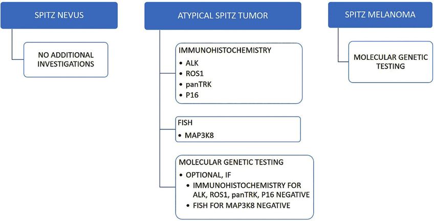
Acta Medica Academica 2021;50(x):xx-xx

Table 4. Spitz Melanocytic Proliferations with ROS1 Fusions      being more common in the pigmented spindle cell
 Morphological         • Junctional component prominent
                                                                 nevus of Reed, a special subtype of Spitz nevus (2,
 features                – Transepidermal elimination of nests   9, 11, 23, 43, 46, 47, 67, 69-74). Only a single case of
                         – Adnexal involvement                   superficial spreading melanoma with an TRAF2-
                       • Epithelioid and spindled melanocytes
                         – Mild to moderate pleomorphism
                                                                 NTRK2 fusion has been reported so far (7).
                         – Mitoses rare                              The NTRK1 fusion partners in Spitz melano-
                         – Pigmentation limited                  cytic proliferations include LMNA (2, 9, 72), TPM3
                         – Low proliferation rate
                         – Maturation present                    (11, 23, 47, 72), TP53 (2, 72), and KHDRBS1 (9,
 Biological behavior   Generally favorable (benign)              72), in descending order of frequency. Even though
                       Regional lymph node deposits              the number of cases for each particular known
                       uncommon                                  NTRK1 fusion partner is relatively low (LMNA was
                       No distant metastases or death from the
                       disease                                   identified in 16 cases, while TP53 and KHDRBS1 in
 Confirmatory test     • Immunohistochemistry                    two cases each), 4 of 5 Spitz melanocytic prolifera-
                         – Staining can be faint                 tions with TPM3-NTRK1 fusion were diagnosed
                       • Next generation sequencing              as Spitz melanomas, all with several additional
                         (preferred)
                       • Fluorescence in situ hybridization      chromosomal aberrations, including (most com-
                                                                 monly) homozygous deletions of CDKN2A (11,
were pigmented spindle cell nevi or Reed nevi (2,                23, 47, 72). Furthermore, Spitz melanoma was also
67), and one was an eruptive Spitz nevus (48). The               diagnosed in one of the two reported cases with an
last was reported in a 49-year old female, who de-               KHDRBS1-NTRK1 fusion (9, 72).
veloped over 100 similar lesions over four years.                    Four different fusion partners have been iden-
Further molecular characterization of the prolifer-              tified so far for NTRK3 fusions, namely MYO5A
ation revealed identical TPM3-ROS1 fusions in the                (67, 69, 70, 73), ETV6 (67, 70, 73), MYH9 (70, 73),
three analyzed lesions (48). Only five Spitz mela-               and SQSTM1 (9), in descending order of frequen-
nomas with ROS1 fusion have been described, and                  cy. While no NTRK1 fusions have been identified
none of them resulted in distant metastases or                   in Reed nevi, they harbor NTRK3 fusions (with
death from the disease (2, 47).                                  MYO5A and rarely ETV6 fusion partners) in up
                                                                 to 57% of cases (67). In addition to pigmented
                                                                 spindle cell nevi of Reed, Spitz proliferations with
NTRK Fusions                                                     NTRK3 fusions are usually diagnosed as Spitz nevi
                                                                 or atypical Spitz tumors and much less frequently
Neurotrophic tyrosine kinase receptor genes
                                                                 as Spitz melanomas (9, 67, 69, 70, 73).
NTRK1, NTRK2 and NTRK3, are oncogenes en-
                                                                     NTRK1 and NTRK3 fusions are also occasion-
coding the Trk family of tyrosine kinase recep-
                                                                 ally detected in non-Spitz melanocytic prolifera-
tors (TrkA, TrkB, and TrkC, respectively) (68).
                                                                 tions, e.g., pigmented epithelioid melanocytomas
These tyrosine kinase receptors are all single-pass
                                                                 (75, 76), acral melanomas (42), and in a wide va-
transmembrane enzymes that stimulate different
                                                                 riety of non-melanocytic tumors, e.g., infantile fi-
pathways once activated, namely the MAPK/ERK,
                                                                 brosarcoma, secretory carcinoma of the breast, se-
PI3K-AKT-mTOR, and phospholipase C-γ path-
                                                                 cretory carcinoma of the salivary gland, congenital
ways (3, 68). In most NTRK fusions identified, the
                                                                 mesoblastic nephroma, lung carcinoma, thyroid
3’ portion encoding the kinase domain is retained,
                                                                 papillary carcinoma and high grade gliomas (77).
and the 5’ portion encoding dimerization domains
                                                                     Histologically, NTRK1-fused Spitz melanocytic
is provided by the fusion partner. The resultant
                                                                 proliferations are characterized by filigree-like rete
chimeric Trk protein is an oncogenic, constitutive-
                                                                 ridges (elongated, thin rete ridges), lobulated der-
ly active tyrosine kinase (69).
                                                                 mal melanocytic nests (composed of smaller nests
    In Spitz melanocytic proliferations, NTRK1 fu-
                                                                 inside the larger ones), and by the formation of
sions predominate over NTRK3 fusions, the latter
Daja Šekoranja et al: Spitz Melanocytic Proliferations – An Update
B
A
C
Figure 4. Spitz nevus with NTRK fusion. A. A symmetrical compound melanocytic proliferation associated with hyperplasia
of the epidermis is depicted in this photo. B. Kamino bodies are often numerous in NTRK1-fused Spitz melanocytic prolifera-
tions. C. pan-TRK immunohistochemistry.
pseudorosettes (23, 43, 72). Exaggerated matura- ple, MYO5A-NTRK3 chimeric protein is localized
tion of spindled and/or epithelioid melanocytes to cell dendritic processes and is associated with
displaying mild to moderate nuclear pleomor- a fusiform/spindled morphology of melanocytes
phism is also a characteristic finding (23, 43, 72). with a fascicular or sometimes plexiform or syn-
Mild to moderate and sometimes even marked cytial growth pattern (70). Besides, the formation
lymphocytic infiltrate is often present (2, 46, 72). of pseudo-Verocay bodies or pseudorosettes is as-
While the pagetoid spread of melanocytes has sociated with a more neuroid appearance of these
been observed in up to 25% of cases, Kamino bod- proliferations (70). In contrast, the ETV6-NTRK3
ies are variably present (Figure 4) (2, 43, 46, 72). chimeric protein is localized to both the nucleus
NTRK1 fused Spitz melanocytic proliferations can and cytoplasm of melanocytes and is linked to
occasionally resemble those with ALK fusions, ex- an epithelioid morphology with well-defined cell
hibiting an intersecting fascicular growth pattern borders. Melanocytes have abundant, glassy cyto-
in the dermis (46). plasm and somewhat large, pleomorphic nuclei.
Pigmented spindle cell nevus of Reed is a pro- They are arranged in large coalescing and also lob-
totype of Spitz melanocytic proliferation harbor- ulated nests. Signs of maturation are fairly discrete
ing an NTRK3 fusion (67). While MYO5A-NTRK3 (70). Finally, Spitz melanocytic proliferations with
fusions are the most common driver genetic ab- MYH9-NTRK3 fusion are distinguished by fibrotic
errations in this subgroup of Spitz nevi, NTRK1 stroma and peripheral collagen trapping (70).
fusions are generally absent (67). It has been re- Immunohistochemistry with pan-TRK an-
cently demonstrated that various NTRK3 fusion tibody can be used to detect the fusion protein
partners have different intracellular localizations, (Figure 4c). Both available clones, clone A7H6R
ultimately determining the morphological charac- (Cell Signaling Technology) and EPR17341 (Ab-
teristics of the Spitz melanocytes (62). For exam- cam/Ventana) are highly sensitive and specific,Acta Medica Academica 2021;50(x):xx-xx
EPR17341 being slightly superior in terms of spec- NTRK fusions and with distant metastases or
ificity (78). The staining pattern can hint at the death from the disease have been reported. In the
presence of the NTRK fusion subtype. However, unlikely event of an NTRK-fused metastatic Spitz
most pan-TRK immunohistochemistry studies in melanoma, specific therapy with TRK inhibitors is
different NTRK fusion subtypes were performed available (81, 82).
on mostly non-Spitz NTRK-fused tumors (79, 80).
Pan-TRK immunohistochemical staining is more
intense and cytoplasmic in NTRK1-fused tumors, RET Fusions
with additional nuclear accentuation in cases with The RET protooncogene resides on chromosome
an LMNA-NTRK1 fusion (79, 80). On the other 10q11.21 and encodes a protein receptor tyrosine
hand, up to 50% of tumors with an NTRK3 fusion kinase involved in the MAPK/ERK, PI3K/AKT/
exhibit a cytoplasmic and nuclear pan-TRK im- mTOR and phospholipase C-γ1 intracellular sig-
munohistochemical reaction (79, 80). However, a naling pathways (2). Only a handful of Spitz mela-
study by de la Fouchardière et al., which included nocytic proliferations with RET fusions have been
only NTRK3-fused Spitz melanocytic prolifera- reported (2, 9, 43, 67, 83). Four different fusion
tions, demonstrated that more intense nuclear and partners have been identified: CCDC6 (9), GOL-
less intense cytoplasmic immunoreactivity is indic- GA5 (2), KIF5B (2), and MYO5A (67). Similar
ative of an ETV6-NTRK3 fusion. At the same time, fusions have also been detected in thyroid cancer
linear staining along dendritic processes can point (84) and lung adenocarcinomas (85).
to the presence of an MYO5A-NTRK3 fusion (70). RET fusions have been reported in the whole
Exceptional cases of non-Spitz (‘spitzoid’) mel- biological spectrum of Spitz melanocytic prolif-
anomas with NTRK fusions have been reported erations, including ordinary Spitz nevi and pig-
resulting in widespread hematogenous metastases mented spindle cell nevus of Reed, atypical Spitz
(7). In contrast, Spitz melanomas with NTRK fu- tumors, and Spitz melanomas (2, 9, 43, 67, 83).
sions do not carry a dismal prognosis since only Although the morphologic features of RET fused
rare metastases to lymph nodes, but not beyond, Spitz melanocytic proliferations lack specificity,
have been described (47, 71). At present, no ex- such proliferations are often well-circumscribed,
amples of Spitz melanocytic proliferations with symmetrical, compound melanocytic prolifera-
tions with a plaque-like silhouette, a nested growth
Table 5. Spitz Melanocytic Proliferations with NTRK1 pattern of small to intermediate-sized epithelioid
Fusions
and spindled melanocytes with only mild cytologi-
Morphological • Filigree-like rete ridges cal atypia (2, 43, 67).
features • Lobulated melanocytic nests RET-fused Spitz melanocytic proliferations
• Rosette-like structures
• Extreme maturation
generally follow an indolent clinical course (2, 9,
• Epithelioid and spindled melanocytes 43, 67, 83). At present, no Spitz melanomas with
– Mild to moderate pleomorphism RET fusion and a dismal outcome have been re-
– Mitoses rare
– Kamino bodies frequent ported in the literature (2, 9, 43, 67, 83). Never-
Biological behavior Generally favorable theless, in the unlikely event of aggressive clinical
Regional lymph node deposits behavior, potential therapy with RET inhibitors is
uncommon available (2).
No distant metastases or death from the
disease in proliferations classified as Spitz
melanomas
Confirmatory test • Immunohistochemistry with pan-TRK
MET Fusions
antibody
• Next generation sequencing The MET protooncogene is localized on chromo-
(preferred) some 7q31.2 and encodes a tyrosine kinase recep-
• Fluorescence in situ hybridization
tor with high affinity for hepatocyte growth factorDaja Šekoranja et al: Spitz Melanocytic Proliferations – An Update
(86). Only eight Spitz melanocytic proliferations, nine protein kinases (87, 88) and is encoded by the
including a Spitz nevus, five atypical Spitz tumors, MAP3K8 gene that resides on chromosome 10p11.
and two Spitz melanomas with MET fusions, have The enzyme consists of a kinase domain encoded
been reported (74, 83, 86). The largest series of six by exons 1-8 of the MAP3K8 gene and an inhibi-
Spitz neoplasms with MET fusions demonstrated tory C-terminal domain encoded by exon 9 of the
a breakpoint in intron 14 in all of the cases (86). MAP3K8 gene. The inhibitory C-terminal domain
The breakpoint event is localized upstream of the covers the kinase domain in its inactive state, pre-
kinase domain-encoding exons 15 to 21, which are venting it from phosphorylating MEK proteins.
fully retained. In contrast, the auto-inhibitory do- The inhibitory C-terminal domain is also essential
main encoded in exon 14 is absent in the chimeric for targeting the MAP3K8 enzyme for proteolytic
protein (86). The N-terminal fusion partners iden- degradation. Fusions involving the MAP3K8 gene
tified in their series were ZKSCAN1, PPFIBP1, and truncation of the MAP3K8 gene follow the
TRIM4, LRRFIP1, EPS15, and DCTN1 (86). same basic mechanism, resulting in a fusion/trun-
MET fusions result in constitutive activation of cated transcript including the intact kinase do-
tyrosine kinase with subsequent activation of the main while lacking the inhibitory C-terminal do-
MAPK/ERK, PI3K/AKT/mTOR, and phospho- main. Consequently, kinase activity is unopposed
lipase C-γ1 pathways, which can be inhibited by by the C-terminal inhibitory action and, at the
cabozantinib (inhibitor of c-MET and VEGFR2) same time, the MAP3K8 is not targeted for proteo-
or PF-04217903 (c-MET inhibitor) (86). The num- lytic degradation, resulting in significant MAP3K8
ber of reported Spitz melanocytic proliferations overexpression and increased phosphorylation of
with MET fusions is too small to conclude specific MEK proteins, which in turn phosphorylate and
morphologic features and prognosis. However, activate ERK1/2 proteins that influence cell pro-
none of the cases with available follow-up resulted liferation, division and differentiation (88-90).
in aggressive clinical behavior (86). Similarly, one of the MEK proteins (MAP2K1,
also called MEK1) has an autoinhibitory domain
in amino acids 98 to 104, and deletions in this re-
Spitz Melanocytic Proliferations with gion (e.g., p.I103_K104del of MAP2K1) also result
Serine/Threonine Kinase Fusions in constitutive activation of downstream ERK1/2
The largest proportion of Spitz melanocytic pro- proteins (91).
liferations with serine/threonine kinase fusions A number of different MAP3K8 fusion part-
involves the MAP3K8 or BRAF genes. Neverthe- ners have been identified, including SVIL (9,
less, other serine/threonine kinase fusions involv- 23, 92, 93), DIP2C (53, 83, 91, 92), UBL3 (9, 83,
ing the RAF1, PRKCA/B, and ARAF genes have 92), SPECC1 (9, 92), STX7 (9, 92), ATP2A2 (91),
exceptionally been reported in a few examples of CCNY (92), CDC42EP3 (92), CUBN (92), GNG2
atypical Spitz tumors and Spitz melanomas (9, 83). (9), LINC00703 (92), MIR3681HG (92), PCDH7
Notably, the vast majority of Spitz melanocytic (91), PIP4K2A (92), PRKACB (9), SFMBT2 (92),
proliferations with serine/threonine kinase fu- SLC4A4 (92), SUBN (9) and ZFP36L1 (23).
sions are classified as atypical Spitz tumors or Spitz MAP3K8 fusions and truncations have also been
melanomas and are infrequently detected in Spitz identified in ovarian, lung, and breast carcinomas,
nevi. mesotheliomas, cutaneous myxoinflammatory fi-
broblastic sarcoma, squamous cell carcinomas, and
melanocytic tumors – in rare acral melanomas and
MAP3K8 Fusions Spitz neoplasms (9, 23, 53, 83, 92-97).
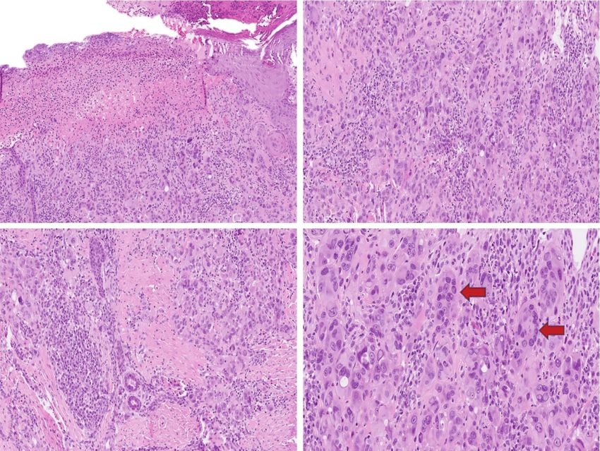
Mitogen-activated protein kinase kinase kinase 8 Morphologically, Spitz proliferations with a
(MAP3K8), also known as Tpl-2 and COT, is an MAP3K8 fusion are often ulcerated tumors (more
enzyme belonging to the group of serine/threo- than 50%) with predominantly epithelioid mor-Acta Medica Academica 2021;50(x):xx-xx A B C D Figure 5. Atypical Spitz tumor. A. This is an example of an ulcerated Spitz melanocytic tumor. B. The lesion is composed of epithelioid melanocytes with moderate cytological atypia. C. No maturation is present. D. Large multinucleated giant me- lanocytes (arrows) can be a morphological clue for the presence of MAP3K8 fusion. phology, moderate to pronounced cytological 53, 91, 92, 98). Houlier et al. reported the larg- atypia, and generally lack maturation (Figure 5). est series of 33 cases of Spitz melanocytic prolif- Additional characteristic features include focal erations with MAP3K8 fusions, of which 13 (40%) hyperpigmented dermal clones and giant multi- were classified as atypical Spitz tumors and 15 nucleated melanocytes. Deep mitotic activity is (45%) as Spitz melanomas (92). Moreover, 77% of not uncommon (9, 23, 53, 91, 92). Furthermore, these atypical Spitz tumors and Spitz melanomas desmoplastic stromal reaction and focal pagetoid harbored CDKN2A (92) inactivation, which was scatter can be seen in 73% and 45% of cases, re- also reported as one of the most common second- spectively (92). ary genetic events in some other series (23, 83, 91). A literature review revealed that most Spitz The biological behavior of Spitz melanocytic pro- proliferations with MAP3K8 fusion or truncations, liferations with a MAP3K8 fusion is variable; it ap- a MAP3K3 fusion, and a MAP2K1 p.I103_K104del pears that the prognosis depends on the presence were classified either as atypical Spitz tumors or of these additional genetic aberrations. Biallelic Spitz melanomas (40% and 52%, respectively). In inactivation of CDKN2A, demonstrated either by comparison, Spitz nevi represented only a small p16 immunohistochemistry (with focal or diffuse portion (8%) of cases in this Spitz subgroup (23, complete loss of p16 expression) or molecular ge-
Daja Šekoranja et al: Spitz Melanocytic Proliferations – An Update

Table 6. Spitz Melanocytic Proliferations with MAP3K8                of autoinhibitory domains results in increased ki-
Fusions                                                              nase activity, evidenced by increased phosphory-
 Morphological         • Ulceration common                           lation and activation of downstream MEK1/2 and
 features              • Epithelioid morphology                      ERK1/2 proteins (4, 5).
                         – Moderate to high grade cytological
                           atypia
                                                                         Numerous fusion partners have been identi-
                         – Lack of maturation                        fied: AKAP9 (9, 10, 101), AGK (10, 102), CLIP2
                         – Mitoses not uncommon                      (102, 103), BAIAP2L1 (11, 47), CEP89 (2), CUX1
                       • Giant multinucleated melanocytes
                       • Focal hyperpigmented dermal clones
                                                                     (10), DYNC1/2 (10), EML4 (11, 47), LSM14A (2),
 Biological behavior   Mostly in atypical Spitz tumors and Spitz
                                                                     MAD1L1 (9), MLANA (102), MYO5A (102), MZT1
                       melanomas                                     (10), NRF1 (23), SKAP2 (102), SLC12A7 (10),
 Confirmatory test     • Next generation sequencing                  SOX6 (23), TRIM24 (10) and ZKSCAN1 (10). In
                       • Fluorescence in situ hybridization          non-Spitz melanoma subtypes, additional fusion
                                                                     partners have been identified, such as KIAA1549
netic techniques, has been detected in about 35%                     in a case of an acral melanoma (8), ZNF767 in two
of all reported cases of this MAP3K8 fused Spitz                     cases of mucosal melanomas (5, 10), PPFIBP2 in a
subgroup of melanocytic proliferation (23, 83, 91,                   case of superficial spreading melanoma (8), GTF2I
92), followed by TERT promotor mutations and                         in a metastatic melanoma of unknown primary
a complex TERT structural rearrangement, albeit                      origin (10), AGAP3, CCDC91, CDC27, PAPSS1,
less frequently (9, 83).                                             RAD18 and TAX1BP1 in melanomas either classi-
    A single case has been reported that resulted                    fied as non-Spitz (spitzoid) or unclassified (4, 10).
in the death of an 11-year old boy, who was diag-                        Similar to the MAP3K8 fused Spitz subgroup
nosed as having a Spitz melanoma with MAP3K8-                        of melanocytic proliferations, the vast majority of
GNG2 fusion, additional complex structural rear-                     Spitz melanocytic proliferations with BRAF fusion
rangement in the TERT gene, and a homozygous                         cluster towards the malignant end of the biologi-
CDKN2A/B deletion (9). Three other Spitz mela-                       cal spectrum, with roughly 45% of published Spitz
nomas with MAP3K8 fusions, all three with ad-                        melanocytic proliferations with BRAF fusion be-
ditional biallelic CDKN2A inactivation, demon-                       longing to atypical Spitz tumor, 41% to Spitz mela-
strated tumor cells’ deposits in at least one lymph                  noma and only 14% to Spitz nevi (2, 9-11, 23, 43,
node. However, none of these cases resulted in                       47, 71, 74, 83, 101-103).
widespread metastatic disease during the 6 to 18                         BRAF fusions are present in various tumors,
months follow up period (91, 92). Two atypical                       including gliomas, thyroid, and pancreatic carci-
Spitz tumors with MAP3K8 fusions locally re-                         nomas, non-small cell lung adenocarcinomas, and
curred, with otherwise no signs of distant metas-                    colorectal carcinomas (10).
tases (83, 91).                                                          Secondary genetic alterations in BRAF fused
                                                                     Spitz melanocytic proliferations were similar to
                                                                     those in other Spitz subgroups. The most common
BRAF Fusions                                                         secondary changes were the homozygous deletion
The BRAF gene encoding a serine-threonine pro-                       of 9p21, TERT promoter mutations, 6p25 gains,
tein kinase is composed of three highly conserved                    and, in single cases, MDM2 amplification and
regions (CRs) (99, 100). CR1 contains N-terminal                     ARID2 p.Q720 mutation (11, 43, 47, 101-103).
RAS-binding and cysteine-rich domains, while                             Histologically, BRAF fused Spitz melanocytic
CR2 contains serine-threonine-rich domains, and                      proliferations are predominantly dermal tumors
both CR1 and CR2 act as auto inhibitors of CR3,                      composed of epithelioid or mixed, epithelioid and
the kinase domain (99, 100). In BRAF fusions, the                    spindled melanocytes with vesicular nuclei and
resulting chimeric protein retains only the intact                   prominent nucleoli, with moderate to high-grade
kinase domain (CR3) of the BRAF gene. The loss                       cytological atypia and somewhat amphophilic
Acta Medica Academica 2021;50(x):xx-xx
A B
Figure 6. Atypical Spitz tumor. A. Low power magnification depicting an ulcerated melanocytic proliferation composed
of epithelioid melanocytes. B. Epithelioid melanocytes with desmoplastic stromal reaction. Although such morphological
changes can be seen in BRAF-fused Spitz melanocytic proliferations, they lack specificity.
Table 7. Spitz Melanocytic Proliferations with BRAF Fusions as a Spitz melanoma harbored concurrent BRAF
Morphological • Epithelioid morphology
fusion, NRAS mutation, and a TERT promoter
features – Moderate to high grade cytological mutation (83).
atypia
– Lack of maturation
– Mitoses not uncommon Conclusion
• Desmoplasia at base
Biological behavior Mostly in atypical Spitz tumors and Spitz Spitz melanocytic proliferations are defined by dis-
melanomas tinctive morphological and molecular genetic fea-
Confirmatory test • Next generation sequencing tures. They encompass the whole biological spec-
• Fluorescence in situ hybridization
trum of proliferations ranging from Spitz nevi,
atypical Spitz tumors to Spitz melanomas. While
cytoplasm (11, 23, 43, 101-103). Interestingly, most Spitz nevi can be reliably diagnosed on mor-
some authors have described a common, distinct phological grounds alone, additional molecular
growth pattern comprising of densely cellular genetic testing is generally necessary to classify
sheet-like proliferation in the superficial part of atypical Spitz tumors and Spitz melanomas and,
the lesion, overlying a less cellular, desmoplastic significantly, to predict their biological behav-
base with prominent dermal sclerosis (Figure 6) ior. The proposed algorithm of how to approach
(2, 43, 101, 102). Spitz melanocytic proliferations is summarized in
Widespread metastatic disease (i.e., metastases Figure 7. Molecular testing includes the detection
beyond the sentinel lymph node) has been de- of different driver fusions and additional genetic
scribed in 19 patients with melanomas with BRAF events associated with biologic behavior. In addi-
fusion (4, 5, 8, 10, 11, 47), ten of which were called tion, since several biological drugs are available to
Spitz melanomas (10, 11, 47). One case of non- treat melanocytic proliferations with aggressive
Spitz metastasizing melanoma in a 54-year-old clinical behavior, characterization of particular
male harbored a BRAF V600E mutation and an driver genetic events and additional genetic ab-
AGAP3-BRAF fusion (10). Another case reported normalities is becoming increasingly important.Daja Šekoranja et al: Spitz Melanocytic Proliferations – An Update
Figure 7. Proposed algorithm for diagnostic work-up of Spitz melanocytic proliferations.
Conflict of Interest: The authors declare that they have no 7. Lezcano C, Shoushtari AN, Ariyan C, Hollmann TJ,
conflict of interest. Busam KJ. Primary and Metastatic Melanoma With
NTRK Fusions. Am J Surg Pathol. 2018;42(8):1052-8.
8. Menzies AM, Yeh I, Botton T, Bastian BC, Scolyer RA,
References Long GV. Clinical activity of the MEK inhibitor tra-
metinib in metastatic melanoma containing BRAF kinase
1. Elder DE, Massi D, Scolyer RA, Willemze R, editors.
fusion. Pigment Cell Melanoma Res. 2015;28(5):607-10.
WHO Classification of Skin Tumours. 4th ed. Lyon: In-
ternational Agency for Research on Cancer; 2018. 9. Newman S, Fan L, Pribnow A, Silkov A, Rice SV, Lee S, et
al. Clinical genome sequencing uncovers potentially tar-
2. Wiesner T, He J, Yelensky R, Esteve-Puig R, Botton T, Yeh
getable truncations and fusions of MAP3K8 in spitzoid
I, et al. Kinase fusions are frequent in Spitz tumours and
and other melanomas. Nat Med. 2019;25(4):597-602.
spitzoid melanomas. Nat Commun. 2014;5:3116.
10. Ross JS, Wang K, Chmielecki J, Gay L, Johnson A, Chud-
3. Wiesner T, Kutzner H, Cerroni L, Mihm MC Jr, Busam KJ,
novsky J, et al. The distribution of BRAF gene fusions in
Murali R. Genomic aberrations in spitzoid melanocytic
solid tumors and response to targeted therapy. Int J Can-
tumours and their implications for diagnosis, prognosis
cer. 2016;138(4):881-90.
and therapy. Pathology. 2016;48(2):113-31.
11. Wu G, Barnhill RL, Lee S, Li Y, Shao Y, Easton J, et al.
4. Hutchinson KE, Lipson D, Stephens PJ, Otto G, Lehmann
The landscape of fusion transcripts in spitzoid melanoma
BD, Lyle PL, et al. BRAF fusions define a distinct molecu-
and biologically indeterminate spitzoid tumors by RNA
lar subset of melanomas with potential sensitivity to MEK
sequencing. Mod Pathol. 2016;29(4):359-69.
inhibition. Clin Cancer Res. 2013;19(24):6696-702.
12. Bastian BC, Wesselmann U, Pinkel D, Leboit PE. Molecu-
5. Kim HS, Jung M, Kang HN, Kim H, Park CW, Kim SM,
lar cytogenetic analysis of Spitz nevi shows clear differ-
et al. Oncogenic BRAF fusions in mucosal melanomas
ences to melanoma. J Invest Dermatol. 1999;113(6):1065-
activate the MAPK pathway and are sensitive to MEK/
9.
PI3K inhibition or MEK/CDK4/6 inhibition. Oncogene.
2017;36(23):3334-45. 13. Bastian BC, LeBoit PE, Pinkel D. Mutations and copy
number increase of HRAS in Spitz nevi with distinctive
6. Lazova R, Pornputtapong N, Halaban R, Bosenberg
histopathological features. Am J Pathol. 2000;157(3):967-
M, Bai Y, Chai H, et al. Spitz nevi and Spitzoid melano-
72.
mas: exome sequencing and comparison with conven-
tional melanocytic nevi and melanomas. Mod Pathol. 14. van Engen-van Grunsven AC, van Dijk MC, Ruiter DJ,
2017;30(5):640-9. Klaasen A, Mooi WJ, Blokx WA. HRAS-mutated Spitz
tumors: A subtype of Spitz tumors with distinct features.
Am J Surg Pathol. 2010;34(10):1436-41.Acta Medica Academica 2021;50(x):xx-xx
15. Maldonado JL, Timmerman L, Fridlyand J, Bastian BC. spitzoid tumours and their possible pathogenetic signifi-
Mechanisms of cell-cycle arrest in Spitz nevi with consti- cance. Br J Dermatol. 2009;161(2):364-72.
tutive activation of the MAP-kinase pathway. Am J Pathol. 30. Harvell JD, Bastian BC, LeBoit PE. Persistent (recurrent)
2004;164(5):1783-7. Spitz nevi: a histopathologic, immunohistochemical, and
16. Ross AL, Sanchez MI, Grichnik JM. Molecular nevogen- molecular pathologic study of 22 cases. Am J Surg Pathol.
esis. Dermatol Res Pract. 2011;2011:463184. 2002;26(5):654-61.
17. Bos JL. ras oncogenes in human cancer: a review. Cancer 31. Sabater Marco V, Escutia Muñoz B, Morera Faet A, Mata
Res. 1989;49(17):4682-9. Roig M, Botella Estrada R. Pseudogranulomatous Spitz
18. Hand PH, Vilasi V, Thor A, Ohuchi N, Schlom J. Quan- nevus: a variant of Spitz nevus with heavy inflammatory
titation of Harvey ras p21 enhanced expression in hu- infiltrate mimicking a granulomatous dermatitis. J Cutan
man breast and colon carcinomas. J Natl Cancer Inst. Pathol. 2013;40(3):330-5.
1987;79(1):59-65. 32. Gerami P, Kim D, Compres EV, Zhang B, Khan AU, Sun-
19. Riou G, Barrois M, Sheng ZM, Duvillard P, Lhomme C. shine JC, et al. Clinical, morphologic, and genomic find-
Somatic deletions and mutations of c-Ha-ras gene in hu- ings in ROS1 fusion Spitz neoplasms. Mod Pathol. 2020.
man cervical cancers. Oncogene. 1988;3(3):329-33. 33. Roh MR, Eliades P, Gupta S, Tsao H. Genetics of melano-
20. van Dijk MC, Bernsen MR, Ruiter DJ. Analysis of muta- cytic nevi. Pigment Cell Melanoma Res. 2015;28(6):661-
tions in B-RAF, N-RAS, and H-RAS genes in the differen- 72.
tial diagnosis of Spitz nevus and spitzoid melanoma. Am J 34. Iwahara T, Fujimoto J, Wen D, Cupples R, Bucay N, Ara-
Surg Pathol. 2005;29(9):1145-51. kawa T, et al. Molecular characterization of ALK, a recep-
21. Bender RP, McGinniss MJ, Esmay P, Velazquez EF, Rei- tor tyrosine kinase expressed specifically in the nervous
mann JD. Identification of HRAS mutations and absence system. Oncogene. 1997;14(4):439-49.
of GNAQ or GNA11 mutations in deep penetrating nevi. 35. Hallberg B, Palmer RH. Mechanistic insight into ALK re-
Mod Pathol. 2013;26(10):1320-8. ceptor tyrosine kinase in human cancer biology. Nat Rev
22. Kiyohara T, Takata M, Itoh H, Kawami K, Yasuta M, Hay- Cancer. 2013;13(10):685-700.
akawa K, et al. HRAS-mutated Spitz nevus on the cheek in 36. Pulford K, Morris SW, Turturro F. Anaplastic lymphoma
a middle-aged man. Acta Derm Venereol. 2012;92(3):326- kinase proteins in growth control and cancer. J Cell Physi-
7. ol. 2004;199(3):330-58.
23. Raghavan SS, Peternel S, Mully TW, North JP, Pincus LB, 37. Slupianek A, Nieborowska-Skorska M, Hoser G, Morri-
LeBoit PE, et al. Spitz melanoma is a distinct subset of one A, Majewski M, Xue L, et al. Role of phosphatidylino-
spitzoid melanoma. Mod Pathol. 2020;33(6):1122-34. sitol 3-kinase-Akt pathway in nucleophosmin/anaplastic
24. Li JY, Berger MF, Marghoob A, Bhanot UK, Toyohara lymphoma kinase-mediated lymphomagenesis. Cancer
JP, Pulitzer MP. Combined melanocytic and sweat gland Res. 2001;61(5):2194-9.
neoplasm: cell subsets harbor an identical HRAS muta- 38. Zamo A, Chiarle R, Piva R, Howes J, Fan Y, Chilosi M, et
tion in phacomatosis pigmentokeratotica. J Cutan Pathol. al. Anaplastic lymphoma kinase (ALK) activates Stat3 and
2014;41(8):663-71. protects hematopoietic cells from cell death. Oncogene.
25. Nemeth K, Szabo S, Cottrell CE, McNulty SM, Segura A, 2002;21(7):1038-47.
Sokumbi O, et al. Mosaic pathogenic HRAS variant in a 39. Falini B, Bigerna B, Fizzotti M, Pulford K, Pileri SA, Del-
patient with nevus spilus with agminated Spitz nevi and sol G, et al. ALK expression defines a distinct group of T/
parametrial-uterine rhabdomyosarcoma. Br J Dermatol. null lymphomas (“ALK lymphomas”) with a wide mor-
2018;178(3):804-6. phological spectrum. Am J Pathol. 1998;153(3):875-86.
26. Pontoizeau J, Stefan A, Comoz F, Houlier A, Haddad V, 40. Doyle LA, Mariño-Enriquez A, Fletcher CD, Hornick JL.
Pissaloux D, et al. Agminated Spitz nevus arising in nor- ALK rearrangement and overexpression in epithelioid fi-
mal skin with redundant HRAS mutation. Eur J Derma- brous histiocytoma. Mod Pathol. 2015;28(7):904-12.
tol. 2017;27(1):73-4. 41. Niu HT, Zhou QM, Wang F, Shao Q, Guan YX, Wen XZ,
27. Porubsky C, Teer JK, Zhang Y, Deschaine M, Sondak VK, et al. Identification of anaplastic lymphoma kinase break
Messina JL. Genomic analysis of a case of agminated Spitz points and oncogenic mutation profiles in acral/mucosal
nevi and congenital-pattern nevi arising in extensive ne- melanomas. Pigment Cell Melanoma Res. 2013;26(5):646-
vus spilus. J Cutan Pathol. 2018;45(2):180-3. 53.
28. Sarin KY, Sun BK, Bangs CD, Cherry A, Swetter SM, 42. Yeh I, Jorgenson E, Shen L, Xu M, North JP, Shain AH, et
Kim J, et al. Activating HRAS mutation in agminated al. Targeted Genomic Profiling of Acral Melanoma. J Natl
Spitz nevi arising in a nevus spilus. JAMA Dermatol. Cancer Inst. 2019;111(10):1068-77.
2013;149(9):1077-81. 43. Amin SM, Haugh AM, Lee CY, Zhang B, Bubley JA,
29. Da Forno PD, Pringle JH, Fletcher A, Bamford M, Su Merkel EA, et al. A Comparison of Morphologic and Mo-
L, Potter L, et al. BRAF, NRAS and HRAS mutations inDaja Šekoranja et al: Spitz Melanocytic Proliferations – An Update
lecular Features of BRAF, ALK, and NTRK1 Fusion Spit- dant ALK immunohistochemistry results. Hum Pathol.
zoid Neoplasms. Am J Surg Pathol. 2017;41(4):491-8. 2018;80:99-103.
44. Chung CT, Marrano P, Swanson D, Dickson BC, Thorner 56. Perron E, Pissaloux D, Charon Barra C, Karanian M, La-
PS. Fusion of ALK to the melanophilin gene MLPH in pe- mant L, Parfait S, et al. Melanocytic Myxoid Spindle Cell
diatric Spitz nevi. Hum Pathol. 2019;87:57-64. Tumor With ALK Rearrangement (MMySTAR): Report
45. Kastnerova L, Martinek P, Grossmann P, Steiner P, of 4 Cases of a Nevus Variant With Potential Diagnostic
Vanecek T, Kyclova J, et al. A Clinicopathological Study Challenge. Am J Surg Pathol. 2018;42(5):595-603.
of 29 Spitzoid Melanocytic Lesions With ALK Fusions, 57. Rousi EK, Koskivuo IO, Juteau SM, Talve LAI, Hern-
Including Novel Fusion Variants, Accompanied by Fluo- berg MM, Vihinen PP, et al. Different expression of
rescence In Situ Hybridization Analysis for Chromosomal BRAFV600E, ALK and PD-L1 in melanoma in children
Copy Number Changes, and Both TERT Promoter and and adolescents: a nationwide retrospective study in Fin-
Next-Generation Sequencing Mutation Analysis. Am J land in 1990-2014. Acta Oncol. 2021;60(2):165-72. Epub
Dermatopathol. 2020;42(8):578-92. 2020 Aug 20.
46. Kiuru M, Jungbluth A, Kutzner H, Wiesner T, Busam KJ. 58. Brown RA, Wang JY, Raghavan SS, Zhang J, Wan DC,
Spitz Tumors: Comparison of Histological Features in Re- Born D, et al. ALK-positive compound Spitz nevus with
lationship to Immunohistochemical Staining for ALK and extensive perineural and intraneural neurotropism. J Cu-
NTRK1. Int J Surg Pathol. 2016;24(3):200-6. tan Pathol. 2021;48(1):154-9. Epub 2020 Nov 8.
47. Lee S, Barnhill RL, Dummer R, Dalton J, Wu J, Pappo A, 59. Ogawa K, Fukumoto T, Azukizawa H, Takeda M, Asada
et al. TERT Promoter Mutations Are Predictive of Aggres- H. First Japanese case report of atypical Spitz tumor with
sive Clinical Behavior in Patients with Spitzoid Melano- an ALK rearrangement. J Dermatol. 2017;44(12):e342-3.
cytic Neoplasms. Sci Rep. 2015;5:11200. 60. Busam KJ, Vilain RE, Lum T, Busam JA, Hollmann TJ,
48. Raghavan SS, Kapler ES, Dinges MM, Bastian BC, Yeh I. Saw RP, et al. Primary and Metastatic Cutaneous Mela-
Eruptive Spitz nevus, a striking example of benign metas- nomas Express ALK Through Alternative Transcriptional
tasis. Sci Rep. 2020;10(1):16216. Initiation. Am J Surg Pathol. 2016;40(6):786-95.
49. Rand AJ, Flejter WL, Dowling CA, Brooke LM, Bo- 61. Wiesner T, Lee W, Obenauf AC, Ran L, Murali R, Zhang
land GM, Kroshinsky D, et al. Atypical ALK-positive QF, et al. Alternative transcription initiation leads to
Spitz tumors with 9p21 homozygous deletion: Report expression of a novel ALK isoform in cancer. Nature.
of two cases and review of the literature. J Cutan Pathol. 2015;526(7573):453-7.
2018;45(2):136-40. 62. Farah M, Nagarajan P, Curry JL, Tang Z, Kim TB, Aung
50. Yeh I, de la Fouchardiere A, Pissaloux D, Mully TW, Gar- PP, et al. Spitzoid melanoma with histopathological fea-
rido MC, Vemula SS, et al. Clinical, histopathologic, and tures of ALK gene rearrangement exhibiting ALK copy
genomic features of Spitz tumors with ALK fusions. Am J number gain: a novel mechanism of ALK activation in
Surg Pathol. 2015;39(5):581-91. spitzoid neoplasia. Br J Dermatol. 2019;180(2):404-8.
51. Busam KJ, Kutzner H, Cerroni L, Wiesner T. Clinical and 63. Dunn ALJ, Gardner JM, Kaley JR, Bellamy W, Shalin SC.
pathologic findings of Spitz nevi and atypical Spitz tumors ALK Rearrangements Are Infrequent in Cellular Blue Ne-
with ALK fusions. Am J Surg Pathol. 2014;38(7):925-33. vus and Deep Penetrating Nevus. Am J Dermatopathol.
52. Melchers RC, Willemze R, van Doorn R, Jansen PM, 2018;40(7):469-78.
Cleven AHG, Solleveld N, et al. Corresponding ana- 64. Drilon A, Jenkins C, Iyer S, Schoenfeld A, Keddy C, Da-
plastic lymphoma kinase-tropomyosin 3 (ALK-TPM3) vare MA. ROS1-dependent cancers - biology, diagnostics
fusion in a patient with a primary cutaneous anaplastic and therapeutics. Nat Rev Clin Oncol. 2020.
large-cell lymphoma and a Spitz nevus. JAAD Case Rep. 65. Donati M, Kastnerova L, Martinek P, Grossmann P, Sti-
2019;5(11):970-2. cová E, Hadravský L, et al. Spitz Tumors With ROS1 Fu-
53. Newman S, Pappo A, Raimondi S, Zhang J, Barnhill R, sions: A Clinicopathological Study of 6 Cases, Including
Bahrami A. Pathologic Characteristics of Spitz Melanoma FISH for Chromosomal Copy Number Alterations and
With MAP3K8 Fusion or Truncation in a Pediatric Co- Mutation Analysis Using Next-Generation Sequencing.
hort. Am J Surg Pathol. 2019;43(12):1631-7. Am J Dermatopathol. 2020;42(2):92-102.
54. Saraggi D, Salmaso R, Zamuner C, Munari G, Lanza C, 66. Mitsui Y, Ogawa K, Takeda M, Nakanishi T, Azukizawa H,
Alaibac MS, et al. Prevalence of ALK gene alterations Asada H. First Japanese case of atypical Spitz tumor exhib-
among the spectrum of plexiform spitzoid lesions. J Am iting ROS1 rearrangement. J Dermatol. 2018;45(9):e248-
Acad Dermatol. 2018;79(4):728-35. 9.
55. Fujimoto M, Togashi Y, Matsuzaki I, Baba S, Takeuchi 67. VandenBoom T, Quan VL, Zhang B, Garfield EM,
K, Inaba Y, et al. A case report of atypical Spitz tumor Kong BY, Isales MC, et al. Genomic Fusions in Pig-
harboring a novel MLPH-ALK gene fusion with discor- mented Spindle Cell Nevus of Reed. Am J Surg Pathol.
2018;42(8):1042-51.Acta Medica Academica 2021;50(x):xx-xx
68. Rubin JB, Segal RA. Growth, survival and migration: the 82. Forschner A, Forchhammer S, Bonzheim I. NTRK gene
Trk to cancer. Cancer Treat Res. 2003;115:1-18. fusions in melanoma: detection, prevalence and po-
69. Wang L, Busam KJ, Benayed R, Cimera R, Wang J, Denley tential therapeutic implications. J Dtsch Dermatol Ges.
R, et al. Identification of NTRK3 Fusions in Childhood 2020;18(12):1387-92.
Melanocytic Neoplasms. J Mol Diagn. 2017;19(3):387-96. 83. Quan VL, Zhang B, Zhang Y, Mohan LS, Shi K, Wagner A,
70. de la Fouchardière A, Tee MK, Peternel S, Valdebran M, et al. Integrating Next-Generation Sequencing with Mor-
Pissaloux D, Tirode F, et al. Fusion partners of NTRK3 af- phology Improves Prognostic and Biologic Classification
fect subcellular localization of the fusion kinase and cyto- of Spitz Neoplasms. J Invest Dermatol. 2020;140(8):1599-
morphology of melanocytes. Mod Pathol. Epub 2020 Sep 608.
23. 84. Rabes HM, Demidchik EP, Sidorow JD, Lengfelder E,
71. Lee CY, Sholl LM, Zhang B, Merkel EA, Amin SM, Gui- Beimfohr C, Hoelzel D, et al. Pattern of radiation-induced
tart J, et al. Atypical Spitzoid Neoplasms in Childhood: RET and NTRK1 rearrangements in 191 post-chernobyl
A Molecular and Outcome Study. Am J Dermatopathol. papillary thyroid carcinomas: biological, phenotypic, and
2017;39(3):181-6. clinical implications. Clin Cancer Res. 2000;6(3):1093-
72. Yeh I, Busam KJ, McCalmont TH, LeBoit PE, Pissaloux 103.
D, Alberti L, et al. Filigree-like Rete Ridges, Lobulated 85. Lipson D, Capelletti M, Yelensky R, Otto G, Parker A, Ja-
Nests, Rosette-like Structures, and Exaggerated Matura- rosz M, et al. Identification of new ALK and RET gene fu-
tion Characterize Spitz Tumors With NTRK1 Fusion. Am sions from colorectal and lung cancer biopsies. Nat Med.
J Surg Pathol. 2019;43(6):737-46. 2012;18(3):382-4.
73. Yeh I, Tee MK, Botton T, Shain AH, Sparatta AJ, Gagnon 86. Yeh I, Botton T, Talevich E, Shain AH, Sparatta AJ, de la
A, et al. NTRK3 kinase fusions in Spitz tumours. J Pathol. Fouchardiere A, et al. Activating MET kinase rearrange-
2016;240(3):282-90. ments in melanoma and Spitz tumours. Nat Commun.
74. Zarabi SK, Azzato EM, Tu ZJ, Ni Y, Billings SD, Arbes- 2015;6:7174.
man J, et al. Targeted next generation sequencing (NGS) 87. Hagemann D, Troppmair J, Rapp UR. Cot protooncop-
to classify melanocytic neoplasms. J Cutan Pathol. rotein activates the dual specificity kinases MEK-1 and
2020;47(8):691-704. SEK-1 and induces differentiation of PC12 cells. Onco-
75. Friedman BJ, Hernandez S, Fidai C, Jiang A, Shwayder gene. 1999;18(7):1391-400.
TA, Carskadon S, et al. A pediatric case of pigmented epi- 88. Salmeron A, Ahmad TB, Carlile GW, Pappin D, Narsim-
thelioid melanocytoma with chromosomal copy number han RP, Ley SC. Activation of MEK-1 and SEK-1 by Tpl-
alterations in 15q and 17q and a novel NTRK3-SCAPER 2 proto-oncoprotein, a novel MAP kinase kinase kinase.
gene fusion. J Cutan Pathol. 2020;47(1):70-5. Embo j. 1996;15(4):817-26.
76. Isales MC, Haugh AM, Bubley J, Verzì AE, Zhang B, Ku- 89. Ceci JD, Patriotis CP, Tsatsanis C, Makris AM, Kovatch
dalkar E, et al. Genomic Assessment of Blitz Nevi Suggests R, Swing DA, et al. Tpl-2 is an oncogenic kinase that is
Classification as a Subset of Blue Nevus Rather Than Spitz activated by carboxy-terminal truncation. Genes Dev.
Nevus: Clinical, Histopathologic, and Molecular Analysis 1997;11(6):688-700.
of 18 Cases. Am J Dermatopathol. 2018;40(2):118-24. 90. Gándara ML, López P, Hernando R, Castaño JG, Alemany
77. Cocco E, Scaltriti M, Drilon A. NTRK fusion-positive S. The COOH-terminal domain of wild-type Cot regu-
cancers and TRK inhibitor therapy. Nat Rev Clin Oncol. lates its stability and kinase specific activity. Mol Cell Biol.
2018;15(12):731-47. 2003;23(20):7377-90.
78. Uguen A. Spitz Tumors With NTRK1 Fusions: TRK-A 91. Quan VL, Zhang B, Mohan LS, Shi K, Isales MC, Panah
and pan-TRK Immunohistochemistry as Ancillary Diag- E, et al. Activating Structural Alterations in MAPK Genes
nostic Tools. Am J Surg Pathol. 2019;43(10):1438-9. Are Distinct Genetic Drivers in a Unique Subgroup Of
79. Gatalica Z, Xiu J, Swensen J, Vranic S. Molecular char- Spitzoid Neoplasms. Am J Surg Pathol. 2019;43(4):538-
acterization of cancers with NTRK gene fusions. Mod 48.
Pathol. 2019;32(1):147-53. 92. Houlier A, Pissaloux D, Masse I, Tirode F, Karanian M,
80. Hechtman JF, Benayed R, Hyman DM, Drilon A, Zehir Pincus LB, et al. Melanocytic tumors with MAP3K8 fu-
A, Frosina D, et al. Pan-Trk Immunohistochemistry Is an sions: report of 33 cases with morphological-genetic cor-
Efficient and Reliable Screen for the Detection of NTRK relations. Mod Pathol. 2020;33(5):846-57.
Fusions. Am J Surg Pathol. 2017;41(11):1547-51. 93. Quan VL, Panah E, Zhang B, Shi K, Mohan LS, Gerami
81. Drilon A, Laetsch TW, Kummar S, DuBois SG, Lassen P. The role of gene fusions in melanocytic neoplasms. J
UN, Demetri GD, et al. Efficacy of Larotrectinib in TRK Cutan Pathol. 2019;46(11):878-87.
Fusion-Positive Cancers in Adults and Children. N Engl J 94. Clark AM, Reynolds SH, Anderson M, Wiest JS. Muta-
Med. 2018;378(8):731-9. tional activation of the MAP3K8 protooncogene in lung
cancer. Genes Chromosomes Cancer. 2004;41(2):99-108.You can also read