Inducing the attachment of cable bacteria on oxidizing electrodes
←
→
Page content transcription
If your browser does not render page correctly, please read the page content below
Biogeosciences, 17, 597–607, 2020
https://doi.org/10.5194/bg-17-597-2020
© Author(s) 2020. This work is distributed under
the Creative Commons Attribution 4.0 License.

Inducing the attachment of cable bacteria on oxidizing electrodes
Cheng Li, Clare E. Reimers, and Yvan Alleau
College of Earth, Ocean and Atmospheric Sciences, Oregon State University, Corvallis, Oregon 97331, USA

Correspondence: Cheng Li (cheng.li@oregonstate.edu)

Received: 22 August 2019 – Discussion started: 2 September 2019
Revised: 17 December 2019 – Accepted: 14 January 2020 – Published: 6 February 2020

Abstract. Cable bacteria (CB) are multicellular, filamentous       membrane cytochromes, and/or mineral nanoparticles to
bacteria within the family of Desulfobulbaceae that transfer       connect extracellular electron donors and acceptors (Li et al.,
electrons longitudinally from cell to cell to couple sulfide ox-   2017; Lovley, 2016). Recently, a novel type of LDET ex-
idation and oxygen reduction in surficial aquatic sediments.       hibited by filamentous bacteria in the family of Desulfobul-
In the present study, electrochemical reactors that contain        baceae was discovered in the uppermost centimeters of var-
natural sediments are introduced as a tool for investigating       ious aquatic, but mainly marine, sediments (Malkin et al.,
the growth of CB on electrodes poised at an oxidizing poten-       2014; Trojan et al., 2016). These filamentous bacteria, also
tial. Our experiments utilized sediments from Yaquina Bay,         known as “cable bacteria” (CB), electrically connect two spa-
Oregon, USA, and we include new phylogenetic analyses of           tially separated redox half reactions and generate electrical
separated filaments to confirm that CB from this marine loca-      current over distances that can extend to centimeters, which
tion cluster with the genus “Candidatus Electrothrix”. These       is an order of magnitude longer than previously recognized
CB may belong to a distinctive lineage, however, because           LDET distances (Meysman, 2017).
their filaments contain smaller cells and a lower number of           The unique ability of CB to perform LDET creates a spa-
longitudinal ridges compared to cables described from other        tial separation of oxygen reduction in oxic surface layers
locales. The results of a 135 d bioelectrochemical reactor ex-     of organic-rich sediment from sulfide oxidation in subsur-
periment confirmed that these CB can migrate out of reduc-         face layers (Meysman, 2017). The spatial separation of these
ing sediments and grow on oxidatively poised electrodes sus-       two half reactions also creates localized porewater pH ex-
pended in anaerobic seawater. CB filaments and several other       tremes in oxic and sulfidic layers, which induces a series of
morphologies of Desulfobulbaceae cells were observed by            secondary reactions that stimulate the geochemical cycling
scanning electron microscopy and fluorescence in situ hy-          of elements such as iron, manganese, calcium, phosphorus,
bridization on electrode surfaces, albeit in low densities and     and nitrogen (Kessler et al., 2018; Rao et al., 2016; Seitaj
often obscured by mineral precipitation. These findings pro-       et al., 2015; Sulu-Gambari et al., 2016a, b). In addition to
vide new information to suggest what kinds of conditions           altering established perceptions of sedimentary biogeochem-
will induce CB to perform electron donation to an electrode        ical cycling and microbial ecology (Meysman, 2017; Nielsen
surface, further informing future experiments to culture CB        and Risgaard-Petersen, 2015), CB also possess intriguing
outside of a sediment matrix.                                      structural features that may inspire new engineering applica-
                                                                   tions in areas of bioenergy harvesting and biomaterial design
                                                                   (Lovley, 2016; Meysman et al., 2019).
                                                                      Much is still unknown about the basic mechanism(s)
1   Introduction                                                   that CB use to perform LDET. It has been suggested that
                                                                   when long filaments form, a chain of cells at the sulfidic
Long-distance electron transfer (LDET) is a mechanism              terminal catalyzes anodic half reactions (e.g., 0.5 H2 S +
used by certain microorganisms to generate energy through          2H2 O → 0.5 SO2−          −      +
                                                                                     4 + 4e + 5H ), while a cathodic half re-
the transfer of electrons over distances greater than a cell-                       −       +
                                                                   action (O2 + 4e + 4H → 2H2 O) is completed by cells at
length. These microorganisms may pass electrons across dis-        the oxic terminal. Electron transfer then occurs along the lon-
solved redox shuttles, nanofiber-like cell appendages, outer-

Published by Copernicus Publications on behalf of the European Geosciences Union.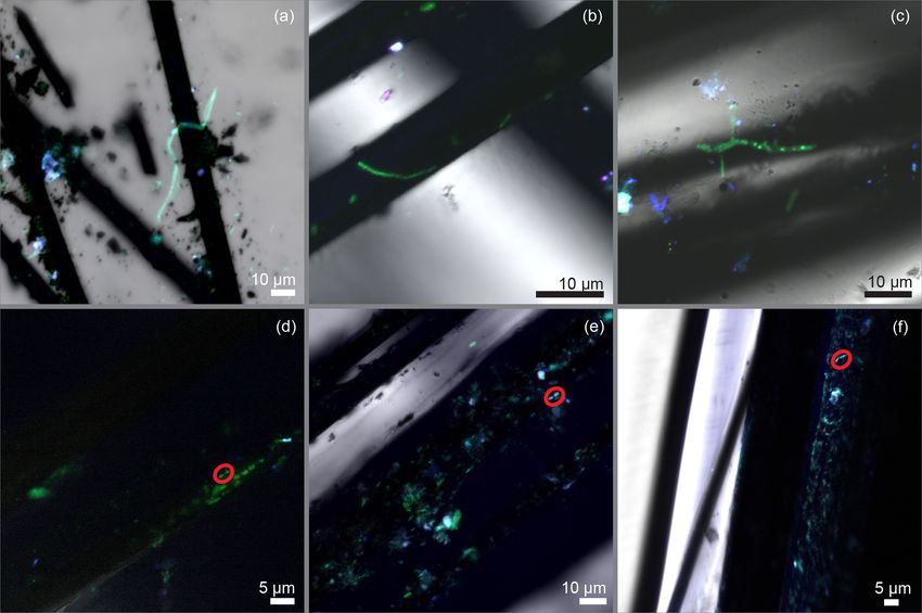
598                                           C. Li et al.: Inducing the attachment of cable bacteria on oxidizing electrodes

gitudinal ridges of CB filaments via electron hopping pro-         2.2   Sediment incubation
moted by extracellular cytochromes positioned within a re-
dox gradient or via conductive electronic structures such as       To cultivate CB of Yaquina Bay, IMF sediment was initially
pili (Bjerg et al., 2018; Cornelissen et al., 2018; Kjeldsen et    incubated for 60 d. These first incubations were started 2 d
al., 2019; Meysman et al., 2019; Pfeffer et al., 2012). These      after collection and performed after homogenizing the sieved
hypotheses await further verification, and CB remain uncul-        sediments under a flow of N2 and then packing the sediment
tured and difficult to grow outside of sediment. This difficulty   into triplicate polycarbonate tubes (15 cm height and 9.5 cm
complicates efforts to study them using different techniques,      inner diameter). These cores were submerged in an aquar-
such as electrochemical assays and metatranscriptomics.            ium containing aerated seawater collected from Yaquina Bay
   In a previous benthic microbial fuel cell (BMFC) experi-        and held at 15 ◦ C, a temperature that is about average for the
ment in a marine estuary (Reimers et al., 2017), we serendip-      mudflats of Yaquina Bay (Johnson, 1980). Once a distinc-
itously observed the attachment of CB to carbon fibers serv-       tive suboxic layer was evident from color changes in the top
ing as an anode in an anaerobic environment above sedi-            centimeters of the cores, profiles of porewater pH, O2 , and
ments. This finding suggested that CB possess the ability to       H2 S were measured to 2–3 cm depth with commercial mi-
donate electrons to solid electron acceptors, and it indicated     croelectrodes (Unisense A.S., Aarhus, Denmark) to confirm
a range of cathodic potentials favorable for electron trans-       geochemical evidence of CB activity (see below). Multiple
fer (Reimers et al., 2017). However further investigations are     small sub-cores (0.5 cm diameter, 3 cm in length) were then
still needed to study the conditions that allow the attachment     taken out from each incubated core using cut-off syringes.
of CB to a poised electrode and to document electron transfer      Some of these sediment plugs were washed gently to reduce
mechanisms at their cathodic terminus. In the present study,       the volume of fine particles, and CB biomass was further sep-
we first clarify the phylogenetic placement of CB found in         arated out from the sediment matrix by using custom-made
sediments from Yaquina Bay, Oregon, where the BMFC was             tiny glass hooks following Malkin et al. (2014). Sediment
previously deployed. Then, we describe the design of a bio-        plugs and separated filamentous biomass were frozen or fixed
electrochemical reactor configured to mimic the environment        for subsequent phylogenetic and microscopic characteriza-
in the anodic chamber of a BMFC and verify conditions that         tions.
can induce CB attachment on electrodes. Results assert that
when oxygen is not available, CB can glide through sedi-           2.3   Reactor configuration and operation
ments and seawater to an electrode poised at oxidative poten-
tials. Thus, the present study provides new information about      To mimic the conditions where CB were found attached to
the chemotaxis of CB in environments other than sediments,         electrode fibers in a BMFC (Reimers et al., 2017), a bio-
revealing key conditions for their attachment to surfaces and      electrochemical reactor was assembled from a polycarbonate
growth in both natural and engineered environments.                core tube (15 cm height and 11.5 cm inner diameter, Fig. 1)
                                                                   as a second phase of this research. A lid, a center rod to locate
                                                                   and support the electrodes, and a perforated bottom partition
2     Materials and methods                                        were made from polyvinyl chloride (PVC, McMaster-Carr,
                                                                   Elmhurst, IL). Three carbon brush electrodes, which would
2.1    Study site and sediment collection
                                                                   serve as two anodes and a control electrode (Mill-Rose, Men-
Several studies suggest that CB may be found widely in             tor, OH, 2 cm in diameter and 8.9 cm total length), were in-
coastal sediments possessing high rates of sulfide genera-         serted through septa within holes in the core lining to meet
tion coupled with organic matter mineralization (Larsen et         the center rod and were spaced radially at 120◦ angles from
al., 2015; Malkin et al., 2014; Pfeffer et al., 2012). There-      each other.
fore, to initiate this enquiry, sediment with these two char-         To initiate the experiment, the reactor was placed inside an
acteristics was collected from Yaquina Bay, Oregon, USA,           8 L plastic beaker (with perforated walls) containing 3 cm of
using a hand shovel at a site on an intertidal mud flat (IMF,      IMF sediments at the bottom. Enough additional IMF sed-
44◦ 370 30 N, 124◦ 000 26 W). The IMF site is located about        iment was then placed inside the reactor to form an 8 cm
3 km upstream from the site where the BMFC was deployed            thick layer after settling and compacting. In this configura-
in the abovementioned study (Reimers et al., 2017). The top        tion, the sediment–water interface was approximately 1 cm
20 cm of these sediments were sieved through a 0.5 mm mesh         away from the lower extent of the carbon brush electrodes.
size metal screen to remove macrofauna and shell debris.           The beaker was then gently lowered inside an aquarium filled
Then the sieved sediments were allowed to settle and stored        with Yaquina Bay seawater until fully submerged, and the re-
in sealed buckets in a cold room at 5 ◦ C.                         actor was left uncapped. Seawater in the aquarium was main-
                                                                   tained at 15 ◦ C and bubbled to maintain air saturation. A fuel
                                                                   cell circuit was completed by placing a 10 cm long carbon-
                                                                   fiber brush cathode (Hasvold et al., 1997) and a reference
                                                                   electrode (Ag/AgCl [3 M KCl], MI-401F, Microelectrodes,

Biogeosciences, 17, 597–607, 2020                                                        www.biogeosciences.net/17/597/2020/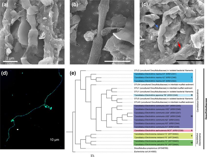
C. Li et al.: Inducing the attachment of cable bacteria on oxidizing electrodes                                                       599

Figure 1. Schematic of the bioelectrochemical reactor design used in this study: (a) lateral view of the reactor, (b) electrical circuit of
the reactor, and (c) bird’s-eye view of the reactor cap and electrode arrangement. Dimensions are in cm. A1 and A2 represent the current
monitored in duplicate anodes, V1 represents the potential monitored between the duplicate anodes and cathode, and V2 and V3 represent
potentials monitored between the duplicate anodes and the reference electrode.

Inc., Bedford, NH) into the seawater outside the reactor tube           through the ports in the reactor lid (Fig. 1c). The pH micro-
(Fig. 1a).                                                              electrode was broken at the start of this profile and there-
   The reactor was monitored in an open-circuit state for 31 d          fore no pH or calculated total sulfide are reported (only H2 S).
to allow the development of a CB population within the top              On day 135, the anodes and control electrode were extracted
centimeters of sediment as had been observed in the previous            through the side openings in the bioreactor tube for SEM
incubations. Microelectrode profiling was used to character-            (scanning electron microscopy) and CARD-FISH (catalyzed
ize the vertical distribution of porewater pH and concentra-            reporter deposition–fluorescence in situ hybridization) anal-
tions of O2 and H2 S on day 13 and 24 of reactor incubation.            yses (described below). At the experiment’s end, the final pH
On day 31, carbon fiber samples were trimmed off the un-                of the seawater inside of the anodic chamber was measured
poised anode brushes as initial reference samples, and the              by microelectrode (see below).
reactor was sealed to create fully anoxic conditions. Begin-
ning on day 44, under seal, cathode versus anode potentials             2.4   Microelectrode measurements
were poised at 300 mV by regulating two of the three anode
carbon brushes with an individual custom-designed poten-                The sediments incubated in open cores and in the bioelectro-
tiostat circuit board (NW Metasystems, Bainbridge Island,               chemical reactor were each profiled with O2 , pH, and H2 S
WA) (Fig. 1b). The third brush was kept at open circuit as a            microelectrodes to show through geochemical signatures ev-
continuing control. Electrode potentials of the anode (versus           idence of CB activity (Malkin et al., 2014). Microelectrodes
reference) and whole cell and the current flow between an-              had tip diameters of 100 µm. The O2 microelectrodes were
odes and cathode were monitored and recorded every 7 min                calibrated in air-purged seawater (as 100 % air saturation)
with a multichannel data logger (Agilent Technologies, Santa            and in a solution of sodium ascorbate and NaOH (both to
Clara, CA, model 34970A fitted with two 34901A multi-                   a final concentration of 0.1 M, as 0 % O2 saturation). Verti-
plexer modules) wired to the potentiostat outputs. The elec-            cal oxygen microprofiles were recorded starting from 2 mm
trodes were poised for more than 3 months. On day 48 mi-                above either the sediment–water interface or in the reactor
croelectrode profiling was repeated by lowering the sensors             above the carbon brush, at a step size of 400 µm. Vertical
                                                                        pH and H2 S microprofiles were measured concurrently at

www.biogeosciences.net/17/597/2020/                                                                Biogeosciences, 17, 597–607, 2020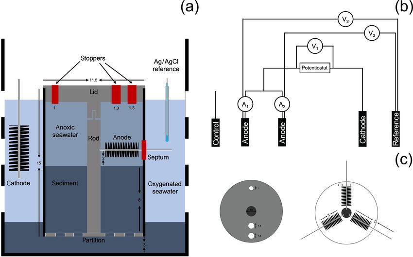
600                                           C. Li et al.: Inducing the attachment of cable bacteria on oxidizing electrodes

the same spatial interval. The pH microelectrode was cal-          sachusetts, USA) was deposited on samples in the presence
ibrated by using standard pH 4, 7, and 10 buffer solutions         of 0.15 % H2 O2 .
(Ricca Chemical, Arlington, Texas, USA). H2 S microelec-               Two-color CARD-FISH was performed on some car-
trodes were calibrated by generating an 11-point calibration       bon fiber samples to look for a previously observed co-
relationship by standard addition, from 0 to 7.48 µM H2 S at       occurrence of CB and other electroactive bacteria on elec-
pH = 1.6. A 3 mM standard solution was made from crystal           trode surfaces (Reimers et al., 2017). To perform the CARD-
Na2 S9H2 O (> 98.0 %, MilliporeSigma, Burlington, MA) in           FISH, horseradish peroxidases on the hybridized DSB706
an anoxic glove box. Total sulfide concentration at each pro-      probes were inactivated by 0.15 % H2 O2 . The inactivated
file depth was derived from pH and H2 S according to equi-         samples were then hybridized with a Desulfuromonadales-
librium relationships given in Millero et al. (1988).              specific oligonucleotide probe (DRM432; 50 -CTT CCC CTC
                                                                   TGA CAG AGC-30 ) modified with horseradish peroxidase in
2.5   SEM                                                          standard hybridization buffer at 46 ◦ C with 40 % formamide
                                                                   for 5 h and sequentially stained with Alexa Fluro 555 (Ther-
                                                                   moFisher, Waltham, Massachusetts, USA). A counter stain,
To confirm the presence of CB and to examine the charac-
                                                                   40 ,6-diamidino-2-phenylindole (DAPI), was applied to all
teristic longitudinal ridges and cell–cell junctions of CB, fil-
                                                                   samples after the deposition of fluorescent probe(s). Hy-
aments extracted from the sediments and carbon fibers from
                                                                   bridization samples were visualized using confocal laser
the reactor electrodes were visualized by scanning electron
                                                                   scanning microscopy (CLSM) (LSM 780, Zeiss, Jena, Ger-
microscopy (SEM). Samples were dehydrated in a graded
                                                                   many).
series of ethanol solutions from 10 % to 100 %. Specimens
were then mounted on aluminum SEM stubs with double-               2.7    Microbial community characterizations
sided carbon tape, critical-point dried using an EMS 850
Critical Point Dryer, and sputter-coated with gold and pal-        To investigate the phylogeny of the CB discovered in
ladium using a Cressington 108 sputter coater. The resul-          Yaquina Bay, genomic DNA was extracted from three sed-
tant specimens were observed under a FEI Quanta 600FEG             iment plug samples and from two separated filamentous
ESEM at 5–15 kV. This instrument also provided elemental           biomass samples using a MoBio PowerSoil DNA Ex-
spectra by X-Ray Energy Dispersive Spectrometry (EDS).             traction Kit. To avoid insufficient cell lysis, all sam-
                                                                   ples went through five to seven freeze–thaw cycles be-
2.6   CARD-FISH                                                    fore the use of the extraction kit (Roose-Amsaleg et
                                                                   al., 2001). Bacterial 16S rRNA genes were amplified by
Catalyzed reporter deposition–fluorescence in situ hybridiza-      polymerase chain reaction (PCR) with random primers
tion (CARD-FISH) was used to microscopically iden-                 357wF (50 -CCTACGGGNGGCWGCAG-30 ) and 785R (50 -
tify Desulfobulbaceae filaments using a Desulfobulbaceae-          GACTACHVGGGTATCTAATCC-30 ). Amplification and se-
specific oligonucleotide probe (DSB706; 50 -ACC CGT ATT            quencing of DNA (Illumina MiSeq Reagent Kit v3, 2 ×
CCT CCC GAT-30 ) labeled with horseradish peroxidase               300 bp) was performed by the Center of Genome Research
(Lücker et al., 2007). In preparation for CARD-FISH, sed-          and Biocomputing at Oregon State University. Sequences
iment samples were fixed with a 1 : 1 (vol : vol) ethanol and      were processed using DADA2 (v.1.10) in R (3.5.0), as de-
phosphate-buffered saline solution and stored at −20 ◦ C until     scribed in a previous study (Callahan et al., 2016). Sequences
analysis. Extracted bacterial filaments and carbon fibers cut      were aligned to the Silva SSU Ref NR database (v.132) and
from the carbon brush electrodes were treated with a fixa-         clustered into operational taxonomic units (OTUs) at 97 %
tive solution containing 1.25 % glutaraldehyde and 1.3 % os-       similarity. Representative sequences classified into the fam-
mium tetroxide. Fixed samples were stored at −20 ◦ C un-           ily of Desulfobulbaceae were tagged and aligned to 16S
til analysis. Sediment and bacterial filament samples were         rRNA gene sequences from previously identified CB (Tro-
first retained on polycarbonate membrane filters and then          jan et al., 2016). A phylogenetic tree was constructed using
mounted onto a glass slide by using 0.2 % agarose (Malkin          RaxML with 1000 bootstraps (Stamatakis, 2014). Sequences
et al., 2014). Carbon fiber samples were mounted directly          from this study were deposited to the Genbank’s Sequence
onto a glass slide without first retaining on a filter. Mounted    Read Archive (MK388690-MK388723, PRJNA587126).
samples were sequentially permeabilized by 10 mg mL−1
of lysosome (2 h at 37 ◦ C) and achromopeptidase (1 h at           3     Results and discussion
37 ◦ C). After permeabilization, glass slides were incubated
in H2 O2 (0.15 % in methanol) for 30 min at room tempera-          3.1    Cable bacteria activity in the sediments of Yaquina
ture (∼ 25 ◦ C) to inactivate the endogenous peroxidases. The             Bay
hybridization process was performed in a standard hybridiza-
tion buffer at 46 ◦ C with 45 % formamide for 7 h (Wende-          During the initial open incubations of IMF sediment, the top
berg, 2010). Alexa Fluro 488 (ThermoFisher, Waltham, Mas-          centimeter of each core changed from dark to light gray, and

Biogeosciences, 17, 597–607, 2020                                                        www.biogeosciences.net/17/597/2020/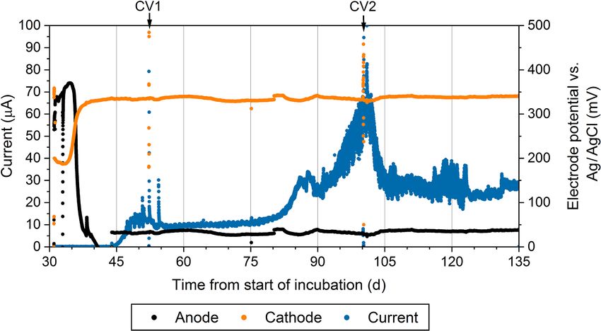
C. Li et al.: Inducing the attachment of cable bacteria on oxidizing electrodes                                                   601

Figure 2. Representative microelectrode depth profiles of oxygen (blue), pH (red), and 6H2 S or H2 S (yellow) in (a) IMF sediment after
53 d of incubation and in the bioelectrochemical reactor at (b) day 13, (c) day 24, and (d) day 48.

a brownish layer formed from the sediment–water interface             tous biomass samples. The most abundant Desulfobulbaceae
to ∼ 0.2 cm depth. Hallmarks of the activity of CB were doc-          OTUs within these samples were aligned with a previously
umented by microelectrode profiling after 53 d of culture.            established taxonomy framework of CB (Trojan et al., 2016)
These hallmarks were a sulfide-free suboxic zone and op-              (Fig. 3e). Partial 16s rRNA sequences of CB have been dis-
posing pH extremes at approximately 0.2 cm and 1–1.5 cm               covered in sediment samples from the US East Coast, Gulf of
deep (Fig. 2a). Although a faint smell of sulfide was detected        Mexico, and certain sites on the US West Coast from SILVA
during collection of the sediment, total sulfide concentrations       or GenBank databases (Trojan et al., 2016). Our studies have
detected by microelectrode profiling were low compared to             provided the first combined microscopic and genetic obser-
previous studies of marine sediments hosting CB (Malkin et            vations of CB in sediments from the northeastern Pacific
al., 2014). The pH minimum within the anoxic layers of cul-           coast of the United States, reinforcing the suggestion that
tured sediment was 6.0, indicating acidification coupled to           CB bacteria are distributed globally. This result also indicates
sulfide or iron sulfide oxidation.                                    that Yaquina Bay, OR, where we deployed the BMFC, indeed
   SEM revealed that cells within extracted filaments were            harbors a rich population of CB.
0.5 to 1.2 µm wide and 2 to 3 µm long (Fig. 3a, b, c). Typical
morphological features of CB including longitudinal ridges            3.2   Encouraging the growth of cable bacteria on poised
and cell–cell junctions were observed, though a smaller num-                electrodes
ber of ridges (8–10) were usually spotted compared to 16–
58 in other characterizations (Malkin et al., 2014). Certain          Geochemical hallmarks of CB developed within 2 weeks of
filaments extracted from sediments were covered by het-               culture within the bioelectrochemical reactor (Fig. 2b). The
erogeneous coatings of mineral particles as was observed              pH minimum within the sulfidic layer and the pH maxi-
recently by Geerlings et al. (2019) (Fig. 3c). These parti-           mum in the oxic layer of sediment became more extreme
cles have similar elemental compositions to some authigenic           by day 24 (Fig. 2c), indicating that a CB population was
clays (Burdige, 2006), showing enrichments of silicon, alu-           actively mediating electrogenic sulfide oxidation and trans-
minum, magnesium, and iron. In our open incubation sam-               porting electrons to reduce oxygen. After sealing the reactor,
ples, some thinner filaments were also seen that displayed            oxygen concentration in the overlaying seawater dropped be-
no obvious longitudinal ridges, although cell–cell junctions          low detection limits and the open-circuit anode potential fell
were still visible (Fig. 3b). Extracted filaments reacted pos-        to −104 mV (versus Ag/AgCl). Once poised with the poten-
itively to the DSB 706 probe and DAPI (Figs. 3d and S1 in             tiostat, the cathode and anode potentials became stable at ap-
the Supplement).                                                      proximately 330 and 30 mV versus Ag/AgCl, respectively.
   When analyzing the 16S rRNA gene sequence data, we                 When microelectrode profiling was performed on day 48,
found that one of the candidate CB genera, “Candidatus                the measurements indicated that the overlying seawater was
Electrothrix”, was relatively abundant in sediment plug sam-          anoxic (except right below the sample port) and that free H2 S
ples (2.9 %) and predominant (83.5 %) in separated filamen-           was detectable right below the sediment–water interface but
                                                                      not in the water column (Fig. 2d). The pH of the seawater

www.biogeosciences.net/17/597/2020/                                                             Biogeosciences, 17, 597–607, 2020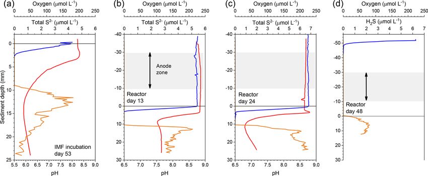
602                                              C. Li et al.: Inducing the attachment of cable bacteria on oxidizing electrodes

Figure 3. Cable bacteria filaments recovered from Yaquina Bay sediments. (a) A cable bacteria filament under SEM. (b) A thin type of
cable bacteria filament under SEM. (c) Multiple filaments of cable bacteria clumped together under SEM. The blue arrow indicates a section
of cable, and the red arrow indicates a cable bacteria filament covered with a mineral coating. (d) Identification of the filaments belonging
to Desulfobulbaceae using catalyzed reporter deposition–fluorescence in situ hybridization (DSB 706 probe + Alexa Fluor 488 in green
and DSB DAPI in blue). (e) Phylogenetic tree of Desulfobulbaceae 16s rRNA gene sequences recovered from IMF sediment and extracted
biomass samples. Color boxes indicate previously recognized species of cable bacteria. The scale bar shows 5 % sequence divergence.

measured inside of the reactor chamber at the experiment’s               the underlying sediment, changes in the anodic biofilm, and
end was 6.2, consistent with sulfide oxidation under anaero-             finally loss of anode surface area due to mineral deposi-
bic conditions within the anolyte seawater. Current collection           tion induced by microbial activity and/or the applied elec-
started to increase once the anodic potential became stable,             trical potential. Coatings containing iron, phosphorus, sul-
indicating that the anode brushes were being used as an elec-            fur, silicon, and aluminum are often found on anode surfaces
tron acceptor. Current records collected from duplicate elec-            of BMFCs in marine environments and were seen by SEM
trodes were similar, and the current in each steadily increased          in the present study (see below). Cyclic voltammetry (CV,
to ∼ 30.5 ± 2.5 µA by day 86, stabilized, then rose again to             Fig. S2a) performed on the anode brushes at day 52 and 100
a peak of ∼ 75 ± 8 µA on day 101. After this maximum, cur-               yielded broad and poorly defined electrochemical signals.
rent decreased and restabilized at ∼ 30 ± 5 µA. These elec-              The interpretation of such voltammograms may be compli-
trochemical results are portrayed in Fig. 4. The cause of the            cated by a high uncompensated resistance between work-
current rise and subsequent fall (Fig. 4) is unknown but is a            ing electrode and reference electrode (Babauta and Beyenal,
common occurrence in marine BMFC experiments (Nielsen                    2015). While an oxidation peak can be clearly identified at
et al., 2009; Ryckelynck et al., 2005). It is likely that such           potentials near where the anode was held, the peak current
behavior is a result of a varying supply of reductants from              did not increase with an increase in scan rate (Fig. S2b). The

Biogeosciences, 17, 597–607, 2020                                                                www.biogeosciences.net/17/597/2020/
C. Li et al.: Inducing the attachment of cable bacteria on oxidizing electrodes 603
Figure 4. The current production (blue), the anodic potential (black), and cathodic potential (orange) over time during the reactor experiment.
The reference electrode was an Ag/AgCl electrode with a saturated KCl filling solution. This figure only shows measurements associated
with one of the duplicate electrodes.
peak oxidation current also did not change much between rus, oxygen, and silicon (Fig. S3b). The control electrode
day 52 and day 100. This CV behavior suggests that any cur- that was not positively poised displayed no mineral deposi-
rent generated by the biomass of electroactive bacteria, in- tion and nearly no cell growth (Fig. 5i). Thirdly, most of the
cluding CB, was obscured during scans by current arising bacterial filaments on the poised electrode surfaces reacted
from irreversible redox reactions, such as oxidation of dis- positively with the Desulfobulbaceae-specific probe. CARD-
solved iron. A reduction peak was unidentifiable through- FISH performed in the present study revealed that the an-
out the scans, a common phenomenon in sediment MFCs odic carbon fibers harbored many short bacterial filaments,
(Babauta and Beyenal, 2015). Taken together, these results as well as colonies belonging to the family of Desulfobul-
demonstrate that the electrode surface was altered during the baceae (Fig. 6a, b, c, d, e, f). Clear cell–cell junctions were
course of the bioreactor experiment by mineral and chemical observed along many of the fluorescent filaments. However,
precipitate deposition (Imran et al., 2019). the complexity of the carbon fiber samples often hampered
clear microscopic visualization of fluorescent cells. Applica-
3.3 Examining the attachment of cable bacteria on the tion of an additional Desulfuromonadales-specific oligonu-
anode cleotide probe (DRM432) confirmed the presence of other
known electrogenic bacteria on the carbon fibers near Desul-
The hypothesis that led to the bioreactor experiments in this fobulbaceae cells as well (Fig. 6b, c, d).
study was that an electrode poised at an oxidative potential Though a global occurrence has recently been indicated
can produce redox conditions and geochemical gradients that (Malkin et al., 2014), CB successfully evaded microbiologi-
attract CB and that will lead to their electron donation to an cal survey for quite a long time. One of the reasons is likely
electrode. Several observations that were made on harvested that the phylogeny of CB is overshadowed by the broad fam-
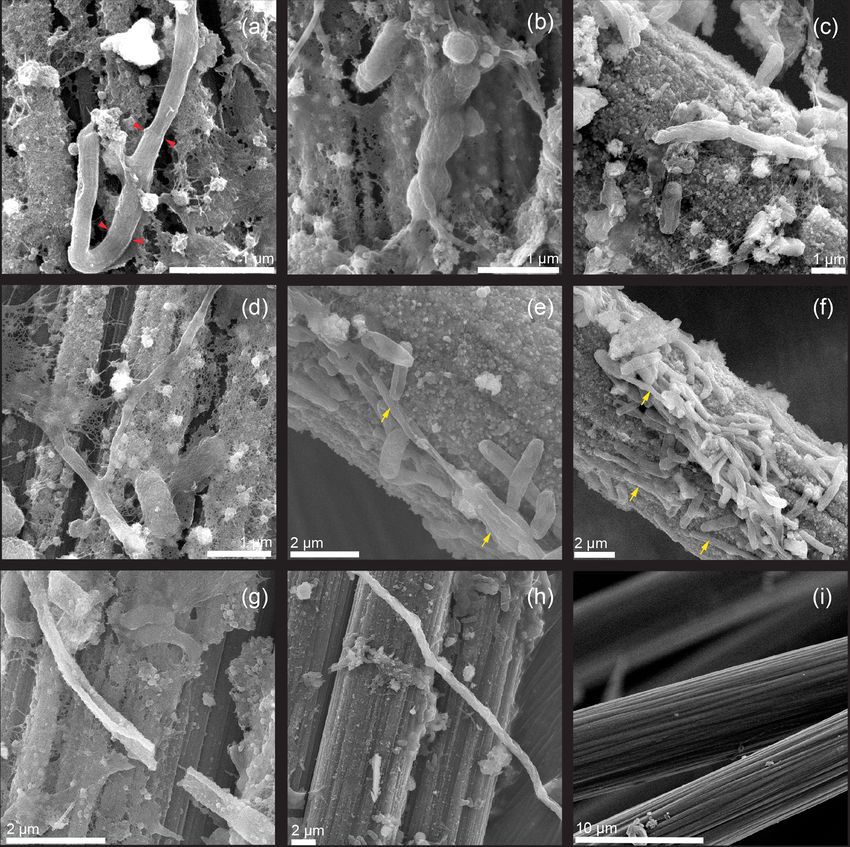
electrodes affirm this hypothesis. Firstly, under SEM, bac- ily of Desulfobulbaceae, which are often highly abundant in
teria filaments with visible longitudinal ridges and cell–cell marine sediments (Kuever, 2014). Another reason may be a
junctions were found integrated into biofilms on the surfaces resistance of the cells of CB to routine cell lysing techniques
of poised electrodes (Fig. 5a, b). As observed in the initial that have been used with many DNA extraction kits (Trojan
IMF sediment examinations, filaments appeared to contain a et al., 2016). Therefore, their identification in various stud-
smaller number of ridges (8 to 10) compared to previously ies has relied on microscopic observations of their unique
reported CB filaments and others were without pronounced filamentous form and morphological features (ridges and
ridges along their longitudinal axes. The latter examples did cell–cell junctions), combined with fluorescence in situ hy-
show cell–cell junctions and appeared to have wrinkled sur- bridization labeling (Malkin et al., 2014, 2017; Malkin and
faces (Fig. 5c, d, e, f). Secondly, many of the bacteria fil- Meysman, 2015). The electrochemical reactor in this study
aments observed on the electrode surfaces were encrusted, was anoxic for more than 100 d. The observation of CB on
suggesting mineral deposition similar to that observed at the anodic carbon fibers at the end of this experiment con-
the oxic terminal of CB filaments in sediments (Fig. 5g, h). firms that, although they may not have been abundant, they
EDS indicated that these deposits contained iron, phospho-
www.biogeosciences.net/17/597/2020/ Biogeosciences, 17, 597–607, 2020604 C. Li et al.: Inducing the attachment of cable bacteria on oxidizing electrodes Figure 5. SEM images illustrating (a, b) cable bacteria filaments with visible ridges and cell–cell junctions incorporated into the biofilms on carbon fiber electrode surfaces. Red pointers indicate cell–cell junctions. (c, d, e, f) Short bacterial filaments without typical morphological features of cable bacteria. Yellow arrows indicate the locations of elongated cells. (g, h) Mineral-encrusted bacterial filaments. (i) Image of control electrode surface after culture. can survive under such conditions and were likely using the sociated with the tube worm Chaetopterus variopedatus, in anode as an electron acceptor (as suggested previously by which redox conditions often oscillated between oxic and hy- Reimers et al., 2017). Besides the recognized forms of CB, poxic, CB were present predominately in short filaments. As- short filaments within the family of Desulfobulbaceae that suming the CB can use an electrode as an electron acceptor, possessed different morphologies were also observed on the the distance between the electron donor and acceptor utilized anode surface. Aller et al. (2019) suggest that redox envi- by CB may be short, reducing the advantage of forming long ronment may play an important role in controlling the length filaments. of CB filaments. For example, in the bioturbated zone as- Biogeosciences, 17, 597–607, 2020 www.biogeosciences.net/17/597/2020/
C. Li et al.: Inducing the attachment of cable bacteria on oxidizing electrodes                                                         605

Figure 6. (a–c) Confocal microscope images illustrating cable bacteria filaments on the carbon fibers that served as an anode. (d–f) Colonies
of cells belonging to Desulfobulbaceae. Red circles indicate a possible doublet of the long cells. Cells were visualized using catalyzed
reporter deposition–fluorescence in situ hybridization (DSB 706 probe + Alexa Fluor 488, green; DRM 432 + Alexa Fluor 555, red; and
DAPI, blue).

   The closest culturable relative to CB, Desulfobulbus pro-             confirm that an active population of filamentous CB are
pionicus, can utilize an electrode as an electron acceptor to            present in Yaquina Bay, Oregon, USA, where CB were pre-
oxidize S0 , H2 , and organic acids like pyruvate, lactate, and          viously found attached to carbon-fiber electrode surfaces
propionate (Holmes et al., 2004). While CB appear to pos-                within a BMFC (Reimers et al., 2017). Moreover, by incu-
sess features like motility and an ability to form loops and             bating intertidal sediment collected from Yaquina Bay in a
bundles that are similar to large sulfur bacteria (but distinct          reactor mimicking the anodic chamber of a BMFC, we ob-
from the D. propionicus), our SEM and CARD-FISH exami-                   served that CB can be drawn to electrodes at oxidative elec-
nations suggest that CB on oxidative electrode surfaces may              trical potentials. Thus, we have further evidence that CB can
produce extracellular structures to transfer electrons to an             survive under anoxic conditions in the presence of an oxida-
electrode and/or to insoluble Fe(III)-oxides similar to D. pro-          tive electrode serving as an electron acceptor. The bioelec-
pionicus (Bjerg et al., 2016; Holmes et al., 2004; Jørgensen,            trochemical reactor study also showed attachment of CB to
2010; Pfeffer et al., 2012). Admittedly, indisputable proof of           an oxidative electrode when the surrounding seawater was
electron transfer from CB to electrodes still awaits growth in           stripped of hydrogen sulfide and at a pH ∼ 6.2. However,
purer biofilms and cultures.                                             the observed CB density and the overall density of recog-
                                                                         nizable cells were both relatively low on electrode surfaces,
                                                                         as the respirable surface area appeared to become limited by
4   Summary and implications for the                                     the deposition of mineral coatings. More work is needed to
    electrode-associated growth of cable bacteria                        determine conditions or experimental designs that may at-
                                                                         tract CB to an electrode while not also leading to excessive
The present study introduces bioelectrochemical reactors as              mineral precipitation on electrode surfaces. Developing ex
an approach to investigate filamentous cable bacteria and                situ culture techniques of CB and using these approaches to
their unique ability to transfer electrons. Furthermore, we

www.biogeosciences.net/17/597/2020/                                                                  Biogeosciences, 17, 597–607, 2020
606 C. Li et al.: Inducing the attachment of cable bacteria on oxidizing electrodes
gain insight into their electron transfer will contribute to the Bjerg, J. T., Damgaard, L. R., Holm, S. A., Schramm, A., and
overall understanding of this group of bacteria, their genomic Nielsen, L. P.: Motility of electric cable bacteria, Appl. Environ.
makeup, and their survival in both natural and engineered en- Microbiol., 82, 3816–3821, 2016.
vironments. Bjerg, J. T., Boschker, H. T. S., Larsen, S., Berry, D., Schmid, M.,
Millo, D., Tataru, P., Meysman, F. J. R., Wagner, M., Nielsen,
L. P., and Schramm, A.: Long-distance electron transport in indi-
vidual, living cable bacteria, P. Natl. Acad. Sci. USA, 115, 5766–
Data availability. Partial 16S rRNA gene and raw sequences are
5791, https://doi.org/10.1073/pnas.1800367115, 2018.
deposited in the Genbank’s Sequence Read Archive (MK388690-
Burdige, D. J.: The components of marine sediments, in Geochem-
MK388723, PRJNA587126).
istry of Marine Sediments, Princeton University Press, Princeton,
NJ, USA, 5–24, 2006.
Callahan, B. J., McMurdie, P. J., Rosen, M. J., Han, A. W., John-
Supplement. The supplement related to this article is available on- son, A. J. A., and Holmes, S. P.: DADA2: high-resolution sample
line at: https://doi.org/10.5194/bg-17-597-2020-supplement. inference from Illumina amplicon data, Nat. Methods, 13, 581–
583, 2016.
Cornelissen, R., Bøggild, A., Thiruvallur Eachambadi, R., Kon-
Author contributions. CL and CER conceived the study. YA de- ing, R. I., Kremer, A., Hidalgo-Martinez, S., Zetsche, E.-M.,
signed and assembled the bioelectrochemical reactor. CL per- Damgaard, L. R., Bonné, R., Drijkoningen, J., Geelhoed, J. S.,
formed the microscopic examinations and microprofiling and ana- Boesen, T., Boschker, H. T. S., Valcke, R., Geerlings, N. M.
lyzed the microbial community and phylogenies. CL also wrote the J., Zetsche, E.-M., Hidalgo Martinez, S., Middelburg, J. J., and
manuscript, with major rewrites and editing contributed by CER. Meysman, F. J. R.: Mineral formation induced by cable bacte-
ria performing long-distance electron transport in marine sedi-
ments, Biogeosciences, 16, 811–829, https://doi.org/10.5194/bg-
Competing interests. The authors declare that they have no conflict 16-811-2019, 2019.
of interest. Geerlings, N. M. J., Zetsche, E.-M., Hidalgo Martinez, S., Mid-
delburg, J. J., and Meysman, F. J. R.: Mineral formation
induced by cable bacteria performing long-distance electron
Acknowledgements. This research was funded through grant transport in marine sediments, Biogeosciences, 16, 811–829,
N00014-17-1-2599 from the Office of Naval Research to https://doi.org/10.5194/bg-16-811-2019, 2019.
Clare E. Reimers. We thank Teresa Sawyer, the Electron Mi- Hasvold, Ø., Henriksen, H., Melvir, E., Citi, G., Johansen,
croscopy Facility Instrument Manager at Oregon State University, B. Ø., Kjønigsen, T., and Galetti, R.: Sea-water battery
for assistance with the SEM imaging and Anne-Marie Girard for for subsea control systems, J. Power Sources, 65, 253–261,
valuable advice on the confocal microscope imaging. The authors https://doi.org/10.1016/S0378-7753(97)02477-4, 1997.
wish to acknowledge the Confocal Microscopy Facility of the Cen- Holmes, D. E., Bond, D. R., and Lovley, D. R.: Elec-
ter for Genome Research and Biocomputing at Oregon State Uni- tron transfer by Desulfobulbus propionicus to Fe(III) and
versity, which is supported in part by an award (no. 1337774) from graphite electrodes, Appl. Environ. Microbiol., 70, 1234–1237,
the National Science Foundation. We also thank our lab members https://doi.org/10.1128/AEM.70.2.1234-1237.2004, 2004.
for providing critical comments on the manuscript. Imran, M., Prakash, O., Pushkar, P., Mungray, A., Kailasa,
S. K., Chongdar, S., and Mungray, A. K.: Perfor-
mance enhancement of benthic microbial fuel cell by
Financial support. This research has been supported by the Office cerium coated electrodes, Electrochim. Acta, 295, 58–66,
of Naval Research (grant no. N00014-17-1-2599) and the National https://doi.org/10.1016/J.ELECTACTA.2018.08.158, 2019.
Science Foundation (grant no. 1337774). Johnson, J. K.: Effects of temperature and salinity on production
and hatching of dormant eggs of Acartia californiensis (Cope-
poda) in an Oregon estuary, Fish. Bull., 77, 567–584, 1980.
Jørgensen, B. B.: Big sulfur bacteria, ISME J., 4, 1083,
Review statement. This paper was edited by Jack Middelburg and
https://doi.org/10.1038/ismej.2010.106, 2010.
reviewed by two anonymous referees.
Kessler, A. J., Wawryk, M., Marzocchi, U., Roberts, K. L., Wong,
W. W., Risgaard-Petersen, N., Meysman, F. J. R., Glud, R. N.,
and Cook, P. L. M.: Cable bacteria promote DNRA through
References iron sulfide dissolution, Limnol. Oceanogr., 64, 1228–1238,
https://doi.org/10.1002/lno.11110, 2018.
Aller, R. C., Aller, J. J., Zhu, Q., Heilbrun, C., Klingensmith, I., Kjeldsen, K. U., Schreiber, L., Thorup, C. A., Boesen, T., Bjerg,
and Kaushik, A.: Worm tubes as conduits for the electrogenic J. T., Yang, T., Dueholm, M. S., Larsen, S., Risgaard-Petersen,
microbial grid in marine sediments, Sci. Adv., 5, eaaw3651, N., Nierychlo, M., Schmid, M., Bøggild, A., van de Vossenberg,
https://doi.org/10.1126/sciadv.aaw3651, 2019. J., Geelhoed, J. S., Meysman, F. J. R., Wagner, M., Nielsen, P.
Babauta, J. T. and Beyenal, H.: Introduction to electrochemi- H., Nielsen, L. P., and Schramm, A.: On the evolution and phys-
cally active biofilms, in Biofilms in Bioelectrochemical Systems: iology of cable bacteria, P. Natl. Acad. Sci. USA, 116, 19116–
From Laboratory Practice to Data Interpretations, John Wiley & 19125, https://doi.org/10.1073/pnas.1903514116, 2019.
Sons, Hoboken, NJ, USA, 1–35, 2015.
Biogeosciences, 17, 597–607, 2020 www.biogeosciences.net/17/597/2020/C. Li et al.: Inducing the attachment of cable bacteria on oxidizing electrodes 607 Kuever, J.: The Family Desulfobulbaceae, in: The Prokary- Pfeffer, C., Larsen, S., Song, J., Dong, M., Besenbacher, F., otes: Deltaproteobacteria and Epsilonproteobacteria, edited by: Meyer, R. L., Kjeldsen, K. U., Schreiber, L., Gorby, Y. A., Rosenberg, E., DeLong, E. F., Lory, S., Stackebrandt, E., and El-Naggar, M. Y., Leung, K. M., Schramm, A., Risgaard- Thompson, F., Springer Berlin Heidelberg, Berlin, Heidelberg, Petersen, N., and Nielsen, L. P.: Filamentous bacteria trans- 75–86, 2014. port electrons over centimetre distances, Nature, 491, 218–221, Larsen, S., Nielsen, L. P., and Schramm, A.: Cable bacteria as- https://doi.org/10.1038/nature11586, 2012. sociated with long-distance electron transport in New England Rao, A. M. F., Malkin, S. Y., Hidalgo-Martinez, S., and salt marsh sediment, Environ. Microbiol. Rep., 7, 175–179, Meysman, F. J. R.: The impact of electrogenic sulfide ox- https://doi.org/10.1111/1758-2229.12216, 2015. idation on elemental cycling and solute fluxes in coastal Li, C., Lesnik, K. L., and Liu, H.: Stay connected: electrical conduc- sediment, Geochim. Cosmochim. Ac., 172, 265–286, tivity of microbial aggregates, Biotechnol. Adv., 35, 669–680, https://doi.org/10.1016/j.gca.2015.09.014, 2016. https://doi.org/10.1016/j.biotechadv.2017.07.010, 2017. Reimers, C. E., Li, C., Graw, M. F., Schrader, P. S., and Wolf, M.: Lovley, D. R.: Happy together: microbial communities The identification of cable bacteria attached to the anode of a that hook up to swap electrons, ISME J., 11, 327–336, benthic microbial fuel cell: evidence of long distance extracel- https://doi.org/10.1038/ismej.2016.136, 2016. lular electron transport to electrodes, Front. Microbiol., 8, 2055, Lücker, S., Steger, D., Kjeldsen, K. U., MacGregor, B. J., Wag- https://doi.org/10.3389/fmicb.2017.02055, 2017. ner, M., and Loy, A.: Improved 16S rRNA-targeted probe Roose-Amsaleg, C., Garnier-Sillam, E., and Harry, M.: Extraction set for analysis of sulfate-reducing bacteria by fluorescence and purification of microbial DNA from soil and sediment sam- in situ hybridization, J. Microbiol. Methods, 69, 523–528, ples, Appl. Soil Ecol., 18, 47–60, https://doi.org/10.1016/S0929- doi10.1016/j.mimet.2007.02.009, 2007. 1393(01)00149-4, 2001. Malkin, S. Y. and Meysman, F. J. R.: Rapid redox sig- Ryckelynck, N., Stecher III, H. A., and Reimers, C. E.: Understand- nal transmission by “cable bacteria” beneath a photosyn- ing the anodic mechanism of a seafloor fuel cell: interactions be- thetic biofilm, Appl. Environ. Microbiol., 81, 948–956, tween geochemistry and microbial activity, Biogeochemistry, 76, https://doi.org/10.1128/AEM.02682-14, 2015. 113–139, https://doi.org/10.1007/s10533-005-2671-3, 2005. Malkin, S. Y., Rao, A. M. F., Seitaj, D., and Vasquez-Cardenas, Seitaj, D., Schauer, R., Sulu-Gambari, F., Hidalgo-Martinez, S., D.: Natural occurrence of microbial sulphur oxidation by long- Malkin, S. Y., Burdorf, L. D. W., Slomp, C. P., and Meysman, F. range electron transport in the seafloor, ISME J., 8, 1843–1854, J. R.: Cable bacteria generate a firewall against euxinia in season- https://doi.org/10.1038/ismej.2014.41, 2014. ally hypoxic basins, P. Natl. Acad. Sci. USA, 112, 13278–13283, Malkin, S. Y., Seitaj, D., Burdorf, L. D. W., Nieuwhof, S., https://doi.org/10.1073/pnas.1510152112, 2015. Hidalgo-Martinez, S., Tramper, A., Geeraert, N., De Stigter, Stamatakis, A.: RAxML version 8: a tool for phylogenetic anal- H., and Meysman, F. J. R.: Electrogenic sulfur oxidation by ysis and post-analysis of large phylogenies, Bioinformatics, cable bacteria in bivalve reef sediments, Front. Mar. Sci., 4, 30, 1312–1313, https://doi.org/10.1093/bioinformatics/btu033, https://doi.org/10.3389/fmars.2017.00028, 2017. 2014. Meysman, F. J. R.: Cable bacteria take a new breath us- Sulu-Gambari, F., Seitaj, D., Meysman, F. J. R., Schauer, ing long-distance electricity, Trends Microbiol., 26, 411–422, R., Polerecky, L., and Slomp, C. P.: Cable bacteria con- https://doi.org/10.1016/j.tim.2017.10.011, 2017. trol iron–phosphorus dynamics in sediments of a coastal Meysman, F. J. R., Cornelissen, R., Trashin, S., Bonné, R., Mar- hypoxic basin, Environ. Sci. Technol., 50, 1227–1233, tinez, S. H., van der Veen, J., Blom, C. J., Karman, C., Hou, J.-L., https://doi.org/10.1021/acs.est.5b04369, 2016a. Eachambadi, R. T., Geelhoed, J. S., Wael, K. De, Beaumont, H. Sulu-Gambari, F., Seitaj, D., Behrends, T., Banerjee, D., Meysman, J. E., Cleuren, B., Valcke, R., van der Zant, H. S. J., Boschker, H. F. J. R., and Slomp, C. P.: Impact of cable bacteria on T. S., and Manca, J. V: A highly conductive fibre network enables sedimentary iron and manganese dynamics in a seasonally- centimetre-scale electron transport in multicellular cable bac- hypoxic marine basin, Geochim. Cosmochim. Ac., 192, 49–69, teria, Nat. Commun., 10, 4120, https://doi.org/10.1038/s41467- https://doi.org/10.1016/j.gca.2016.07.028, 2016b. 019-12115-7, 2019. Trojan, D., Schreiber, L., Bjerg, J. T., Bøggild, A., Yang, T., Millero, F. J., Plese, T., and Fernandez, M.: The dissociation of Kjeldsen, K. U., and Schramm, A.: A taxonomic framework hydrogen sulfide in seawater, Limnol. Oceanogr., 33, 269–274, for cable bacteria and proposal of the candidate genera Elec- https://doi.org/10.4319/lo.1988.33.2.0269, 1988. trothrix and Electronema, Syst. Appl. Microbiol., 39, 297–306, Nielsen, L. P. and Risgaard-Petersen, N.: Rethinking sediment bio- https://doi.org/10.1016/j.syapm.2016.05.006, 2016. geochemistry after the discovery of electric currents, Ann. Rev. Wendeberg, A.: Fluorescence in situ hybridization for the identi- Mar. Sci., 7, 425–442, https://doi.org/10.1146/annurev-marine- fication of environmental microbes, Cold Spring Harb. Protoc., 010814-015708, 2015. https://doi.org/10.1101/pdb.prot5366, 2010. Nielsen, M. E., Wu, D. M., Girguis, P. R., and Reimers, C. E.: Influ- ence of substrate on electron transfer mechanisms in chambered benthic microbial fuel cells, Environ. Sci. Technol., 43, 8671– 8677, https://doi.org/10.1021/es9013773, 2009. www.biogeosciences.net/17/597/2020/ Biogeosciences, 17, 597–607, 2020
You can also read