HHS Public Access Author manuscript Depress Anxiety. Author manuscript; available in PMC 2018 January 01.
←
→
Page content transcription
If your browser does not render page correctly, please read the page content below
HHS Public Access
                                Author manuscript
                                Depress Anxiety. Author manuscript; available in PMC 2018 January 01.
Author Manuscript

                    Published in final edited form as:
                     Depress Anxiety. 2017 January ; 34(1): 9–24. doi:10.1002/da.22556.

                    Defining biotypes for depression and anxiety based on large-
                    scale circuit dysfunction: A theoretical review of the evidence
                    and future directions for clinical translation
                    Leanne M Williams, PhD*
                    Department of Psychiatry and Behavioral Sciences, Stanford University, CA
Author Manuscript

                    Abstract
                         Complex emotional, cognitive and self-reflective functions rely on the activation and connectivity
                         of large-scale neural circuits. These circuits offer a relevant scale of focus for conceptualizing a
                         taxonomy for depression and anxiety based on specific profiles (or biotypes) of neural circuit
                         dysfunction. Here, the theoretical review first outlined the current consensus as to what constitutes
                         the organization of large-scale circuits in the human brain identified using parcellation and meta-
                         analysis. The focus is on neural circuits implicated in resting reflection (“default mode”), detection
                         of “salience”, affective processing (“threat” and “reward”), “attention” and “cognitive control”.
                         Next, the current evidence regarding which type of dysfunctions in these circuits characterize
                         depression and anxiety disorders was reviewed, with an emphasis on published meta-analyses and
                         reviews of circuit dysfunctions that have been identified in at least two well-powered case:control
                         studies. Grounded in the review of these topics, a conceptual framework is proposed for
Author Manuscript

                         considering neural circuit-defined “biotypes”. In this framework, biotypes are defined by profiles
                         of extent of dysfunction on each large-scale circuit. The clinical implications of a biotype
                         approach for guiding classification and treatment of depression and anxiety is considered. Future
                         research directions will develop the validity and clinical utility of a neural circuit biotype model
                         that spans diagnostic categories and helps to translate neuroscience into clinical practice in the real
                         world.

                    INTRODUCTION
                                     We are experiencing a paradigm shift in psychiatry and the integration of psychiatry with the
                                     neurosciences. This integration is motivated by the search for a model that connects a
                                     neurobiological understanding of mental disorder with clinical phenomenology, in order to
Author Manuscript

                                     improve the precision of classification and treatment decisions. Major disorders of mood and
                                     anxiety impair the very functions that enable us to live productive and satisfying lives. Each
                                     year, an estimated 16 million American adults have at least one episode of major depressive
                                     disorder (MDD) and 40 million experience an anxiety disorder[1]. These disorders are the

                    *
                    Corresponding author: Leanne M Williams, PhD, Department of Psychiatry and Behavioral Sciences, Stanford University, 401
                    Quarry Road, California, 94134-5717, leawilliams@stanford.edu, Phone: 650 723 3579.
                    Disclosures
                    LMW has received fees from Brain Resource and Humana in projects not related to the present work.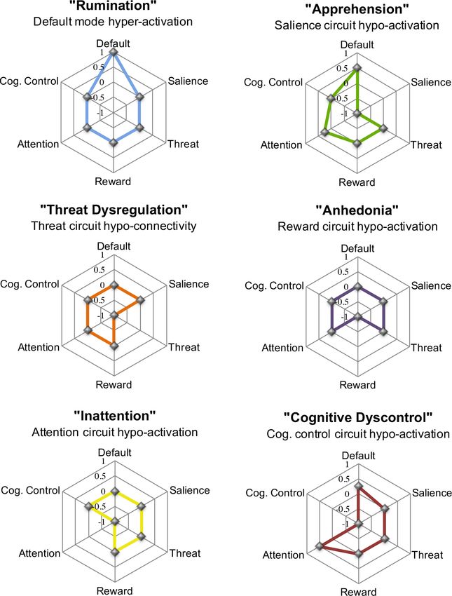
Williams                                                                                              Page 2

                                leading causes of disability and lost productivity, with a staggering economic cost of $42-
                                $53 billion per year[2].
Author Manuscript

                       CONCEPTUALIZING DEPRESSION AND ANXIETY AS “NEURAL CIRCUIT” DISORDERS
                                “Brain disorder” is typically used to refer a neurological condition associated with a lesion
                                or degenerative process in discrete brain regions. Mental disorders such as depression and
                                anxiety may not typically have been considered “brain disorders” due to our limited
                                understanding of the organization of human neural circuits and the way in which
                                disorganization of these circuits may disrupt normal cognitive, emotional and self-reflective
                                functions. Now that we have brain imaging techniques with sufficient spatial and temporal
                                resolution to quantify such neural circuit organization in vivo, we have the opportunity to
                                reformulate our neurobiological understanding of mental disorders. This vision is at the
                                heart of NIMH’s goal to “map the connectomes for mental illness” (NIMH’s Strategic Plan,
Author Manuscript

                                Strategy 1.3 under Objective 1). It is also consistent with the cross-cutting themes of the
                                “Research Domain Criteria” (RDoC) project[3].

                                The term “neural circuit” has typically referred to how one neuron communicates with
                                another through synaptic connections and transmission[4]. Here, the term “large-scale neural
                                circuit” is used to refer to the macroscale of neural organization. At the macroscale, vast
                                numbers of interconnected neurons constitute anatomical and functional circuits that make
                                up the “connectome” of the brain[5,6]. These vast sets of neurons can be probed by
                                noninvasive brain imaging to visualize the activation and structure of specific regions, the
                                structural communication between regions and functionally correlated regions of activation
                                at rest or during task-evoked situations[7–9]. In brain imaging studies, these macroscale
                                circuits have commonly been referred to as “networks” (for example, the “default mode
                                network”[10,11].
Author Manuscript

                                 Neural circuits can be considered a pertinent scale of measurement from which to delineate
                                 a neurobiology of human mental disorder. Circuits integrate across different levels and
                                 measures of brain function, but still reflect the complexity of the brain. Circuits are engaged
                                 by specific human cognitive, emotional and self-reflective functions, and offer promise for
                                 defining appropriate animal homologues. It is likely that most of the human brain involves
                                 multiple parallel circuits that are interdigitated such that each cortical lobe contains
                                 components of multiple circuits[12,13]. This organization may have occurred with the
                                 expansion of the association cortex in humans relative to non-human primates[12]. Mood and
                                 anxiety disorders may be possible maladaptive consequences of this expansion.

                       METHODS
Author Manuscript

                       LITERATURE SEARCH
                                To search the literature we used PubMedR and PsycINFOR and combined three sets of
                                search terms as follows:

                                     1.       Disorder class. The search was by classes that span the DSM-5 diagnoses of
                                              mood and anxiety disorders and also compatible with previous editions of DSM;
                                              Depressive Disorders (including Major Depressive Disorder, Disruptive Mood

                                    Depress Anxiety. Author manuscript; available in PMC 2018 January 01.
Williams Page 3
Dysregulation Disorder); Anxiety Disorders (including Agoraphobia,
Author Manuscript
Generalized Anxiety Disorder, Social Anxiety Disorder); Obsessive-Compulsive
and Related Disorders; and Trauma-and-Stressor-Related Disorders (including
Post-Traumatic Stress Disorder, Unspecified Trauma-and Stressor-Related
Disorder). This trans-diagnostic approach also took into account the substantial
comorbidity (at least 40%) of mood and anxiety disorders[14]. To consider future
implications for the wider transdiagnostic relevance of the research we also
included the term “bipolar disorder”.
2. Neural circuit terms and metrics. “Disorder class” terms were combined with the
following neural circuit terms: “neural circuit”, “network”, “functional brain
imaging”, “activation”, “connectivity”, “resting” and “task” and “grey matter”
and “white matter”.
3. Specific neural circuits. The focus was on six large-scale circuits identified using
Author Manuscript
the terms “default mode”, “salience”, “affective”, “positive emotion”, “negative
emotion”, “threat”, “reward”, “attention”, “frontoparietal”, “cognitive control”
and “central executive”.
Searches were restricted to articles written or translated into English from December 1990 to
June 30, 2016, consistent with the period in which functional brain imaging has emerged for
the study of psychiatric disorder. The focus of this review was on functional activation and
connectivity, as reflected in the blood oxygen level-dependent (BOLD) signal of functional
magnetic resonance imaging (fMRI) technology. However, studies involving the structural
neuroanatomy and white matter connectivity of depression and anxiety were also included as
a secondary focus. The reason for this secondary focus was to consider areas of evidence for
which depression and/or anxiety are associated with both functional and structural
Author Manuscript
impairments in the same circuit, which may be indicative of a more trait-like biotype,
NEURAL CIRCUIT SELECTION
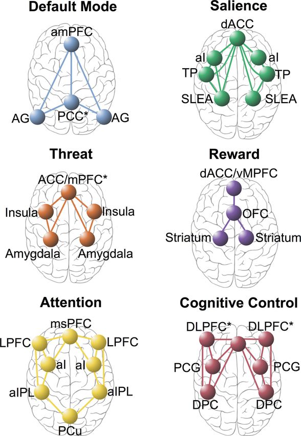
Researchers have identified intrinsic neural circuits that support domain-general processes of
self-reflection, salience perception and alertness (Figure 1; [15]) as well as conflict
monitoring, attention, sensori-motor, visual and auditory processes (Supplemental Figure
1)[16–24]. The intrinsic architecture has been demonstrated using large-scale functional
connectivity analysis of hundreds of brain regions that have been identified using
parcellation and meta-analysis and that define major brain systems at rest and across many
task-evoked states (e.g., [25]). These circuits may be observed in the task-free state and
during task-evoked conditions. During rest the default mode circuit tends to be up-regulated
and other circuits, down-regulated[16,25]. Specific task states (such as those designed to
Author Manuscript
probe reactivity to potential threat or reward) engage more specialized functional
components of these circuits (e.g.[26–31]) (Figure 1).
Given the wide scope of the search and the theoretical nature of the review, the focus of
interpretation was on areas in which there is a convergence of evidence. Thus, the emphasis
was on reviews and meta-analyses and circuit dysfunctions that have been identified in at
least two well-powered case:control studies. With this emphasis, we focused on six circuits
Depress Anxiety. Author manuscript; available in PMC 2018 January 01.Williams Page 4
in particular: default mode, salience, negative affect, positive affect, attention and cognitive
Author Manuscript
control.
RESULTS
The results of this theoretical review are organized as follows:
1. The current consensus as to what constitutes the organization of large-scale
circuits in the human brain identified using parcellation and meta-analysis.
2. The current evidence regarding which type of dysfunctions in these circuits
characterize depression and anxiety disorders was reviewed, with an emphasis on
published meta-analyses and reviews of circuit dysfunctions that have been
identified in at least two well-powered case:control studies.
Table 1 provides a complementary summary of the extant studies identified by these
Author Manuscript
searches.
“Default Mode” circuit
The default mode circuit (more typically known as the “default mode network”) is defined
by the anterior medial prefrontal cortex (amPFC), posterior cingulate cortex (PCC) and
angular gyrus[11,32] (Figure 1; Table 1). This circuit is observed when the brain is at rest
under task-free conditions and typically when participants are instructed to reflect on their
own spontaneously generated thoughts (Table 1). Independent components analysis suggests
that the anterior and posterior regions define sub-networks of the default mode circuit (for
review;[33]). This circuit also has a basis in structural white matter connections between the
same regions[34,35]. Evidence from a twins samples indicates that the default mode circuit is
engaged even during “rest” periods that occur between task stimuli, and this circuit is
Author Manuscript
genetically heritable[35]. The “default mode” is currently listed under the Arousal and
Regulatory systems domain of RDoC1.
“Salience” circuit
The “salience” circuit is defined by core nodes in the anterior cingulate cortex (ACC),
anterior insula (aI) and sublenticular extended amygdala[19,21] (Figure 1; Table 1). The
salience network is implicated in the detection of salient changes in the environment, both
interoceptive and external, and signals the need for cognitive control (Table 1, [21]).
Increased functioning of this network may result in a maladaptively low threshold to alter
cognitive control[36]. The salience circuit is consistent with the RDoC construct of “arousal”
listed under the Arousal and Regulatory Systems domain[3]2.
Author Manuscript
1http://www.nimh.nih.gov/research-priorities/rdoc/arousal-and-regulatory-systems-workshop-proceedings.shtml
2In addition to the salience circuit, a distinct “cingulo-opercular” circuit has also been defined (Supplementary Figure 1). The circuit is
defined by nodes in the amPFC, dorsal ACC, aI/frontal operculum and anterior thalamus37 and is involved in the detection of potential
mismatches and conflict21. These regions and functions show overlap with the default mode and salience circuits even though the
cingulo-opercular circuit is articulated as a distinct circuit21.ibid.
Depress Anxiety. Author manuscript; available in PMC 2018 January 01.Williams Page 5
Negative Affect circuit: “Threat”
Author Manuscript
Affective circuits are robustly activated by biologically salient stimuli such as facial
expressions signaling potential threat (fear, anger) and social reward (happy). Affective
circuits for processing threat and reward are consistent with the RDoC domains of Negative
Valence and Positive Valence systems3.
Threat processing components of the affective circuits comprise the amygdala,
hippocampus, insula, and both dorsal and ventral portions of the prefrontal cortex, including
the dorsal medial prefrontal cortex (dmPFC) and its dorsal ACC connections, and the ventral
mPFC (vMPFC) and its ventral (subgenual and pregenual)-rostral ACC connections ([38,39];
Figure 1; Table 1). The dorsal prefrontal sub-circuit has been preferentially implicated in
appraisal and expression of emotion and may be considered an “aversive amplification” sub-
network[39] that serves to boost the processing of signals of potential threat [39].
Author Manuscript
Complementing this function, the ventral sub- circuit has been implicated in automatic
regulation of negative emotion[38,40] (for review, [41]; for meta-analysis [38]). These
components overlap with components of the salience circuit, and they may both be engaged
by the arousal and valence properties of threat stimuli respectively.
These sub-networks may be engaged even in the absence of conscious sensory awareness[27]
(for meta-analysis, [38]). In light of their commonly observed co-activation[38], the negative
affect circuit might subserve the perception of negative emotion cues and the salience
circuit, the arousal aspects of feeling these emotions.
Direct activation of the amygdala and prefrontal regions to which it projects may occur
automatically even in the absence of explicit conscious evaluation ([27,42–44] for meta-
analysis, [38]). Similar bottom-up amygdala reactivity has been observed for masked
Author Manuscript
presentations of other threat stimuli such as phobic-relevant cues[44].
Positive Affect circuit: “Reward”
Reward processing components of the affective circuits are defined by the striatal nucleus
accumbens and ventral tegmental areas (collectively referred to as “the striatum”) and their
projections to the orbitofrontal cortex (OFC) and mPFC ([45]; Table 1). These regional
components are preferentially engaged by different types of reward processing, including
sensitivity to the presence of salient reward stimuli and the anticipation of these stimuli
(Table 1). There are also connections between the striatum and the amygdala, consistent
with interactions between the processing of threat and reward and of significant stimuli that
encompass multiple valences[28].
Author Manuscript
Attention circuit
The frontoparietal “attention” circuit is defined by nodes in the medial superior frontal
cortices, anterior insula, anterior inferior parietal lobule and precuneus[46,47]; Table 1). This
circuit is implicated in alertness, sustained attention and the support of recollection ([17,47];
3http://www.nimh.nih.gov/research-priorities/rdoc/negative-valence-systems-workshop-proceedings.shtml and http://
www.nimh.nih.gov/research-priorities/rdoc/positive-valence-systems-workshop-proceedings.shtml
Depress Anxiety. Author manuscript; available in PMC 2018 January 01.Williams Page 6
Table 1). It interacts closely with the default mode circuit to configure the switching from
resting to task-context processing[47,48]. The attention circuit may be considered relevant to
Author Manuscript
the attention construct listed under the RDoC Cognitive Systems domain4.
Cognitive Control circuit
The “cognitive control” circuit comprises the DLPFC, ACC, dorsal parietal cortex (DPC)
and precentral gyrus (Table 1). Together these regions and their interconnectivity are
implicated in the support of higher cognitive functions such as working memory and
selective attention (for meta-analysis; [49], evidence from convergent neuroimaging
methods; [50]). Under task-specific demands the cognitive control circuit is implicated in
cognitive flexibility[51].
Types of neural circuit dysfunction underlying depression and anxiety
Author Manuscript
Three themes emerge from previous research on depression and anxiety. Previous research
has focused mainly and appropriately on case:control comparisons of diagnostic groups of
mood and anxiety disorder defined by traditional checklists of observed symptoms. These
previous studies have also focused on activation within specific brain regions of interest and
typically on one imaging modality at a time. While the emphasis has been on regional
activation, there has been a recent escalation in structural and functional connectivity
investigations of depression and anxiety. This escalating interest in connectivity in part
reflects the advances in precision imaging and analysis techniques, including from the
Human Connectome Project[7–9,17,52–54].
The findings from previous case:control studies of depression and anxiety tend to be
inconsistent, revealing profiles of neural hypo-reactivity and hyper -reactivity, and both
hypo-connectivity and hyper-connectivity, in people diagnosed with mood and anxiety
Author Manuscript
disorders compared to their healthy peers.
Default Mode circuit disruptions in depression and anxiety
Several resting state studies have reported on functional over-activation and hyper-
connectivity of the default mode circuit in depression[22,55] (for review of meta-
analyses; [56]) (Table 1). Hyper-functioning of the default mode circuit in MDD has been
associated with higher levels of maladaptive rumination about depressive thoughts and with
lower levels of more adaptive self-reflection[56]. Hyper-activation of the frontal and anterior
insula cortices in particular has been associated with maladaptive rumination[57].
Anatomical abnormalities might contribute to default mode circuit hyper-function.
Structurally, MDD has been associated with decreased regional grey matter connectivity[58]
and loss of white matter connectivity[59] within the default mode circuit, particularly within
Author Manuscript
the posterior sub-network. Widespread reductions in grey matter have also been observed
across regions of the default mode circuit and in nodes within interacting circuits[60].
Specifically, MDD patients show reduced grey matter volume in ACC and anterior medial
regions of the prefrontal cortex and in parietooccpital regions consistent with components of
4http://www.nimh.nih.gov/research-priorities/rdoc/constructs/attention.shtml
Depress Anxiety. Author manuscript; available in PMC 2018 January 01.Williams Page 7
the default mode circuit, as well as in striatal and limbic components of the affective
circuits[61].
Author Manuscript
Other studies have reported evidence for hypo-connectivity of the default mode circuit in
MDD that is correlated with clinical indicators of over-general autobiographical memory[61]
and some suggestion of treatment sensitivity[62]. Hypo-connectivity of the default mode,
specifically involving the MPFC and angular gyrus, has also been observed in social anxiety
disorder[63]. This reduction in MPFC-angular gyrus connectivity has been interpreted as a
possible neural basis for impairments in the perception of socially relevant emotional states
and self-related mental representations[63].
Salience circuit disruptions in depression and anxiety
Studies of the salience circuit in depression and anxiety have focused on insula activation
and connectivity in particular.
Author Manuscript
Insula hyper-reactivity has been observed in MDD under stimulus-evoked conditions of
processing sadness and disgust[64,65] (for review[66]) (Table 1). Heightened insula reactivity
is positively correlated with severity of depressive symptoms[67], suggesting a bias toward
salient and mood-congruent stimuli. Individuals with generalized social anxiety disorder
also show exaggerated insula reactivity when attending to salient emotional faces[68]. These
functional activation differences might be due in part to structural deficits. For example,
MDD patients show a loss of insula gray matter, which is negatively correlated with
symptom severity[64].
In regard to functional connectivity, profiles of both hyper- and hypo-connectivity have been
observed in depression and anxiety. Insula hypo-connectivity within the salience circuit has
Author Manuscript
been observed in depression, social anxiety disorder and in panic disorder (for
review, [33,69]) (Table 1). Insula hypo-connectivity has been inversely associated with
symptom severity[33]. In generalized anxiety a weakening of the normal connectivity
between the insula and the ACC has been observed, specifically when the patient is required
to focus attention on salient emotional faces presented among neutral stimuli (such as
shapes)[68].
Hypo-connectivity between the insula and amygdala has also been reported in MDD[55] and
correlated with overall symptom severity[70] (for review, [33]; Table 1). Amygdala hypo-
connectivity has been more specifically correlated with avoidance symptoms in social
anxiety disorder[71]. Correspondingly, hypo-connectivity between the amygdala and ACC
has also been observed in social anxiety disorder[72].
Author Manuscript
Hyper-connectivity between the insula and anterior nodes of the default mode circuit has
been positively correlated with symptoms of nervous apprehension[73] and reported in both
depression (for review, [33]) and social anxiety disorder (for review, [69]). Dorsal nodes of the
salience circuit show both hyper- and hypo-connectivity with the posterior precuneus node
of the attention circuit (for meta-analysis, [74]; Figure 2 reflects a profile of hyper-
connectivity). The direction of altered connectivity between salience and other circuits may
fluctuate with the nature of interoceptive or external events, consistent with the idea that the
Depress Anxiety. Author manuscript; available in PMC 2018 January 01.Williams Page 8
salience circuit guides how attention is switched flexibly between rest and stimulus-evoked
Author Manuscript
processing.
Affective circuit disruptions in depression and anxiety
Threat—Altered threat processing, involving amygdala-ACC activation and connectivity
have been observed across multiple diagnostic categories (for review [41,75,76]). Amygdala
over-reactivity elicited by implicit or non-conscious processing of threat-related stimuli has
been reported in current depressive disorder[77–81] (for review, [82]), generalized anxiety
disorder[83], generalized social phobia/anxiety disorder[83–85], specific phobia[86], and panic
disorder[83,86]. Excessive amygdala activity elicited by masked threat faces has also been
associated with a dimension of trait anxiety and with neuroticism in otherwise healthy
people[87] consistent with a trait-like phenotype of hyper-reactivity to sources of threat
(Table 1). Hypo-activation of the ACC has been observed in generalized anxiety
Author Manuscript
disorder[40,88,89] and generalized social anxiety[89] (Table 1).
While it is commonly presumed that the amygdala is engaged by potential threat, it is also
more generally engaged by biologically significant emotion. In addition to the findings for
threat, MDD has also been associated with mood-congruent hyper-reactivity of the
amygdala evoked by sad faces [90–92].
Alterations in activation may also reflect a reduction in connectivity between the amygdala
and subgenual/ventral ACC, observed during implicit processing of threat-related faces in
unmedicatd MDD [93,94], generalized social anxiety disorder[95] and generalized anxiety
disorder[40]. A lack of connectivity elicited during the conscious evaluation of threat has also
been observed between the amygdala and prefrontal regions including the ACC[96], OFC[97],
mPFC[98] and [95] for social anxiety disorder (Table 1).
Author Manuscript
Disruptions in amygdala-ACC functional connectivity might also have a basis in disruptions
to white matter connectivity. For example, MDD has been associated with reduction in the
uncinate fasciculus white matter connections that support functional communication
between the amygdala and ACC [99]. An ongoing state of poor emotion regulation might
also contribute to the often-observed loss of hippocampal grey matter in depression and
anxiety (for meta-analysis; [100]).
Reward circuit disruptions in depression and anxiety
Across studies hypo-activation of the striatum has been identified as a robust characteristic
of at least some patients with depression, especially those who report experiences of
anhedonia (for meta-analysis[101]; for review; [102]) (Table 1). Such hypo-activation in
Author Manuscript
depression is elicited not only by primary signals of social reward (such as happy faces) but
also by tasks that rely on reward-motivated decision-making [102]. Striatal hypo-activation
also characterizes adolescents at risk of depression[103], suggesting that a trait-like
disruption to reward circuits may contribute to the development of mood disorder.
Consistent with the possibility of a trait-like biotype for altered reward circuitry and
anhedonia, depression has also been associated with grey matter loss in the striatum [104,105].
In addition, depression has been associated with increased white matter connectivity in
Depress Anxiety. Author manuscript; available in PMC 2018 January 01.Williams Page 9
bilateral corticospinal tracts, a structural alteration that might underlie some aspects of the
striatal and motor functional disruptions in this disorder [106].
Author Manuscript
For social reward (happy faces) hypo-activation of the amygdala has also been observed in
unmedicated MDD[77], generalized anxiety disorder[107], panic disorder[108] and obsessive
compulsive disorder (OCD)[109], and may reflect a further neural characteristic of trans-
diagnostic anhedonia. Frontally, in remitted depression, hyper-activation of the OFC, medial
prefrontal/midfrontal regions and ACC has also been observed in response to happy
faces[110,111], reward outcomes[112] and reward anticipation (for meta-analysis)[113]. Frontal
hyper-activation might reflect an adaption accompanying striatal hypo-activation. However,
the opposing finding of medial frontal hypo-activation for positive valence processing in
anhedonic female patients has also been observed [110,111]
Attention circuit disruptions in depression and anxiety
Author Manuscript
There has also been relatively little work on disruptions to the frontoparietal attention circuit
in depression and anxiety. However, several studies have observed hypo-connectivity within
the attention circuit in MDD and in social anxiety[55,63,71]. Such hypo-connectivity within
the attention circuit has been correlated with a specific behavioral profile of false alarm
errors (e.g., responding to “no go” stimuli as if they are “go” stimuli) in anxiety disorder[36],
suggesting a biotype of poor sustained attention and vigilance.
Cognitive control circuit disruptions in depression and anxiety
Dysfunction of the cognitive control circuit may be elicited by tasks that require effortful
selective processing of relevant stimuli and inhibition of irrelevant stimuli, such as in a
working memory task. Hypo-activation of the DLPFC and dorsal anterior cingulate cortex
(dACC) during cognitive tasks, and in stress-induced situations, has been found in depressed
Author Manuscript
patients and in social anxiety [114–117] (Table 1). Induced anxious mood has also been
related to persistent DLPFC hypo-activation during working memory performance[118].
Hypo-activity in defining nodes of the cognitive control circuit has been observed in
adolescents with depression and found to persist after recovery in adult and later-life
depression[117,119–121], suggesting that this type of dysfunction may have a trait-like status.
This trait-like status is also suggested by the presence of reductions in grey matter volume of
the same DLPFC and ACC regions in younger and older adults with MDD[60,122].
Cognitive control problems in depression may also involve problems suppressing default
ruminative thoughts, reflected in positive correlations (rather than anti-correlation) between
DLFPC cognitive control regions and posterior cingulate default mode regions[22,123].
Author Manuscript
Suggesting a second type of cognitive control circuit dysfunction, some depressed patients
show hyper- (rather than hypo-) activation of the DLPFC during working memory and
executive function tasks. DLPFC hyper-activation has been observed in depression during
tasks with an increasing cognitive demand but in the absence of behavioral deficits in
performing the task[121,124–129]. In this context, hyper-activation may reflect an attempt at
compensation to retain normal cognitive behavior[121,129]. Over-activity in both the rostral
and dorsal portions of the ACC[121,125,130]), as well as DLPFC-ACC hyper-connectivity, has
also been observed in MDD when participants are performing similarly to controls. Hyper-
Depress Anxiety. Author manuscript; available in PMC 2018 January 01.Williams Page 10
activation in regions of the cognitive control circuitry has been observed in adolescents with
depression[121] and in medicated[121,124,130] and unmedicated[126] individuals with MDD,
Author Manuscript
and it persists in the ACC after remission[131]. Hyper-connectivity of the DLPFC and
cingulate has also been observed in MDD during working memory tasks.
Considering clinical translatable profiles of neural circuit dysfunction
Based on the variability of existing findings we might envision a theoretical taxonomy of
neural circuit biotypes, each of which may cut across the traditional diagnostic categories of
mood and anxiety disorder. One possible explanation for the variability in the extant
literature is that multiple types of dysfunction are conflated within the one group average in
case: control studies. Because existing studies have understandably focused on particular
diagnostic groups it is also unclear which particular types explain dysfunctions at the
individual patient level both within and across diagnostic categories. Arguable, a taxonomy
Author Manuscript
based on brain dysfunctions provides us with one cohesive way to identify neurobiologically
valid types that may then be mapped onto the specific symptoms that are the expression of
these dysfunctions.
In future clinical translational applications, we will likely continue to rely on a system for
classifying individual patients. Yet, a classification system may also consider dysfunction
(and symptoms) along dimension of severity. For example, based on the existing evidence
we might conceptualize six types of circuit dysfunction that contribute to the variability in
depression and anxiety (Figure 2). Each of these six types represents a profile of dysfunction
defined by the extent of dysfunction on dimensions of activation and connectivity within
each neural circuit.
Given evidence from multiple resting state studies that depression (in at least some patients)
Author Manuscript
is characterized by default mode hyper-activation associated with excessive ruminative
thought we might consider a “Rumination” type defined by over-engagement of the default
mode circuit (Figure 2). The consistent findings regarding the salience circuit, in particular
hypo-activation of the insula, suggest a complementary “Apprehension” type defined by
over-engagement of the salience circuit and its clinical manifestations in anxious avoidance
and stimulus overload (Figure 2). In the threat circuit dysfunctions defined by altered
amygdala-ACC activation and connectivity suggest a “Threat Dysregulation” type that may
contribute to arousal systems across depression and anxiety diagnoses (Figure 2). The
consistent findings of striatal hypo-activation in the complementary reward circuit suggest
an addition type characterized clinically by “Anhedonia” and a loss of sensitivity to reward
stimuli (Figure 2). In the attention circuit, findings of general hypo-connectivity suggest an
additional “Inattention” type that is expressed clinically as hypo-vigilance and loss of
Author Manuscript
alertness such that false alarm errors may occur (Figure 2). Sixth, multiple studies reporting
hypo-connectivity of the DLPFC and major regions of the cognitive control circuit suggest a
phenotype in which cognitive control over concentration is weakened, suggestive of a
“Cognitive Dyscontrol” (Figure 2).
The intention of this theoretical taxonomy is to suggest that a combination of such types is
one possible explanation of the heterogeneity and mixed findings within our current
knowledge base. This suggestion is certainly not the only possible explanation but it
Depress Anxiety. Author manuscript; available in PMC 2018 January 01.Williams Page 11
suggests a direction for future research. In this theoretical taxonomy I have considered
Author Manuscript
distinct profiles (or subtypes) of dysfunction without also considering how they might
combine within an individual. Very few studies to date have considered multiple circuits and
resting as well as task conditions within the same sample of patients, in order to delineate
combinations of dysfunctions within and between circuits. To envision a taxonomy suitable
for future clinically use we might consider how brain circuit information could guide
classification. For example, within each circuit individual patients might be classified based
on whether they are at the extremes of hypo- or hyper-activation and hypo- or hyper-
connectivity along dimensions of activation and connectivity. To combine information across
circuits we might envision a profile taxonomy in which an individual patient is classified
according to the degree to which they exhibit dysfunctions on each circuit (for example;
Figure 3). Intriguing existing evidence from resting studies suggests that some regions may
act as hubs for the combinatorial effect of circuit dysfunction. For example, depressed
Author Manuscript
individuals have been found to show hyper-connectivity between default mode, threat and
cognitive control circuits via the dorsal mPFC (“dorsal nexus”)[22]. This dorsal nexus is a
potential mechanism by which these networks are “hotwired” and generate a phenotype of
multiple symptoms that include rumination, emotional dysregulation and poor cognitive
performance. Increasingly detailed information about how circuit dysfunctions map to
behavior and symptoms, and ultimately to treatment outcomes, will be essential in testing
the clinical utility of taxonomy based on these dysfunctions.
A mechanistic basis for conceptualizing depression and anxiety as neural circuit disorders
Given the weight of available evidence, this review has focused on types of neural circuit
dysfunction that characterize the overt expression of depression and anxiety. An important
future direction would be to investigate the mechanisms by which pathological neural circuit
Author Manuscript
types develop. A dimensional framework is pertinent for conceptualizing these mechanisms.
Within a dimensional framework, we could consider neural circuit disorders of depression
and anxiety as being analogous to systemic illness. Variables of neural activation and
connectivity may be considered dimensionally distributed. These variables may also be
considered to serve a dual function. On the one hand they contribute to normal variation in
brain capacities and on the other they also confer vulnerability to mental disorder. In
systemic illness, an analogous variable would be blood pressure. Blood pressure has a fairly
wide range of normal distribution. At higher levels, blood pressure may be expressed as
hypertension and confer vulnerability to a pathological condition such as ischemic
stroke[134]. Although this analogy does not capture the complexity of brain circuits and their
interaction, it serves as an illustration for how extremes of hypo- or hyper-activation and
activity can produce identifiable failures of function. Observable discontinuities in behavior
Author Manuscript
may occur when neural trait vulnerabilities are coupled with other risk factors, such as
environmental hazards (stress, etc.), and pushed to their extreme. To continue the cardiology
analogy, observable discontinuities such as stroke may occur when high blood pressure
(producing hypertension) is coupled with the effects of other risk factors (stress, obesity etc).
In our current classification system, the DSM, discontinuities in behavior (or function) are
categorized according to diagnostic thresholds. To incorporate information on neural circuit
Depress Anxiety. Author manuscript; available in PMC 2018 January 01.Williams Page 12
dysfunction it will be important to establish metrics as to what constitutes the normative
Author Manuscript
distribution of neural circuit function in healthy individuals, and what thresholds constitute
heightened vulnerability through overt disorder and failures of function.
FUTURE CLINICAL DIRECTIONS
As highlighted in this review, advances in brain imaging offer a paradigm shift in how we
conceptualize depression and anxiety. Despite these advances, however, we still lack an
integrated neural circuit understanding of depression and anxiety that is suited to clinical
translation. Several areas of investigation might be pursued in order to accelerate our
progress toward incorporating neural circuit information into a clinically viable taxonomy
for depression and anxiety. First, it will be important to supplement diagnostically focused
studies with trans-diagnostic investigations that consider a wider spectrum of disorders.
Author Manuscript
For example, additional anxiety disorders such as obsessive compulsive disorder (OCD),
trauma disorders such as Post-traumatic Stress Disorder (PTSD) and emotion dysregulation
disorders, such as bipolar disorder, share common neural circuit dysfunctions. OCD has
been associated with hyper-activation of the default mode circuit (for review, [69,135]) and
with altered connectivity of the reward circuit[136]. In PTSD several studies have highlighted
disruptions to the resting activation and connectivity of intrinsic default mode, salience and
attention (or central executive) circuits (for review, [137,138]). PTSD has also been associated
specifically with ACC hypo-activation along with amygdala hyper-activation during task-
evoked threat processing, consistent with a threat dysregulation biotype (for review, [139], for
meta analysis, [140–142]. The observation of amygdala hyper-activation is common across
other disordered emotional states as reviewed above, although the combination of ACC and
amygdala disturbances might be specific to trauma states (for meta-analysis, [141]). This
Author Manuscript
profile in PTSD is observed for implicit processing of threat[143,144], and may extend to the
salience circuit[144] (for meta-analysis, [140,141]). Threat dysregulation has also been found
to characterize bipolar disorder[141]. Bipolar disorder has also been associated with
alterations in the intrinsic default mode, salience and attention circuits at rest (for
review, [145,146]) and with disruptions to both reward circuits (for review, [147]) and cognitive
control circuits (for review, [148]) under task-evoked conditions.
A trans-diagnostic approach will help to characterize the neural circuit dysfunctions that cut
across diagnostic boundaries, and define new types that are agnostic to these boundaries.
Second, brain imaging studies will also need to consider multiple neural circuits within the
same patient samples, the variance explained by between-circuit interactions
(e.g., [22,70,72,149]) as well as within circuit dysfunctions, and the effect of resting versus
task-evoked conditions. Such an undertaking is likely to be possible only if standardized
Author Manuscript
imaging protocols are used. Standardization will also facilitate data sharing and the viability
of future clinical translation[150]. Third, to test the fit of neural circuit taxonomies based on
trans-diagnostic, multi-circuit datasets we will need to take advantage of modern
computational tools that can handle such multi-dimensional information. The translational
relevance of computationally-defined neural circuit types will depend on how well we
determine which of these types are clinically (and not just statistically) meaningful. A useful
taxonomy would enable us to classify the onset of a disorder, identify its biological cause
Depress Anxiety. Author manuscript; available in PMC 2018 January 01.Williams Page 13
and select treatment accordingly. Existing evidence points to the likely utility of neural
Author Manuscript
circuit dysfunctions for helping to guide treatment choice. For example, amygdala hyper-
reactivity to threat stimuli attenuates in responders to the selective serotonin reuptake
inhibitors escitalopram and paroxetine[151,152], while hyper-reactivity to sad faces predicts
non-response to venlafaxine[77]. In seminal prediction studies insula hyper-reactivity
(assayed by positron emission tomography) has been identified as a differential biomarker of
remission on citalopram (versus cognitive behavior therapy)[153,154]. Drugs that bind more
selectively to dopamine receptors, such as pramipexole, have antidepressant
efficacy [155–157] and modulate striatal function relevant to reward circuit
dysfunction [158,159] and anhedonia[160,161]. Neural circuit dysfunctions are also viable
biomarkers for guiding treatment selections for emerging new treatments such as
neuromodulation. For example, resting state hyper-connectivity within the default mode
circuit and hypo-connectivity within the parietofrontal attention circuit are predictive of
response to repetitive Transcranial Magnetic Stimulation (for review[162]).
Author Manuscript
CONCLUSION
With advances in human neuroimaging we have the tools for in vivo quantification of large-
scale neural circuits that govern our core functions of self-reflection, emotional reactivity
and regulation, attention and cognitive control. Research with patients experiencing a
spectrum of mood and anxiety disorders has identified distinctive disruptions in the
activation and connectivity of these circuits. Existing knowledge is based understandably on
group average data within diagnostic categories, typically focusing on a particular circuit or
on rest or task-evoked conditions. With a foundation in existing knowledge now is the right
time to develop an integrative translational approach in which we recruit patients across
multiple diagnoses (particularly commonly comorbid diagnoses), multiple circuits and both
Author Manuscript
task-free and task-evoked conditions. By integrating these sources of information we will be
in a position to parse the neural circuit dysfunctions that define distinct types of depression
and anxiety within and across diagnostic boundaries. To be clinically useful a taxonomy of
neural circuit dysfunctions will depend on our capacity to map dysfunctions onto profiles of
observable symptoms, and to demonstrate the benefit of using neural circuit information to
help guide treatment selections and to improve functional outcomes.
Supplementary Material
Refer to Web version on PubMed Central for supplementary material.
Acknowledgments
Author Manuscript
This work was supported by NIMH GRANT R01MH101496. I acknowledge the contributions to manuscript
preparation of Andrea Goldstein-Piekarski, Ph.D. (Stanford, CA, USA), Nowreen Chowdhry, B.Sc. (Stanford, CA,
USA), and Katherine Grisanzio, B.Sc. (Stanford, CA, USA),
REFERENCES
1. Substance Abuse and Mental Health Services Administration. Results from the 2012 National
Survey on Drug Use and Health: Mental Health Findings. Rockville, MD: 2013.
Depress Anxiety. Author manuscript; available in PMC 2018 January 01.Williams Page 14
2. Whiteford HA, Degenhardt L, Rehm J, et al. Global burden of disease attributable to mental and
substance use disorders: findings from the Global Burden of Disease Study 2010. Lancet. 2010;
Author Manuscript
382(9904):1575–1586.
3. Insel T, Cuthbert B, Garvey M, et al. Research domain criteria (RDoC): toward a new classification
framework for research on mental disorders. Am J Psychiatry. 2010; 167(7):748–751. [PubMed:
20595427]
4. Yuste R. From the neuron doctrine to neural networks. Nat Rev Neurosci. 2015; 16(8):487–497.
[PubMed: 26152865]
5. Leergaard TB, Hilgetag CC, Sporns O. Mapping the connectome: multi-level analysis of brain
connectivity. Front Neuroinform. 2012; 6:14. [PubMed: 22557964]
6. Bullmore E, Sporns O. Complex brain networks: graph theoretical analysis of structural and
functional systems. Nat Rev Neurosci. 2009; 10(3):186–198. [PubMed: 19190637]
7. Van Essen DC, Smith SM, Barch DM, et al. The WU-Minn Human Connectome Project: an
overview. Neuroimage. 2013; 80:62–79. [PubMed: 23684880]
8. Sotiropoulos SN, Jbabdi S, Xu J, et al. Advances in diffusion MRI acquisition and processing in the
Human Connectome Project. Neuroimage. 2013; 80:125–143. [PubMed: 23702418]
Author Manuscript
9. Barch DM, Burgess GC, Harms MP, et al. Function in the human connectome: task-fMRI and
individual differences in behavior. Neuroimage. 2013; 80:169–189. [PubMed: 23684877]
10. Raichle ME. The Brain’s Default Mode Network. Annu. Rev. Neurosci. 2015; 38(1):
150504162358003.
11. Greicius MD, Krasnow B, Reiss AL, Menon V. Functional connectivity in the resting brain: a
network analysis of the default mode hypothesis. Proc Natl Acad Sci U S A. 2003; 100(1):253–
258. [PubMed: 12506194]
12. Mesulam MM. From sensation to cognition. Brain. 1998; 121(Pt 6):1013–1052. [PubMed:
9648540]
13. Felleman DJ, Van Essen DC. Distributed hierarchical processing in the primate cerebral cortex.
Cereb Cortex. 1991; 1(1):1–47. [PubMed: 1822724]
14. Kessler RC, Barber C, Beck A, et al. The World Health Organization Health and Work
Performance Questionnaire (HPQ). J. Occup. Environ. Med. 2003:156–174. [PubMed: 12625231]
15. Buckner RL, Krienen FM, Yeo BT. Opportunities and limitations of intrinsic functional
Author Manuscript
connectivity MRI. Nat Neurosci. 2013; 16(7):832–837. [PubMed: 23799476]
16. Fox MD, Snyder AZ, Vincent JL, et al. The human brain is intrinsically organized into dynamic,
anticorrelated functional networks. Proc Natl Acad Sci U S A. 2005; 102(27):9673–9678.
[PubMed: 15976020]
17. Gordon EM, Laumann TO, Adeyemo B, et al. Generation and Evaluation of a Cortical Area
Parcellation from Resting-State Correlations. Cereb Cortex. 2014; 26(1):288–303. [PubMed:
25316338]
18. Lindquist KA, Barrett LF. A functional architecture of the human brain: emerging insights from the
science of emotion. Trends in Cognitive Sciences. 2012; 16(11):533–540. [PubMed: 23036719]
19. Oosterwijk S, Lindquist KA, Anderson E, et al. States of mind: emotions, body feelings, and
thoughts share distributed neural networks. Neuroimage. 2012; 62(3):2110–2128. [PubMed:
22677148]
20. Power JD, Cohen AL, Nelson SM, et al. Functional network organization of the human brain.
Neuron. 2011; 72(4):665–678. [PubMed: 22099467]
Author Manuscript
21. Seeley WW, Menon V, Schatzberg AF, et al. Dissociable Intrinsic Connectivity Networks for
Salience Processing and Executive Control. The Journal of Neuroscience. 2007; 27(9):2349–2356.
[PubMed: 17329432]
22. Sheline YI, Price JL, Yan Z, Mintun MA. Resting-state functional MRI in depression unmasks
increased connectivity between networks via the dorsal nexus. Proc Natl Acad Sci U S A. 2010;
107(24):11020–11025. [PubMed: 20534464]
23. Spreng RN, Sepulcre J, Turner GR, et al. Intrinsic architecture underlying the relations among the
default, dorsal attention, and frontoparietal control networks of the human brain. J Cogn Neurosci.
2013; 25(1):74–86. [PubMed: 22905821]
Depress Anxiety. Author manuscript; available in PMC 2018 January 01.Williams Page 15
24. Yeo BT, Krienen FM, Sepulcre J, et al. The organization of the human cerebral cortex estimated by
intrinsic functional connectivity. J Neurophysiol. 2011; 106(3):1125–1165. [PubMed: 21653723]
Author Manuscript
25. Cole MW, Bassett DS, Power JD, et al. Intrinsic and task-evoked network architectures of the
human brain. Neuron. 2014; 83(1):238–251. [PubMed: 24991964]
26. Castelli F, Happe F, Frith U, Frith C. Movement and mind: a functional imaging study of
perception and interpretation of complex intentional movement patterns. Neuroimage. 2000; 12(3):
314–325. [PubMed: 10944414]
27. Williams LM, Das P, Liddell BJ, et al. Mode of functional connectivity in amygdala pathways
dissociates level of awareness for signals of fear. J Neurosci. 2006; 26(36):9264–9271. [PubMed:
16957082]
28. Haber SN, Knutson B. The reward circuit: linking primate anatomy and human imaging.
Neuropsychopharmacology. 2010; 35(1):4–26. [PubMed: 19812543]
29. White SJ, Coniston D, Rogers R, Frith U. Developing the Frith-Happé animations: a quick and
objective test of Theory of Mind for adults with autism. Autism Res. 2011; 4(2):149–154.
[PubMed: 21480540]
30. Rushworth MFS, Mars RB, Sallet J. Are there specialized circuits for social cognition and are they
Author Manuscript
unique to humans? Curr. Opin. Neurobiol. 2013; 23(3):436–442. [PubMed: 23290767]
31. Touroutoglou A, Andreano JM, Barrett LF, Dickerson BC. Brain network connectivity-behavioral
relationships exhibit trait-like properties: Evidence from hippocampal connectivity and memory.
Hippocampus. 2015
32. Greicius MD, Supekar K, Menon V, Dougherty RF. Resting-state functional connectivity reflects
structural connectivity in the default mode network. Cereb Cortex. 2009; 19(1):72–78. [PubMed:
18403396]
33. Mulders PC, van Eijndhoven PF, Schene AH, et al. Resting-state functional connectivity in major
depressive disorder: A review. Neurosci Biobehav Rev. 2015; 56:330–344. [PubMed: 26234819]
34. Horn DI, Yu C, Steiner J, et al. Glutamatergic and resting state functional connectivity correlates of
severity in major depression - the role of pregenual anterior cingulate cortex and anterior insula.
Frontiers in Systems Neuroscience. 2010:4.
35. Korgaonkar MS, Ram K, Williams LM, et al. Establishing the resting state default mode network
derived from functional magnetic resonance imaging tasks as an endophenotype: A twins study.
Author Manuscript
Hum Brain Mapp. 2014; 35(8):3893–3902. [PubMed: 24453120]
36. Sylvester CM, Corbetta M, Raichle ME, et al. Functional network dysfunction in anxiety and
anxiety disorders. Trends Neurosci. 2012; 35(9):527–535. [PubMed: 22658924]
37. Raichle ME. The restless brain. Brain Connect. 2011; 1(1):3–12. [PubMed: 22432951]
38. Kober H, Barrett LF, Joseph J, et al. Functional grouping and cortical-subcortical interactions in
emotion: a meta-analysis of neuroimaging studies. Neuroimage. 2008; 42(2):998–1031. [PubMed:
18579414]
39. Robinson OJ, Krimsky M, Lieberman L, et al. The dorsal medial prefrontal (anterior cingulate)
cortex-amygdala aversive amplification circuit in unmedicated generalised and social anxiety
disorders: an observational study. The Lancet Psychiatry. 2014; 1(4):294–302. [PubMed:
25722962]
40. Etkin A, Prater KE, Hoeft F, et al. Failure of anterior cingulate activation and connectivity with the
amygdala during implicit regulation of emotional processing in generalized anxiety disorder. Am J
Psychiatry. 2010; 167(5):545–554. [PubMed: 20123913]
41. Phillips ML, Drevets WC, Rauch SL, Lane R. Neurobiology of emotion perception II: Implications
Author Manuscript
for major psychiatric disorders. Biol Psychiatry. 2003; 54(5):515–528. [PubMed: 12946880]
42. Liddell BJ, Brown KJ, Kemp AH, et al. A direct brainstem-amygdala-cortical ‘alarm’ system for
subliminal signals of fear. Neuroimage. 2005; 24(1):235–243. [PubMed: 15588615]
43. Williams LM, Liddell BJ, Kemp AH, et al. Amygdala-prefrontal dissociation of subliminal and
supraliminal fear. Hum Brain Mapp. 2006; 27(8):652–661. [PubMed: 16281289]
44. Carlson JM, Reinke KS, Habib R. A left amygdala mediated network for rapid orienting to masked
fearful faces. Neuropsychologia. 2009; 47(5):1386–1389. [PubMed: 19428403]
45. Berridge KC, Kringelbach ML. Affective neuroscience of pleasure: reward in humans and animals.
Psychopharmacology (Berl). 2008; 199(3):457–480. [PubMed: 18311558]
Depress Anxiety. Author manuscript; available in PMC 2018 January 01.Williams Page 16
46. Gordon EM, Laumann TO, Adeyemo B, et al. Generation and Evaluation of a Cortical Area
Parcellation from Resting-State Correlations. Cereb Cortex. 2014
Author Manuscript
47. Fornito A, Harrison BJ, Zalesky A, Simons JS. Competitive and cooperative dynamics of large-
scale brain functional networks supporting recollection. Proc. Natl. Acad. Sci. U.S.A.: Acad
National Sciences. 2012:12788–12793.
48. Vincent JL, Kahn I, Snyder AZ, et al. Evidence for a frontoparietal control system revealed by
intrinsic functional connectivity. J Neurophysiol. 2008; 100(6):3328–3342. [PubMed: 18799601]
49. Niendam TA, Laird AR, Ray KL, et al. Meta-analytic evidence for a superordinate cognitive
control network subserving diverse executive functions. Cogn Affect Behav Neurosci. 2012; 12(2):
241–268. [PubMed: 22282036]
50. Cole MW, Schneider W. The cognitive control network: Integrated cortical regions with dissociable
functions. Neuroimage. 2007; 37(1):343–360. [PubMed: 17553704]
51. Roalf DR, Ruparel K, Gur RE, et al. Neuroimaging predictors of cognitive performance across a
standardized neurocognitive battery. Neuropsychology. 2014; 28(2):161–176. [PubMed:
24364396]
52. Glasser MF, Sotiropoulos SN, Wilson JA, et al. The minimal preprocessing pipelines for the
Author Manuscript
Human Connectome Project. Neuroimage. 2013; 80:105–124. [PubMed: 23668970]
53. Marcus DS, Harms MP, Snyder AZ, et al. Human Connectome Project informatics: quality control,
database services, and data visualization. Neuroimage. 2013; 80:202–219. [PubMed: 23707591]
54. Van Essen DC, Ugurbil K, Auerbach E, et al. The Human Connectome Project: a data acquisition
perspective. Neuroimage. 2012; 62(4):2222–2231. [PubMed: 22366334]
55. Veer IM, Beckmann CF, van Tol MJ, et al. Whole brain resting-state analysis reveals decreased
functional connectivity in major depression. Front Syst Neurosci. 2010:4.
56. Hamilton JP, Farmer M, Fogelman P, Gotlib IH. Depressive Rumination, the Default-Mode
Network, and the Dark Matter of Clinical Neuroscience. Biol Psychiatry. 2015; 78(4):224–230.
[PubMed: 25861700]
57. Hamilton JP, Furman DJ, Chang C, et al. Default-mode and task-positive network activity in major
depressive disorder: implications for adaptive and maladaptive rumination. Biol Psychiatry. 2011;
70(4):327–333. [PubMed: 21459364]
58. Singh MK, Kesler SR, Hadi Hosseini SM, et al. Anomalous gray matter structural networks in
Author Manuscript
major depressive disorder. Biol Psychiatry. 2013; 74(10):777–785. [PubMed: 23601854]
59. Korgaonkar MS, Fornito A, Williams LM, Grieve SM. Abnormal structural networks characterize
major depressive disorder: a connectome analysis. Biol. Psychiatry. 2014:567–574. [PubMed:
24690111]
60. Grieve SM, Korgaonkar MS, Koslow SH, et al. Widespread reductions in gray matter volume in
depression. Neuroimage Clin. 2013; 3:332–339. [PubMed: 24273717]
61. Zhu X, Wang X, Xiao J, et al. Evidence of a dissociation pattern in resting-state default mode
network connectivity in first-episode, treatment-naive major depression patients. Biol Psychiatry.
2012; 71(7):611–617. [PubMed: 22177602]
62. Dichter GS, Gibbs D, Smoski MJ. A systematic review of relations between resting-state
functional-MRI and treatment response in major depressive disorder. J Affect Disord. 2014; 172c:
8–17.
63. Qiu C, Liao W, Ding J, et al. Regional homogeneity changes in social anxiety disorder: a resting-
state fMRI study. Psychiatry Res. 2011; 194(1):47–53. [PubMed: 21831605]
64. Sprengelmeyer R, Steele JD, Mwangi B, et al. The insular cortex and the neuroanatomy of major
Author Manuscript
depression. J Affect Disord. 2011; 133(1–2):120–127. [PubMed: 21531027]
65. Suslow T, Konrad C, Kugel H, et al. Automatic mood-congruent amygdala responses to masked
facial expressions in major depression. Biol Psychiatry. 2010; 67(2):155–160. [PubMed:
19748075]
66. Stuhrmann A, Suslow T, Dannlowski U. Facial emotion processing in major depression: a
systematic review of neuroimaging findings. Biol Mood Anxiety Disord. 2011; 1(1):10. [PubMed:
22738433]
Depress Anxiety. Author manuscript; available in PMC 2018 January 01.You can also read