Morphometric Study of Rat Sciatic Nerve Recovery after Three Nerve Repair Techniques: Epineural Suture, Polyethylene Glycol Hydrogel and Fibrin ...
←
→
Page content transcription
If your browser does not render page correctly, please read the page content below
Int. J. Morphol.,
39(3):677-682, 2021.

      Morphometric Study of Rat Sciatic Nerve Recovery after
         Three Nerve Repair Techniques: Epineural Suture,
          Polyethylene Glycol Hydrogel and Fibrin Sealant

    Estudio Morfométrico de la Recuperación del Nervio Ciático de Rata después de Tres Técnicas
       de Reparación Nerviosa: Sutura Epineural, Hidrogel de Polietilenglicol y Sellante de Fibrina

         Goncharuk Oleksii O.1; Savosko Serhii I.2; Petriv Taras I.1; Medvediev Volodymyr V.1 & Tsymbaliuk Vitaly I.1

GONCHARUK, O. O.; SAVOSKO, S. I.; PETRIV, T. I.; MEDVEDIEV, V. V. & TSYMBALIUK, V. I. Morphometric study of rat
sciatic nerve recovery after three nerve repair techniques: Epineural suture, polyethylene glycol hydrogel and fibrin sealant. Int. J. Morphol.,
39(3):677-682, 2021.

         SUMMARY: The effectiveness of microsurgical technique has a direct impact on the recovery of the injured peripheral nerve. The
aim of our study was to investigate the result of sciatic nerve regeneration in rats after complete neurotomy and after nerve repair techniques
including: 1) epineural suture; 2) polyethylene glycol hydrogel (PEG) (DuraSeal); 3) fibrin sealant (Tisseel). The cross-section of distal
sciatic nerve was studied at 14th, 30th and 60th days after nerve repair. Morphometry of myelinated nerve fibers in the distal stump of the
sciatic nerve was performed. A significant increase in the number of myelinated nerve fibers was found, especially between 14 and 30 days.
The density of myelinated nerve fibers in the distal stump at day 60 was significantly higher after using nerve repair technique including
PEG and fibrin versus epineural suture (29.2 % and 32.1 % versus 21.5 %, P
GONCHARUK, O. O.; SAVOSKO, S. I.; PETRIV, T. I.; MEDVEDIEV, V. V. & TSYMBALIUK, V. I. Morphometric study of rat sciatic nerve recovery after three nerve repair techniques:
Epineural suture, polyethylene glycol hydrogel and fibrin sealant. Int. J. Morphol., 39(3):677-682, 2021.
Autologous fibrin glue was originally the only The surgery was performed under anesthesia
available and easy to create tissue glue, but some technical (xylazine 15 mg / kg and ketamine 70 mg / kg,
circumstances of its preparation (centrifugation of blood intraperitoneally). Firstly approach to the sciatic nerve was
components, addition of thrombin) prompted the made, then the nerve was completely cut, after that specific
development of a simpler form of use that would operation depending on the group it was performed, and in
completely eliminate these disadvantages. Commercial the end suturing the wound in layers with monofilament
fibrin gel has become the most commonly used gel in polyamide thread 4/0 was done.
surgery, such as Tisseel (Isaacs et al., 2008).
Bioethics. All experimental procedures were conducted
Unlike suture materials, hydrogels are less according of current standards of bioethics (EU Directive
traumatic, but the strength of nerve connections, 2010/63/EU “on the protection of animals used for scientific
biodegradation, and elimination of gels remain unclear purposes” (1986), European Convention for the Protection
(Isaacs et al.). PEG and FG can equally connect the nerve of Vertebrate Animals Used for Experimental and Scientific
stumps; they do not interfere with nerve regeneration and Purposes (1986), Law of Ukraine of February 21, 2006 No.
should not cause connective tissue development, as fixation 3447-IV “About protection of animals against ill treatment”
of the nerve with paraneural tissues impairs the efficiency (2006)). The protocol of the study was approved by the
of recovery (Tse & Ko, 2012). In this paper, we consider bioethical commission of Bogomolets National Medical
the possibility of additional coaptation of damaged nerve University (protocol 113).
stumps with polyethylene glycol (PEG) and fibrin glue
(FG), counting on additional adhesion of the nerve sheath Light Microscopy. Longitudinal cryo-sections were made
(epineurium) of the two stumps of the nerve. from the suture area, the material was pre-fixed in 10 %
phosphate buffered formalin. Slices stained by PicroSirius
The aim of this study was to evaluate the Red (0.5 g Direct Red 80 (Magnacol Ltd, UK) in 500 ml of
regeneration of sciatic nerve after microsurgical techniques saturated picric acid) dehydrated and placed in the medium
including epineurial suture, PEG and FG. (Merck, Germany).
Distal sciatic nerve was fixed in 2.5 % solution of
MATERIAL AND METHOD glutaraldehyde in phosphate buffer with 1 % osmium
tetrachloride, dehydrated in increasing concentrations of
ethanol and acetone. The distal nerve samples we embedded
Experimental animals. The experiments were performed in the Epone-Araldite mixture. To get the ultrathin slices,
on white outbred male rats (250 ± 25 g, 5-6 months). we applied an ultratome (Reihart). The semi-thin sections
Rats were divided into groups of 5 animals in the group: were stained with toluidine blue (Hayat, 2000), and then
group 1 – control (intact rats), group 2 –sham-operated were studied under a light microscope (Olympus BX 51)
animals, where only approach to the sciatic nerve was for histological and morphometrical examination.
done without nerve damage, group 3 – animals, where
sciatic nerve was transected and then connected with an For morphometric examination, Carl Zeiss software
epineural suture“end-to-end” (4-6 epineural sutures with (AxioVision SE64 Rel.4.9.1) and a camera attachment were
polyamide thread 10 / 0), group 4 –animals, where sciatic used. Distal sciatic nerve samples for each rat were
nerve was transected and then connected with examined at high (×1000) magnification. The mean
polyethylene glycol hydrogel (PEG) (DuraSeal®, numerical density of the myelinated axons was estimated
Covidien LLC, USA) and 2 “fixating” epineural sutures, in photo (216×138 µm, average 0.03 mm2), amount of
group 5 –animals, where sciatic nerve was transected and sampling photo are 10-15 (2/3-3/4 of cross-section of
then connected with fibrin glue (FG) (Tisseel) and 2 nerve). The mean diameter (µm) of the myelinated axons
“fixating” epineural sutures. was estimated by average of large and small diameters per
individual fiber.
DuraSeal® dural sealant system is composed of
two solutions: 1) a polyethylene glycol (PEG) ester The statistical analysis was done by StatPlus version
solution and 2) a trilysine amine solution (referred to as 7.0 (Microsoft). The data are expressed as mean ± stan-
the blue and the clear precursors, respectively). Tisseel dard error of mean (SEM). The results were analyzed and
sealant system is composed of two solutions: 1) Human compared using analysis of variance (one-way ANOVA)
fibrinogen, Synthetic Aprotinin and 2) a Human thrombin, followed by Bonferroni’s post hoc test. Differences were
Calcium chloride dihydrate. considered significant at PGONCHARUK, O. O.; SAVOSKO, S. I.; PETRIV, T. I.; MEDVEDIEV, V. V. & TSYMBALIUK, V. I. Morphometric study of rat sciatic nerve recovery after three nerve repair techniques:
Epineural suture, polyethylene glycol hydrogel and fibrin sealant. Int. J. Morphol., 39(3):677-682, 2021.
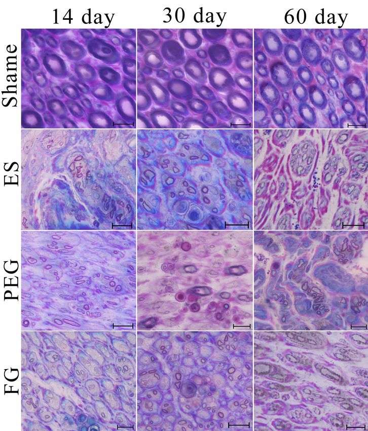
RESULTS AND DISCUSSION detected both with control and in three periods of the
experiment. On days 14, 30, and 60 after repair, thin
regenerating myelinated nerve fibers were found in the distal
Nerve suture area. The sutures were surrounded by stump of the sciatic nerve (Fig. 1). In the ES-group, the
connective tissue and this has increased the focal thickness number of nerve fibers increased between 14 and 60 days
of the epineurium (Table I). Between days 14 and 60, nerve (Table I). The diameter of the myelinated nerve fibers did
thickness, which was calculated on longitudinal histological not differ. At 60th day, the appearance of thick fibers was
sections, increased statistically (neuroma formation and detected, but this did not affect the overall assessment (ave-
epineurial connective tissue enlargement). PEG encapsulation rage diameter of most fibers was 4-6 µm, whereas in the
(focal clusters of PEG from 90 to 1600 µm) was detected, control 8-10 µm).
and focal clusters of fibrin were smaller and multifocal (focal
clusters of FG from 100 to 200 µm). In the PEG and FG group In the group with PEG (PEG-group), the density of
epineurial thickness increased by 60th day, but remained nerve fibers increased significantly compared with the ES-
within the statistical error compared to the epineural suture group at 30 and 60 days. The number of nerve fibers in distal
group (ES-group) (Table I). There were no inflammatory stump significantly increased compared to day 14. The
leukocyte infiltration in areas with PEG and FG. diameter of myelinated nerve fibers also increased, especially
by day 30 (the appearance of fibers up to 16 µm in diameter,
Fascicles changes. At the site of the suture, the integrity of although 4-6 µm dominated), and the number of thin fibers
the fascicles was changed, and regenerative neuroma was further increased.
formed. In all studied samples, the distal stump of the nerve
retained the fascicular structure. PEG and FG did not affect In the FG-group, the density of myelinated nerve
the change of nerve fascicles. The thickness of the distal fibers in distal stump significantly increased by 30 and 60
stump of the nerve also did not change. days, and the fiber diameter was significantly larger than in
the epineural suture group by day 14 and was smaller by
Regeneration of myelinated nerve fibers in distal day 60 than in the PEG-group (most with a diameter of 4-6
stump. In the group of sham operated animals, no difference µm at all periods; the fiber density did not differ relative to
in density and diameter of myelinated nerve fibers was the PEG-group).
Table I. Morphometric data for rats sciatic nerve on 14, 30 and 60 day after neurorrhaphy.
P arameter Day Control Shame-operated Epineural suture PEG FG
Thickness of the 14 - 1534.1±222.6 1560.1±148.9 1598.9±179.3
nerve in sutured 30 - 1943.7±63.1 1619.3±102.2 1542.2±133.3
site (µm) 60 2260.8±165.2@ 2261.5±101.9 1997.0±80.8
Thickness of the 14 1122.8±53.3 986.9±68.4 1006.8±45.4 1001.4±52.1
distal nerve 30 1139.7±96.8 1142.8±32.8 992.7±27.0 1017.8±87.1 1208.1±172.2
(µ m) 60 1150.2±63.0 1024.1±85.2 1101.0±75.4 985.4±57.6
14 2.6±0.2 2.6±0.2 2.8±0.2 2.6±0.2
Number of
30 2.8±0.3 3.0±0.3 2.8±0.2 2.4±0.2 3.0±0.3
fascicle
60 2.8±0.2 2.8±0.4 3.0±0.4 2.8±0.2
14 202.8±12.06 25,29±1,77* 30,4±1,45* 31,7±1,47*
Number
30 200.0±13.14 197.4±10.71 38.5±2.40* 48.5±2.78*^ 56.8±4.39*^
(fiber/test-zone
60 199.5±10.76 43,15±2,04* 58,5±2,66*^@** 64,2±2,20*@**
14 5643.4±209.4 720.6±48.5* 868.1±39.7* 905.4±40.2*
Density
30 5703.6±256.0 5734.4±211.4 1076.8±61.7* 1384.7±77.6*^@ 1618.8±121.7*^@
(fiber/mm2)
60 5445.7±177.3 1229.8±55.8*@ 1675.9±72.8*^@ 1829.8±60.1*^@
Diameter of 14 15.16±0.37 4,75±0,06 5,20±0,15^ 5,54±0,18^
myelinated 30 15.28±0.36 15.81±0.35 4.82±0.05* 8.58±0.18*^ 5.23±0.06*
nerve fiber (µm) 60 16.24±0.34 6,20±0,12* 6,50±0,12* 5,74±0,27*,***
Myelin 14 3.71±0.13 0.74±0.02* 0.81±0.02* 0.82±0.02*
thickness 30 3.74±0.12 3.72±0.18 0.98±0.10*@*** 1.65±0.10*@ 1.21±0.02*@***
(µ m) 60 3.78±0.15 1.27±0.04*@** 1.34±0.04*@** 1.32±0.05*@
Data expressed as mean ± SEM * P < 0.05 in comparison with the control and shame-operated group; ^ P < 0.05 in comparison with the ES; @ in
comparison with 14day; ** in comparison with 1 m; *** P < 0.05 in comparison with the PEG.
679GONCHARUK, O. O.; SAVOSKO, S. I.; PETRIV, T. I.; MEDVEDIEV, V. V. & TSYMBALIUK, V. I. Morphometric study of rat sciatic nerve recovery after three nerve repair techniques:
Epineural suture, polyethylene glycol hydrogel and fibrin sealant. Int. J. Morphol., 39(3):677-682, 2021.
Fig. 1. Cross section of in distal sciatic nerve stained by M.A. Hayat method: SHAME – shame-operated; ES
– epineurial suture; PEG – polyethylene glycol; FG – fibrin glue. Scale bar: 20µm.
Morphometric analysis showed a significantly 60 days in each group, the difference was 1.78, 1.65 and
higher number of myelinated nerve fibers in the distal 1.65 times (PGONCHARUK, O. O.; SAVOSKO, S. I.; PETRIV, T. I.; MEDVEDIEV, V. V. & TSYMBALIUK, V. I. Morphometric study of rat sciatic nerve recovery after three nerve repair techniques:
Epineural suture, polyethylene glycol hydrogel and fibrin sealant. Int. J. Morphol., 39(3):677-682, 2021.
significant difference was found in the regeneration of electrophysiological values after suture and PEG
nerve fibers, although PEG contributed to the regeneration (DuraSeal), instead the latter gave better results compared
of larger diameter myelinated fibers, indicating the to fibrin glue. But due to sutures and PEG, the thickness
acceleration of regeneration, similarly, the regeneration of the nerve has increased, which is absent or at a lesser
density increases after the application of FG. extent develops after the application of fibrin glue. PEG
had a higher adhesive capacity compared to FG, so in the
There is interest in the development of surgical latter occurred a diastase (gap). After PEG, myelinated
adhesives for gluing nerves, which would be non-toxic, fibers of a larger diameter were regenerating, as described
biocompatible, had adhesion to epineurium and at the same in later articles (Lin et al., 2019) and noted in our study.
time did not cause fibrotic changes and nerve fixation with Moreover, there is evidence that acceleration of peripheral
paraneural tissues. The main purpose of their use is to re- nerve regeneration after PEG administration prevents
duce the trauma of the connecting nerve stumps. gastrocnemius muscle atrophy (Lin et al., 2019).
Substances in the form of glues and gels partially realize
this task. This study compared nerve regeneration after In conclusion, adhesive sealants based on PEG and
neuroraphy with the use of 4-6 epineural sutures, and nerve fibrin can be an alternative to sutures in restoring peripheral
repair techniques including 2 sutures withPEG and FG. nerves, it reduces coaptation time and trauma, creates an
The last two were still fixated with 2 sutures, because in environment of glue around the injured nerve.
our opinion the adhesive characteristics of PEG and FG
are still insufficient for a strong connection of the nerve
stumps. Similarly, other authors believe, in particular, the
effect of combining PEG with spastin (Lin et al., 2019) or GONCHARUK, O. O.; SAVOSKO, S. I.; PETRIV, T. I.;
PEG with suture (Ghergherehchi et al., 2019) was better MEDVEDIEV, V. V. & TSYMBALIUK, V. I. Estudio
morfométrico de la recuperación del nervio ciático de rata
than PEG alone.
después de tres técnicas de reparación nerviosa: sutura
epineural, hidrogel de polietilenglicol y sellante de fibrina.
According to the results of the work, the coaptation
Int. J. Morphol., 39(3):677-682, 2021.
of the transected sciatic nerve in 3 ways was equally
sufficient for the formation of regenerative neuroma and RESUMEN: La efectividad de la técnica
regeneration of nerve fibers in the distal stump of the nerve. microquirúrgica tiene un impacto directo en la recuperación
At day 60, focal PEG and FG remained, FG underwent del nervio periférico lesionado. El objetivo de nuestro estu-
greater elimination than PEG, and caused less connective dio fue investigar el resultado de la regeneración del nervio
tissue growth at the suture site. Importantly, the fascicular ciático en ratas después de una neurotomía completa y des-
structure of the nerve and endoneural tubes in the distal pués de técnicas de reparación nerviosa que incluyeron: 1)
stump of the nerve remained preserved, and no difference sutura epineural; 2) hidrogel de polietilenglicol (PEG)
in the morphology of the distal nerve between the (DuraSeal); 3) sellante de fibrina (Tisseel). La sección trans-
comparison groups was found. Preservation of endoneural versal del nervio ciático distal se estudió a los 14, 30 y 60
tubes is important for nerve regeneration because días después de la reparación del nervio. Se realizó la
regenerating nerve fibers have adhesion to collagen and morfometría de fibras nerviosas mielinizadas en el muñón
endoneural fibroblasts, as well as to other extracellular distal del nervio ciático. Se observó un aumento significati-
vo en el número de fibras nerviosas mielinizadas, especial-
matrix proteins. The absence of damaging effect on the
mente entre los 14 y 30 días. La densidad de las fibras ner-
nerve from PEG and FG, sufficient nerve fixation, and the
viosas mielinizadas en el muñón distal en el día 60 fue
acceleration of nerve fibers regeneration at day 60 after
significativamente mayor después de usar una técnica de re-
microsurgical nerve repair is an advantage over multiple paración nerviosa que incluye PEG y fibrina en compara-
nerve sutures. ción con la sutura epineural (29,2 % y 32,1 % versus 21,5 %,
PGONCHARUK, O. O.; SAVOSKO, S. I.; PETRIV, T. I.; MEDVEDIEV, V. V. & TSYMBALIUK, V. I. Morphometric study of rat sciatic nerve recovery after three nerve repair techniques:
Epineural suture, polyethylene glycol hydrogel and fibrin sealant. Int. J. Morphol., 39(3):677-682, 2021.
REFERENCES
Gaiovych, I.; Savosko, S.; Labunets, I.; Utko, N.; Makarenko, A. & Corresponding author:
Chaikovsky, Y. Sciatic nerve regeneration after autografting and Savosko Serhii I.
application of the bone marrow aspirate concentration. Georgian Med.
Associate professor, PhD
News, (295):145-52, 2019.
Ghergherehchi, C. L.; Mikesh, M.; Sengelaub, D. R.; Jackson, D. M.; Smith,
department of histology and embryology
T.; Nguyen, J.; Shores, J. T. & Bittner, G. D. Polyethylene glycol (PEG) Bogomolets National Medical University
and other bioactive solutions with neurorrhaphy for rapid and dramatic Kyiv
repair of peripheral nerve lesions by PEG-fusion. J. Neurosci. Methods, UKRAINE
314:1-12, 2019.
Hayat, M. A. Principles and Techniques of Electron Microscopy: Biological
Applications. Cambridge, Cambridge University Press, 2000. pp.543. E-mail: s.i.savosko@gmail.com
Isaacs, J. E.; McDaniel, C. O.; Owen, J. R. & Wayne, J. S. Comparative
analysis of biomechanical performance of available "nerveglues". J.
Hand Surg. Am., 33(6):893-9, 2008.
Khalifeh, J. M.; Dibble, C. F.; Dy, C. J. & Ray, W. Z. Cost-effectiveness Received: 25-01-2021
analysis of combined dual motor nerve transfers versus alternative Accepted: 21-02-2021
surgical and nonsurgical management strategies to restore shoulder
function following upper brachial plexus injury. Neurosurgery,
84(2):362-77, 2019.
Kim, P.; Kim, D. H.; Kim, B.; Choi, S. K.; Lee, S. H., Khademhosseini, A.;
Langer, R. & Suh, K. Y. Fabrication of nanostructures of polyethylene
glycol for applications to protein adsorption and cell adhesion.
Nanotechnology, 16(10):2420-6, 2005.
Kornfeld, T.; Vogt, P. M. & Radtke, C. Nerve grafting for peripheral nerve
injuries with extended defect sizes. Wien Med. Wochenschr., 169(9-
10):240-51, 2019.
Lee, S. H.; Park, C. W.; Lee, S. G. & Kim, W. K. Postoperative cervical
cord compression induced by hydrogel dural sealant (DuraSeal®).
Korean J. Spine, 10(1):44-6, 2013.
Lin, K. L.; Yang, D. Y.; Chu, I. M.; Cheng, F. C.; Chen, C. J.; Ho, S. P. &
Pan, H. C. DuraSeal as a ligature in the anastomosis of rat sciatic nerve
gap injury. J. Surg. Res., 161(1):101-10, 2010.
Lin, Y. F.; Xie, Z.; Zhou, J.; Chen, H. H.; Shao, W. W. & Lin, H. D. Effect
of exogenous spastin combined with polyethylene glycol on sciatic
nerve injury. Neural Regen. Res., 14(7):1271-9, 2019.
Molotkovets, V. Y.; Medvediev, V. V.; Korsak, A. V.; Chaikovsky, Y. B.;
Marynsky, G. S. & Tsymbaliuk, V. I. Restoration of the integrity of a
transected peripheral nerve with the use of an electric welding
technology. Neurophysiology, 52:31-42, 2020.
Puttaswarmy, S. V.; Bandla, A.; Fishlock, S. J.; Lee, S.; Lee, C. &
McLaughlin, J. Hydrogel as a Nerve Guide and Biocompatible Glue
for Neural Applications. Cork, 2018 IEEE 18th International Conference
on Nanotechnology (IEEE-NANO), 2018. pp.1-4.
Sexton, K. W.; Pollins, A. C.; Cardwell, N. L.; Del Corral, G. A.; Bittner,
G. D.; Shack, R. B.; Nanney, L. B. & Thayer, W. P. Hydrophilic polymers
enhance early functional outcomes after nerve autografting. J. Surg.
Res., 177(2):392-400, 2012.
Tse, R. & Ko, J. H. Nerve glue for upper extremity reconstruction. Hand
Clin., 28(4):529-40, 2012.
682You can also read