Gene Therapy in Cancer Treatment: Why Go Nano? - MDPI
←
→
Page content transcription
If your browser does not render page correctly, please read the page content below
pharmaceutics
Review
Gene Therapy in Cancer Treatment: Why Go Nano?
Catarina Roma-Rodrigues 1 , Lorenzo Rivas-García 1,2 , Pedro V. Baptista 1, * and
Alexandra R. Fernandes 1, *
1 UCIBIO, Departamento de Ciências da Vida, Faculdade de Ciências e Tecnologia, Campus de Caparica,
2829-516 Caparica, Portugal; catromar@fct.unl.pt (C.R.-R.); lorenrivas@ugr.es (L.R.-G.)
2 Biomedical Research Centre, Institute of Nutrition and Food Technology, Department of Physiology,
Faculty of Pharmacy, University of Granada, Avda. del Conocimiento s/n. 18071 Armilla, Granada, Spain
* Correspondence: pmvb@fct.unl.pt (P.V.B.); ma.fernandes@fct.unl.pt (A.R.F.);
Tel.: +351-212-948-530 (P.V.B. & A.R.F.)
Received: 29 January 2020; Accepted: 3 March 2020; Published: 5 March 2020
Abstract: The proposal of gene therapy to tackle cancer development has been instrumental for the
development of novel approaches and strategies to fight this disease, but the efficacy of the proposed
strategies has still fallen short of delivering the full potential of gene therapy in the clinic. Despite the
plethora of gene modulation approaches, e.g., gene silencing, antisense therapy, RNA interference,
gene and genome editing, finding a way to efficiently deliver these effectors to the desired cell and
tissue has been a challenge. Nanomedicine has put forward several innovative platforms to overcome
this obstacle. Most of these platforms rely on the application of nanoscale structures, with particular
focus on nanoparticles. Herein, we review the current trends on the use of nanoparticles designed for
cancer gene therapy, including inorganic, organic, or biological (e.g., exosomes) variants, in clinical
development and their progress towards clinical applications.
Keywords: gene therapy; gene delivery; tumor microenvironment; nanoparticles; nanomedicine
1. Introduction
According to the World Health Organization, cancer is the second leading cause of death
worldwide, accounting for 9.6 million deaths in 2018 [1]. The global efforts in cancer prevention, early
diagnosis, screening and treatment, have been challenged by the complexity and variability of tumors
(reviewed in [2]). The genomic instability of tumor cells and a pro-inflammatory environment are key
factors for tumor growth [3]. Regardless of the monoclonal origin of the neoplasia, the interplay between
tumor cells and the surrounding environment results in a complex tumor microenvironment (TME) that
supports tumor intra-heterogeneity, with spatially different and phenotypically distinct subclones [2].
Nonetheless, major common features of tumor cells include continuous proliferative signaling, evasion
of growth suppressors, resisting cell death, replicative immortality, deregulating cellular energetics,
promoting angiogenesis, activating invasion and metastasis, and avoiding immune destruction [3].
These features sustain the foundation of a TME composed by a characteristic extracellular matrix
(ECM), cancer-associated fibroblasts (CAFs), mesenchymal stromal cells, endothelial cells and pericytes,
and immune system cells, such as macrophages, T and B lymphocytes and natural killer cells (reviewed
in [4]). TME composition dictates tumor progression, chemotherapeutic efficacy and prognosis [5–7].
The mounting knowledge on the characteristics of tumor cells and surrounding TME have
sparked the use of gene therapy to tackle cancer molecular mechanisms. Gene therapy consists of the
introduction of exogenous nucleic acids, such as genes, gene segments, oligonucleotides, miRNAs or
siRNAs into cells envisaging a target gene edition, expression modulation of a target gene, mRNA
or synthesis of an exogenous protein [8–19]. Gene transfer into tumor cells has been demonstrated
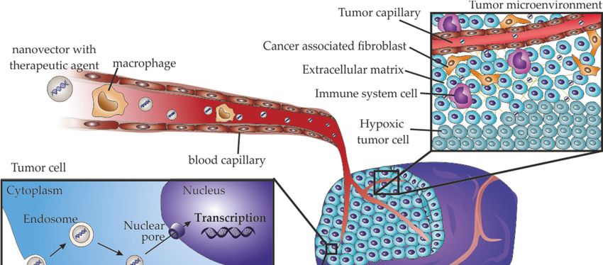
via administration of therapeutic nucleic acids (TNAs) ex vivo and/or in vivo (Figure 1) [20]. In the
Pharmaceutics 2020, 12, 233; doi:10.3390/pharmaceutics12030233 www.mdpi.com/journal/pharmaceutics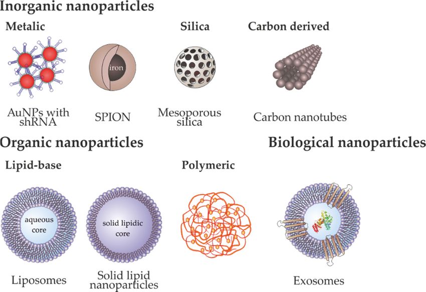
Pharmaceutics 2020, 12, 233 2 of 34
Pharmaceutics 2019, 11, x 2 of 36
ex vivo approach, patient-derived tumor cells are collected, propagated usually as 2D monolayers,
manipulated genetically
administration and then
of therapeutic introduced
nucleic back into
acids (TNAs) the and/or
ex vivo host [20]. In the
in vivo in vivo
(Figure approach,
1) [20]. In the exTNAs
may vivo
be introduced in loco into the tumor
approach, patient-derived tumorcellscells,
aresystemically via intravenous
collected, propagated usually administration,
as 2D monolayers, or in a
manipulated
pre-systemic genetically
manner through and oral,
then introduced back into the
ocular, transdermal orhost [20].
nasal In the inroutes,
delivery vivo approach,
depending TNAsof the
may
specific be introduced
localization in loco into
of tumors andthe tumor
disease cells, systemically
progression [20–22].viaInintravenous administration,
the case of systemic or in a
and pre-systemic
pre-systemic manner through oral, ocular, transdermal or nasal delivery routes,
deliveries, the administration of naked TNAs is hindered by biological barriers, nuclease susceptibility, depending of the
specific localization of tumors and disease progression [20–22]. In the case of systemic and pre-
phagocyte uptake, renal clearance and/or immune response stimulation [23]. Hence, the use of stable
systemic deliveries, the administration of naked TNAs is hindered by biological barriers, nuclease
carriers/vectors that protect the nucleic acid cargo from circulatory nucleases, avoid the immune system,
susceptibility, phagocyte uptake, renal clearance and/or immune response stimulation [23]. Hence,
and ensure the efficient targeting of the therapeutic vector into the tumor cells, without dissipation
the use of stable carriers/vectors that protect the nucleic acid cargo from circulatory nucleases, avoid
in thethebody through
immune system,lymphatic
and ensureand the blood
efficientsystems
targetingandof theavoiding non-target
therapeutic vector into cells
the is required
tumor cells, [21].
Despite the apparent
without limitations
dissipation in the body the in vivo
ofthrough approach,
lymphatic it is less
and blood invasive
systems and and morenon-target
avoiding suitable for cancer
cells
treatment than ex
is required vivo
[21]. approaches,
Despite sincelimitations
the apparent the latter ofrequire
the in avivo
proliferative
approach, itadvantage of transfected
is less invasive and more cells,
which suitable for cancer
is antagonist to thetreatment than ex vivo
major objectives approaches,
of cancer since the latter
gene therapeutics that require
mainly aaimsproliferative
to inhibit the
advantage of transfected cells, which is antagonist to the major objectives of cancer
tumor progression by tackling the tumor cell division ability [21,24–26]. Nevertheless, it is important gene therapeutics
that mainly
to highlight the aims to inhibit
relevance thevivo
of ex tumor progression
therapy by tackling
in indirect the tumor
immune cell division
gene-based ability (detailed
therapies [21,24– in
26]. Nevertheless, it is important to highlight the relevance of ex vivo therapy in indirect immune
Section 2.7). In these ex-vivo approaches, immune cells are collected from the patient’s blood and
gene-based therapies (detailed in Section 2.7). In these ex-vivo approaches, immune cells are collected
genetically engineered to tackle the tumor cells (reviewed in [27,28]).
from the patient’s blood and genetically engineered to tackle the tumor cells (reviewed in [27,28]).
Figure 1. Delivery strategies used for gene therapy directly targeting tumor cells or tumor
Figure 1. Delivery strategies used for gene therapy directly targeting tumor cells or tumor
microenvironment components, their major advantages (preceded by a green checkmark) and
microenvironment components, their major advantages (preceded by a green checkmark) and
disadvantages (preceded by a red cross).
disadvantages (preceded by a red cross).
The success of cancer gene therapy relies on a safe, effective and controllable vector [25]. Viral
The success of cancer gene therapy relies on a safe, effective and controllable vector [25]. Viral
vectors were the first platform proposed for gene therapy [29]. Indeed, the nature and properties of
vectors weremade
viruses the first
themplatform
temptingproposed forRNA
vectors for geneand
therapy
DNA [29]. Indeed,
delivery the nature
to human and properties
cells, with multiple of
viruses made them tempting vectors for RNA and DNA delivery to human cells, with
clinical trials that ended-up in clinical approved of some gene therapy drugs (reviewed in [29]). multiple clinical
trialsHowever,
that ended-up in clinical approved
the immunogenicity, limited of some genecancer
genetic-load, therapy
riskdrugs
due to(reviewed
therapeuticin [29]). However,
payload insertion the
immunogenicity,
near genes that limited
controlgenetic-load, cancer
cell growth, and risk duemass-production
constrained to therapeutic payload insertion
of viral vectors near genes
prompted the that
development
control cell growth, and engineering
and of non-viral
constrained vectors, supported
mass-production of viral by nanomedicine
vectors prompted [25,30].
the development and
engineering of non-viral vectors, supported by nanomedicine [25,30].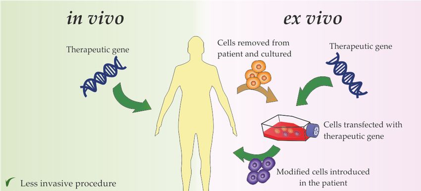
Pharmaceutics 2020, 12, 233 3 of 34
Pharmaceutics 2019, 11, x 3 of 36
Nanotechnology refers to the area of science focused on the study of the synthesis, characterization
and application of materials and functional systems of particles whose size is between 1-100 nm [31,32].
Nanotechnology refers to the area of science focused on the study of the synthesis,
Nowadays, the interest
characterization andinapplication
these materials is not only
of materials due to their
and functional smallof
systems size, but also
particles to their
whose size unique
is
physical
between 1-100 nm [31,32]. Nowadays, the interest in these materials is not only due to their small to
(electric, optical, magnetic) and chemical properties at these dimensions (in comparison
the same material
size, but also toattheir
the unique
macroscopic
physicalscale), conveying
(electric, a more scalable
optical, magnetic) interaction
and chemical with
properties cells and
at these
dimensionsThe
biomolecules. (in comparison
applicationtoofthe same material at to
nanotechnology thethe
macroscopic scale),(nanomedicine)
medical field conveying a moreenhanced
scalable the
interactionofwith
development newcells
and andmorebiomolecules. The application
effective diagnostics of nanotechnology
and therapies, particularly to the medical field
in complex diseases,
(nanomedicine) enhanced the development of new and more effective
such as diabetes, Parkinson and cancer [33,34]. The application of nanoparticles as carriers in diagnostics and therapies,
gene particularly
therapy is in onecomplex diseases, such as diabetes, Parkinson and cancer [33,34]. The application of
of the most promising technologies in biomedical research due to its ease and
nanoparticles as carriers in gene therapy is one of the most promising technologies in biomedical
straightforward synthesis and functionalization with different moieties, low immunogenicity and
research due to its ease and straightforward synthesis and functionalization with different moieties,
toxicity [35]. One of the interests is focused on the development of biocompatible and more effective
low immunogenicity and toxicity [35]. One of the interests is focused on the development of
transfection systems
biocompatible and [36,37] to vectorize
more effective TNAs to
transfection cells and
systems tissues,
[36,37] such as
to vectorize DNA
TNAs (e.g.,and
to cells plasmid
tissues,DNA,
antisense oligonucleotides (ASO)) or RNA (e.g., microRNA (miRNA),
such as DNA (e.g., plasmid DNA, antisense oligonucleotides (ASO)) or RNA (e.g., microRNA short hairpin RNA (shRNA),
small(miRNA),
interfering RNA
short (siRNA))
hairpin RNAinto cells [36,37].
(shRNA), Some of RNA
small interfering the limitations
(siRNA)) intoin the
cellsefficiency of transfection
[36,37]. Some of the
limitations
of naked plasmid in the
DNA efficiency
(pDNA) of or
transfection
siRNAs can of naked plasmid by
be improved DNAthe(pDNA)
applicationor siRNAs can be
of functionalized
improved[38].
nanoparticles by the application
However, of technology
this functionalized nanoparticles
still faces several[38]. However, this
shortcomings technology
that need to bestilladdressed,
faces
several shortcomings that need to be addressed, including lower transfection
including lower transfection efficiency when compared to viral vectors, or blood clearance before efficiency when
compared to viral vectors, or blood clearance before reaching the target site in the case of systemic
reaching the target site in the case of systemic administration [39].
administration [39].
Effective TNA agents require a vector that can travel through the circulatory system, accumulate
Effective TNA agents require a vector that can travel through the circulatory system, accumulate
in theintumor, enter target cells via endosome pathway and be able to escape the endosome to efficiently
the tumor, enter target cells via endosome pathway and be able to escape the endosome to
accomplish cargo
efficiently deliverycargo
accomplish (Figure 2) [21,23,25].
delivery (Figure 2) [21,23,25].
Figure 2. Barriers that nanoparticles must overcome for effective cancer gene delivery. In a systemic
Figure 2. Barriers that nanoparticles must overcome for effective cancer gene delivery. In a systemic
administration, nanoparticles should travel through the blood circulatory system, avoiding the
administration, nanoparticles should travel through the blood circulatory system, avoiding the immune
immune system. The accumulation at the tumor occurs through passive targeting by the enhanced
system. The accumulation at the tumor occurs through passive targeting by the enhanced permeability
permeability and retention effect. Nanoparticles also have to penetrate into the most inaccessible areas
and retention effect.
of the tumor Nanoparticles
to reach the hypoxicalso
tumorhave to penetrate
region into the most
with low oxygenation andinaccessible areas ofmatrix.
dense extracellular the tumor
to reach
After reaching tumor cells, nanoparticles should be internalized, which is mainly accomplishedreaching
the hypoxic tumor region with low oxygenation and dense extracellular matrix. After via
tumorendocytosis,
cells, nanoparticles shouldfrom
and then escape be internalized,
the endosomewhich is mainly
to efficiently accomplished
deliver viathe
the cargo into endocytosis,
cytoplasm, and
then escape from theRNA,
when targeting endosome toto
or travel efficiently deliver
the nucleus, whenthe cargo into
targeting DNA. the cytoplasm, when targeting RNA,
or travel to the nucleus, when targeting DNA.
Pharmaceutics 2020, 12, 233                                                                      4 of 34

      Indeed, systemic administration of the therapy implies that the vector can travel through the
blood circulation, with consequent interaction with blood cells, including phagocytic cells, proteins
and lipids [40]. The surface, size and shape of the nanoparticles are preponderant for their endurance
across the circulatory system (reviewed in [40]). Neutral or slightly negative surfaces assure low
adsorption to blood proteins, such as opsonins, and avoid phagocytosis (reviewed in [40,41]). Hence,
neutralization of charged nanoparticles may be achieved by coating with hydrophilic polymers such
as polyethylene glycol (PEG), polyglycerol (PG), or polysaccharides, such as heparan or chitosan, with
zwitterionic ligands, such as carboxybetaines or sulfobetaines, with mercaptoalkyl acid ligands, such
as 11-mercaptoundecanoic acid (MUA), or even with proteins and lipids (reviewed in [40,42]).
      Concerning tumor accumulation, nanomedicine design often takes advantage of the natural
accumulation at the tumor location – passive targeting [40,43,44]. In fact, the characteristic immature
phenotype of the tumor vasculature, characterized by leaky vessels with chaotic branching, together
with poor lymphatic system, renders an enhanced permeability and retention (EPR) of nanoparticles
at the TME (reviewed in [43–45]). As the EPR effect is dependent of the tumor in terms of the
anatomical location, tumor size, stage and type, the properties of the nanoparticles (size, shape and
surface charge) should be optimized (reviewed in [40]). As an example, pegylation of drug-loaded
liposomes not only improved their blood circulation, but also increased the accumulation of the drug
in the tumor [43]. Furthermore, active targeting of the nanomedicines improves greatly their efficacy
(reviewed in [40,46–48]). With that purpose, several biological ligands could be bind to nanomaterials,
including antibodies, such as cetuximab, an FDA approved antibody against anti-epidermal growth
factor receptor (EGFR) used in clinical practice for cancer treatment; glycoproteins, such as the
iron-binding transferrin; polysaccharides, such as hyaluronic acid for CD44 targeting; peptides, such
as arginylglycylaspartic acid (RGD) for integrins targeting; aptamers, such as AS-1411 G-rich DNA
aptamer for nucleolin targeting; or other small molecules, such as folate ([49–60], reviewed in [48]).
      After reaching the tumor, another bottleneck that nanoparticles must overcome is the penetration
into the TME to reach regions with low or without vascularization, low interstitial fluid pressure and
dense ECM [61–63]. To achieve higher diffusion, the nanoparticles size, functionalization and tumor
modulation ability have been extensively studied and modulated (reviewed in [63,64]). This is often
accomplished by altering the nanoparticles’ properties, such as size and surface hydrophobicity, after
they reach the tumor using stimulus-triggering strategies, including light, ultrasound or magnetic
fields, or taking advantages of the TME properties, such as hypoxia, acidity, and the overexpression of
metalloproteinases (reviewed in [40,63]).
      Additionally, for gene therapy it is necessary that the vector and payload pass across the
complex hydrophobic layer of the tumor cell membrane [41]. This mainly occurs via endocytosis
mediated by ligand-receptor specific, using active targeting, or non-specific, such as electrostatic or
hydrophobic, interactions with the cell membrane (reviewed in [40,65,66]). Once more, nanoparticle
size, shape and surface are preponderant for an efficient cellular internalization [67–73]. The
most suitable features are dependent on type of particle, for example, Xue et al. observed
an improved internalization when polypeptide-based nanoparticles composed by mixtures of
FITC-poly(γ-benzyl-L-glutamate)-block-PEG and polystyrene (PS) had smaller size, rod-like shape
and helical/striped surface morphology [68]. On the other side, a study from Bandyopadhyay et al.
revealed that the pillow-shape and irregular structure of gold nanoparticles resulted in a higher
cellular uptake, when compared with small spherical nanoparticles [70]. Once nanoparticles enter
cells via endocytosis, they must escape from the endosome to avoid degradation in lysosomes or
exocytosis [65]. The endosomal escape could occur through membrane destabilization, proton sponge
or photochemical internalization [40,65,74]. Liposomes and lipid-based nanoparticles mainly escape
via membrane destabilization due to direct contact between the nanoparticles lipid layer with the
endosomal membrane, resulting in the release of the nanoparticles content into cytoplasm [65]. The
lipidic nanoparticles transport of membrane-disruptive peptides induce the formation of a pore
in the endosome, enhancing the endosomal escape [74]. The proton sponge escape occurs when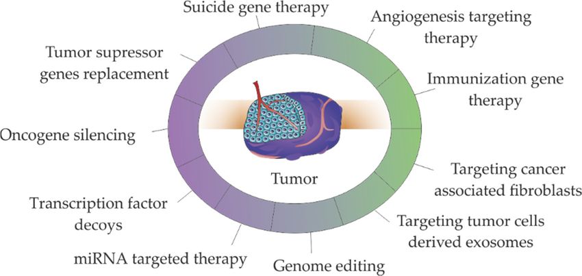
2019, 12,
Pharmaceutics 2020, 11, 233
x 36
5 of 34
(PAMAM) based dendrimers, are protonated during acidification of the endosome, resulting in an
nanoparticles containing
increased osmotic pressure amino
due groups, suchofaschloride
to an influx polyethylenimine (PEI) or polyamidoamine
ions and consequent (PAMAM)
swelling of the endosome
based dendrimers, are protonated during acidification of the endosome,
with nanoparticles release into the cytoplasm [65,74]. In the photochemical internalization, resulting in an increased
osmotic pressure
nanoparticles are due to an influxwith
functionalized of chloride ions and consequent
photosensitizers that after light swelling of thegenerate
activation endosome with
reactive
nanoparticles release into the cytoplasm [65,74]. In the photochemical
oxygen species (ROS) that rupture the endosomal membrane [74]. The most suitable properties of internalization, nanoparticles
are functionalized
nanoparticles that with
enhancephotosensitizers
endocytosis and thatendosomal
after light activation
escape was generate reactive
extensively oxygeninspecies
reviewed recent
(ROS)
papersthat rupture the endosomal membrane [74]. The most suitable properties of nanoparticles that
[65,74,75].
enhance endocytosis
Considering all and endosomal escape
the characteristics for anwasideal
extensively reviewedeffective
vector towards in recentgene
papers [65,74,75].
therapy and the
Considering
advantages posed all
by the characteristics
nanoparticles fordelivery
as gene an idealsystems,
vector towards
the presenteffective
review gene
willtherapy and the
first address the
advantages posed by nanoparticles as gene delivery systems, the present review
different non-viral gene therapy strategies used in cancer, followed by the application of the different will first address the
different non-viral
nanoparticles gene therapy
as vectors for cancerstrategies
therapy,used in cancer,
together with followed
their wayby tothe
theapplication
clinics. of the different
nanoparticles as vectors for cancer therapy, together with their way to the clinics.
2. Gene Therapy Focused on Cancer
2. Gene Therapy Focused on Cancer
Delivery of TNAs, such as genes, oligonucleotides, miRNAs or siRNAs to cancer cells has
Delivery
allowed to tackleof TNAs,
cancersuchvia as
thegenes, oligonucleotides,
silencing oncogenes ormiRNAs restoringorthe siRNAs to cancer
expression cells has
of tumor allowed
suppressor
to tackle
genes cancerMost
[8–19]. via theofsilencing oncogenes or
these approaches restoring
(e.g., the expression
antisense therapy, RNA of tumor suppressor(RNAi),
interference genes [8–19].
gene
Most of these approaches (e.g., antisense therapy, RNA interference
editing) aim at gene alteration/modulation [16–19,76–81] - see Figure 3. The immunization (RNAi), gene editing) aim at gene
gene
alteration/modulation
therapies, particularly [16–19,76–81]
chimeric antigen - seereceptor
Figure 3.(CAR)The immunization
in T cells (CAR-T gene cells)
therapies,
based particularly
therapies,
chimeric antigen receptor (CAR) in T cells (CAR-T cells) based therapies, stand-out
stand-out since they represent the higher number of therapeutic strategies in clinical practice. since they representIt
the higher number of therapeutic strategies in clinical practice. It should
should be noted that some of the presented strategies such as genome editing or miRNA/siRNA be noted that some of the
presented strategies
targeted therapy are such as TME
used in genome editing
targeting orangiogenesis
via miRNA/siRNA andtargeted
immune therapy
therapiesare used or
[82,83] in CAFs
TME
targeting via angiogenesis
targeting [84–86]. and immune therapies [82,83] or CAFs targeting [84–86].
Figure 3. Major strategies used in non-viral gene therapies for cancer treatment. Therapies targeting
Figure 3. Major strategies used in non-viral gene therapies for cancer treatment. Therapies targeting
the tumor
the tumormicroenvironment
microenvironment(in(in green),
green), including
including angiogenesis
angiogenesis targeting
targeting therapy,
therapy, immunization
immunization gene
therapy, targeting cancer associated fibroblasts and targeting tumor cells derived exosomes, alsoalso
gene therapy, targeting cancer associated fibroblasts and targeting tumor cells derived exosomes, use
use described
the the described molecular
molecular strategies
strategies (in such
(in purple), purple), suchreplacement,
as genes as genes replacement, gene
gene silencing, silencing,
transcription
transcription
factor decoys,factor
miRNA decoys, miRNA
targeted targeted
therapy therapyediting.
and genome and genome editing.
2.1. Oncogene Silencing via RNAi
Gene silencing
silencingconsist in the
consist in delivery of nucleic
the delivery of acids intoacids
nucleic tumorinto
cells tumor
that endcells
up inthat
downregulation
end up in
of specific genes [24,37,87,88].
downregulation Gene[24,37,87,88].
of specific genes silencing therapy
Gene is usually accomplished
silencing therapy is usuallyby introducing
accomplishedsiRNA
by
or shRNA in tumor cells designed to target a specific complementary sequence
introducing siRNA or shRNA in tumor cells designed to target a specific complementary sequence to messenger RNA
(mRNA) of a selective
to messenger gene, inducing
RNA (mRNA) its degradation
of a selective or by blocking
gene, inducing protein synthesis
its degradation [89]. Oncogenes,
or by blocking protein
such as cMYC
synthesis KRAS, and such
[89].orOncogenes, genesasinvolved
cMYC or in KRAS,
drug-resistance
and genes such as multi-drug
involved resistance 1 (MDR1)
in drug-resistance such as
are temptingresistance
multi-drug targets for1tumor
(MDR1) therapy using RNAi
are tempting [14,24,90–94].
targets for tumor Major
therapy challenges faced[14,24,90–94].
using RNAi by RNAi are
related to target specificity,
Major challenges off-target
faced by RNAi RNAi activity,
are related to targetdissipation
specificity,inoff-target
circulation,
RNAicellular internalization
activity, dissipation
in circulation, cellular internalization and endosomal escape [95]. Strategies used to surpass these
limitations were extensively reviewed in [12,95].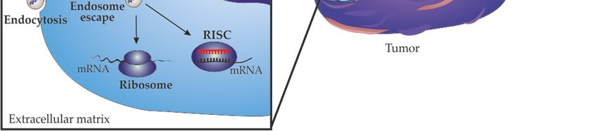
Pharmaceutics 2020, 12, 233                                                                        6 of 34

and endosomal escape [95]. Strategies used to surpass these limitations were extensively reviewed
in [12,95].

2.2. Tumor Suppressor Genes Replacement
      Gene replacement can be accomplished by gene transduction, maintenance of stability and
full expression of the gene, or by correcting gene mutations into its wild-type form (reviewed
in [96]). Tumor suppressor genes, such as TP53, P21 or PTEN are major targets for gene replacement
therapy [25,26,97–102]. Due to the central role of P53 protein in cell cycle regulation, DNA repair,
apoptosis, senescence and/or autophagy, TP53 gene is a major target for gene therapy [100]. The
first commercial gene therapy product was gendicine in 2003, a recombinant human P53 adenovirus
commercialized by SiBiono Gene Technologies, approved by the Chinese Food and Drug Administration
for head and neck squamous cell carcinoma [103]. As the bottlenecks of gene editing are transversal
for RNA delivery, DNA delivery should also overcome the barrier of the passage through the nucleus
membrane [75]. The entry of nucleic acids into the nucleus occur through channels of the nuclear pore
complex (NPC, Figure 2), that allow the passage of linear DNA with maximum 200-300 bp, posing
a challenge for the nuclear entry of therapeutic gene expression cassettes with few kilobase pairs
(kbp) [75]. Strategies to improve the DNA entry into the nucleus involve nuclear-targeted delivery
with nuclear localization signals or inclusion of nucleotide sequences in DNA [75]. As the mentioned
strategies require activation of signaling pathways that limit their application for cancer therapeutics,
another strategy for gene editing therapeutics take advantage of the nuclear envelope disruption during
mitosis, which require the presence of intact foreign DNA near the chromatin [75]. The bottlenecks
and strategies for gene editing based on nucleic acid delivery were recently reviewed in [75].

2.3. microRNA Targeted Therapy
     In cancer, some miRNAs are overexpressed promoting tumor development (oncomirs), and
others are downregulated bypassing the inhibitory control over oncogenes, or the control of cell
differentiation and apoptosis (tumor suppressor miRNAs) [104]. The miRNA targeted therapy consists
in the repositioning of the levels of miRNAs in cells. The silence mechanism of miRNA is similar to the
RNAi, however, miRNA are complementary or semi-complementary sequences to the 30 untranslated
region (30 -UTR) of a specific mRNA target or to several mRNAs involved in a particular cellular
process [12,19]. The levels of miRNAs altered under pathological conditions could be restored to normal
physiological conditions using miRNA-duplexes, to replace the levels of underexpressed miRNAs, or
siRNA complementary to the seed sequence of the miRNA of interest [20]. Several studies proposed
the reposition of miRNAs envisaging cancer therapy, including for example, by adding the tumor
suppressor miRNA Let-7c for prostate cancer treatment, by silencing the oncomirs miR-21 in breast
cancer, or by over-expressing miR-143 in colon cancer to overcome oxaliplatin resistance [13,104,105].
The systemic administration of free miRNAs for therapy has been challenged by single stranded or
double stranded miRNA degradation in the circulatory system or in the endosome, potential off-target
effects, miRNA-mediated toxicity and poor delivery [106–108]. The knowledge of the miRNAs
metabolic modulation in targeted and non-targeted cells is preponderant to avoid off-target effect
that occurs due to partial complementarity with non-targeted transcripts, or by leading to undesired
effects by regulation of metabolic processes in non-targeted cells [107]. To circumvent the degradation
limitations, miRNA may be modified: miRNA mimics are mainly modified by methylation of the
passenger strand and locked nucleic acids (LNA) chemistry is used for modification of anti-miRNA
(reviewed in [106]). Another strategy involves the delivery of miRNAs in nanoparticles able to perform
endosomal escape, reviewed in Section 3, “Nanoparticles for gene delivery: fostering gene therapy”.

2.4. Transcription Factor Decoys
     Transcription factor decoys (TFD) are double stranded oligodeoxynucleotides (ODN) designed to
inhibit specific regulatory pathways (reviewed in [109]). The TFD-ODNs are short double-stranded
Pharmaceutics 2020, 12, 233 7 of 34
DNA molecules with the sequence of a transcription factor of a particular gene, or the consensus DNA
recognition motif of the transcription factor, competing with specific binding sites of transcription
factors [109]. TFDs designed envisaging cancer therapy include TFDs targeting NF-KB for inhibition of
metastasis, signal transducer and activator of transcription 3, or STAT3, to induce apoptosis and cell
cycle arrest in ovarian, glioblastoma, lung and neck cancers (reviewed in [110]). Major challenges for
the application of TFD-ODNs in cancer therapy include the design of TFDs, and stability in circulatory
system and endosome [110]. The design of TFDs require the exact sequence of the transcription
factor binding site, which may be a problem due to the mismatch between the available information
in databases, and requiring the performance of rigorous but costly techniques such as chromatin
immunoprecipitation, and further confirmation of accurate targeting, usually using reporter genes, like
luciferase, and usage of scrambled decoys [110]. Peptide nucleic acid (PNA), LNA or phosphorotioate
(PS) chemical modifications of the TFD-ODNs could improve their half-life, increase resistance to
serum nucleases and decrease the interaction with DNA binding proteins [110]. Nevertheless, the most
promising techniques for in vivo TFD-ODN delivery involves their nanoparticle transport [110].
2.5. Genome Editing
Genome editing therapy consists in the modification of intracellular DNA in a sequence specific
manner, by insertion, deletion, integration or sequence substitution [111,112]. Three major nucleases
have been used for this purpose, zinc finger nucleases (ZFN), transcription activator-like effector
nucleases (TALEN), meganucleases, and CRISPR/Cas9 system (reviewed in [111]). The efficiency of the
genome editing therapy depend on the specificity of the DNA cleavage together with the prevention
of collateral damage to the rest of the genome. The CRISPR/Cas9 system was proven as a suitable
tool for stable and efficient genome editing as well as for high-throughput screening of mutations
involved in oncogenesis and tumor progression [112–115]. The mostly used CRISPR/Cas9 system is the
CRISPR system of Streptoccocus pyogenes (SpCas9), that recognize the short sequence 50 -NGG, where N
represents any nucleotide and G represents guanine, and Cas9 is an nuclease guided by a single guide
RNA (sgRNA) mediated by paring to the target sequence (reviewed in [116]). The CRISPR/Cas9 system
is delivered as plasmid or linear DNA encoding Cas9 and sgRNA [116,117]. When delivered as linear
DNA, it must enter the nucleus for transcription, while the plasmid DNA allow a stable and prolonged
gene expression [116]. The challenges in the delivery of the CRISPR/Cas9 system are similar to other
gene editing strategies detailed in Section 2.2. “Tumor suppressor genes replacement”. Moreover,
challenges faced by genome editing based on this system are due to prolonged exposure of the genome
to endonuclease activity that result in the cleavage of off-target sites [116,117]. The expression of
CRISPR/Cas9 system in non-target tissues should be minimized to avoid off-target mutagenesis [116].
The approaches currently used for in vivo delivery of CRISPR/cas9 system were recently reviewed
in [116,117].
2.6. Suicide Genes
The concept of suicide gene therapy was originally proposed for cancer treatment. Consist
in inserting in tumor cells a gene encoding a cytotoxic protein by applying two main strategies:
(i) direct gene therapy, by introducing in tumor cells a toxin gene that reduce the viability of the
cells, (ii) indirect gene therapy, by introducing a gene encoding an enzyme into tumor cells that
is able to convert a non-toxic prodrug into a cytotoxic drug [25,118,119]. The first proposal of
suicide gene therapy was made in 1983 by inserting in BALB/c murine cell lines the herpes virus
thymidine kinase gene, and then generate tumors with these cells in BALB/c mice [120,121]. Ganciclovir
(9-([2-hydroxymethyl)ethoxy]methyl)guanine) was then administered to the mice, and metabolization
of ganciclovir by herpes virus thymidine kinase at the tumor cells resulted in tumor regression [120,121].
The potential of this therapeutic strategy motivated its application in several clinical trials for treatment,
e.g., liver (NCT02202564) or colorectal (NCT00012155) cancer. The issues inherent to suicide genePharmaceutics 2020, 12, 233 8 of 34
therapy are related to gene editing that must result in tumor-specific high expression of the gene,
preferentially under control of tumor-specific promoters (reviewed in [122]).
2.7. Immunization Gene Therapy
The immunization gene therapy consists in the enhancement of the immune system efficacy
towards TME cells, with major focus on tumor cells. Three major approaches are applied, cytokine
gene therapy, tumor vaccine therapy and CAR-T cells therapy.
2.7.1. Tumor Vaccines
Tumor vaccination relies on presenting tumor-related antigens to the immune system, triggering
an immunological response against cancer antigens/markers (reviewed in [119]). Tumor-related
antigens may consist of proteins over-expressed in cancer cells, such as prostate-specific antigen
(PSA), differentiation antigens, such as glycoprotein 100, or tumor-specific epitopes [120,121]. The
genomic instability in tumor cells result in an alteration of proteins sequence creating new epitopes
specific to the tumor, neoepitopes, that can be recognized by T cells [122]. The advent of Next
Generation Sequencing (NGS) allowed to obtain a comprehensive mapping of the mutations at the
in a specific tumor and prediction of neoepitopes for personalized cancer therapy [122]. This can be
accomplished by vaccination the patient with neoepitopes to stimulate the adaptive immune system
against tumor cells [25,122]. Vectors for neoepitope presentation include synthetic peptides, mRNA,
pDNA, viral vectors, engineered attenuated bacterial vectors or genetically modified APCs, including
dendritic cells (DCs), macrophages and activated B cells [122]. DCs showed to be the most promising
vaccination vectors, with one DCs-based vaccine approved by the FDA, Sipuleucel-T (Provenge,
Dendreon Corporation), for treatment of castrate resistant prostate cancer [123]. However, the tumor
point mutations complexity poses limitations for the identification of a neoepitope that will elicit an
effective immune response. This subject was recently reviewed in [124,125].
2.7.2. CAR-T Cells Therapy
CAR-T cells therapy is in line with the strategy used in tumor vaccine therapy. In this approach,
T cells retrieved from a patient or a healthy donor are genetically engineered to produce antigens
against neoepitopes and then are transferred back to the patient [126]. Major implementations of
CAR-T cell therapies for tumor treatment are limited by two main factors, the target miss effect,
since target antigens could not be highly expressed in tumor cells or be present in normal cells,
and the over-activation of immune system, that could induce T-cell death and excessive cytokine
production, resulting in nausea, fatigue, anorexia and high fever [25,126]. However, the CAR-T cells
therapeutic approach showed promising results for treatment of aggressive B-lymphoma and B-cell
precursor acute lymphoblastic leukemia, with two CAR-T cells based viral therapies approved by
the European Commission for treatment of hematological neoplasms, tisagenlecleucel (Kymriah,
Novartis) and axicabtagene ciloleucel (Yescarta, Gilead) (reviewed in [127]). Despite the enthusiasm
of the scientific community, the associated costs limit its widespread implementation (reviewed
in [28,128]). Another limitation is the need of large-scale production of viral vectors and associated
quality control performed by highly competent technicians [28,128]. To surpass these bottlenecks,
non-viral technologies, including Sleeping Beauty and PiggyBac transposon-based vectors [129,130],
pDNA transfection [131,132], or different nanoformulations (reviewed in [35]), are being pursued.
2.7.3. Cytokine Genes
The fundamentals of cytokine gene therapy relies on the increase of cytokine levels with anti-tumor
properties, including interleukin-2 (IL-2), IL-4, IL-6, IL-12, IL-24, interferon-alpha (IFN-α), IFN-γ,
IFN-β or tumor necrosis factors (TNF) TNF-α and TNF-β [25]. The interaction of IL-12 with its receptor
results in activation of the JAK-STAT signaling pathway, and activation of IFN-α, with consequent
activation of innate and adaptive immune responses [133]. The severe toxic effects experienced byPharmaceutics 2020, 12, 233 9 of 34
cancer patients after systemic administration of IL-12 lead to the development of in vivo and ex
vivo approaches using viral and non-viral vectors to induce expression of the cytokine at the TME
(reviewed in [133]). Regardless of the described challenges for gene induced expression mediated
by nanoparticles, that end up in modest antitumor effects, the observed severe toxicity related with
increased IL-12 concentration in serum triggered the re-focusing towards anticancer therapies that
combine the effect of IL-12 with other antitumor strategies, e.g., synergistic effect of IL-12 with other
cytokines, such as TNF-α, or GM-CSF, using anti-angiogenic factors, such as VEGF inhibitors, suicide
gene therapy or chemotherapy [133–135].
2.8. Targeting Angiogenesis
The hypoxia experienced in the tumor induced by the uncontrolled growth of tumor cells, induce
the secretion of angiogenesis signals, such as vascular endothelial growth factor (VEGF), fibroblast
growth factor-2 (FGF-2), angiopoietins or IL-8, to assure oxygen and nutrient supply [136,137]. Two
major strategies are being pursued to tackle tumor angiogenesis, 1) down-regulation of pro-angiogenic
factors expression, such as VEGF; and 2) up-regulation of expression of anti-angiogenic factors, such
as angiostatin, endostatin or TSP-1s (reviewed in [25]). The potential use of angiogenesis targeting
for cancer treatment is mainly focused on the administration of engineered antibodies that interfere
with angiogenic signals and is limited by the complexity of angiogenic pathway (reviewed in [137]).
Indeed, targeting one angiogenic key player could induce other angiogenesis pathways or even induce
alternate endothelial-like vascular channels [137].
2.9. Targeting Cancer Associated Fibroblasts
Inflammation at the TME renders cancer as a “wound that never heal”, inducing the differentiation
of fibroblasts into myofibroblasts, termed as CAFs in the tumor context [6,138,139]. CAFs are a
heterogeneous population resultant from different stimulus at the TME including local hypoxia,
oxidative stress and growth factors secreted by tumor cells and cells from the immune system
(reviewed in [140]). Regarding tumor progression, CAFs stimulate the growth of tumor cells, induce
an immunosuppressive TME and stimulate an increased desmoplasia of the ECM [139,140]. Several
anti-CAF immunotherapeutic approaches were proposed in the last years for cancer therapy, including
elimination or silence of the fibroblast activator protein+ (FAP+) or targeting of the CAF-derived ECM
proteins and associated signaling pathways (revised in [140]). FAP is a type 2 dipeptidyl peptidase
expressed in CAFs in most solid tumors but also have important roles in the maintenance of normal
muscle mass and hematopoiesis [141]. Hence, while FAP targeting CAR-T cells therapy resulted in
tumor regression due to enhanced anti-tumor immunity, it also may cause failure of immunosurveillance
and alterations in normal tissues, resulting in lethal toxicity anemia and cachexia [84–86].
2.10. Targeting Tumor Cells Derived Exosomes
Exosomes are nanovesicles synthesized in the endosomal pathway of cells with important roles
in inter-cellular communications. They are composed by a lipid membrane and an exosomal lumen
composed by proteins and nucleic acids, including mRNA and miRNAs, and their content is dependent
of the cell of origin as well as in its physiological condition [142]. Importantly, after internalization by a
secondary cell, exosomes induce phenotypical alterations dependent of the exosomal cargo [16,143,144].
Generally, tumor cells secrete higher quantities of exosomes than normal cells, and tumor cells derived
exosomes promote tumor progression by inducing malignant transformation in normal cells, tumor
escape to immune system, CAF transformation, angiogenesis and metastasis [7]. Hence, efforts are
being made to inhibit tumor cells derived exosomes release and uptake [145]. Interestingly, silencing in
melanoma cells of Rab27, a protein involved in the transport of the late endosome from the nucleus to
the plasmatic membrane, induced miR-494 accumulation, with consequent suppression of malignant
phenotype by apoptosis induction [11]. Exosomes can also be used as antigens for tumor vaccination
and inhibit cancer progression. An interesting study of Squadrito et al. described a lentivirus-basedPharmaceutics 2020, 12, 233 10 of 34
extracellular vesicle internalizing vector (EVIR) that promoted the selective uptake of extracellular
vesicles by DCs that successfully presented the tumor antigens to T cells [146].
3. Nanoparticles for Gene Delivery: Fostering Gene Therapy
Cationic lipids, such as Lipofectamine, and biocompatible polymers, are broadly used for
intracellular nucleic acid delivery due to their transfection efficacy and ease of production in large-scale.
Nevertheless, their low storage stability, lack of targeting capability, and reduced in vivo monitoring,
limiting their application in the clinics [19]. Also, the limitations of viral vectors, such as immunogenicity,
insertional mutagenesis, poor selectivity and poor efficiency of delivery, lead to the design and
development of additional delivery systems. As explained in Section 1, nanoparticles emerged as
promising vectors for gene and drug delivery. One of the major advantages of nanomedicines in
cancer is that these nanosystems use the tumor tissue physiopathology characterized by a poor
lymphatic drainage and a leaky vasculature, with broader fenestrations, facilitating the extravasation
of nanoparticles from the surrounding vessels into its interior. This abnormal structure leads to
an increased vessels’ permeability and accumulation of nanoparticles in the tumor by passive
targeting–EPR effect [19]. Additionally, the ease of functionalization of these nanostructures with
different biocompatible molecules, such as PEG and targeting moieties (e.g., antibodies) promotes
the active targeting of these moieties to the specific cancer cells with low toxicity [19]. The following
section summarizes the most used nanovectors for gene delivery, their advantages and disadvantages
and applicability for cancer therapy (resumed in Table 1 and Figure 4).
Table 1. Advantages and disadvantages of nanoparticles used for gene delivery.
Composition Advantages Disadvantages Reference
Inorganic Nanoparticles
Multiple forms (spherical,
More information about uptake,
nanorods, triangles)
Metallic biocompatibility and low
Biocompatibility [19,147]
(Gold, iron) cytotoxicity are required for
Tunable size
clinical translation
Straightforward functionalization
More information about
The structure could add high
cytotoxicity, biodistribution and
Silica amounts of drugs and genes [148]
biocompatibility are required for
Tunable pore size
clinical translation
Large surface area
High loading capacity
Carbon-derived Few in vivo studies developed [149–151]
Vast numbers of possibilities for
surface modification
Organic nanoparticles
Low toxicity
Moderate loading capacity
Lipid-base Biodegradable
Could crystallize after prolonged [152,153]
(Liposomes) Can transport hydrophobic and
storage conditions
hydrophilic molecules
Non-degradable polymers tend to
accumulate in tissues
Biodegradable properties
Promote allergic reactions
Polymeric Good tissue penetration [154]
In vivo metabolism and
Ease manipulation
elimination routes are not
elucidated
Biological nanoparticles *
Reduced immune response
Protection of circulating genetic
Exosomes Limited transfection efficiency [155]
material
Possibility of cell targeting
* Viruses were not included since they are out of the scope of the review.Pharmaceutics 2020, 12, 233 11 of 34
Pharmaceutics 2019, 11, x 11 of 36
Figure 4.
Figure 4. Nanoparticles
Nanoparticles used used for gene delivery.
delivery. Examples
Examples of of metallic
metallic nanoparticles
nanoparticles are goldgold
nanoparticles (AuNPs) that can be be functionalized
functionalized with several
several molecules,
molecules, e.g., short hairpin RNARNA
(shRNA) for
(shRNA) for gene
gene silencing.
silencing. Other examples of inorganic
inorganic nanoparticles
nanoparticles are
are superparamagnetic
superparamagnetic ironiron
oxide nanoparticles
oxide nanoparticles (SPION)
(SPION) containing
containing ancore
an iron iron corewith
coated coated with biocompatible
biocompatible polymers,
polymers, mesoporous
mesoporous
silica silicaornanoparticles
nanoparticles carbon nanotubes.or carbon
Examples nanotubes.
of organicExamples of are
nanoparticles organic nanoparticles
polymeric nanoparticles,are
polymeric
and nanoparticles,
liposomes and liposomes
and solid lipid and (SLNs),
nanoparticles solid lipid nanoparticles
which (SLNs),
are lipid-base which are that
nanoparticles lipid-base
differ
nanoparticles
mainly that differ
in the aqueous andmainly
lipidicincore
theand
aqueous and lipidic
the number core
of lipid and the
layers. numberare
Exosomes of nanovesicles
lipid layers.
Exosomes
secreted by are nanovesicles
eukaryotic secreted by
cells composed bya bi-lipidic
eukaryoticmembrane
cells composed
containingby membrane
a bi-lipidic membrane
proteins, that
surround
containinganmembrane
aqueous lumen containing
proteins, proteins an
that surround andaqueous
nucleic acids.
lumen containing proteins and nucleic
acids.
3.1. Inorganic Nanoparticles
3.1. Inorganic
Inorganicnanoparticles
nanoparticles, due to their low cost, ease of synthesis and good tolerance in biological
systems makes nanoparticles,
Inorganic them as one of due
the most used
to their type
low of ease
cost, nanomaterial employed
of synthesis in nanomedicine,
and good namely
tolerance in biological
as carriers for the cellular delivery of various moieties such as drugs, genes and/or proteins
systems makes them as one of the most used type of nanomaterial employed in nanomedicine, [156].
namely as carriers for the cellular delivery of various moieties such as drugs, genes and/or proteins
3.1.1. Metallic Nanoparticles
[156].
One of the most used metals in biomedicine is gold due to its benefits in treating inflammation,
3.1.1. Metallic
infection Nanoparticles
and tuberculosis [157]. Gold nanoparticles (AuNPs) can be easily synthesized using distinct
protocols (the most frequent is the reduction of HAuCl4 ), attaining different sizes and shapes like
One of the most used metals in biomedicine is gold due to its benefits in treating inflammation,
nanorods or nanoshells [158]. Moreover, they can be easily functionalized with different moieties
infection and tuberculosis [157]. Gold nanoparticles (AuNPs) can be easily synthesized using distinct
improving biocompatibility and internalization and their optical properties at the nanoscale makes
protocols (the most frequent is the reduction of HAuCl4), attaining different sizes and shapes like
possible to track their intracellular localization [17,158,159]. In the last years, several different
nanorods or nanoshells [158]. Moreover, they can be easily functionalized with different moieties
applications of AuNPs as carriers in cancer therapy have been described [17,160–162]. Indeed, AuNPs
improving biocompatibility and internalization and their optical properties at the nanoscale makes
functionalized with novel drugs/compounds have been described to increase drug efficacy and tumor
possible to track their intracellular localization [17,158,159]. In the last years, several different
reduction [49,160,163,164]. Recently, Coelho et al. developed a drug delivery nanosystem based on
applications of AuNPs as carriers in cancer therapy have been described [17,160–162]. Indeed, AuNPs
pegylated AuNPs loaded with doxorubicin and varlitinib, an anthracycline and a tyrosine kinase
functionalized with novel drugs/compounds have been described to increase drug efficacy and
inhibitor respectively, for a combined approach against pancreatic cancer cells [165]. AuNPs have
tumor reduction [49,160,163,164]. Recently, Coelho et al. developed a drug delivery nanosystem
been also applied for simultaneous gene and antimicrobial therapy by Peng and collaborators, by
based on pegylated AuNPs loaded with doxorubicin and varlitinib, an anthracycline and a tyrosine
conjugating antimicrobial peptides with cationic AuNPs for gene delivery to mesenchymal stem
kinase inhibitor respectively, for a combined approach against pancreatic cancer cells [165]. AuNPs
cells [166].
have been also applied for simultaneous gene and antimicrobial therapy by Peng and collaborators,
The delivery of TNAs to cells has been a focus of high expectations due to the possibility to
by conjugating antimicrobial peptides with cationic AuNPs for gene delivery to mesenchymal stem
treat many human diseases by giving a functional copy of a defective gene or by delivering miRNA,
cells [166].
The delivery of TNAs to cells has been a focus of high expectations due to the possibility to treat
many human diseases by giving a functional copy of a defective gene or by delivering miRNA,Pharmaceutics 2020, 12, 233 12 of 34
shRNA, ASOs and siRNA to cells [36]. The ideal transfection reagent must protect TNAs from nuclease
degradation allowing their release within the nucleus. That is one of the advantages of binding nucleic
acids to AuNPs’ surface since, due to steric hindrance the nucleic acid is protected from degradation by
nucleases [160]. AuNPs are progressively being used in vitro and in vivo for gene therapy purposes
due to their high payload (due to large specific surface area), low toxicity, enhanced uptake, fast
endosomal escape, increased half-life; efficient and selective gene silencing [160,167]. For instance,
Ryou and collaborators used AuNPs to deliver RNA aptamers, specific to the β-catenin gene, into the
nucleus of cancer cells. This strategy, efficiently promoted the inhibition of β-catenin transcriptional
activity in the nucleus of lung cancer cells, inducing apoptosis [168]. Moreover, AuNPs have been used
as vectors for siRNA delivery, which do not need genome integration for its action, interacting with high
specificity to its target and promoting a silencing complex [19,169–171]. Additionally, different types of
functionalization, like cationic quaternary ammonium or cationic lipid bilayer, allows a more effective
siRNA delivery [172]. Some of the obstacles that have limit the application of siRNA conjugated with
AuNPs is their aggregation after binding with nucleic acids, reducing their efficacy. Consequently,
Elbakry et al., designed a new assembly procedure that consisted in the deposit of siRNA on gold
in a layer-by-layer approach [173]. This technique increased the specificity of silencing activity and
increased the size uniformity [173]. Furthermore, gold nanorods were used to decrease the expression
of some proteins as DARPPP-32, ERK and protein phosphatase in the dopaminergic signaling pathway
in the brain, which represent a change in some cancer and drug addiction therapies [174].
Gold nanoconjugates conjugated with oligonucleotides have also demonstrated their effective
application in gene therapy [19]. Indeed, Vinhas and collaborators have demonstrated that AuNPs
functionalized with an antisense oligonucleotide against BCR-ABL mRNA, a fusion mRNA that when
translated gives rise to a constitutively active tyrosine kinase that plays a central role on leukemogenesis,
induces an effective silencing and increase in K562 cell death [15]. Also, Cordeiro and collaborators
demonstrated the applicability and efficiency of Au-nanobeacons for in vivo silencing of a fli-enhanced
green fluorescence protein (fli-EGFP) transgenic zebrafish embryos [175].
Abrica-Gonzalez and collaborators analyzed the efficiency of DNA transfection in HEK-293 cells
using AuNPs functionalized with chitosan, acylated chitosan and chitosan oligosaccharide. The highest
efficiency was obtained with the chitosan oligosaccharide nanoconjugates [176].
Another important type of inorganic nanoparticles used in cancer are iron oxide nanoparticles
(IONP) and the superparamagnetic iron oxide nanoparticles (SPION) (reviewed in [177]). In both
cases, when an external magnetic field is applied, the particles are attracted to it resulting in the
modification of their distribution in the organism [177]. As AuNPs, iron oxide nanoparticles have low
toxicity, efficient biodegradability, low cost of production and ease of surface modification [177]. Iron
oxide nanoparticles are mainly synthetized by iron coprecipitation in water, obtaining an iron oxide
nucleus, and as in the case of AuNPs, they should be covered by amphipathic molecules to improve
the biocompatibility [177]. Then, the nanoparticles can be capped with genetic material and allowed to
interact with the target cells [177]. There are, however, other methods to synthesize the iron oxide
nanoparticles such as reverse micelle mechanism and chemical vapor condensation [177].
There are several applications of iron magnetic nanoparticles in cancer diagnostics and
therapy [178–180]. Traditionally these nanosystems are used as contrast agents to improve magnetic
resonance imaging [181]. Nevertheless, their possible application as carriers in gene therapy is
increasing [182,183]. To improve internalization and lysosomal release, functionalization with PEI has
been used [184]. However, other authors proposed to transfer DNA coated by nude nanoparticles using
an intelligent colloidal nanovector for transfection in equine peripheral blood-derived mesenchymal
stem cells with success [185]. Moreover, the efficiency of DNA transfer increases using a magnetic
field which delivers the nanoparticles through the cell compartments increasing the DNA delivery
efficiency [186]. For this reason, magnetic iron nanoparticles loaded with DNA were employed in
mitochondrial therapies with the objective to induce cell death by interacting with the mitochondrial
translocation protein [186]. Kim and co-workers evaluated the effect of magnetism and gene silencingYou can also read


