CCI 101 Understanding Craniocervical Instability and the Road to Recovery - Christopher J. Centeno, M.D - Regenexx
←
→
Page content transcription
If your browser does not render page correctly, please read the page content below
CCI 101
Understanding Craniocervical Instability
      and the Road to Recovery

      Christopher J. Centeno, M.D.

                   1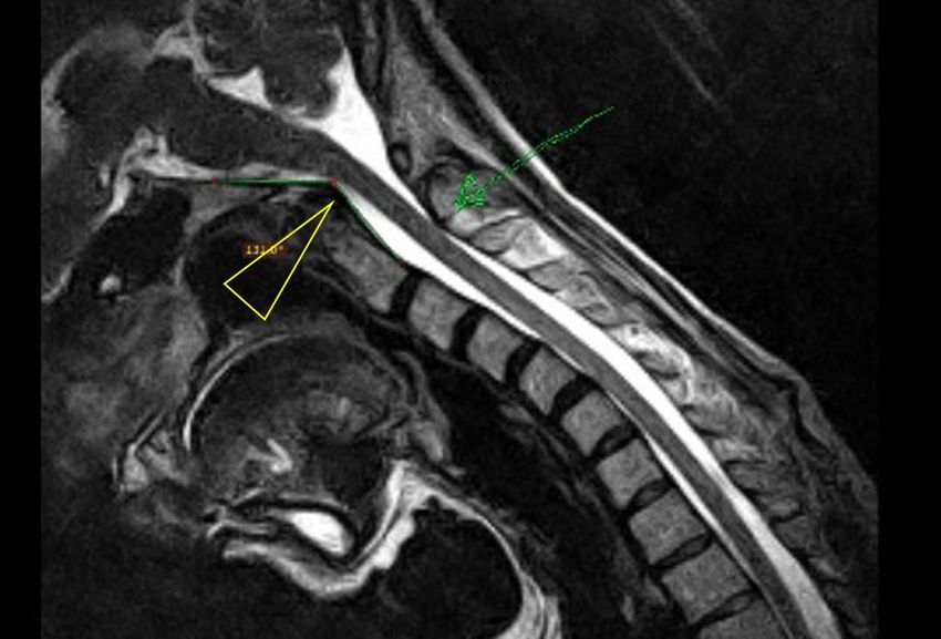
Copyright 2021

The Centeno-Schultz Clinic

     Colorado, USA

            2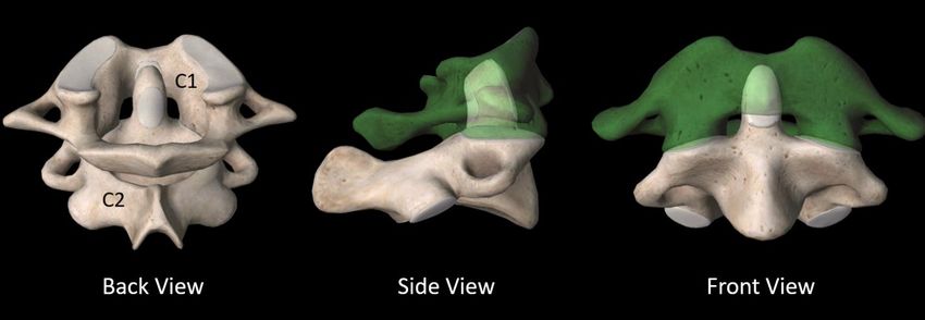
Table of Contents

➢   Forward and History-Page 1
➢   Chapter 1-What is CCI?-Page 6
➢   Chapter 2-Getting to a Diagnosis-Page 8
➢   Chapter 3- Chapter 3-Upper Cervical Anatomy and Basic CCI Concepts-Page 12
➢   Chapter 4_Imaging-Page 25
➢   Chapter 5-Treatment-Page 33
➢   Chapter 6-Is There Another Way? PICL-Page 38
➢   Chapter 7-Surgery-Page 52
➢   Chapter 8- Chapter 8-Other Issues Surrounding CCI-Page 67

                                             3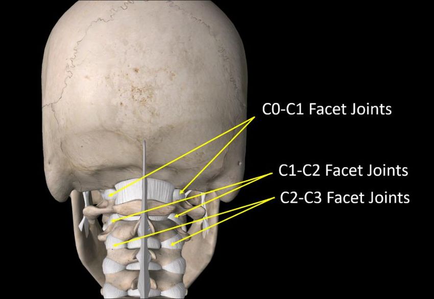
Forward and History

Way back when, when I was a young doctor with brown hair (it’s grey now) and more enthusiasm than
common sense, I had a patient who was different. She had headaches, but also dizziness and brain fog.
In looking through the chart, I saw an ENG study by a local audiologist Ed Jacobsen, Ph.D., so I called him
and that began an odyssey that ultimately led to a new way to treat CCI.

The audiologist I met that day told me about patients he was seeing for dizziness and imbalance that his
ENG tests told him had neck issues as the cause. I hadn’t really heard about anything like that, but after
some on-line research (back then you had to have software to search the National Library of medicine), I
saw that he was right. This neck and balance connection went way back to the 1920s when two
physicians (Barre and Lieou), one French and one Chinese, independently described the syndrome. At
the time they thought that this collection of symptoms which included headaches, imbalance, brain fog
and other things was caused by damage to the cervical sympathetic chain (posterior cervical
sympathetic syndrome). However, what Barre and Lieou syndrome are now credited for is figuring out
that headaches and other symptoms can come from the neck.

The next big advance in neck and balance came during NASA research in the 1960s as scientists tried to
figure out what damage to the front neck muscles would do to astronauts exposed to levels of
acceleration never before experienced by humans. They began to cut the sternocleidomastoid muscles
of primates and found that the monkeys lost their balance and would bang into walls.

Then in the 1990s, a new procedure to help neck pain was developed called radiofrequency ablation.
The procedure used a radiofrequency probe placed using x-ray guidance to burn the nerves taking pain
from the neck joints and reduce neck pain. However, when the early doctors began to treat the C2-C3
joint in the neck, some patients got permanently dizzy. While later advancements in the technique
solved this issue, the upper neck was now firmly implicated in balance.

After confirming what Dr. Jacobsen had told me I soon began treating the upper necks of these patients
who had whiplash injuries and also had headaches and dizziness. At first with simple muscle trigger
point injections and then with upper neck facet injections into the C2-C3 neck joints. Many of them got
better. Hence, as more physicians and colleagues learned that this was becoming an area of expertise
for our clinic, they referred more of these patients.

However, in this group of patients, we had some people who never got better. When a local
chiropractor, Evan Katz came to Boulder and began using a DMX (Digital Motion X-ray), we both began
to see that these were those patients who had excessive motion due to damaged upper neck ligaments.
Hence, my interest in craniocervical instability (CCI) was born. While more aggressively treating the
posterior ligaments helped a bit, most remained largely untreatable.

What was available to these patients? First, many were often bounced from specialist to specialist
without answers or any diagnosis. Second, almost all of them would flare up in physical therapy, so
many physicians would blame the lack of progress on that patient’s lack of effort. Finally, the few that
got a diagnosis were just beginning to be offered upper neck fusion, a procedure that in the past would
have only been offered to those with neck fractures or severe life-threatening dislocations of the upper
neck bones.

                                                     4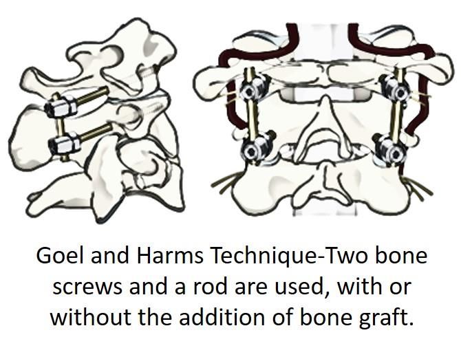
Then one day in 2013, I began to play with a model we had of the upper neck bones and ligaments. I
literally had it on my desk and would look at it periodically to see if we could access these ligaments.
While some of my spinal interventionalist colleagues thought it might be possible to access the
ligaments that hold the head on (alar and transverse) from the back, after trying this in a few patients I
concluded that it couldn’t be done safely as the spinal cord was in the way. It then dawned on me that
injections from the front might work as there was a tiny bony tunnel between the C1 and C2 vertebrae.

I then spent months reviewing the anatomy of this area to make sure we wouldn’t injure someone and
going back and forth with experts on this issue. I tried my first patients in 2015 and by 2016 had enough
dialed in that we were starting to see amazing results in these formerly untreatable patients. We added
many procedural improvements as the years went on and the rest as they say, is history.

I’ve written this book to help CCI patients. My goal is to go over everything they need to know about all
of their options. So, I’ll cover anatomy, diagnosis, conservative care, surgery, and the procedure we
developed. Why take the time to do this as a busy physician? Because patients who know more, in my
experience, are the ones that can successfully navigate our medical system to get the best possible
results.

However, please realize that in writing this book, I’ve also tried to “thread the needle” between a
patient book and one with enough detail so that patients can give it to their physicians. Why? There is
still a serious and devastating lack of knowledge on CCI and this adversely impacts patients in all sorts of
ways. Hence, oftentimes patients need a resource that they can hand to their doctors so that the
physician can understand what’s wrong with the patient.

                                                     5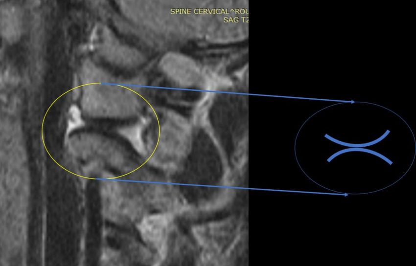
Chapter 1-What is CCI?

CCI stands for craniocervical instability. This is the area where the head meets the top of the neck also
called the craniocervical junction or CCJ. Instability means that the head or upper neck bones move
around too much due to lose ligaments that hold the head in place. Patients often complain of
headaches, dizziness, visual problems, and many other problems. We’ll get into that all later.

Patients who are ultimately diagnosed with CCI can have a broad swath of disability. I’ve seen everything
from high-functioning patients who can still exercise with symptoms to those who are completely
disabled an must lie down most of the day or wear a cervical collar to function.

What Exactly Is Instability?

All of your joints are built to move within certain defined directions. The two things that make that
happen are ligaments and muscles. Let’s dive in.

Ligaments make sure that your joints can’t move too much. When they get damaged, the joint moves in
ways in which it wasn’t designed. What does this do? It places excessive forces on the joint and bones
and damages them. In the upper neck, many other structures can get harmed. That includes the facet
joints, nerves, cranial nerves, muscles, etc… As the book progresses, we’ll go into all of this anatomy.

CCI Due to Fractures vs. Ligament Laxity

Many surgeons have been taught more about CCI due to upper neck fractures than ligament laxity.
These are classical “hangman” type fractures where the upper neck bones are no longer able to protect
the spinal cord. This is a surgical emergency as unless something is done to stabilize this area surgically,
turning the head could literally cause a spinal cord injury. This type of CCI is VERY RARE.

The type of CCI covered in this book is caused by damaged ligaments and intact bones. This is much
more common, but much more mysterious to classically trained spine surgeons. Hence, specialists in
this more common type of CCI are few and far between.

The Causes of CCI

Here we can break CCI into two camps: traumatic and congenital. Traumatic is caused by some injury
mechanism like a car crash, a manipulation of the head and neck gone bad, or a blow to the head.
Congenital means that the patient has a condition that would predispose them to CCI. The most
common is EDS, or Ehlers Danlos Syndrome where the patient is born with ligaments that are too
stretchy. In that type of CCI, while there can also be an inciting episode, the symptoms can just begin
without an event.

Traumatic CCI

I’ve heard many different causes of traumatic CCI that my patients through the decades have relayed:

    •   Car crash where the head was turned, hit something (like the back of a pick-up truck window),
        or without head trauma
    •   Forceful manipulation of the head during a therapeutic manipulation
    •   Something striking the head
    •   Falling or inadvertently diving on the head

                                                      6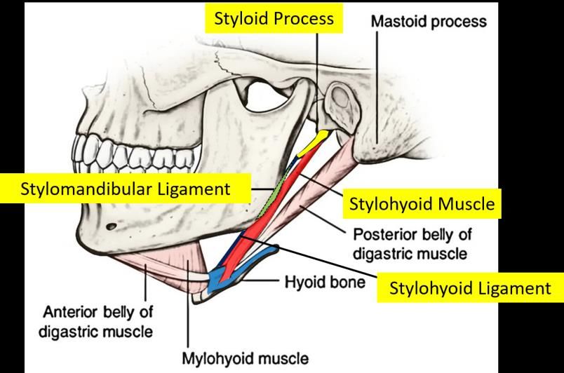
•   A history of old head and neck trauma when younger

There are many more, but these are the most common ones I hear from patients.

One issue to bring up is that some patients report having CCI after being involved in what are classified
by insurance companies as low damage crashes. How is this possible?

Cars are designed to deform to absorb force and keep it away from the occupants. However, there are
three phases to absorbing forces. The first is, as you see above, when the car is elastic (not much
damage), since the car is not absorbing force, it gets transferred to the occupant. This is where many
patients get injured. Next, in crashes where the car starts to deform, the forces on the occupant goes
down because now the car is now absorbing the brunt of the force. Finally, in crashes where big damage
happens, you get immense forces on the occupant. My friend and college Michael freeman, a forensic
epidemiologist recently published an excellent paper that quantified why the insurance company
position that patients can’t be injured in a low damage crash is not supported by crash test data (26).

Congenital CCI

When a patient is born with ligaments that are too stretchy, this condition is known as Ehlers Danlos
Syndrome or EDS. In this condition, the body makes collagen with the wrong mix of components. This is
a spectrum disorder from milder ligament laxity to more severe. As patients with more severe ligament
laxity get older, all of the extra wear and tear and accumulated injuries over time begin to cause pain
and disability. One such area where some patients’ manifest problems is the cranio-cervical junction.

Because of the overly stretchy ligaments that hold the head on, EDS patients have a lower threshold for
injury here. So, while all of the above listed traumas can cause CCI, lesser things like banging the head
on a car while getting out can cause it as well. Other times there is no specific trauma that these
patients can point to, the CCI symptoms just begin to manifest.

                                                    7
Chapter 2-Getting to a Diagnosis

The diagnostic journey for CCI is often long and arduous. Few patients get to the right specialists quickly
and get a concrete diagnosis within months of the onset of symptoms. In fact, many end up going down
unfruitful and costly rabbit holes as the medical care system focuses on only one symptom and works
that up. So, for example, here are common unfruitful journeys for CCI patients based on their
symptoms:

    •   Headaches that are caused by the CCI end up getting worked up and treated by a neurologist as
        migraines which are a different animal involving blood vessels in the brain.
    •   Dizziness/Imbalance/Lightheadedness caused by the bad position sense information coming in
        from the damaged upper neck is often worked up as a vestibular problem by an ENT doctor with
        tests like a VNG.
    •   Rapid heart rate caused by Vagus nerve irritation is often worked up by a cardiologist with a
        Holter or other wearable monitor to rule out supraventricular tachycardia.
    •   Brain fog or problems concentrating caused by the weird inputs coming in from the damaged
        neck is often worked up by a neurologist as a brain injury, stroke, or a seizure disorder.

We use many different things to get to a CCI diagnosis. I’ll cover these here:

    •   History of Onset-see above in causes of CCI section
    •   Symptoms
    •   Response to treatment
    •   Imaging

The Symptoms of CCI

There has never been a formal published survey of CCI symptoms. Hence, I complied the symptoms of
80 patients who we conformed as having CCI and who were about to undergo a PICL procedure. This
was the word cloud of what they reported:

The bigger the word, the more often it was reported. Based on this data and decades of experience
treating these patients, this how symptoms break down.

                                                     8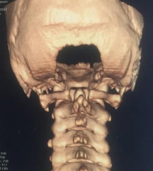
These are classical symptoms that almost all CCI patients would have like:

    •   Headaches
    •   Upper neck pain
    •   Dizziness/Imbalance/lightheadedness
    •   Brain fog
    •   Clicking/popping in the neck (crepitus)

Then there are a collection of symptoms shared by most CCI but not all patients:

    •   Ringing in ears (tinnitus)
    •   Shoulder pain
    •   Rapid heart rate (tachycardia)-POTS and/or increased anxiety
    •   TMJ
    •   Memory Loss
    •   Wandering pain or numbness/tingling in various areas of the body
    •   Gastric disturbances
    •   Clicking or popping in the upper neck with movement
    •   Fatigue
    •   Nausea

Then there are a collection of symptoms shared by some CCI patients:

    •   Facial pain
    •   Tongue symptoms
    •   Loss of consciousness episodes
    •   Sweating

Hence, diagnosing CCI by symptoms alone can be very difficult.

A Word of Advice on Symptoms

As you can see, CCI patients can have a large number of symptoms. During this book I’ll go through
various rabbit holes of misdiagnosis to avoid. One tip here is that patients these days often find like
patients on social media and begin self-diagnosing. When they see a doctor, they begin using more
diagnostic terms like “atypical tachycardia” rather than just saying that when my neck is bad my heart
races. This can often confuse the diagnosis for the doctor. Another thing they do is to begin listing every
possible symptom they believe is related to their CCI rather than just the main symptoms they know are
for sure related. This also can make the doctor confused. So when you talk to a doctor about your
problem, keep it focused on what you experience and keep it streamlined and simple.

Response to Treatment

CCI patients have a few classical responses to treatment. One is that active physical therapy focused on
strengthening tends to make them worse. Why? The upper neck ligaments are just too loose. Hence,
when they try to strengthen the neck muscles, the upper neck moves around too much and irritates
joints, nerves, tendons, etc…

                                                     9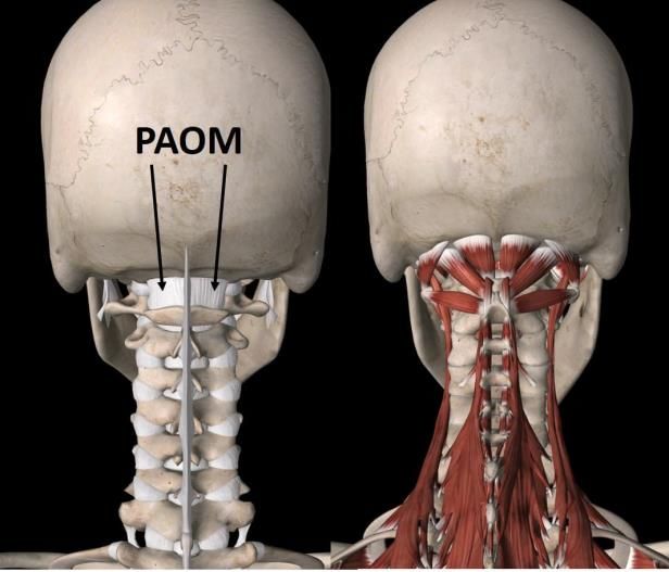
On the other hand, many patients find good temporary relief from upper cervical chiropractic
adjustments (NUCCA or AO chiropractic). They have improved symptoms that can last for hours to
weeks. This can also be highly dependent on finding the right chiropractic expert. Meaning not all upper
cervical chiropractors have success in all CCI patients and some patients find only a handful of expert
chiros who can handle their case.

Some quick definitions here:

    •     NUCCA (National Upper Cervical Chiropractic Association) Chiropractic-focused on gentle
          manual adjustments of the upper neck bones that are guided through precise x-ray
          measurements.
    •     AO (Atlas Orthogonal) Chiropractic-Very similar to NUCCA, but the x-rays are used to program a
          vibration device which performs the adjustment.
    •     Blair Chiropractic-Also focused on gentle manual adjustments of the upper neck as well as
          diagnostic imaging where angles are calculated to drive what needs to be adjusted. There is also
          a leg length test that’s commonly used to see how the upper neck is impacting the rest of the
          body, all the way down where the feet meet the ground.

Other types of treatments where we look for a response are injections. For example, the upper neck
instability can cause the upper neck facet joints to get beat up and hurt. Hence, in many CCI patients,
injecting the C0-C3 facet joints will help their pain. Or for example, occipital nerve blocks which can help
headache pain. The focus here is to determine if there are structures in the upper neck causing pain as
that helps the doctor pinpoint the area of the problem.

Imaging

There are many ways to try to image the problem of CCI. However, before we get into that in chapter 3,
we need to learn a little anatomy.

Examples of How These Pieces Come Together

You now have the diagnostic categories, which to review are:

    •     History of Onset-see above in causes of CCI section
    •     Symptoms
    •     Response to treatment
    •     Imaging

So now let’s review some examples. While we still have some things to learn as we move forward, this is
just to show you how a CCI diagnosis would be made.

Example 1-A patient was in a car crash where they were rear ended while their head was turned. Now
let’s plus some things in:

    •     History of Onset-The problem began at the car crash, which was sufficient to cause this kind of
          injury.
    •     Symptoms-Headache, dizziness, brain fog, and upper neck pain. Hence the symptoms match CCI.

                                                     10
• Response to treatment-Physical therapy makes the symptoms worse. An upper cervical facet
injection decreased headache pain for 3 weeks. Both of these things fit the profile of how a CCI
patient would respond to treatment.
• Imaging-The patient’s DMX study shows 6 mm of overhang which fits with instability.
All of this shows that we’re 4/4 in determining f this patient has CCI.
Example 2-A patient who has EDS bangs their head on a the roof of a cab while getting out. Here’s the
analysis:
• History of Onset-The patient has EDS, which means that they have a major risk factor for CCI.
The new problems began getting out of the car, which could injure the upper neck ligaments in
an EDS patient who already has stretchy ligaments.
• Symptoms-Headache, neck pain, rapid heart rate, GI problems, and brain fog. Hence, the
symptoms match.
• Response to treatment-Upper cervical chiropractic (NUCCA) helps the symptoms for days at a
time, but then they return. Home strengthening makes the patient worse. Both of these
responses to treatment match what a typical CCI patient would report.
• Imaging-The patient’s flexion-extension MRI shows that their Grabb-Oakes measurement
increase from 9 mm to 12 mm with flexion. This matches CCI.
Again, another 4/4 match.
Example 3-Now we have a patient who has no history of trauma whose symptoms just began without
any warning:
• History of Onset-No trauma, no risk factors for CCI.
• Symptoms-Brain fog, rapid heart rate, GI problems, and jaw pain. This may or may not be a
match.
• Response to treatment-Upper cervical chiropractic (NUCCA) doesn’t help the symptoms.
Strengthening is not a problem, it also doesn’t help. This doesn’t match CCI.
• Imaging-There are no concrete measurements that match CCI other than one measurement
which is borderline.
So we have a 1.5/4 here, so this is less likely to be CCI.
11Chapter 3-Upper Cervical Anatomy and Basic CCI Concepts
While this next chapter may seem tedious, it’s essential for anyone with CCI to understand how the
basic anatomy works. I’ve purposefully made this chapter a bit more complex that the average patient
may want to know, to again, thread that needle between a book that can help patients versus one that’s
detailed enough to also help their doctors understand CCI. If it gets too intense for you as a patient, then
read as much as you want to know and skip forward to the next chapter.
Bones
The head on the neck is like a bowling ball on a stick, meaning it’s inherently unstable. Hence, we have
two things that evolved to hold it on. Tough ligaments and muscles. In addition, the upper neck bones
are specially adapted to help this effort as well. Let us begin there.
The neck bones are numbered. C0 is the skull and C1 is called the atlas while C2 is called the axis. Both
are ring shaped bones that I have simplified above. The peg of C2 fits into C1 and the skull rests upon C1.
There are also joints connecting the skull to C1 and C1 and C2 (not shown here). These are called facet
joints.
Here’s what the bones really look like:
Now back to our simplified drawings. The peg in C2 (Dens) is there so the head and the atlas can rotate
around it:
12Ligaments
How would we keep these two bones together? That’s where ligaments start factoring in. The big one
here is the transverse ligament (aka the transverse band of the cruciform ligament):
If this transverse ligament is loose, it allows too much movement of C1 on C2 when the head is flexed
forward.
However, how do we keep these two bones firmly attached to the skull? The alar ligament does that job:
This ligament goes from the dens to the skull base, kind of like arms reaching up from the dens to the
skull. Hence, it binds C2 to the skull and indirectly holds C1 in place as well by sandwiching it between C2
and the skull. When this ligament is loose or damaged, it can allow C1 to rotate on C2 because it no
longer applies that sandwich force to help keep C1 in place. In addition, when damaged, it can also allow
13the skull and C1 to slide sideways when the head is side bent. We measure this as C1-C2 “over-hang” on
a DMX study (more on that later).
It should also be note that when viewed above, one band of the alar ligament travels toward the front
as shown here. Remember that this ligament goes the base of the skull. Hence, then the head and neck
bend forward, the weight of the head is partially supported by this ligament.
There’s another part of the alar ligament which extends downward between the alar ligament and C2
which is called the accessory ligament as shown below (in this picture I made the C1 and C2 bones
transparent (except for the dens)):
If we look from the side at the whole neck, we see that there are lots of ligaments in the back or
posterior part:
14These are the nuchal, VDL (Vertebro-dural Ligament), supraspinous, and interspinous. They all help
prevent the vertebrae from moving too far forward and are very active when you look down. The VDL is
special because it connects directly to the dura, which is the covering of the brain and spinal cord.
Hence, it is believed that damage to this ligament may cause headaches.
Finally, there are deeper cervical ligaments to consider, which are shown below:
These include from front to back:
15• SAAOL-Superficial Anterior Atlantooccipital Ligament (this is the extension of the anterior
longitudinal ligament upwards. This connects the front the cervical vertebral column with the
skull.
• AAOM-Anterior Atlantooccipital membrane-This connects C1 to the skull.
• Apical-This extends from the dens of C2 to the skull.
• Cruciate-This is a cross shaped ligament that has as its horizontal part the transverse ligament
discussed above, but has parts that go up and down as well.
• Tectorial-This is the upward extension of the posterior longitudinal ligament that connect the
back of the vertebral column to the skull.
• PAOM-This is the upward extension of the ligamentum flavum that connects the back part of
the spinal canal to the skull.
The front ligaments help to stabilize your head on your neck when you look up, the middle ones help to
hold your head on, and the back ligaments keep everything stable when you look down.
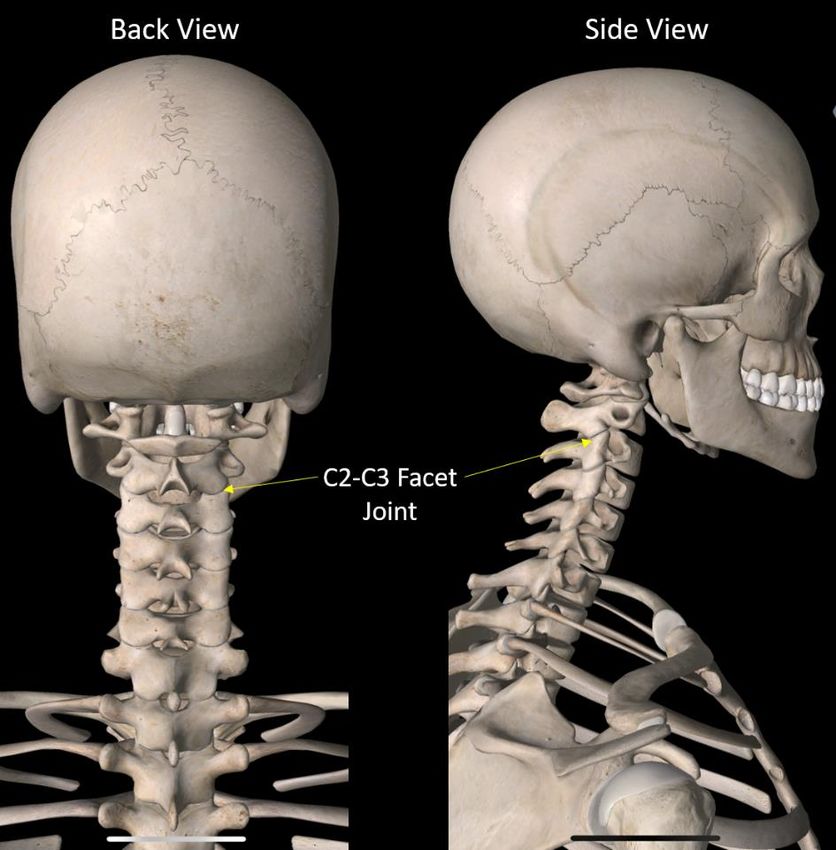
Facet Joints
Your upper neck (and the rest of it as well), has finger size joints called facets. Their purpose is to help
your neck move normally. When the ligaments are damaged, the joints can sustain too much wear and
tear and begin to get injured.
This is how those joints really look:
16Here’s a simplified diagram:
These upper neck joints are
• C0-C1-This joint allows the nodding movement of the head. If injured or painful, this joint can
cause pain in the back of the head.
• C1-C2-This joint allows for about half of the rotation of your head. This joint can causes pain to
the back of your upper neck where your head and neck meet.
• C2-C3-This joint allows for a little rotation of the head and a little forward bending of the neck. It
causes pain in the upper neck and the back of the head.
These are the referred pain patterns for these joints (24):
17The red area is where pain from the C0-C1 joints is felt. The yellow/green is where pain from the C1-C2
joint localizes. Finally, the blue area is where pain coming from the C2-C3 joint travels.
Hence, these upper neck joints can cause headache. In addition, all of them also provide information to
brain about head, neck, and eye position. This happens through tiny sensors present in the joint
capsules called proprioceptors. Hence, if these joints are injured they can cause dizziness, imbalance, or
visual problems.
C1-C2 and Inherent Instability
The C1-C2 facet joint is responsible for 50% of the total neck rotation. Like all facet joints, it has cartilage
and a joint capsule. What makes this joint very unique is the shape of its internal joint surfaces:
Above is an MRI side view of the C1-C2 facet joint. Its surfaces are shown to the right. Note that they are
both convex, which is unusual in the body. Why? Usually, the joint surfaces fit together like a puzzle
piece with one surface being convex and the other being concave. That makes them inherently stable.
However, the C1-C2 joint is inherently unstable.
What does that mean? If we have a convex/concave joint like the one below and there are no muscles
or ligaments acting on it, it’s not going anywhere. Meaning the joint surfaces stay together. The
opposite is true if the joint surfaces are convex sitting on top of convex:
18So the C1-C2 joint is inherently unstable. This explains why we have an entire field of chiropractic care
devoted to this part of the neck! However, the C0-C1 joint above it is that convex/concave type, so it is
inherently more stable.
In the section above, we discussed how the atlas (C1) was sandwiched between the skull and C2 and
held in just the right amount of tension by the alar ligaments. These ligaments help to maintain the
concave joint surfaces in approximation as you move your neck. However, if this ligament is damaged,
these joint surfaces cause the bones to rotate (25).
Why Inherent C1-C2 Instability Can Wreak Havoc with Patients
Given that the C1-C2 joint is inherently unstable, if it doesn’t have ligaments and muscles to hold it in
place, it tends to move out of place. This results in the C1 bone rotating on the C2. In fact, this is why an
entire type of chiropractic (NUCCA) is devoted to trying to get C1-C2 back in place. So if you have C1-C2
instability due to damaged ligaments (Craniocervical Instability or CCI) it’s easy to see why this joint
would not be “in place”.
What happens when this joint gets out of whack? The capsule is rich in position sensors that help
maintain the position of the body relative to the neck, so your body will feel out of whack as well. In
addition, the joint, when painful, refers its pain to the head. So you can also have headaches and other
symptoms (see below).
In addition, it’s possible for this joint to be out of whack without having damaged ligaments. This is
where a NUCCA chiropractor or an experienced manual physical therapist can make a big difference.
Symptoms here include headaches, dizziness/imbalance, visual disturbances, brain fog, or spasm, or
pain at the back of the head.
Muscles
The muscles in this area all control the fine movements of bowling ball in the stick:
The RCP (Rectus Capitis Posterior) major and minor connect the C2 and C1 muscles to the skull and can
help you look up. The RCP minor is unique in that it connects directly to the covering of the brain and
spinal cord (dura), so it’s implicated in headaches. The obliquus capitis muscles connect C1 to C1 and the
skull and help with bending your head sideways on your neck.
19The deep muscles in the front of the neck look like this:
The longus capitis and colli live just in front of the spinal column and help to flex the neck and maintain
the curve.
As we move outward, we find the strap muscles in front including the SCM:
The SCM is a critical muscle up front as it is the prime mover for rotating the head. This is important for
CCI as C1-C2 is a critical joint that’s responsible for 50% of your ability to rotate.
There are a slew of other muscles like the upper trapezius, levator scapula, and paraspinals muscles that
usually get in on the act in CCI, but we’ll discuss those later.
20Nerves
Pissed off upper neck nerves tend to cause headaches. The nerves that are critical for CCI live in both the
back and front of the neck. Here are the ones in the back of the neck:
Realize that since these upper neck muscles help to hold and stabilize the head, when they spasm and
go into overdrive to help stabile the head on the neck, they can irritate these nerves can cause
headache. There are also nerves that live and exit in front of and behind the SCM:
• TON-Third Occipital Nerve
• GON-Greater Occipital Nerve
• LON-Lesser Occipital Nerve
21The TCN is the transverse cervical nerve (also called the superficial cervical plexus) and the TA is the
temporal-auricular nerve (also called auriculo-temporal nerve). The TCN nerves live behind the SCM
muscles, so they can become irritated as those muscles get tight in C1-C2 instability. They can cause
headaches, as well as jaw, side of head and face pain. Note that the TA nerve also travels right behind
the jaw, so any extra motion or overload of the jaw can cause it to be irritated and this can lead to
headaches at the side of the head. you
Cranial Nerves
The cranial nerves come out the skull and head south, as shown below:
Given that the skull moves too much on the upper neck and these nerves also are close to the atlas bone
which can move around too much as well, these nerves can get irritated. The more common cranial
nerves are:
• CN10-Vagus Nerve-This is literally the brakes on the heart, lungs, and digestive tract. If the nerve
gets irritated the patient can get rapid heart rate, anxiety, and digestive problems.
• CN11-Spinal Accessory Nerve-Controls the upper trapezius and sternocleidomastoid (SCM)
muscles. When it gets irritated these muscles can go into spasm.
• CN12-Cranial Nerve 12-Hypoglossal-Controls the tongue muscles. This can cause spasm in the
tongue area.
• CN V and VII-These both go to the face.
Foramen Magnum
The base of the skull has a big hole in it and the name for that in Latin is “foramen magnum”, which is
what we doctors call it. Realize that this area has quite a bit of room for the spinal cord, which is great as
that means that quite a bit of motion can happen here before there is a spinal cord injury:
22The black arrows one either side of the yellow spinal cord show that room, but also note the yellow line
which is the covering of the spinal cord and brain (the dura) which is closely adhered to ligaments. So if
that area gets irritated due to CCI (too much skull movement the dark blue arrows), then while there is
no spinal cord injury, there can be all sorts of referred pain in all sorts of different places in the body. In
addition, in many CCI patients the dens (here marked C2) can move backwards due to a loose transverse
ligament, which can cause pressure on the dura in the front and possibly the spinal cord (also called
cervical medullary syndrome).
Cervical Medullary Syndrome
As discussed above, when there’s instability, the skull moves too much against the spine and as a result,
the dens can move too far backward (yellow arrowhead) and irritate the top part of the spinal cord or
brainstem (medulla). When that happens, as shown above, that’s called a cervical medullary syndrome.
Also realize that the white stuff you see above on either side of the spinal cord is cerebrospinal fluid
(CSF). This circulates around the brain and acts as the waste removal system for chemicals produced in
the brain. This circulates all the way down to the neck, upper back, and lower back. The flow of that fluid
23can get obstructed by a cervical medullary syndrome. We’ll go into more detail later on how that’s
measured using specialized MRI.
24Chapter 4-Imaging
Imaging in medicine means that some technology is used to visualize the deep structures of the body.
This is always an interesting topic with CCI patients. What I mean is that this is such a huge topic that it’s
tough to cover all of it. In the meantime, many CCI patients see surgeons who use some of the metrics
I’ll cover here to determine if they’re surgical candidates, so I want to make sure to cover those
measurements. However, to make this part of the blook less cluttered, I’ll give brief descriptions of the
measurements and then point to a YouTube video or reference that goes into more depth.
Imaging 101
X-ray Exposure
It’s important to understand a bit about common types of imaging discussed here and x-ray exposure.
For example, a simple neck x-ray has about the same about of amount of x-ray exposure as a chest x-ray,
which is about the same amount natural background radiation that most people get in 10 days of
normal living. However, a CT scan exposes you to about 8 years of background radiation exposure! (27)
Hence, we generally DO NOT recommend CT scans to diagnose CCI.
MRI Imaging Pitfalls
Before we get into the topic of imaging and MRIs, it’s critical to point out that a routine cervical MRI has
limitations in CCI patients. Let’s explore that a bit.
An MRI (Magnetic Resonance Imaging) machine is a big magnet. It applies a huge magnetic field which
aligns the small molecules in your body and then removes that magnetic field and lets these flip back to
their normal state. When that happens, these molecules produce a radio signal that is reconstructed by
the computer into an image.
The Good News
Static measurements like Grabb-Oakes and the CXA can be measured on most routine cervical MRIs.
This may help make the diagnosis of CCI. The problem is that, as you’ll see below, movement like flexing
your neck forward and backward usually enhances the information gathered. That kind of motion can
only be picked up on a specialized “Stand-up” MRI unit and not on the standard “lie face up in a tube”
type. More on that below.
The Bad News
What an MRI can detect depends on the specific coil used. This is something placed around the body
part that acts an antenna to pick up the signals coming out of your body that are generated by the big
magnet. There is a different coil used for each body part.
The usual cervical MRI only images from C2-T1 because that’s what the neck coil is optimized to image.
However, the pathology in a CCI patient is above C2. In fact, to get good MRI pictures above C2, a head
coil is often needed. Hence, having your doctor write a script for a routine cervical MRI may not be
helpful in getting to a diagnosis.
Imaging Resolution and Strength
25It’s also critical to understand that imaging strength is directly related to the resolution or quality of an
image. Hence, whatever type of static MRI you get, you should make sure that you get the best possible
image. In addition, getting imaged on a more or less powerful MRI machine is agnostic to your insurance
benefits. Meaning, getting imaged on a 2 million dollar 3.0 Tesla MRI is covered by insurance just as
much as a getting imaged on an old-school 0.3T open MRI that someone would pay you to haul away for
parts.
MRI imaging strength is measured by a unit called a Tesla (T). That’s for early 20th century electrical
engineering genius Nicolai Tesla and NOT the car company. One Tesla is the magnetic field strength of
the earth. Hence, 0.3T means about 1/3 that amount.
Here is how they rate out:
• Worst quality images-0.3-0.6T
• Good quality images-1.5T
• Best quality images-3.0T
You can find this out by asking the center the “field strength” of the machine. They should report it as
above.
Realize that any open or Stand-up MRI has a lower field strength than most high-quality lying down
MRIs. This is a regrettable trade off. For example, in order to be able to image someone in a machine
that allows for motion, you need more room, and hence the magnetic field will have lower strength to
make that work. However, more information about an instability condition like CCI can be gathered
when patients move. Hence while field strength may be important in a static MRI, we’re never going to
see a Stand-up MRI with 3.0T.
The “Best” Type of Imaging for CCI?
In our clinic, we like using DMX (discussed below), which allows for movement that approximates
reality. Our second choice would be movement-based MRI or a rotational CT scan. Static MRI of the
upper neck can sometimes be helpful and there are times that we can see abnormal measurements on a
routine neck MRI.
However, this preference for DMX is not universal. For example, many neurosurgeons prefer routine
static MRIs where some of the measurements shown below can be measured.
A Gold Standard?
While it’s always great if any diagnosis has a gold standard test to determine if you have or don’t have
the disease, that doesn’t exist in CCI. Hence, different physicians all use different tests to get to a
diagnosis. While that makes it harder for patients, who understandably want certainty, it’s reality.
Types of Imaging
Imaging can be broken down into two main categories: static and dynamic. Static means that the patient
doesn’t move during the imaging. This can be a problem for diagnosing instability as that, by definition,
involves movement. To solve that issue, more recently applied imaging techniques use movement and
this is called dynamic imaging.
26Before I jump into explanations of all of the different types of measurements and imaging used, realize
that this chapter is yet another example of “threading the needle” between a book that can be read by
patients, but that has enough detail to be handed to a physician that needs to be educated. Hence, it’s
OK if you jump through parts of this chapter that are in too much detail for you.
Static Measurements
Grabb-Oakes
This is the distance between a line drawn from the front of the spinal canal (basion) and the back
inferior corner of the C2 bone and the back of the dens. Given that the C2 bone (axis) is held against the
C1 bone (atlas) by the transverse ligament, a high Grabb-Oakes measurement can be due to a lax
transverse ligament. An abnormal measurement is often considered to be 9 mm or more (2,5). With the
advent of flexion-extension MRI, it’s easier to measure the G-O measurement when the patient moves,
which provides more information. See this video for more on the G-O measurement
[https://youtu.be/4f-Yi9fuKD0].
CXA (Clivo-Axial Angle)
This is the angle between the clivus (the inside front area on the bottom of the skull) and the back of the
dens (C2). The problem being measured here is the skull is falling forward on the upper neck which can
cause irritation of the front of the brainstem and upper spinal cord. This movement is controlled by
strong ligaments in the back of the neck such as the nuchal, supraspinous, and interspinous ligaments.
It’s also controlled by the posterior atlantoaxial membrane (PAOM, a ligament at the back of the spinal
canal) and to a lesser extent the transverse ligament. Abnormal is a bit different when reported by
different authors. Less than 150 degrees (4) was originally reported with others stating that normal is
between 145° to 160° in the neutral position (3). The differences in opinion are likely due to the fact that
things like kyphosis (forward head posture) and male/female sex can change the angle.
27Power’s Ratio
This is another measurement that determines if the head has moved forward on the upper neck. This
one is more complex, requiring two lines (one between the basion and the back of the spinal canal at C1
and another between the opisthion and the posterior aspect the front of the atlas) and then some
division is applied. If the calculated measurement is less than 1, the ratio is normal, if it’s >1 then it’s
abnormal (2). The Powers Ratio is measuring whether the head is aligned properly on the upper neck
bones. For example, if the number if greater than 1, the head is too far forward on the spine and
multiple ligaments might be injured.
See my video here for more information [https://youtu.be/mQw7Sx5QA2c].
BAI (Basion-Axial Interval)
This is the distance between the basion (front of the skull) to the back of the dens of C2 (axis). The
problem here is that CT scan studies have shown the usual normal of 12mm to be unreliable with
28normal ranging from 9-26 mm. This can also be measured in flexion and extension where >4mm
difference between the two positions is considered abnormal enough to warrant surgery. The ligament
most responsible for keeping this measurement in check is the transverse ligament. This is again a
measurement that can determine if the head if too far forward on the spine.
BDI (Basion-Dens interval)
This measurement focuses on vertical instability. This would be damage to the ligaments preventing
vertical translation of the head like the PAOM, tectorial membrane, AAOM, apical, and SAAOL. The
normal value is less than 12 mm on x-rays and 8.5 mm on CT scan (2). Like the BAI, it can also be
measured in flexion and extension. The BDI change can also be measured as traction is applied to the
head.
29Chamberlain Line
This is a line drawn from the back of the hard palate of the skull to the back of the hole in the skull
(opisthion). If the dens of C2 is more than 3mm above this line, this indicates cranial settling or that the
skull is too low on the cervical spine. This is also called basilar invagination. Both of these conditions are
more extreme than are usually seen in most CCI patients. Meaning in in purest neurosurgical sense, both
cranial settling and basilar invagination have the dens touching the brain stem.
ADI (Atlantodental Interval)
This is the distance between the front of the C1 (atlas) and the dens of C2. Normal is 2mm (2). If this is
greater than 2mm, the transverse ligament may be injured. Like many of these measurements, the ADI
can be measured in flexion as well to see if it increases, as that stresses the transverse ligament.
30Upper Cervical MRI
Note that a routine neck MRI rarely shows these upper cervical structures. Much of that has to do with
the wrong type of coil used to collect the information which is designed to an image from the neck and
not the upper neck/head. This means that ligaments like the alar, transverse, and others just don’t show
up on the usual cervical MRI.
However, if a specialized MRI is taken, the upper neck ligaments can be imaged. This requires a head coil
and other specialized image sequences. Once we have those images, you can look at how the ligaments
appear and grade them based on a scale published by Krakenes (6). While we look at this information,
the problem is that it’s not showing whether those ligaments work functionality to stabilize the head on
the neck. That would require movement-based imaging. If you want to learn more, see my video
[https://youtu.be/ZQaztACMzbk].
CSF Flow Imaging
One of the more promising new technologies out there is using a specialized MRI to measure the flow of
cerebral spinal fluid though the upper neck. This is critical because many problems that surround CCI can
restrict the normal flow of the brain’s fluid through this area. The world’s expert in this area is Scott
Rosa, DC of upstate New York. He’s been using this technology in CCI patients for years and can show
improvements of CSF flow through the upper neck before and after specialized chiropractic adjustments
(see http://rosaclinic.com/)
Dynamic Imaging
Flexion-Extension X-rays
These are x-rays where the patient is asked to look down and then an image is taken and then they are
asked to bend their neck back and look up and another picture is taken. The goal is to see if the neck
vertebrae move too much against one another, which they can only do if the ligaments are damaged.
For CCI, the one measurement than can be looked at here, is the ADI when the neck is in flexion. In
addition, many CCI patients have also damaged other neck ligaments in the lower neck that can be
evaluated here.
One of the biggest issues with this type of imaging is that many radiology techs don’t get enough
movement out of the patient to allow proper measurements to made. For these films to be diagnostic,
the patient needs to flex and extend as far as possible, despite the instructions from the tech taking the
picture. Rad techs often tell people to move minimally because it’s easier for the tech, but make sure
you’re a rebel here and get as much motion as possible!
Upright MRI
MRI of the neck is usually performed with the patient lying face up in a tube (supine). Upright MRI
allows a patient to be imaged in a weight bearing position. This places more natural stress on the upper
neck and other ligaments, so it’s possible that measurements that weren’t indicative of CCI lying face up
in the usual MRI scanner could be become positive when imaging the patient this way. Having said that,
normal values obtained while imaging people supine may not apply to upright MRI.
31Upright MRI also allows for motion. Hence, the patient can do things like bend or extend the neck. This
can give valuable information about whether the ligaments perform normally or allow too much motion.
To learn more, see my video here [https://youtu.be/rpHv0AalJ4Y].
Functional CT (C1-C2 Rotatory CT)
This is a specialized CT scan where the patient rotates their head and the movement between
the skull, C1, and C2 is measured (7). Instability is indicated by rotation of the skull on the atlas
of more than 7 degrees, and of the C1/C2 joint of more than 54 degrees.
DMX (Digital Motion X-ray)
DMX uses a fluoroscope which is real time x-ray imaging and captures video of how the upper neck
bones move. The technology is the only one that allows for natural movement from the patient as all
others are contrived and artificial in some way. DMX allows a patient to move in one direction at various
velocities and accelerations. The patient then must quickly stop and go another direction. This is very
different than a neck flexion-extension x-ray where the patient only moves in one direction maximally
and then a picture is taken.
The measurements that are important for CCI patients here are C1-C2 overhang and ADI. C1-C2
overhang means that the patient bends their head to the side and the doctor looks to see if C1 slips
sideways against C2. If that happens beyond 3mm with also a difference in the distance between the
dens and the atlas, then the alar ligament on the same side may be injured. The ADI can also be
measured. To learn more, see my videos [https://youtu.be/nPBW4Bk-8l4 and
https://youtu.be/6MRnZw2BpV8].
32Chapter 5-Treatment
Understanding the Ladder of Invasiveness
How do physicians look at medical care for their own families? They use “the ladder”. What’s that?
If you want to keep patients as safe as possible, you only expose them to the least risk needed to get the
job done. For CCI, that system creates a ladder that looks like this:
You’ll notice at the bottom I have listed exercises. Things like physical therapy and alternative healing
techniques like craniosacral would be in this category.
Next up would be manipulation of the upper neck. These are things like NUCCA or AO chiropractic.
These are slightly more invasive than physical therapy.
Then we get into posterior injections (from the back), which could be prolotherapy of the ligaments in
the back, facet injections, epidurals, etc. This is more invasive than manipulation.
Next up are anterior (from the front) injections which includes the PICL procedure. This means injecting
the deep ligaments that hold the head on. Since this is a newer procedure where fewer have been done
than posterior injections, it’s automatically considered higher risk than more commonly performed
posterior injections.
Finally, at the top of the list are spine surgeries like dithering, decompression, and fusion. For example, a
surgical detethering procedure would be more invasive than PICL since it involves open surgery and
33destroys a part of the spine. Even more invasive would be fusion which is where screws, rods, or plates
are placed to make sure parts of the spine or a joint can never move again.
So how can you use the ladder like I would to protect my wife and kids? Anything lower on the ladder
that applies to that injury or problem that may work needs to be tried first before moving up the ladder.
That way you don’t get exposed to more risk than you need.
Conservative Care
Conservative care for CCI breaks into a couple of common areas:
• Bracing
• Physical therapy
• Chiropractic
• Curve restoration
• Cranio-sacral
Bracing
Neck bracing is an interesting topic in the CCI community. On the one hand, you have patients that find
the right neck brace and swear by it. On the other hand, you have patients who can’t find a brace that
will keep their head/neck in the position where it’s happy with less pain. For those who like braces, this
is a two-edged sword. Why? Because stability requires normal ligaments and muscles. Hence, using a
neck brace for an extended period of time can make the neck muscles weaker, which can make the
instability worse. So if you use a brace, use it only for short periods. For example, some patients will only
use one when travelling or exercising. Some need it more often or they can’t function. Just remember,
like all CCI treatments there are advantages and disadvantages.
Physical Therapy
One of the things that’s almost diagnostic for CCI is that patients do poorly with active physical therapy
that focuses on strengthening. This response can change if the patient has had their upper neck
stabilized through the PICL or a fusion procedure. At that point, especially with the first two treatments,
exercises are encouraged to slowly help the patient build back the upper neck muscles that stabilize the
head on the neck. Of mote, if the patient is fused, normally some muscles are destroyed by the
procedure. More on this later.
However, there are physical therapists with specialized manual knowledge who can help CCI patients.
Meaning that these therapists have years of additional training, so they represent less than 1 in 100 of
all PTs. A helpful guide to finding an upper cervical expert is finding a therapist who knows the upper
neck. Here are some resources:
• IPA Physical Therapy [https://instituteofphysicalart.com/]
• Ola Gimsby Manual Therapy [https://www.olagrimsby.com/]
Other types of PT to consider that don’t focus on the upper neck, but do focus extensively on postural
correction that may help the upper neck include:
• Feldernkrais [https://feldenkrais.com/]
34• Egoscue [https://www.egoscue.com/]
Chiropractic
While physical therapists have few experts who know how to treat this specialized area of the body,
that’s not true for chiropractors. In particular, there is NUCCA [https://nucca.org/], which is a national
upper cervical chiropractic association that qualifies and trains these providers. Many of my patients
swear by NUCCA chiropractors who are able to help their symptoms. Usually, if they find their way to
me, they will find one who can get temporary relief from hours to days to weeks, but then things come
out of place. On the other hand, for other patients, NUCCA is not helpful.
Another type of upper neck chiropractic is Atlas Orthogonal (AO). This is very similar to NUCCA, but
instead of a hands-on manual adjustment, a specific low force instrument is used to precisely move the
atlas back into place.
How do both of these differ from traditional chiropractic? Many chiropractors will perform “long lever
arm” neck manipulations where they suddenly twist the skull. NUCCA instead uses a precise, non-
invasive spinal adjusting technique. This uses precise and objective x-ray views of the head and neck,
mathematical measurement and analysis to plan the specific low force adjustments used by these
providers.
Curve Restoration
Your neck is born with a natural shallow c-shaped curve. This distributes the forces from your head to
the discs in the front of the neck and the facet joints in the back. In our modern society, looking down at
computers and phones or neck trauma can cause the neck can straighten or develop a reverse curve.
That puts too much force on the discs in front and can cause too much force on the upper neck
ligaments as well. Hence, we’ve seen several CCI patients get improvements with curve restoration.
There is an online credentialing organization where providers can be found [www.idealspine.com]
Curve restoration involves forward traction to increase the curve by pulling the neck forward (not to be
confused with axial traction that pulls your head upwards). Specialized machines are used to do this and
at home devices are also available. It should be noted that only some CCI patients find this helpful
(usually fewer than find NUCCA or AO to be helpful).
Injections
This is a big topic. Before I get too far into it, it’s good to spend a few minutes on how different injection
procedures stack based on complexity:
35Why? We often see patients who get confused that Level 1 injections as shown have something to do
with Level 5 injections. For example, blindly injecting prolotherapy solution into the back of the neck has
little to do with using endoscopy and fluoroscopy to inject stem cells into the alar and transverse
ligaments (PICL). To use another analogy, the skill level needed to do the level 1 injection can be taught
in a few hours and the skill level needed to perform a level 5 procedure would require many years to
master.
For CCI patients, the different skill levels break down as follows:
Level 1-Posterior prolotherapy. This is injecting the easy to reach ligaments usually with no imaging
guidance (blind or palpation guided). Sometimes the doctor will use a fluoroscope, but not in the same
way a more experienced and highly trained doctor performing higher level injections would use that
machine. The Level 1 doctor is just using that fluoroscope to make sure they have hit certain bone
landmarks, whereas the higher-level physician with more training is using that machine to make sure
that they are in certain joints by injecting radiographic contract. I could teach the first technique in a few
hours, but the second takes months to years to master.
This is not to say that in the right patients with general neck instability, posterior injections are
worthless. They can be very helpful. However, the vast majority of CCI patients that try posterior
injections don’t get relief and functional benefit. This is a concern as MANY CCI patients spend
thousands to tens of thousands of dollars on posterior injections with prolotherapy, platelet-rich
plasma, or bone marrow stem cells that ultimately don’t work. This is despite the promises of a few
practitioners that these procedures will be highly effective.
Why won’t posterior injections help most CCI patients? Because the ligaments that need the most
attention can’t be reached via this approach.
Level 2-Lower neck facet or epidural injections. This is using x-ray or ultrasound guidance to inject the
C2-C7 facets or spinal nerve levels. These procedures can usually be performed by any competent
interventional pain physician.
Level 3-This is a bit more intense and for CCI patients, this might include injecting the C2-C7 neck discs if
needed.
36Level 4-For CCI patients, a major issue is finding a physician who has the rare experience of injecting the
upper neck facet joints (C0-C2). Why? These are much more technically demanding and thus more rarely
preformed.
There are only a handful of US physicians who have injected these joints more than a few dozen times.
Since repetition in medicine breeds competency, if you’ve only every done something a dozen or two
times, you’re not an expert. Hence, these are physicians who have injected these joints at least 100
times or more. For comparison, our clinic physicians have injected them much more than 1,000 times.
Level 5-Finally, now we’re at the level of a physician who would be capable of performing the PICL
procedure which will be discussed later. This would only be a handful of experienced interventional
spine physicians who would also be willing to take the time to learn how to treat this patient population.
At the time of this writing, the only site that is qualified to perform this procedure is the Centeno-Schultz
clinic in Colorado.
37Chapter 6-Is There Another Way? PICL
What if, instead of fusing the upper neck with hardware, we could instead prompt the damaged and
loose ligaments to heal? That’s the concept behind the PICL procedure. This acronym stands for
Percutaneous Implantation of the Craniocervical Ligaments.
Who Is a Candidate for this Procedure?
Patients with CCI who continue to have disability despite conservative care and who have been qualified
as discussed in earlier chapters. We tend to use a DMX study the most often to nail the CCI diagnosis
and less often, a movement based MRI.
Procedure Summary
This is a procedure where we use endoscopy and fluoroscopy to precisely guide a needle into the
craniocervical ligaments from the front. We start by imaging the back of the throat using endoscopy and
preparing a sterile field. A special 3D printed mouthpiece is also used to keep the mouth open and the
tongue depressed. A needle is then placed on either side of the uvula and fluoroscopy is then used to
guide the needle into the craniocervical ligaments. Bone marrow concentrate is injected to help
ligaments heal.
What’s Injected?
We use bone marrow concentrate prepared in a cGMP class clean room. This means that we start the
procedure with a bone marrow aspiration. Most patients want to be put asleep with an IV for this part
with some just getting local numbing. The bone marrow aspirate is then processed in our lab to
concentrate the stem cell fractions.
Two Types of PICL Procedure
Under the Atlas
38You can also read