The UCLH Education Centre Saturday 10th July 2021 www.ucl.ac.ukanaesthesia/education/AnaesthesiaIntr oduction
←
→
Page content transcription
If your browser does not render page correctly, please read the page content below
A Practical Introduction to Anaesthesia

                                          Saturday 10th July 2021

                                                                                                    The UCLH Education Centre

                                                                                                        Saturday 10th July 2021

                                                                    www.ucl.ac.uk/anaesthesia/education/AnaesthesiaIntroduction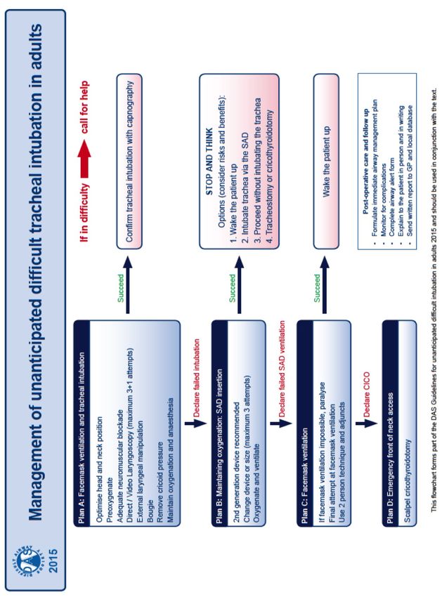
WELCOME AND INTRODUCTION

Dear Candidate

Welcome to “An Introduction to Anaesthesia‟. This is intended to be a one day
survival guide for those of you new to Anaesthesia, or who want to know more
about a career in Anaesthesia.

This handbook is intended to supplement the lectures. As this is the first course for
those of you new to Anaesthesia, we really want your feedback! Is there anything
you’d like in, anything not done well, or anything that was great? In addition coffee
and lunch should give you a chance to discuss with the course tutors any questions
you might have.

We’d recommend you start reading as soon as you start your post, as it all seems
quite new. To start try using the following three books:

       Anaesthesia and Intensive Care A to Z: An Encyclopedia of Principles and
       Practice 3rd Edition (Yentis, Hirsch & Smith)

       Drugs in Anaesthesia & Intensive Care (Sasada and Smith)

       Respiratory Physiology: The Essentials (John West)

We also think you should sign up to

       Join the Association of Anaesthetists of Great Britain and Ireland
       (http://www.aagbi.org )

       e-learning for Anaesthesia (www.e-lfh.org.uk/projects/ela/index.html)

       Check out the Royal College of Anaesthetists (www.rcoa.ac.uk ) website
       especially their e-learning site at http://www.rcoa.ac.uk/e-la

Good luck

The Course Directors
Rob, Mo, Hannah and Anita

If you’d like to find out more about our research and educational activities, we would love to hear from you,
please contact us via the website: www.ucl.ac.uk/anaesthesia/education/AnaesthesiaIntroduction or Rob via
www.ucl.ac.uk/anaesthesia/people/stephens

                                         Introduction to Anaesthesia

                                                    Page | 2
TABLE OF CONTENTS

THE ABC OF Anaesthesia ......................................................................................... 5

  General Anaesthesia (GA) .................................................................................................................... 5

     Induction .......................................................................................................................................... 5

     Maintenance .................................................................................................................................... 6

     Emergence ....................................................................................................................................... 7

  Complications of General Anaesthesia ................................................................................................ 7

A is for AIRWAY ...................................................................................................... 9

  Airway assessment............................................................................................................................... 9

  The difficult airway .............................................................................................................................. 9

  Airway management .......................................................................................................................... 10

  When to intubate – a decision-making framework: .......................................................................... 11

  Non-technical skills of airway management ...................................................................................... 12

  Extubation………………………………………………………………………………………………………………………………………12

B is for BREATHING ........................................................................................... 1414

  Assessing the adequecy of breathing ................................................................................................ 16

  Ventilation during general anaesthesia…. ……………………………………………………………………………………..17

  Causes of hypoxaemia……………………………………………………………………………………………………………………19

C is for Circulation ................................................................................................. 20

  3 Key physiology equations ............................................................................................................... 20

  Effects of anaesthesia + surgery on the cardiovascular system......................................................... 20

  Key Cardiovascular stages in the ‘Anaesthesia Journey’.................................................................... 21

D IS FOR Anaesthetic Drugs ................................................................................... 23

  Non-anaesthetic drugs (usual medication) ........................................................................................ 23

  Premedication.................................................................................................................................... 23

  General Anaesthesic Agents .............................................................................................................. 24

  Muscle Relaxants ............................................................................................................................... 26

                                                      Introduction to Anaesthesia

                                                                     Page | 3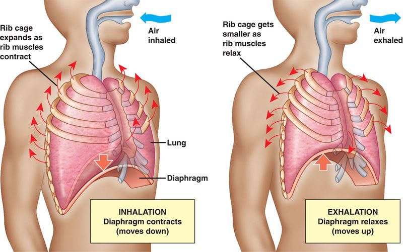
Uppers and Downers ......................................................................................................................... 27

   Pain Killers.......................................................................................................................................... 27

   Anti-emetics ....................................................................................................................................... 28

P is for preassessment .......................................................................................... 29

  Building a rapport…………………………………………………………………………………………………………………………..29

   Assessing risk ..................................................................................................................................... 29

   Structured approach to patient assessment...................................................................................... 31

   Perioperative planning....................................................................... Error! Bookmark not defined.33

R is for Regional anaesthesia and Pain .................................................................. 35

   Why manage pain? ............................................................................................................................ 35

   Methods of managing pain ................................................................................................................ 36

E is for Emergencies .............................................................................................. 40

  Anaphylaxis ......................................................................................................................................... 43

  Bradycardia ......................................................................................................................................... 44

  Laryngospasm ..................................................................................................................................... 45

F is for Fluid and Blood transfusion ....................................................................... 46

   Transfusion Triggers........................................................................................................................... 46

   Blood Component Therapy ................................................................................................................ 47

   Massive Haemorrhage ....................................................................................................................... 49

   Crystalloids + Colloids and contoversies ............................................................................................ 51

   Crystalloids vs Colloids ....................................................................... Error! Bookmark not defined.52

   Hypovolaemia .................................................................................................................................... 54

Appendix 1 Failed Intubation Procedure ............................................................... 54

Appendix 2 Management of LA toxicity.................................................................56

Appendix 3 Malignant Hyperthermia Guidelines………………………………………………….57

Appendix 4 Machine Checklist …………………………………………………………………………….58

                                                        Introduction to Anaesthesia

                                                                       Page | 4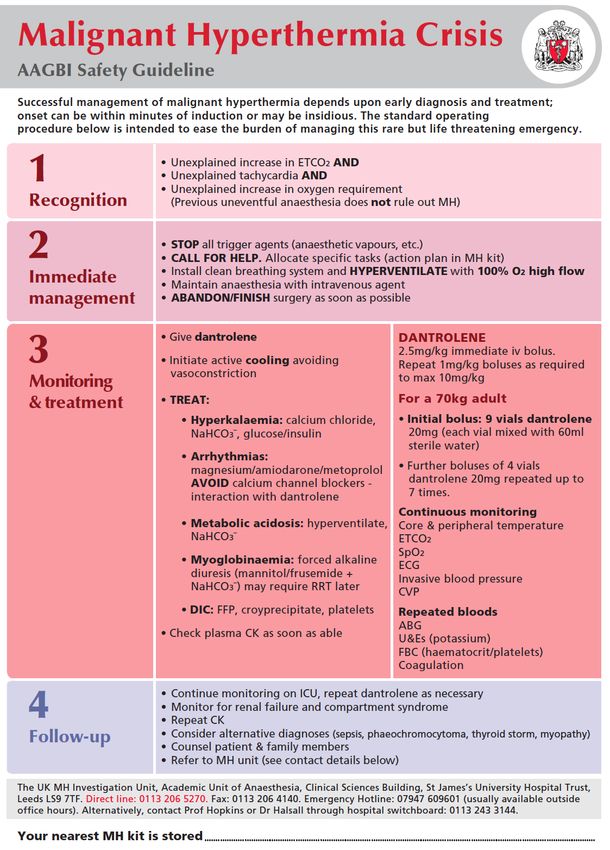
THE ABC OF ANAESTHESIA

                                       A PRACTICAL CONDUCT
                                         DR ELISA BERTOJA

Anaesthesia: an-aesthesia from Greek meaning “without sensation”

The practical conduct of anaesthesia is just a brick in the anaesthetic wall.

Anaesthesia starts well before the anaesthetic room. Our role in the patient pathway can be divided
in three stages:

              PRE OPERATIVE CARE

              INTRA OPERATIVE CARE

              POST OPERATIVE CARE

INTRA OPERATIVE CARE is only one component of our job. Thanks to our knowledge of anatomy,
physiology, pharmacology, physics and years of experience, we can run safe and effective anaesthesia
in several different ways. The primary distinction is between:

              GENERAL ANAESTHESIA

              REGIONAL ANAESTHESIA

GENERAL ANAESTHESIA (GA)

We can divide the conduct of a GA in three main components

              INDUCTION

              MAINTENANCE

              EMERGENCE

INDUCTION

This is the first part of the intra operative time. It usually happens in the anaesthetic room, sometimes
in theatre, and in emergencies in A and E or on the Wards. By administering selective drugs we induce
a loss of consciousness (hypnosis), which is temporary and reversible. The loss of consciousness can
be achieved via intra venous drugs or inhalational agents.

Together with the loss of consciousness comes the loss of the airway, and loss of the airway
protective reflexes. Therefore a patient undergoing GA needs an airway device to maintain airway
patency (A) and to be able to self ventilate or to be ventilated by a machine (B).

The drugs used to induce and maintain anaesthesia do have an impact on the patients’ cardiovascular
system(C). One of our roles is to preserve the patients’ cardiovascular stability.

                                      Introduction to Anaesthesia

                                                 Page | 5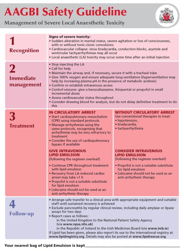
Inducing anaesthesia means that we willingly take a patient from a self sufficient and safe condition to
a state of dependency (on us, our drugs and our machines). This is pretty unique in medicine!

Lack of awareness does not imply lack of response to pain. Just because the patient is asleep does not
mean that he/she is pain free. Before surgery can start we need to administer drugs that blunt the
body response to pain.

MAINTENANCE

During the maintenance of anaesthesia we have to achieve two goals:

MAINTAIN SAFE AND STABLE PATIENT CONDITIONS

IMPLEMENT CONDITIONS FAVOURABLE TO THE SURGEON

A SAFE AND STABLE PATIENT

During anaesthesia a patient and his/her body do not respond to stimuli as if he/she is awake. On top
of that the anaesthetic and surgical procedures implement acute changes to patients’ physiology
(induction agents, surgical insult, blood loss etc). As Anaesthetists, we are in charge of maintaining
patients’ homeostasis. For this reason we always use a variety of monitoring devices.

A patient under GA has the following standard monitoring (Association of Anaesthetists of Great
Britain and Ireland (AAGB) standard guidelines)

ECG
Pulse oximeter
Blood pressure cuff
Inspiratory and expiratory airway gases, including oxygen (O2), carbon dioxide (CO2), and anaesthetic
vapours.
Ventilatory parameters : Tidal Volume, Minute ventilation, Airways pressures,
Other devices are available to the expert Anaesthetist during major or specialized surgical procedures
(Invasive pressures measurement, Doppler, Arterial Blood Gas) and Depth of anaesthesia monitoring
(BI Spectral Index etc).

All procedures and data is documented on the Anaesthetic chart for intraoperative, post-operative,
and future references. This is a legal document.

CONDITIONS FAVOURABLE TO THE SURGEON

Anaesthetists and surgeons work as a team for the patients’ benefit. We can improve the surgical
result by liaising with the surgeon before the beginning of the operation, choosing the appropriate
anaesthetic technique (e.g. avoid nasal intubation for a nasal polyp extraction!), interfering with
patient physiology to improve surgical conditions. (e.g. Blood pressure, muscle relaxation etc.)

                                     Introduction to Anaesthesia

                                               Page | 6
EMERGENCE

The importance of emergence from anaesthesia is frequently underestimated by non-anaesthetists. It
is a delicate time of the anesthetic conduit. It can be a dangerous time.

Our main goals during emergence are for the patient to:

    •         Regain consciousness

    •         Regain control on his/her airway and ventilation

    •         Gain satisfactory pain control

It is a challenging task and there is not just one way to achieve it. As Anaesthetists, we gradually hand
over the control of the A B C back to the patient. As much as the induction, emergence can be a grey
area, whereby complications (fail to extubate, sudden increase in BP/ HR etc.) can occur.

COMPLICATIONS OF GENERAL ANAESTHESIA

COMMON

Post Operative Nausea and Vomiting (PONV) (1 in 3)

Sore throat (1 in 4)

Chest infection (1in 5)

Cognitive dysfunction including, confusion and dizziness (1 in 5)

Muscle weakness, Aches and pain

Shivering or Itching

UNCOMMON

Awareness (1.5 in 1,000 – 1 in 42,000)

Teeth and lips damage (1 in 4500)

Breathing difficulties

Nerve Damage

RARE

Death due to Anaesthesia (1 in 185,000)

Serious allergic reaction (1: 10,000- 1:20,000)

Equipment failure

Damage to eyes

                                      Introduction to Anaesthesia

                                                  Page | 7
SUMMARY
TAKE HOME MESSAGE:
ü THE ANAESTHETISTS’ WORK BEGINS WELL BEFORE THEATRE
ü EACH ANAESTHESIA IS TAILORED FOR EACH PATIENT AND PROCEDURE
ü WE WORK TOGETHER WITH THE SURGEON
ü PATIENT SAFETY IS PARAMOUNT
Introduction to Anaesthesia
Page | 8A IS FOR AIRWAY
DR ED BURDETT
DR RAVI ALAGAR
One of the consequences of general anaesthesia and deep sedation is that the patient is unable to
maintain a patent airway - Anaesthetists are necessarily airway specialists.
In the emergency scenario, the airway always comes first. This is because resuscitation is futile unless
you can delivery oxygen to the lungs.
A ‘difficult airway’ is one in which ventilating an anaesthetized patient with a mask or intubating
them is difficult. Airways can be difficult at extubation too.
AIRWAY ASSESSMENT
History: Previous difficult airway
Other disorders (see below)
Examination: not very sensitive or specific
Neck flexion and extension
Mouth opening
Mallampati score (MP) Range 1-4
Thyromental distance
Atlanto-axial range of movement
Investigations: Imaging e.g. CT; flexion/extension cervical spine x-ray
Obstructive lung defects
THE DIFFICULT AIRWAY= DIFFICULT VIEW OR VENTILATION
Beware the following patients:
The multisystem disorders
Bony disorders - rheumatoid arthritis; ankylosing spondylitis
Soft tissue disorders – marfans, acromegaly scleroderma
The airway disorders:
Previous head/neck radiotherapy or surgery
Airway pathology e.g. tonsils, oral abscess, throat cancer
Introduction to Anaesthesia
Page | 9The acutely unwell:
Bowel obstruction – aspiration can be huge
Facial/mouth trauma or burns
Anyone in an unfamiliar area e.g. the ward, radiology etc.
Confused or aggressive patients
The others:
Pregnant women
Children with syndromes
Celebrities
This list is not exhaustive, but you get the idea.
AIRWAY MANAGEMENT
‘Managing’ a patient’s airway is necessary if, the patient cannot maintain a patent airway, they are in
respiratory failure, or they require a general anaesthetic. It includes
Supplemental oxygen
Non-invasive Manoeuvres:
Chin lift, jaw thrust
Suction, oral toilet, removal of FB etc.
Invasive manoeuvres:
Supra-glottic airways
a. Oropharyngeal / Nasopharyngeal airway
b. Laryngeal mask airway and similar
Endotracheal
a. Oral/nasal endotracheal tube
b. Tracheostomy
Introduction to Anaesthesia
Page | 10WHEN TO INTUBATE – A DECISION-MAKING FRAMEWORK:
The airway must be managed if:
Severe respiratory/systemic illness
Cannot maintain airway eg neurological (GCSNON-TECHNICAL SKILLS OF AIRWAY MANAGEMENT
Airway management is usually technically easy, but like any high-risk task performed under time
pressure it can be stressful.
Here are a few ways of making airway management smoother.
Ensure that you and your team are aware of your limitations, both your level of experience and
acutely (tired, hungry, unfamiliar with surroundings etc.)
Preparation is everything:
Consider your options - have a plan B and plan C if plan A fails. You should be calling for help
before plan C.
Get ready: equipment, drugs, and anything you might need if things don’t go well
Check the patient is optimised - best possible oxygenation and position
NG tube if any risk of full stomach
Check oxygen, monitoring, suction, machine and IV access beforehand
Communicate well: this is surprisingly difficult when you’re under pressure. Ensure that everyone else
in the team is briefed and aware of their roles.
EXTUBATION
What happens after removing your chosen airway device (extubation) can be risky - even more than
intubation.
Respiratory (e.g. coughing, laryngospasm, desaturation) and cardiovascular complications
(tachycardia, hypertension, arrhythmia) may happen more at extubation than at intubation.
Complication rates during intubation have fallen over recent years due to our use of a systematic
approach. However there’s now much more focus on optimising the way we extubate.
Consider Identifying who is at high risk of problems during extubation?
• Known difficult airway
• Deteriorating airway post-intubation (oedema/bleeding)
• Aspiration risk
• Cardiovascular /respiratory comorbidity
Introduction to Anaesthesia
Page | 12Optimisation
• Patient position
• Equipment ready
• Skilled assistance available- and a senior anaesthetist
• Awake or deep extubation
Handover and monitoring after extubation
The intubation, management and extubation guidelines by DAS, the Difficult Airway Society below
are useful
https://www.das.uk.com/guidelines/downloads.html
Introduction to Anaesthesia
Page | 13B IS FOR BREATHING
DR IRENE BOURAS
WHY WE BREATHE
We breathe to enable gas exchange ie the transfer of O2 from the air to the tissues and the removal of
CO2 from the tissues back into the air. The average adult consumes 250mls oxygen per minute and
produces 200mls CO2 per minute. Air contains 21% O2.
OXYGEN DELIVERY
The delivery of O2 to the tissues depends on a number of variables
O2 delivery = arterial blood O2 content x Hb x cardiac output
A change in any one of these variables will affect the amount of O2 reaching the tissues
HOW WE BREATHE
There are 3 respiratory control centres in the brain which control and co-ordinate breathing. They are
the:
Medullary inspiratory area
The pneumotactic area
The apneustic area
These respiratory centres receive inputs from 3 sets of sensory neurons. The central and peripheral
chemoreceptors, which monitor plasma pH and CO2 and O 2 concentrations and the stretch receptors
located in bronchial walls are activated when the lungs are expanded and trigger cessation of
inspiration.
MECHANICS OF BREATHING
Inspiration:
Inspiration occurs when the inspiratory muscles (diaphragm & external intercostals) contract. This
elevates the ribs and sternum and causes an increase in the size of the thoracic cavity. The sudden
increase in the relative size of the thoracic cavity creates negative intra-thoracic pressure which
causes air to move into the chest (from an area of high pressure to an area of lower pressure).
Inspiration is an active process i.e. requires energy.
Expiration:
Expiration occurs when the diaphragm and external intercostal muscles relax. This causes the lungs to
recoil back to their original volume. The reduction in volume results in a pressure increase within the
lungs and expiratory gases being expelled from the lungs. Under resting conditions expiration is an
entirely passive process. However, during more laboured breathing the intercostal and abdominal
muscles are used which requires energy.
Introduction to Anaesthesia
Page | 14LUNG VOLUMES AND CAPACITIES
The most important lung volumes are:
Tidal Volume (the amount of air inspired during breathing at rest, approximately 500mls)
Functional Residual Capacity (the amount of air remaining in the lungs after normal expiration,
approximately 2.5L). Whilst breathing room air 79% of the gas in the FRC is nitrogen, if a patient is
given 100% O2 to breathe for several minutes then all of this nitrogen will be replaced with O2. The
2.5L O2 in the FRC is enough O2 to last for 10 minutes.
The reserve volumes are the volumes that can be inspired/expired over and above a normal tidal
volume breath.
Introduction to Anaesthesia
Page | 15Not all of the inspired air takes part in gas exchange, only the air which is in the alveoli does. The air in
the bronchi, bronchioles and trachea which does not contribute to gas exchange is known as dead
space.
RESPIRATORY FAILURE/INSUFFICIENCY
As breathing is regulated by the nervous system and requires the use of the respiratory muscles and a
patent airway any disorders which affect these may impair breathing. Problems which prevent
effective gas exchange in the lungs either by preventing the inspired oxygen diffusing into the blood
stream or by limiting the blood supply to the lungs (hypoperfusion) will impair ventilation. These may
be congenital (e.g. motor neuron disease) or acquired (e.g. pneumonia).
Problems with breathing can occur preoperatively, intra-operatively and post-operatively. Intra and
post operative breathing problems can also be attributed to:
Patient factors - a pre-existing problem which may have been worsened e.g. COPD
Anaesthetic factors – e.g. excess opiates or a high epidural block which prevents the muscles of
respiration working appropriately
Surgical factors – eg diaphragmatic splinting caused by a distended abdomen or a CO2 gas embolism
during laparoscopic surgery
ASSESSING ADEQUACY OF BREATHING
This can be done either clinically, at the bedside, or with more invasive procedures. You may need to
assess breathing pre-op, intra-op or post-op.
CLINICALLY
Does the breathing pattern and rate look comfortable and normal? Are accessory muscles of
ventilation being used? Are they cyanosed? Is there evidence of airway obstruction? Does the chest
sound normal on auscultation?
Causes of tachypnoea: compensating a metabolic acidosis (eg sepsis, DKA), hypoxeia, hypercapnia,
pain, anxiety
Causes of bradypnoea: excess sedatives or opioids, CNS pathology
Oxygen saturations
Tell you only about oxygenation not ventilation; if a patient is on supplemental oxygen, oxygen
saturations may mask worsening respiratory failure and hypercarbia (high CO2).
Peak Expiratory Flow Rate/Bedside Spirometry
These tests may be useful if done serially but rarely done in practice.
Introduction to Anaesthesia
Page | 16CLINICAL DEFINITION OF RESPIRATORY FAILURE
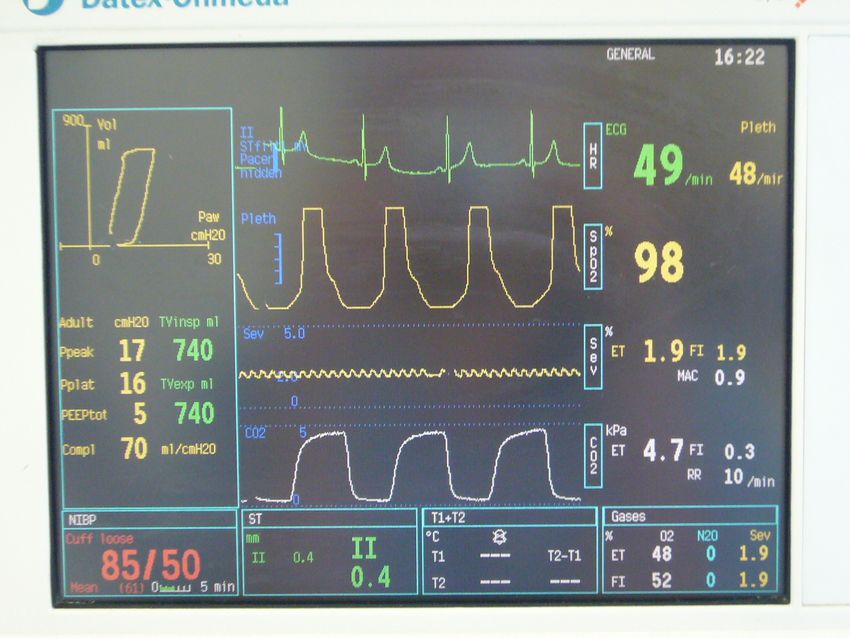
Type 1 respiratory Failure = PaO2MONITORING VENTILATION
This is a picture of a
typical patient monitor
on an anaesthetic
machine. It includes an
ECG and BP as well as
oxygen saturations and
capnography. The box
to the left of the
picture includes a
flow-volume curve
and also gives an
estimate of lung
compliance i.e. the
pressure required to
inflate the lungs.
CLINICAL
During anaesthesia the anaesthetist constantly observes for any change in breathing pattern, rate and
asymmetrical movement of the chest. Common problems include hypoventilation and unilateral chest
movement if the tracheal tube has been inserted too far into either the left or right main bronchus.
CAPNOGRAPHY
The white waveform circled in the diagram on the previous page represents end-tidal CO2 (ETCO2).
This is the concentration of CO2 which the patient breathes out. Changes in ETCO2 may represent
hyperventilation (in which case the ETCO2 falls) hypoventilation (in which case the ETCO2 rises).
Sudden falls in ETCO2 maybe one of the first signs of cardiac arrest or severe hypotension; the lack of
lung perfusion in these conditions means that no CO2 is delivered to the lungs and therefore cannot
be exhaled.
OXYGEN SATURATIONS
The yellow waveform circled is the O2 saturations. There must be a good waveform for the saturations
recorded to be accurate. The waveform also gives you the heart rate.
Introduction to Anaesthesia
Page | 18CAUSES OF HYPOXAEMIA
These apply to anaesthetised and non-anaesthetised patients. They relate to the factors that
determine oxygen delivery.
HYPOXIC HYPOXAEMIA: This is when not enough O2 is transported across the lungs. It can be
due to a low concentration of O2 in the inspired air (eg anaesthetic machine failure) or impaired
oxygen transfer across the lungs.
STAGNANT: If there is gross circulatory insufficiency (shock). Then there may not be sufficient
cardiac output to transfer the oxyhaemoglobin to the tissues.
ANAEMIC: If there is insufficient haemoglobin then the O2 will not be transported to the tissues.
CYTOTOXIC: This occurs when O2 arrives at the tissues but they are unable to utilise it. Examples
include cyanide toxicity which causes mitochondrial dysfunction. In this case the oxygen saturations
will be normal.
As anaesthetists we assess breathing pre-operatively, intra-operatively and post-operatively.
Therefore, a knowledge of respiratory physiology and pathology is important to help us optimise our
patients before theatre and to explain any changes in ventilatory function. In patients with severe
lung disease we may try to avoid giving a general anaesthetic by giving a regional anaesthetic (eg. a
spinal). An understanding of the respiratory system along with the cardiovascular system is vital for
anaesthetists as small changes in these systems can cause significant changes to the physiology of the
entire body.
EVIDENCE
We know from ICU studies that over ventilation and lack of PEEP can damage already precariously
functioning lungs. In 2013 Futier published a study in the NEJM ' A Trial of Intraoperative Low-Tidal-
Volume Ventilation in Abdominal Surgery'. He showed that, in patients at risk of respiratory
complications, those randomised to 'lung protective ventilation'
o 6-8ml/kg of ideal body weight
o 6-8cm PEEP
o recruitment manouvres
had less postoperative acute respiratory failure and shorter length of stay. Some of us use the app
MedCalc to work out the ideal 6-8ml/kg values, which depend on gender and height.
Introduction to Anaesthesia
Page | 19C IS FOR CIRCULATION
DR ROBERT CM STEPHENS
The primary purpose of the circulation (heart, vessels & blood) is to supply oxygen to the tissues
and remove waste products from cells. The Circulation in anaesthesia is a large subject: here is an
introduction!
3 KEY PHYSIOLOGY EQUATIONS
Heart work µ Heart oxygen requirements
Heart work µ Preload, Afterload, Contractility, Heart rate
Message: Increasing these 4 factors may increase coronary ischaemia
Mean Arterial Pressure (MAP) = Cardiac Output (CO) x Systemic Vascular Resistance (SVR)
Cardiac Output (CO) = Heart Rate (HR) x Stroke Volume (SV)
Message: This is one way to work out why someone is hypotensive
Oxygen delivery (DO2)= Cardiac Output (CO) x Oxygen content of blood
Oxygen Content = (SaO2 x Haemoglobin x 1.39) + (0.003 x PaO2)
Message: These are the ways to increase systemic oxygen delivery
EFFECTS OF ANAESTHESIA + SURGERY ON THE CARDIOVASCULAR SYSTEM
Blood is pumped (blood flow ~5L/min) down a pressure gradient from the left ventricle to the cells.
Tissues need a constant oxygen delivery to maintain oxidative metabolism.
Blood vessels vasodilate or vasoconstrict when blood pressure changes to maintain a constant blood
flow. This phenomenon is known as autoregulation.
General Anaesthesia on its own (hypnosis, analgesia, paralysis) generally reduces blood pressure by
reducing cardiac output and causing vasodilation. Fluids, vasoconstrictors and inotropes can be given
to correct this. In addition most anaesthetic agents abolish the process of autoregulation, thus oxygen
delivery is determined primarily by perfusion pressure (MAP)
Spinal/Epidural generally cause vasodilation, leading to hypotension and a compensating tachycardia
Introduction to Anaesthesia
Page | 20Stimulation (laryngoscopy or surgical incisions) generally opposes the effects of a general anaesthetic by increasing cardiac output (mostly heart rate) and causing vasoconstriction (see equation). Some procedures can cause vagal stimulation (e.g. dilating the cervix ) which can cause bradycardias, and others have more complex effects eg inflating the peritoneum with CO2 during laparoscopies General and regional (spinal/epidural) anaesthesia both affect the heart and vascular resistance in different ways There is no evidence as to the ‘level’ of blood pressure (mean arterial pressure) considered acceptable under anaesthesia. Patient (Preoperative hypertension, end organ perfusion monitors etc.) and surgical (bleeding) have to be considered. For non-hypertensive patients with normal end organ perfusion under 70 years of age most anaesthetists would be happy with a mean pressure of over 60mmHg. The degree of acceptable heart rate change from baseline mainly depends on the patients’ cardiac condition. E.g. patients with ischaemic heart disease should not have a tachycardia as that increases cardiac O2 demands and reduces time for coronary blood flow. Cardiac monitors: all patients (GA, regional, sedation) should have ECG, Sa02, non-invasive BP. Invasive monitors (arterial line, CVP, cardiac output + urine output) should be placed into patients having major surgery (eg laparotomy or >500ml blood loss) especially if they have limited function (can walk up
SUMMARY

You should consider the effects of…..

                                              Anaesthesia

                                 Patients’ cardiovascular state

                                                 Surgery

                                                  Drugs

Upon the……………..

                                                  Heart

                                                 Vessels

                                                  Blood

                                                   Cell

                                        Introduction to Anaesthesia

                                                Page | 22
D IS FOR ANAESTHETIC DRUGS
DR JAMES HOLDING
DR MARYAM JADIDI
NON-ANAESTHETIC DRUGS (USUAL MEDICATION)
Patients are now encouraged to continue all their usual medications throughout the perioperative
period with only a few exceptions:
DIABETIC MEDICATIONS
The aim is to maintain a perioperative BM of 4-10, and particularly to avoid hypos. And so insulin and
oral hypoglycaemics should be omitted on the morning of surgery (and very long acting drugs from
the evening before), and the BM monitored. Insulin sliding scale may be needed.
ANTICOAGULANTS
Aspirin – is generally now continued perioperatively, the risk of MI / stroke if it is stopped being
greater than the risk of significant bleeding if it isn’t, but it is stopped 7 days before some bleeding
critical operations e.g. neurosurgery, arthroscopy.
Clopidogrel – is generally stopped 7-10 days preoperatively – but not lightly in patients with drug
eluting coronary stents.
Warfarin – is normally stopped 5 days pre-operatively. When the INR is below 2 a daily treatment
dose of unfractionated heparinmay be given for all indications except those on warfarin for simple AF
prophylaxis 'bridging plan'. The last dose of heparin should be given 24 hours before surgery. If there
are no concerns about bleeding post-operatively a smaller dose of unfractionated heparin is given
after surgery, returning to the treatment dose on the first post-op day, which is continued until the
INR has returned to a therapeutic level.
Antihypertensives – continue all, except ACE inhibitors and AT II inhibitors, which should be omitted
on the morning of surgery for patients having laparoscopic surgery -there are reports of profound
hypotension. Some people continue these if the patient has a diagnosis of heart failure.
Dabigatran and Rivaroxaban- newer oral anticoagulants stopped 1-2 days before surgery
PREMEDICATION
In the past all surgical patients received a heavy, long lasting premed combining anxiolysis, analgesia
and a secretion drying agent. The advent of modern anaesthetic drugs and techniques has reduced
the need for this kind of premed.
Introduction to Anaesthesia
Page | 23GENERAL ANAESTHESIC AGENTS
Here is a secret. For most operations anaesthesia is induced with a dose of short acting opiate and an
intravenous induction agent. These drugs ‘buy’ a few minutes of unconsciousness in which time the
patient is set up to breathe some anaesthetic vapour that keeps them asleep (maintenance). At the
end of the operation the anaesthetic vapour is turned off, the patient breathes it out of their system,
and then they wake up. Don’t tell the surgeons, they still think it is some kind of magic.
Induction can also be achieved by just breathing the anaesthetic vapour, which can be useful for
children, and others in whom cannulas are difficult to place. Specific muscle paralysing drugs are
sometimes used to facilitate tracheal intubation, enable the respiration to be controlled, and to allow
surgery within body cavities.
The following list is not exhaustive, but includes the drugs that are commonly used by UK
anaesthetists in 2014. Doses have been omitted on purpose – they can be very variable.
Anaesthetists tend to titrate the dose to effect. (The single bolus dose of most anaesthetic drugs
(except vasopressors and remifentanil) for a standard 80 kg adult is about one ampoule).
INTRAVENOUS INDUCTION AGENTS
A single bolus dose of an induction agent quickly causes brain levels of the drug to rise and results in
almost instantaneous anaesthesia. This is followed by a rapid fall in brain and blood levels as the drug
is redistributed to other tissues. The result is a brief duration of anaesthesia with a rapid recovery.
The emergence from anaesthesia after a single bolus dose is due to redistribution of the drug, not
metabolism and elimination – which may take many hours.
PROPOFOL – which comes as a 1% emulsion (like milk), and produces a smooth and rapid
induction of anaesthesia with greater laryngeal reflex suppression than other agents. It is thought to
have an antiemetic effect, and its metabolism and elimination are little affected by hepatic or renal
failure. The major side effect is a dose-dependant reduction in vascular tone, causing both a
reduction in SVR and CVP (and preload) resulting in a reduced cardiac output and blood pressure. It
can also be painful on injection, although this can be reduced by injecting into a big vein or by adding
some lignocaine to the propofol. Propofol has become the dominant induction agent over the last 10
years.
THIOPENTONE – this was the universal induction agent before propofol came along. Its action is
similar to propofol, although the period of unconsciousness in generally longer and there is more
‘hangover’, but there is less cardiovascular depression. It is the classic drug to be used in a rapid
sequence induction (RSI), and is a classic trigger for porphyria.
ETOMIDATE – a cardio-stable induction agent, which has recently fallen out of favour as it inhibits
corticosteroid and mineralocorticoid synthesis.
KETAMINE – a non-competitive antagonist of the calcium ion channel operated by the excitatory
NMDA glutamate receptor, but also has effects at opiate and mACh receptors. It is a strange drug,
which produces a strange kind of anaesthesia (dissociative) and analgesia. Laryngeal reflexes are
preserved, there is bronchodilatation, and the heart rate and blood pressure are increased. It is the
Introduction to Anaesthesia
Page | 24only induction agent that is also effective if given intra-muscularly (IM). Its use has been limited by
fears of dissociative side-effects on emergence from anaesthesia (hallucinations and nightmares).
It might be a good choice for the induction of patients with bronchospasm (e.g. acute asthma), but is
more commonly used in sub anaesthetic doses for its analgesic effect.
MAINTENANCE AGENTS
The pharmacokinetics of gases and vapours when they enter a body are difficult and non-intuitive.
The important value is the partial pressure, which is the driving force behind gas transfer. A vapour is
a gas, and acts like a gas – but it is below its boiling point. Here are two useful concepts:
If a gas is very insoluble in blood, then not many of the gas molecules have to transfer over the
gas/blood interface in the lung before equilibrium of the partial pressures is reached, and so it will be
reached relatively quickly. And at the end of the operation, when the gas is removed from the
breathing circuit, not many molecules have to move out of the blood before the blood partial
pressure of the gas falls towards zero, and so the patient wakes up relatively quickly.
The minimal alveolar concentration (MAC) has been calculated for all anaesthetic gases / vapours, it is
a measure of potency (the name is an historical error – it should be minimal alveolar partial pressure
– but as atmospheric pressure at sea-level is about 100kPa it doesn’t matter because the values for
pressure and concentration are the same). One MAC is the concentration of anaesthetic agent, which
at equilibrium will prevent a reflex response to a standardised skin incision in 50% of patients. As the
alveolar concentration can be assumed to be the end expiratory concentration, which can be
measured by a gas analyser, we can check that a patient is receiving enough agent to keep them
anaesthetised.
ANAESTHETIC VAPOURS – their clinical effects are all broadly similar. They produce
respiratory depression, bronchodilatation, and reduced blood pressure – through a mixture of
reduced myocardial contractility and vasodilatation. None have analgesic properties. Isoflurane is
the most soluble in blood, followed by sevoflurane and then desflurane, the least soluble
Isoflurane (ISO) – MAC = 1.1
Desflurane (DES) – MAC = 6 The most insoluble – so the fastest to equilibriate – but a respiratory
irritant, so unsuitable for gaseous induction.
Sevoflurane (SEVO) – MAC = 2.2 Used for gaseous induction.
NITROUS OXIDE – a gas. MAC = 105 – so it is unable to be a sole anaesthetic agent, but it can be
a carrier agent for the anaesthetic vapours. As MAC is additive a reduced amount of vapour is needed
when nitrous oxide is used. It is also an analgesic, and is very insoluble in blood. It has become
unfashionable recently due to its side effects of causing gas filled bowel to expand, increasing nausea,
and suppressing bone marrow production of neutophils and platelets.
PROPOFOL – again. Propofol is redistributed and eliminated reliably enough to maintain
anaesthesia when given as an infusion. The pharmacokinetics of infusions of drugs, which are not
metabolised almost immediately, are particularly complex. Maintenance of a steady plasma level
requires a continually changing infusion rate. This is achieved with a computer controlled infusion
pump using an algorithm based on the patient’s sex, age and weight. Unlike with MAC for the
Introduction to Anaesthesia
Page | 25vapours, we are unable to measure in real-time the plasma levels of propofol so can only judge the
effectiveness of anaesthesia by monitoring the clinical effects.
MUSCLE RELAXANTS
These act at the post-junctional nicotinic (nACh) receptors of the skeletal muscle neuromuscular
junction (NMJ). The nACh receptor is a ligand gated sodium ion channel. Binding of ACh (or nicotine)
causes the channel to open, allowing sodium to move into the cell, depolarizing the membrane
potential.
NON-DEPOLARIZING MUSCLE RELAXANTS
These drugs competitively and reversibly bind to the receptor preventing ACh reaching its binding
site. The duration of action of a single bolus dose is: Vecuronium (40mins) > Atracurium (35mins) >
Rocuronium (30mins). Atracurium causes more histamine release, and therefore more
bronchospasm and hypotension, but it spontaneously fragments in the circulation (Hofmann
degradation), and is widely metabolised, so is more suitable for patients with renal or hepatic
impairment.
DEPOLARIZING MUSCLE RELAXANTS
SUXAMETHONIUM mimics the effect of ACh when it binds at the nACh receptor causing
depolarisation of the cell and twitching of the muscle fibres. As these twitches are uncoordinated
they result in fasciculation of the muscle body. Suxamethonium is the fastest acting of the muscle
relaxants (within 30 secs), and the fastest to wear off (within 7 mins), so is the relaxant of choice for
rapid sequence induction. Sux is an old, dirty drug though, with a lot of side effects. It can stimulate
nACh receptors at autonomic ganglia, causing bradycardia, it causes post-op muscle pain and nausea,
results in a rise in plasma potassium and it is associated with a high rate of anaphylaxis. The effect of
sux is terminated by diffusion away from the NMJ and then hydrolysis by plasma cholinesterases.
Plasma cholinesterase deficiency is relatively common, both genetic and acquired, and results in a
prolonged block following a dose of suxamethonium (sux apnoea).
REVERSALS
NEOSTIGMINE is an acetylcholinesterase inhibitor. It is used when a non-depolarizing block has
already begun to wear off to increase the NMJ synaptic concentration of ACh to help overcome the
block and restore muscle power and function. It is another dirty drug, and also acts at autonomic
ganglia, causing bradycardia, bronchoconstriction and smooth muscle peristalsis. To minimise these
effects it is given together with an anti-cholinergic (atropine or glycopyrulate).
SUGAMMADEX is a relatively new drug that is designed to bind with the steroid non-depolarizers
(rocuronium and vecuronium) and stop them acting at the NMJ. It has the potential to reverse even
profound blocks with little stimulation of the autonomic system.
Introduction to Anaesthesia
Page | 26UPPERS AND DOWNERS
The anti-cholinergics atropine and glycopyrulate are used to reduce vagaly mediated bradycardia,
and to dry secretions. Unlike atropine, glycopyrulate does not cross the blood-brain barrier, and does
not cause sedation.
The pure a adreno receptor agonists phenylepherine and metaraminol predominantly cause a
transient rise in blood pressure by peripheral vasoconstriction. The mixed a and b adreno agonist
ephedrine raises BP by a combination of vasoconstriction and increased heart rate.
Most anaesthetists will reduce blood pressure by giving more anaesthetic agent or opiate, although
short acting b-blockers (labetalol, esmolol), GTN, and the a2 agonist clonidine, are also regularly
used.
PAIN KILLERS
OPIOIDS
The side-effects of opioids include respiratory depression, hypotension, bradycardia, delayed gastric
emptying, nausea, constipation, itching and muscle rigidity – but they are still the mainstay of surgical
analgesia. By using clean short acting drugs these side-effects can be minimised. (In brackets is the
equianalgesic dose to 10mg of IV morphine, and the approximate duration of action)
MORPHINE (10mg, 120mins) – metabolism produces active metabolites which can accumulate in
renal failure.
FENTANYL (100mcg, 30 mins)
ALFENTANIL (1mg, 15mins)
REMIFENTANIL – rapidly metabolised by plasma and tissue esterases, and is very short acting. It has
to be given as an infusion, and can cause severe bradycardia if given as a bolus.
CODEINE (30-60mg, 120 mins) – about 10% is metabolised to morphine, although there is genetic
heterogeneity, some people unable to metabolise any, and others metabolising almost 100% to
morphine. Its effects are therefore unpredictable. Use dihydrocodeine- same dose.
TRAMADOL (50-100mg, 120mins) – has some weak effects at opioid receptors, but also seems to
modulate 5-HT, NMDA and noradrenergic receptors. It is not a wonder drug though, its nausea
profile being similar to morphine.
NON-OPIOIDS
These include the usual drugs paracetamol and diclofenac, and the more unusual ketamine and
clonidine.
Introduction to Anaesthesia
Page | 27ANTI-EMETICS
Post-operative nausea and vomiting (PONV) is common with multiple causes (opiates, stimulation of
vagus, hypotension, starvation, ileus) and risk factors (females, children, surgery on the eye, inner ear,
abdomen or laparoscopic). Anti-emetics are given routinely, and often 2 or 3 drugs are used together
as prophylaxis from different groups eg Dexamethasone or Cyclizine and Ondansetron
CYCLIZINE – an anti-histamine (SE – tachycardia and other anti-cholinergic effects)
GRANISETRON AND ONDANSETRON – 5-HT3 receptor antagonists (SE – constipation)
DEXAMETHASONE – a glucocorticoid (SE – deranged glucose control)
PROCHLORPERAZINE (‘stematil’) – a dopamine and mACh receptor antagonist (SE – extrapyramidal)
Introduction to Anaesthesia
Page | 28P IS FOR PREASSESSMENT
DR MARK EDWARDS
Increasing attention has been paid to preoperative assessment over the years and in many cases this
is now performed on an outpatient basis weeks before surgery, followed up with a shorter visit from
the anaesthetist on the day of surgery. So, why bother assessing patients before an anaesthetic?
1. Build a rapport with the patient and reduce patient anxiety
2. Assess risk using a structured method of patient assessment
3. Form a plan for the perioperative period and gain consent for it
BUILDING A RAPPORT
A friendly, confident manner, a clear explanation of the expected events and an opportunity to field
questions are important when seeing patients. Many patients, particularly on the day of surgery, are
understandably nervous and a good preoperative visit can go a long way to reducing this anxiety.
RISK:
All operations and their anaesthetic carry risk, although for healthy patients having minor surgery the
chances of serious adverse events is probably less than the chances of the patient crashing their car
that year. Risk assessment informs the consent process and allows interventions which can reduce
perioperative morbidity and mortality (“optimisation”) to be planned. Thinking of anaesthetic risk in
isolation is unhelpful – without the operation there is no anaesthetic so we should consider the effect
of the whole perioperative insult on the patient and consider the chances of them suffering minor
morbidity (e.g. a chipped tooth, sore throat or nausea) or something more major such as
postoperative respiratory failure, adverse cardiac event or multiorgan dysfunction. So what dictates
the risk attached to a particular operation?
Surgical factors Patient Factors "System" factors
Increasing Risk
Planned
Minor Surgery e.g. on surgery/experienced
Usually fit and well, young
the body surface, short staff/ easy access to high
and active
duration quality post-op critical
care
Some minor illness, well
Moderate surgery
controlled
Major surgery e.g.
Multiple cardiorespiratory Emergency surgery/
intra-
or other morbidities, not junior or fatigued staff/
abdominal/thoracic,
well controlled, elderly no or poor critical care
major orthopaedic,
patients facilities
long duration
Introduction to Anaesthesia
Page | 29SURGICAL FACTORS
Surgery can be divided into minor, intermediate, major and major plus as below, with examples:
Grade 1: Excision skin lesion; drainage breast abscess
Grade 2: Inguinal hernia; varicose veins; tonsillectomy; arthroscopy
Grade 3: Hysterectomy; TURP; lumbar discectomy; thyroidectomy
Grade 4: Joint replacement; thoracic operations; colonic resection; radical neck dissection
A discussion with the surgeon is very helpful in establishing whether they are expecting any particular
difficulties e.g. due to previous surgery, high risk of bleeding etc.
Emergency surgery makes it much more likely that the patient already has a degree of physiological
upset, such as hypovolemia, anaemia or sepsis.
PATIENT FACTORS Many factors can influence patient risk:
AGE Old age on its own does not automatically equate to increased risk, but there is a gradual
physiological decline and the increased likelihood of overt or subclinical comorbidity with advancing
age.
COMORBIDITIES There is not enough space here to describe the effects of all illnesses on the
perioperative phase, but some examples include:
Heart failure – impaired cardiac pump function will be aggravated by most anaesthetic agents,
leading to possible tissue hypoperfusion. Probably the most serious common comorbidity .
Significant respiratory disease - increases the chances of intraoperative problems such as
hypoventilation, quicker desaturation at intubation and extubation, difficulty ventilating the patient,
postoperative respiratory insufficiency and secondary infection.
Hypertension – chronic hypertension means end organs are habituated to higher than normal blood
pressures, and are more likely to suffer inadequate oxygen delivery during anaesthesia-related
hypotension.
Ischaemic heart disease – if significant can lead to perioperative myocardial ischaemia in the face of
the physiological stress of surgery and increased sympathetic nervous system activity.
Diabetes – fasting, omitting normal medication and the glycaemic response to the surgical stress can
all lead to disruption of blood sugar control. Diabetics have a range of associated comorbidities which
can also affect the perioperative course.
MEDICATION: effects of patients’ normal medication.
Some examples include:
Antihypertensives – can exaggerate intraoperative hypotension. Diuretics can cause electrolyte
abnormalities.
Antiplatelets / anticoagulants – increased bleeding risk (although stopping them may lead to
increased risk of cardiovascular events!).
Opiates – patients with chronic pain requiring high doses of opiates will require more
Introduction to Anaesthesia
Page | 30perioperative analgesia.
Steroids – long term use or recent high dose administration blunts the adrenal axis and the
cortisol response to surgical stress. Additional perioperative supplementation is required.
EXERCISE TOLERANCE requires the integrated actions of the respiratory, cardiovascular and
musculoskeletal systems and can be seen as a way of gauging how well a person will stand up to the
stresses of surgery. Assessment can be by patient reported history e.g in terms of metabolic
equivalents of task, 'METs', 1 MET being equivalent to sitting quietly.
1– 4 METs: eating, dressing, dishwashing and walking around the house
4 – 10 METs: Climbing a flight of stairs, walking on level ground at >6km/hr, running briefly and
playing golf
10 METs: strenuous sports, swimming, singles tennis or football
Patients who cannot sustain 4 METs of physical activity may have worse postoperative outcomes. A
more objective test of exercise tolerance is cardiopulmonary exercise testing (CPET); see box below.
This advanced test may be used prior to major surgery or in patients with other risk factors. Poor CPET
performance is associated with more morbidity and mortality after major surgery.
ALLERGIES , or adverse reactions to drugs used intraoperatively.
AIRWAY factors which may make controlling the patients’ airway difficult .
STRUCTURED APPROACH TO PATIENT ASSESSMENT
Taking into account all the patient factors which require assessment a logical approach to the
preoperative visit and assessment process is needed:
HISTORY
Previous anaesthetics – any problems?
Known family problems with anaesthetics? (rare hereditary problem e.g. suxamethonium apnoea or
malignant hyperpyrexia)
Medical history, including an assessment of severity of the comorbidities (e.g. if asthmatic, how many
exacerbations? Restricted functionally? Ever been in ITU? Ventilated? What treatment?)
Effort tolerance
Drugs – prescription, alcohol, nicotine, illegal drugs, allergies
Fasting status – a full stomach may lead to aspiration during the anaesthetic, so no solids or milk for 6
hours preop, and no water for 2 hours preop. Patients should be discouraged from excessive fasting
however.
Introduction to Anaesthesia
Page | 31EXAMINATION
Cardiorespiratory system
Weight and BMI
Airway and dentition
INVESTIGATIONS
Should be triggered by the patient’s age, severity of surgery and identified comorbidities. NICE has
guidelines (www.nice.org.uk) that should be adhered to for simple tests (e.g. FBC, U+E, ECG and urine
dipstick). A young fit male for minor surgery doesn’t really need an ECG or routine blood tests
preoperatively as the chances of finding an abnormality are extremely low, but a female will be
offered Hb and Urine HCG.
ADVANCED TESTS
Imaging – e.g. CXR if active respiratory disease and no recent CXR (not for asthma). C-spine
radiography if bony problem with the airway suspected (e.g. rheumatoid arthritis).
Cardiac testing – if suspected cardiac disease not already diagnosed or known disease without recent
assessment. Echocardiogram? (ventricular function, murmur diagnosis etc.) Popular with
anaesthetists but tells us little about how the heart will perform under stress!). Tests for ischaemic
heart disease such as radionuclide scanning / stress ECHO / angiography in consultation with a
cardiologist- but beware- fixing the heart in general doesn't change postoperative mortality.
Respiratory testing – arterial blood gases, pulmonary function testing. Again, may help quantify the
severity of respiratory disease but are static tests.
Cardiopulmonary exercise testing
• A dynamic, objective test demonstrating how well the patient stands up to physiological
stress.
• Standardized exercise protocol using cycle or treadmill and gradually increasing resistance –
lasts approx 10 minutes.
• ECG, SpO2, BP and inspired / expired O2 and CO2 are measured, allowing continuous
calculation of oxygen uptake (VO2) and CO2 production.
• Key measures – anaerobic threshold (AT); the VO2 at which muscular lactate production
begins. VO2peak – the maximum oxygen uptake achieved by the patient.
• Cardiac insufficiency, ischaemia and respiratory compromise may also be detected.
• ATYou can also read