Multiple system atrophy - a clinicopathological update
←
→
Page content transcription
If your browser does not render page correctly, please read the page content below
Free Neuropathology 1:17 (2020)                                                                                                                                                  Kurt A. Jellinger
doi: https://doi.org/10.17879/freeneuropathology-2020-2813                                                                                                                          page 1 of 28

                                                                                                                                                                                     Review

Multiple system atrophy - a clinicopathological update
Kurt A. Jellinger

Institute of Clinical Neurobiology, Vienna, Austria

Corresponding author:
Kurt A. Jellinger · Institute of Clinical Neurobiology · Alberichgasse 5/13, A-1150 Vienna, Austria
kurt.jellinger@univie.ac.at

Submitted: 25 May 2020                       ·     Accepted: 24 June 2020                    ·     Copyedited by: Nicole Schwab                          ·            Published: 03 July 2020

Abstract
Multiple system atrophy (MSA) is a fatal, adult-onset neurodegenerative disorder of uncertain etiology, clinical-
ly characterized by various combinations of Levo-dopa-unresponsive parkinsonism, and cerebellar, motor, and
autonomic dysfunctions. MSA is an α-synucleinopathy with specific glioneuronal degeneration involving stria-
tonigral, olivopontocerebellar, autonomic and peripheral nervous systems. The pathologic hallmark of this
unique proteinopathy is the deposition of aberrant α-synuclein (αSyn) in both glia (mainly oligodendroglia) and
neurons forming pathological inclusions that cause cell dysfunction and demise. The major variants are stria-
tonigral degeneration (MSA with predominant parkinsonism / MSA-P) and olivopontocerebellar atrophy (MSA
with prominent cerebellar ataxia / MSA-C). However, the clinical and pathological features of MSA are broader
than previously considered. Studies in various mouse models and human patients have helped to better under-
stand the molecular mechanisms that underlie the progression of the disease. The pathogenesis of MSA is
characterized by propagation of disease-specific strains of αSyn from neurons to oligodendroglia and cell-to-
cell spreading in a "prion-like" manner, oxidative stress, proteasomal and mitochondrial dysfunctions, myelin
dysregulation, neuroinflammation, decreased neurotrophic factors, and energy failure. The combination of
these mechanisms results in neurodegeneration with widespread demyelination and a multisystem involve-
ment that is specific for MSA. Clinical diagnostic accuracy and differential diagnosis of MSA have improved by
using combined biomarkers. Cognitive impairment, which has been a non-supporting feature of MSA, is not
uncommon, while severe dementia is rare. Despite several pharmacological approaches in MSA models, no
effective disease-modifying therapeutic strategies are currently available, although many clinical trials targeting
disease modification, including immunotherapy and combined approaches, are under way. Multidisciplinary
research to elucidate the genetic and molecular background of the noxious processes as the basis for develop-
ment of an effective treatment of the hitherto incurable disorder are urgently needed.

Keywords: Multiple system atrophy; α-synuclein; Glio-neuronal degeneration; Animal models; Etiopathogenesis; Prion-like seeding;
Biomarkers; Experimental therapeutics

Copyright: © 2020 The author(s). This is an open access article distributed under the terms of the Creative Commons Attribution 4.0 International License (https://creativecommons.org/licenses/by/4.0/),
which permits unrestricted use, distribution, and reproduction in any medium, provided the original author and source are credited, a link to the Creative Commons license is provided, and any changes
are indicated. The Creative Commons Public Domain Dedication waiver (https://creativecommons.org/publicdomain/zero/1.0/) applies to the data made available in this article, unless otherwise stated.
Free Neuropathology 1:17 (2020)                                                                      Kurt A. Jellinger
doi: https://doi.org/10.17879/freeneuropathology-2020-2813                                              page 2 of 28

Abbreviations                                                thy [4, 7-12]. The aim of the present review is to
                                                             describe recent advances in MSA neuropathology,
     αSyn - α-synuclein, ADNC - Alzheimer disease            clinical diagnosis, neuroimaging, and candidate
neuropathological changes, BG - basal ganglia, CAA           biomarkers. It further provides an overview of the
- cerebral amyloid angiopathy, CI - cognitive im-            mechanisms underlying MSA pathogenesis and of
pairment, CN - caudate nucleus, CNS - central                possible novel therapeutic targets that have
nervous system, CSF - cerebrospinal fluid, DAT -             emerged from animal studies and preclinical inter-
dopamine transporter, FTLD - frontotemporal lobar            ventional trials [13-16].
degeneration, GCI - glial cytoplasmic inclusion,
GDNF - glia-derived neurotrophic factors, GP - glo-          Epidemiology
bus pallidus, GWAS - genome-wide association
study, HPR - hyperintensive putaminal rim, LBD -                  MSA is a rare disease with an estimated inci-
Lewy body disease, LBs - Lewy bodies, MBP - mye-             dence of 0.6-0.7/100,000 person-years [17], alt-
lin basic protein, MCI - mild cognitive impairment,          hough studies from Russia and Northern Sweden
MSA - multiple system atrophy, MSA-C - MSA with              have reported incidences of 0.1 and 2.4/100,000
prominent cerebellar ataxia, MSA-P - MSA with                person-years, respectively [18, 19]. Prevalence
predominant parkinsonism, OPC - olivoponto-                  estimates range from 1.9 to 4.9/100,000 [20] but
cerebellar, OPCA - olivopontocerebellar atrophy,             may reach up to 7.8 after the age of 40 years [21],
OS - oxidative stress, PART - primary age-related            and up to 12/100,000 after the age of 70 [22].
tauopathy, PD - Parkinson disease, PET - positron            MSA-P accounts for 70-80% of cases in the western
emission tomography, PrPC - cellular prion protein,          world [23], whereas MSA-C is more frequent in
PSP - progressive supranuclear palsy, SN - substan-          Asian populations (67-84%) with rather frequent
tia nigra, SND - striatonigral degeneration, tg -            mixed phenotypes [24-27], probably due to genetic
transgenic, TPPP - tubulin polymerization-                   and environmental factors [5].
promoting protein, wt - wild type
                                                             Etiology and genetics
Introduction
                                                                  MSA is generally considered a sporadic dis-
      Multiple system atrophy (MSA) is a rare adult-         ease [17], but MSA pedigrees with both autosomal
onset and lethal neurodegenerative disorder clini-           dominant and autosomal recessive inheritance
cally characterized by rapidly progressing autonom-          patterns have been reported in Europe and Asia
ic and motor dysfunctions. The pathological hall-            [28-33]. A genome-wide association study (GWAS)
mark of MSA, a specific form of α-synucleinopathy,           found an estimated heritability of 2-7% [34], but
is abnormal accumulation of fibrillar α-synuclein            unlike Parkinson disease (PD), no single gene muta-
(αSyn) in oligodendrocytes as glial cytoplasmic in-          tions linked to familial forms and no definite envi-
clusions (GCI) [1], which may represent a primary            ronmental risk factors have been identified [35].
pathologic event [2]. Degeneration of many neu-              Screening for PD causal genes (MAPT, PDYN, Par-
ronal pathways causes multifaceted clinical pheno-           kin, PINK1, and several single nucleotide polymor-
types: a parkinsonian variant (MSA-P), associated            phisms/SNPs) did not reveal any association with
with striatonigral degeneration (SND), and a cere-           MSA [36-38], while LRRK2 exon variants may con-
bellar (MSA-C) variant with olivopontocerebellar             tribute to its susceptibility [39]. Glucocerebrosidase
atrophy (OPCA) [3]. In addition to combined or               (GBA) variants were associated with autopsy-
"mixed" MSA, there are several disease variants [4-          proven MSA [40, 41], particularly with MSA-C [42],
6]. The underlying molecular mechanisms are poor-            while others have found no association [43]. Fur-
ly understood, but converging evidence suggests              thermore, C9ORF72 repeat expansions [44] and
that a "prion-like" spreading of disease-specific            SNCA polymorphisms as risk factors of MSA [45, 46]
αSyn strains is involved in the pathogenic cascade           have not been confirmed [47-49]. No significant
leading to a specific multisystem neurodegenera-             associations of the APOE locus nor the prion PRNP
tion in this (oligodendro)glioneuronal proteinopa-           with risk of MSA was observed [50, 51], and there is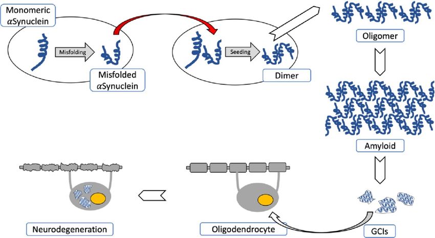
Free Neuropathology 1:17 (2020)                                                                     Kurt A. Jellinger
doi: https://doi.org/10.17879/freeneuropathology-2020-2813                                             page 3 of 28

no evidence of autosomal dominant MSA or of de               tial role in the disease process [10, 76-78]. The im-
novo mutations in this disorder [52]. A British fami-        pact of the neuronal endosomal-lysosomal system
ly with SNCA mutation showed neuropathologic                 in the processing of αSyn in PD is well established,
features of both PD and MSA [53], but they are               while lysosomes contribute to the pathogenesis of
distinct from PD patients carrying the H50Q or               MSA, enabling oligodendroglial and neuronal up-
SNCA duplication [54]. None of the nucleotide pol-           takt of αSyn [79]. Reduced oligodendrocyte-derived
ymorphisms (FBXO47, ELOVL7, EDN1, etc.) reached              enriched microvesicles (OEMVs) could be an im-
genome-wide significance [34], and polymorphisms             portant mechanism related to pathological αSyn
of the LINGO1 and LINGO2 (nogo receptor interact-            aggregation in oligodendrocytes [80]. Although it
ing protein-1 and -2) do not decrease the risk of            has been speculated that primary neuronal pathol-
MSA [55]. The possible involvement of the SNCA,              ogy leads to secondary oligodendroglial degenera-
COQ2, MAPT, GBA1, LRRK2 and C9ORF72 genes in                 tion, as suggested by the widespread occurrence of
MSA pathogenesis was examined recently [56].                 NCIs even in areas lacking GCIs [77], the distribu-
                                                             tion and severity of neurodegeneration reflects
      The link between V393A mutations and the               subregional GCI density and supports the assump-
COQ2 gene, encoding the coenzyme Q10 (COQ10),                tion that MSA is a primary oligodendrogliopathy [2,
and familial or sporadic MSA in Japanese and other           81]. The role of oligodendroglia in introducing the
Asian populations [44, 57-61] has not been con-              neurodegenerative process was confirmed experi-
firmed in other populations [34, 62-64]. Thus,               mentally in transgenic (tg) mice overexpressing
COQ2 polymorphisms may be region-specific and                αSyn in oligodendrocytes [10, 13, 82-84]. These and
may not represent common genetic factors for                 other results highlight the role of endogenous αSyn
MSA. Decreased levels of COQ10 in cerebellum and             and p25α in the formation of αSyn assemblies in
plasma of MSA patients [65, 66] suggest that its             oligodendrocytes and provide in vivo evidence of
deficiency may contribute to pathogenesis due to             the role of oligodendroglial αSyn in the establish-
decreased electron transport in the mitochondria             ment of αSyn pathology in MSA [85]. Early events
and increased vulnerability to oxidative stress (OS)         are an ectopic appearance of αSyn in oligodendro-
[67].                                                        cytes, loss of the cAMP-regulated phosphoprotein
      RNA analyses of MSA brain tissue revealed al-          of 32kDA (DARPP-32), and calbindin indicating cal-
terations in α- and β-immunoglobuline [68],                  cium toxicity and disturbance of phosphorylated
dysregulations of microRNAs that regulate gene               proteins [86]. Recent findings suggest the possibil-
expression in the pons and cerebellum [69, 70], and          ity of endogenous αSyn accumulation in oligoden-
disruption of long intervening non-coding RNAs               drocyte precursor cells that contribute to GCI for-
(lincRNA) in the frontal cortex along with protein           mation and perturbation of neuronal/glia support
coding genes related to iron metabolism and im-              in MSA brain [86a]. Reduced OEMVs could be an
mune response regulation [71, 72]. Epidemiological           important mechanism related to pathological αSyn
studies suggested that epigenetic factors or envi-           aggregates in oligodendroglia, inducing dysfunction
ronmental toxins may be associated with the risk             of the SNARE protein complex, which regulates
for MSA [73], but there are no convincing data cor-          membran fusion in eukaryotic cells. The concentra-
relating increased risk of MSA with exposure to              tions of OEMVs in MSA were significantly reduced
pesticides, solvents, other toxins, or alcohol con-          compared to those in PD [80]. Decreased expres-
sumption [74, 75].                                           sion of glia-derived neurotrophic factors (GDNF) in
                                                             MSA brains [87] indicates that αSyn aggregation in
                                                             oligodendrocytes impacts their trophic transport to
Pathogenesis
                                                             neurons. Oligodendroglial changes are more wide-
                                                             spread than αSyn positive GCIs, suggesting that
    Although our understanding of MSA remains
                                                             oligodendroglial pathology induces degeneration of
incomplete, evidence from animal models and hu-
                                                             the oligodendroglia-myelin-axon-neuron complex
man post mortem studies indicates that the accu-
                                                             [2, 26]. The selectivity of the neurodegeneration in
mulation of misfolded αSyn, particularly in oli-
                                                             MSA is determined by the interaction of multiple
godendrocytes but also in neurons, plays an essen-
                                                             noxious factors. Some of these factors include: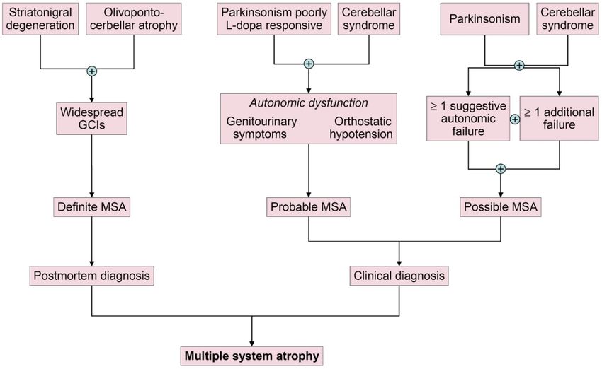
Free Neuropathology 1:17 (2020)                                                                                    Kurt A. Jellinger
doi: https://doi.org/10.17879/freeneuropathology-2020-2813                                                            page 4 of 28

Fig. 1. Pathogenetical features of MSA causing neurodegeneration. Spontaneous misfolding of αSyn results in formation of abnormally
folded dimers and further assembly results in oligomers and amyloid formations. αSyn-rich GCIs involving oligodendroglia result in
demyelization and neurodegeneration. The red arrow shows the “prion-like” cell-to-cell transfer of misfolded αSyn.
Courtesy of Victoria Sidoroff, MD, Dept. of Neurology, Medical University of Innsbruck, Innsbruck, Austria

ectopic αSyn accumulation in oligodendrocytes and                        αSyn, which shows specific conformational strains
neurons, "prion-like" propagation of disease-                            [88, 106] that are primarily generated by neurons,
specific strains of misfolded αSyn [88], targeting                       can be toxic once released to the extracellular envi-
distinct brain regions and cell types [89, 90], im-                      ronment [107] and can spread throughout the
paired protein degradation, proteasomal and mito-                        brain in a "prion-like" manner [9, 108-111]. Extra-
chondrial dysfunctions [91, 92], alterations of the                      cellular αSyn, interacting with neuronal and non-
autophagic pathway [91, 93, 94], perturbed iron                          neuronal cell types, mediates neuroinflammation
homeostasis [95], lipid dysfunction involved in                          and cell-to-cell spread [112, 113]. Neuron-to-
myelin synthesis [96-98], genetic polymorphism                           oligodendrocyte transport of misfolded αSyn plays
[55], microglial activation [97, 99], neuroinflamma-                     a major role in the pathogenesis of MSA [114, 115].
tion [100], proteolytic disturbance, autophagy                           MSA and PD show different phosphorylation signa-
[101], and microRNA dysregulation [102] driving                          tures of αSyn and distinct seed characteristics, indi-
inflammation, disrupting myelin, and contributing                        cating that distinct strains underlie these diseases
αSyn accumulation via the dysregulation of au-                           [90, 116, 117]. After propagation in TgM83 tg mice,
tophagy and prion mechanisms [103]. These and                            strain-specific phenotypic differences are main-
other factors are contributing to OS, which is sug-                      tained after serial transmission, providing evidence
gested to be a major pathogenic factor in MSA and                        that disease heterogeneity among the synucleinop-
related diseases [104]. These multiple mechanisms                        athies is caused by distinct αSyn strains [89]. MSA
interact to result in the system-specific pattern of                     strains show several similarities to PD strains, and
neurodegeneration in MSA (Fig. 1). TNFα-                                 less so with DLB strains, but more potently induce
dependent neuroinflammation may play a key role                          motor deficits, nigrostriatal degeneration, αSyn
in MSA pathogenesis, and its relevance has been                          spreading, and inflammation, reflecting the aggres-
underlined in various models of the disease [105].                       sive nature of this disease [118].

Free Neuropathology 1:17 (2020)                                                                      Kurt A. Jellinger
doi: https://doi.org/10.17879/freeneuropathology-2020-2813                                              page 5 of 28

      Recent animal model studies that only partial-         gy with five types of inclusions: GCIs within oli-
ly replicate the human disorder have provided                godendrocytes (also referred to as Papp-Lantos
some progress in our understanding of MSA patho-             bodies [125], the presence of which is mandatory
genesis [13, 15, 84, 119, 120]. Early accumulation           for the post mortem diagnosis of definite MSA [1])
of p25α (TPPP), a potent stimulator of αSyn aggre-           and less frequently glial nuclear inclusions, neu-
gation, may decrease myelin basic protein (MBP),             ronal nuclear inclusions, astroglial cytoplasmic in-
favoring both the deposition and fibrillation of αSyn        clusions, and neuronal threads, also composed of
and changing myelin metabolism [121]. Relocation             αSyn [126]; (2) selective neuronal loss and axonal
of p25α from the myelin sheaths to the oligoden-             degeneration involving multiple regions of the
droglial soma, due to mitochondrial dysfunction,             nervous system; (3) extensive myelin degeneration
and the formation of cytoplasmic p25α inclusions             with pallor and reduction in MBP with astrogliosis;
precedes the aggregation of transformed αSyn in              and (4) widespread microglial activation [127] and
oligodendrocytes. Endogenous αSyn and p25α in-               neuroinflammation [128, 129], with extensive CD4
duce the formation of pathological αSyn assemblies           and CD8 T-cell infiltration [130]. GCIs and the re-
in oligodendrocytes and provide in vivo evidence of          sulting neurodegeneration show a characteristic
their contribution to the pathogenesis of MSA [85].          distribution, involving not only the striatonigral and
Although large inclusions appear in a later disease          OPC systems, but also cortical regions, autonomic
states, small, soluble assemblies of αSyn, promoted          and motor nuclei in the brainstem, spinal cord,
by p25α, are pathogenic [122]. The source of αSyn            preganglionic autonomic nerve structures [131-
in oligodendroglia is unclear, but it contains αSyn          134], and the peripheral nervous system [135-138],
mRNA expression and αSyn may be secreted by                  characterizing MSA as a multi-system/-organ disor-
neurons and taken up by oligodendrocytes, which              der [2, 77, 139]. Phosphorylated αSyn is accumu-
is facilitated by protein Cx32 via direct protein-           lated in subpial and periventricular astrocytes after
protein interaction in both neurons and oligoden-            long disease duration [140]. However, αSyn-
droglia [115]. 21% of proteins found consistently in         positive astrocytes in subpial and perivascular re-
GCIs and LBs are synaptic vesicle-related, suggest-          gions are seen in both MSA and Lewy body disease
ing that misfolded αSyn may be targeted via vesi-            (LBD), suggesting that this pathology is not a specif-
cle-mediated transport, and may explain the pres-            ic feature of MSA [141].
ence of this neuronal protein within GCIs [123].
Thus, MSA represents a specific form of oligoden-            Inclusion pathology
droglial proteinopathies [124], while others suggest
that it is a primary neuronal disease with secondary               GCIs are argyrophilic, triangular, sickle-/half
accumulation of αSyn in oligodendrocytes [77].               moon-shaped or oval cytoplasmic aggregations,
Induced pluripotent stem cell (iPSC) studies indi-           composed of fibrillar αSyn, ubiquitin and various
cate a pathological phenotype of MSA neurons,                multifunctional proteins, including 14-3-3 protein,
independently from oligodendrocytes. These data              LRRK2, aggressomal proteins, etc. [125] (Fig. 2).
together with findings in animal models suggest              They form a meshwork of loosely packed filaments
that both neurons and oligodendrocytes are affect-           or tubules 15-30 nm in diameter with a periodicity
ed in MSA [91]. The disease is currently viewed as a         of 70-90 nm and straight filaments, both composed
primary synucleinopathy with specific glio-neuronal          of polymerized αSyn granular material and other
degeneration developing via the oligo-myelin-axon-           filaments. The central core contains phosphory-
neuron complex [2, 4].                                       lated (ser129) αSyn Cryo-EM showed that αSyn
                                                             inclusions from MSA are made of two types of fila-
                                                             ments, each of which consists of two different pro-
Histopathology and molecular pa-                             tofilaments. Each type contains non-proteinaceous
thology                                                      molecules at the interface of the two proteofila-
                                                             ments. Thus, they differ from those in DLB brain,
     The histological core features of MSA encom-            which suggests that distinct conformations/strains
pass the following types of different severity:              are characteristic for specific synucleinopathies. In
(1)specific αSyn-immunoreactive inclusion patholo-           addition, αSyn filament extracts from MSA tissue
Free Neuropathology 1:17 (2020) Kurt A. Jellinger
doi: https://doi.org/10.17879/freeneuropathology-2020-2813 page 6 of 28
Fig. 2. (A–C) Glial cytoplamic inclusions in MSA: (A) in globus pallidus (Gallyas silver impregnation), (B) in pontine basis (α-Synuclein) and
(C) in frontal white matter, anti-ubiquitin. (D) Neuronal cytoplasmic inclusion and neurites in pontine basis (α-Synuclein). (A–D) original
magnification 34,000.
From [126].
differ from those formed in vitro using recombinant wide distribution of αSyn oligomers not only in
proteins, which may have implications for the oligodendrocytes but also in neocortical neurons
mechanisms of protein aggregation and neuro- and Purkinje cells, suggesting that αSyn oligomer-
degeneration [142]. Soluble αSyn in GCIs differs mediated toxicity is an early event in MSA, inducing
from the insoluble form in Lewy bodies (LBs) [143]. neuronal loss in MSA [145].
Purification of αSyn containing GCIs revealed 11.9%
αSyn, 2.8% α-β-crystallin, and 1.7% 14-3-3 protein On the other side, interactions exist between ex-
compared to 8.5%, 2.0% and 1.5% in LBs [144]. In tracellular αSyn and each of the major central
the MSA brain, αSyn 140 and 122 isoform levels are nervous system (CNS) cell types. This has thepoten-
tial to contribute to secondary disease processes
increased, whereas αSyn 126 is decreased, in the
such as neuroinflammation, synaptic dysfunc-tion,
substantia nigra (SN), striatum, and cerebellum. In
and cell-to-cell spread, with vehicles such as micro-
early disease states, diffuse αSyn staining in neu-
ronal nuclei and cytoplasm occurs in many gray glia and exosomes that mediate spread of αSyn
matter areas, indicating that aggregation of non- pathology to peripheral brain regions [113]. Ca-
fibrillary αSyn occurs early in neurons [26]. Recent thepsin-D, calpain-1 and kallikrein-6 are elevated in
studies using a proximity ligation assay revealed a the putamen, pontine basis, and cerebellar whiteFree Neuropathology 1:17 (2020) Kurt A. Jellinger
doi: https://doi.org/10.17879/freeneuropathology-2020-2813 page 7 of 28
matter, indicating that αSyn accumulation is not III+SND I/II). These clinicopathological subtypes
due to reduced activity of these proteases, but correlated with initial symptoms and clinical fea-
rather that their upregulation is compensatory to tures of both types. Post mortem MRI changes in
increased αSyn [146]. Iron levels in basal ganglia the putamen (type 1, mild atrophy and isointensity;
(BG) and SN are higher in MSA than in PD and con- type 2, atrophy and diffuse hypointensity with a
trols, indicating perturbed iron homeostasis as a hyperintensive putaminal rim/HPR; type 3, putami-
potential pathogenic factor in MSA neurodegenera- nal atrophy and iso- or hypointensity with HPR)
tion [95]. reflect various degrees of brain damage [156]. In
two large series from the UK and Japan, another
Quantitative analyses of neuronal death and grading system for MSA was proposed [148]: each
GCI density showed a positive correlation with each case of SND and OPCA was divided into three
other, indicating the pivotal role of GCIs in neuronal grades based on semiquantitative assessment of
death [81, 147], and additionally, both lesions in- neuronal loss in regions of interest: for SND, the
crease with disease duration [148-150]. In the SN, putamen, GP and SN; and for OPCA the pontine
severe neuronal loss is accompanied by low GCI nucleus, cerebellar hemisphere and vermis, inferior
density, indicating that this and other areas affect- olivary nucleus and SN. This classification showed
ed in early disease have been burned out [139]. significant clinicopathological correlations. SND
Glial nuclear inclusions show a distinct distri- phenotypes showed more severe bradykinesia, and
bution from GCIs (Fig. 2D), and similarly the density the OPCA phenotype more frequently showed cer-
of neuronal cytoplasmic inclusions (NCIs) and neu- ebellar signs. No patients showed "pure" SND or
ronal nuclear inclusions are unrelated to that of "pure" OPCA. However, there is an increasing over-
GCIs [151]. NCIs are more widespread and show a lap of αSyn pathology with increased duration of
hierarchical pattern related to the duration of dis- the disease the extent of αSyn pathology [157].
ease but are independent of neuronal destruction, Damage to the striatonigral system is most severe
suggesting that other factors may induce the sub- in the dorsolateral caudal putamen and lateral SN,
type-dependent neuronal loss [77]. Region-specific suggesting transsynaptic degeneration of the stria-
astrogliosis is positively correlated with αSyn pa- tonigral fibers.
thology in MSA, in contrast to PD [152], and in gen- Consistently and severely affected areas are
eral parallels the severity of neurodegeneration the putamen, CN, SN, pontine and medullary teg-
[148]. Microglial activation in degenerated regions mental nuclei, inferior olives, and cerebellar white
accompanies GCI pathology and is most abundant matter; moderately affected areas are the motor
in white matter areas with mild to moderate de- cortex and GP, and mild lesions involve the cingular
myelination [153]. In MSA-C, the cerebellar subcor- cortex, hypothalamus, nucleus basalis of Meynert,
tical white matter and cerebellar brainstem projec- thalamus, subthalamus, and pontine tegmentum
tions are the earliest involved, followed by other [158]. Degeneration of the GP and SN leads to dys-
CNS regions. function of these inhibitory nuclei projecting to the
motor thalamus, but the SN loss is of dopamine,
Distribution of lesions
not GABA (gamma aminobutyric acid), neurons.
A grading system for SND was proposed based Stereological studies of the BG revealed a substan-
on semiquantitative assessment of atrophy, neu- tial loss of neurons in the SN, putamen, and GP,
ronal loss, and the presence of GCIs [154]: Neu- whereas astrocytes were more frequent in the pu-
ronal loss in the SN pars compacta is grade 1; ex- tamen and caudate nucleus (CN). Microglia were
tension to the putamen is grade 2; further involve- found in all CNS regions with greatest frequency in
ment of the caudate and globus pallidus (GP) is the, otherwise unaffected, red nucleus. These data
grade 3. Subsequently, the grading system was support the region-specific pattern of pathological
extended for both SND and OPCA [155]. Of 42 pa- changes in MSA [159]. Another neuropathological
tients, 22 were assigned as MSA-P and 20 as MSA- study showed that the striatonigral region was
C, but none displayed "pure" OPCA pathology or most severely affected in 34% of SND and in 17% in
more severe OPCA pathology than SND (i.e., OPCA OPCA cases, while in almost half of them both re-Free Neuropathology 1:17 (2020) Kurt A. Jellinger
doi: https://doi.org/10.17879/freeneuropathology-2020-2813 page 8 of 28
gions were equally affected [133]. In view of the correlates with αSyn pathology and the severity of
frequent overlap and mixed forms, the value of neurodegeneration [153, 173], which is in contrast
grading systems for evaluation of MSA is under to PD [174]. Significant increase of monoaminox-
discussion [139]. idase B (MAO-B), a biomarker of astrogliosis, in the
degenerated putamen (+83%) was associated with
There is widespread involvement of the neo- astrogliosis and showed a positive correlation with
cortex with significant loss of neurons and increase αSyn accumulation [175]. Microglial activation ac-
of astrocytes and microglia in the frontal and parie- companying αSyn pathology and phagocytosing
tal areas, but no change in the total number of degenerating myelin is prominent in all degenerat-
oligodendrocytes [160]. Early degeneration of the ing regions [176], particularly in white matter input
BG drives late onset cortical atrophy due to fronto- tracts to the extrapyramidal system and cerebellum
striatal degeneration [161, 162]. Reduced neuronal [177]. Stereological studies revealed a significant
numbers in the anterior olfactory nucleus and in- increase of microglia in the white matter without
trabulbar part of the primary olfactory (pyriform) concomitant astrogliosis and with absence of signif-
cortex may underlie olfactory dysfunction in MSA icant oligodendroglial degeneration [171], suggest-
[163]. Limbic TDP-43 pathology is rare in MSA, but ing that microglia cells play an important role in the
co-localization with αSyn suggests an interaction initiation and progression of neurodegeneration in
between the two molecules [164-167]; TDP-43 MSA [100, 178]. This is supported by tg mouse
positive cases showed significantly older age at models indicating an active contribution of micro-
death than negative ones, suggesting that TDP-43 glial activation by triggering neuroinflammatory
pathology in MSA is an age-related phenomenon responses in the MSA brain [179].
rather than a disease-specific change [141].
In MSA-C, GCIs are most prominent in the cer-
Demyelination of variable intensity affecting ebellum, pons, and medulla [169]. The cerebellar
all parts of the nervous system [168] is associated Purkinje cells are more severely affected in the
with reduction of MBP by about 50% [96]. GCIs and vermis, with atrophy of olivary nucleus, cerebel-
microglial burden are greatest in mild to moderate lopontine fibers, and pontine basis, causing inter-
white matter lesions and decrease with progression ruption of specific cerebellocortical circuits [180].
of myelin damage that increases with disease dura- The motor subnetwork in MSA-C is significantly
tion [169]. The regional vulnerability of the white altered in both BG and cerebellar connectivity
matter to MSA pathology is poorly understood, but [181], with hyperintensity of the middle cerebellar
recent GWASs revealed dysregulation of various peduncle [182].
methylated loci, including HIP1, LMAN2, MOBP,
and others, giving the first evidence that DNA Involvement of autonomic and peripheral nerv-
methylation changes contribute to the molecular ous systems
processes altered in MSA [170]. Early MSA stages
show increased microglia (about 100%) in the white Degeneration of preganglionic autonomic
matter [127], without concomitant astrogliosis or neurons of the brain stem and spinal cord cause
oligodendroglial degeneration [171]. Both microgli- multidomain autonomic failures in MSA [133, 183,
al activation and αSyn-containing oligodendrocytes 184]. Supraspinal lesions involve cholinergic neu-
trigger neuroinflammation in the white matter rons of the ventrolateral nucleus ambiguous [185,
[128]. 186], tegmental nuclei [187], ventral periaqueduc-
tal dopaminergic neurons [188], medullary and
The loss of tubulin polymerization-promoting
arcuate nucleus, noradrenergic locus ceruleus
protein (TPPP)/p25α immunoreactivity correlated
[134], serotonergic medullary groups, ventrolateral
significantly with the degree of microglial reaction
medulla [189], caudal raphe neurons [190, 191],
and loss of MBP density as a marker of tract de-
catecholaminergic neurons of rostral ventral me-
generation [124]. White matter degeneration caus-
dulla, and noradrenergic neurons of the caudal
es degeneration of neuronal loops, leading to dys-
ventrolateral medulla [185, 192]. The medullary
function of cerebral autoregulation [172]. Gliosis in
serotonergic and catecholaminergic systems are
the degenerated areas of the MSA brain usuallyFree Neuropathology 1:17 (2020) Kurt A. Jellinger
doi: https://doi.org/10.17879/freeneuropathology-2020-2813 page 9 of 28
involved in early stages of MSA [193]. Other in- age age at disease onset is earlier in MSA-C com-
volved areas are the dorsal vagal nucleus [185], pared to MSA-P, the latter leading to more severe
periaqueductal gray [132], the Westphal-Edinger disability [214-216]. Average duration after clinical
nucleus and posterior hypothalamus, the tuber- diagnosis is 6-10 (mean 9.5) years [12, 23], with few
omamillary and suprachiasmatic nuclei [194], and patients surviving more than 15 years [217]. Others
the pontomedullary reticular formation [149]. The have reported a 5 year survival of 78% [218] and a
density of αSyn pathology did not correlate with 43% death rate during 3 years of follow-up [135]. A
neuronal loss, and there was no correlation be- Pan-American multicenter study reported that 68%
tween the αSyn burden and disease duration in of the participants presenting as MSA-P showed an
these regions, indicating that the loss of monoam- age at onset of 61.5 years, and those as MSA-C of
inergic neurons may progress independently from 57.4 years [219], while a prospective cohort in the
αSyn accumulation [195]. Sympathetic preganglion- USA reported a median survival of 9.8 (95% CI 8.8-
ic neurons in the intermediolateral cell columns of 10.7) years [220]. Early autonomic dysfunctions and
the thoracolumbar spinal cord [26, 134, 196] and severity of orthostatic hypertension have negative
sympathetic ganglia and Schwann cells in autonom- impact on both disease progression and survival
ic nerves are involved [197]. Neuronal loss affects [221] and more than triples the risk of shorter sur-
Onuf's nucleus in the sacral region [198], with mi- vival [222, 223], and a meta-analysis identified se-
nor loss of upper and lower motor neurons [26] vere dysautonomia, early combined autonomic and
and variable involvement of anterior horn cells motor failure, and early falls as unfavorable predic-
[134]. Mild degeneration of cardiac sympathetic tors of survival, whereas MSA phenotype and sex
innervation has been reported in some cases of did not predict survival [224].
MSA [199, 200], which accounts for a mild to mod-
erate decrease in the number of tyrosine hydrox- Parkinsonism with rigidity, slowness of move-
ylase, but not of neurofilament-immunoreactive ments, postural instability, gait disability, and a
nerve fibers in the epicardium. However, depletion tendency to fall, characterize the motor presenta-
of cardiac sympathetic nerves is closely related to tion of MSA-P [12]. Parkinsonism is rapidly pro-
the presence of αSyn pathology in the sympathetic gressing to wheelchair confinement within 5 to 10
ganglia of the CNS [200, 201]. The peripheral nerv- years from symptom onset, poorly responsive to L-
ous system shows αSyn deposits in sympathetic dopa, and is often associated with atypical features
[17]. Unilateral parkinsonism occurs in 40% of MSA
ganglia, skin nerve fibers [138, 202, 203], and
patients [220] and typical tremor in 4-10% [225].
Schwann cells [204], but lack of αSyn immunoreac-
tivity in dermal fibers in contrast to PD [203, 205]. Early postural instability and gait difficulties with
Filamentous αSyn aggregates involve the cytoplasm recurrent falls are also seen in MSA [35].
of Schwann cells in cranial, spinal and autonomic Polyminimyoclonus, not included in the current
nerves in MSA [141, 197, 206]. diagnostic criteria of MSA, has now been recog-
nized as a specific clinical feature of MSA.
Clinical features Among motor and non-motor symptoms in
early MSA, dysarthria was the most prevalent fea-
The onset of motor symptoms is 56±9 (mean ± ture (98.4%), followed by sexual dysfunction (95%),
SD) years, with both sexes equally affected [207], RBD (90.2%), constipation (82%), snoring (70.5%),
however 20-75% of MSA patients have a prodro- dysphagia (69%), and stridor (42.6%), which was
mal/preclinical phase with non-motor symptoms. more common in MSA-C than in MSA-P [226].
This phase includes cardiovascular and other auto-
A resting tremor is rare, whereas irregular
nomic failures (urogenital and sexual dysfunctions,
postural and action tremor may occur [227, 228].
orthostatic hypotension, and REM sleep behavior
Cerebellar ataxia, widespread gait, uncoordinated
disorder (RBD), which occurs in 88% or more [208,
limb movements, action tremor, and spontaneous
209]), which may precede the motor presentation
or gaze invoked nystagmus predominate MSA-C
by months to years [210, 211] and indicates more
[35]. Hyperreflexia and a Babinski sign occur in 30-
rapid progression of the disease [212, 213]. Aver-
50% of patients, while abnormal postures, such asFree Neuropathology 1:17 (2020) Kurt A. Jellinger
doi: https://doi.org/10.17879/freeneuropathology-2020-2813 page 10 of 28
bent spine, antecollis, and hand or foot dystonia in the discrimination between MSA and PD or PSP
are rare [229]. Early generalized and rapidly pro- [5, 241, 242]. A recent meta-analysis of available
gressive autonomic failure is typical of MSA [230] CSF data showed that reduction of p-tau, αSyn, Aβ-
and, in the absence of parkinsonism or cerebellar 42 and total tau and elevated NFL are indicators for
signs, indicating pure autonomic failure, which con- MSA [243]. Currently, the most promising approach
verts to MSA within a few years in about 28% [231- is a combination of CSF DJ-1, phospho-tau, light
233]. Among non-motor symptoms observed in 75- chain neurofilament protein (NFL) and Aβ-42 that
95% of patients [234], urinary urgency and in- may be helpful in the differential diagnosis be-
creased frequency are common in early disease tween MSA and other parkinsonian disorders [5,
stages [35]. In a subset of MSA patients with early 240, 243, 244] (Fig. 3). Other studies have shown
urinary retention, the disease may begin in the increased CSF levels of cytokines such as MCP-3,
sacral spinal cord and then spread to other regions MDC, fractalkine, and MIP-1β [246]. Phosphory-
[235]. lated αSyn in red blood cells may be a potential
diagnostic biomarker for MSA [247]. The results of
Orthostatic hypotension with recurrent syn- proteomics for biomarker discovery and mRNA
cope, which occurs after the onset of urogenital expression need further elucidation [248].
symptoms, is a hallmark feature of MSA; less spe-
cific are dizziness and nausea. Other symptoms are Molecular and functional imaging
anhydrosis, gastrointestinal dysfunction with early
dysphagia and constipation [225], pupillary auto- A cardiac sympathetic postganglionic denerva-
nomic involvement with blurred vision and dry tion distinguishes PD from MSA, showing intact
eyes. [236]. innervation. I-123 MIBG (metaiodobenzylguani-
Dysproportional antecollis and Pisa syndrome dine) scintigraphy can help differentiate the two
diseases with a pooled specificity of 77% (95% CI:
are common postural deformities in MSA [35].
68-84%) [199]. Recent meta-analyses suggest that
About 50% of patients with MSA-P develop cerebel-
MIBG imaging is useful to discriminate PD from
lar signs and even a higher proportion of MSA-C
cases develop parkinsonian features [23, 220]. Dys- MSA in moderate to advanced disease stages, but
tonia, repeated falls, drooling, dysphagia, dyspho- unreliable in early stages [199, 249]. However, in-
nia, and pain occur in advanced stages of the dis- teractions with many drugs limit the value of this
ease [237]. Laryngeal stridor is rare [210]. Respira- method [250]. The anteroposterior diameter of the
tory disturbances including diurnal or nocturnal medulla oblongata is a potential imaging marker of
inspiratory stridor and sleep apnea are frequent parasympathetic dysfunction in MSA [251].
[238, 239]. In recent years, several brain magnetic reso-
nance imaging (MRI) features have been described
Diagnostic biomarkers as helpful in the differential diagnosis of parkin-
sonian syndromes. They include atrophy of the
Despite numerous studies, to date there are
putamen, pons, cerebellum, and middle cerebellar
no reliable diagnostic and prognostic biomarkers
peduncle, a dilated fourth ventricle, and various
available. While multimodal imaging of structural
signal intensity variations on MRI [252]. MRI ab-
and functional brain changes gave insight into the
normalities including the "hot-cross bun" sign, a
pathophysiology and may evaluate disease pro-
cruciform hyperintensity in the pons [253], and the
gression, recent studies suggest that the combina-
"putaminal rim sign", which marks hyperintensive
tion of neuroimaging and fluid biomarkers may be
bordering of the dorsolateral margins of the puta
more successful than using single markers to in-
men in T2-weighted MRI reflecting degeneration
crease the accuracy of the clinical (differential)
and iron deposition, may differentiate MSA-P from
diagnosis of MSA [240].
PD [254-258]. They are, however, non-specific signs
Fluid and tissue biomarkers and therefore not included in the recent consensus
criteria [3], in contrast to putaminal atrophy which
Studies of αSyn levels in cerebrospinal fluid
shows 92.3% specificity but low sensitivity (44.4%)
(CSF) and plasma have been shown to not be usefulFree Neuropathology 1:17 (2020) Kurt A. Jellinger doi: https://doi.org/10.17879/freeneuropathology-2020-2813 page 11 of 28 Fig. 3. Candidate biomarkers of multiple system atrophy compared to Parkinson’s disease and controls. MSA: multiple system atrophy; PD: Parkinson’s disease; NfL: neurofilament light chain; FH: complement factor H; C3: complement 3; MHPG: 3-methoxy-4-hydroxyphenylethyleneglycol; IGF-I: insulin-like growth factor I; UCH-L1: ubiquitin carboxy-terminal hydrolase L1; oxDJ: oxidized DJ-1 protein; miRNA: microRNA. Modified from [245]. [259, 260]. Putaminal atrophy together with hy- [265]. Combined use of diffusion ratios and mag- pointense putaminal signal changes on iron- netic susceptibility values/quantitative susceptibil- sensitive routine sequences seem to be specific for ity mapping allowed differentiation of MSA-P and MSA-P [252]. Others showed significantly increased MSA-C from other parkinsonian syndromes with putaminal diffusivity volumes in the small anterior sensitivities and specificities of 81-100% [266]. Hy- region of interest in MSA-P versus PD [261]. Anoth- perintensity of the middle cerebellar peduncle and er distinguishing feature is the extensive and wide- hot cross bun sign should be added into the list of spread volume loss across the entire brain in MSA- additional neuroimaging features of possible MSA- P [262]. In quantitative MRI studies, the bilateral C [182]. Several studies assessed the diagnostic R2* increase in the putamen best separated MSA-P potential of multimodal MRI [267-270]. In conclu- from PD [263]. Putaminal and infratentorial volume sion, the sensitivity of conventional MRI findings in information classified 96.8% of MSA cases [260]. MSA compared to PD and healthy controls is incon- Diffusion tensor imaging permits differentiation sistent (36-83%), the specificity of MRI abnormali- between PD and MSA-P, the latter showing higher ties differentiating MSA from PD is high (88-100%). values of the diffusion coefficient in the inner cap- Automated imaging differentiation in parkinsonism sule, corona radiata, and lateral periputaminal (AID-P) and magnetic resonance Parkinsonism in- white matter [264], while a meta-analysis of pu- dex (MRPI) are robust biomarkers for PD and MSA taminal diffusivity measurements showed sensitivi- [271]. Diffusion weighted images, T2* weighted ty of 90% and specificity of 93% in distinguishing images and proton density weighted images are MSA-P from PD based on putaminal diffusivity useful for diagnosis MSA-P in early stages [272].
Free Neuropathology 1:17 (2020)                                                                                   Kurt A. Jellinger
doi: https://doi.org/10.17879/freeneuropathology-2020-2813                                                          page 12 of 28

     Fluorodeoxyglucose-positron emission tomog-                              Interpretation of tau-PET should be done cau-
raphy (FDG-PET) can distinguish MSA-P from PD,                           tiously, since some MSA cases with severe GCI pa-
showing different patterns of decreased glucose                          thology may be false-positive [281, 282], even
metabolism with a positive predictive value of 97%                       though the affinity of PBB3 is 10 to 50 times less
[273, 274]. Targeting postsynaptic dopaminergic                          than αSyn [283]. 1-(2-chlorophenyl)-N-methyl-N-(1-
functions using 123FβCIT SPECT differentiates PD                         methylpropyl)-3-isoquinoline          carboxamide
(normal or increased signal) from MSA (normal or                         (PK11195) for imaging microglia-mediated process-
increased signal) [275]. Dopamine transporter                            es showed elevated tracer binding in many areas of
(DAT) imaging showed more prominent and earlier                          the MSA brain, consistent with the known neuro-
DAT loss in the anterior caudate and ventral puta-                       pathologic distribution [284].
men in MSA than in PD [276], although normal DAT
imaging does not exclude MSA [277]. In autopsy-                          Diagnostic accuracy and differential
confirmed cases a greater asymmetry of striatal
binding was seen in MSA than in PD [278], but it is
                                                                         diagnosis
highly correlated with SN cell loss [279]. 18F-Dopa-
                                                                              Revised consensus guidelines define 3 degrees
PET showed more widespread BG dysfunction in
                                                                         of certainty of clinical diagnosis of MSA: definite,
MSA than in PD without evidence of early compen-
                                                                         probable and possible [3] (Table 1, Fig. 4).
satory increase in Dopa uptake [280]. Future stud-
ies will be needed to determine the usefulness of                             Definite MSA requires post mortem evidence
tau-PET imaging for the characterization of αSyn                         of widespread αSyn inclusions with concomitant
filaments and the differential diagnosis of atypical                     SND or OPCA [1]. Probable MSA is defined as a spo-
parkisonian disorders.                                                   radic, progressive disorder in adults, clinically char-
                                                                         acterized by severe autonomic failure, urinary dys-

Fig. 4. Diagnostic scheme for MSA according to the current consensus diagnostic criteria.
Free Neuropathology 1:17 (2020) Kurt A. Jellinger
doi: https://doi.org/10.17879/freeneuropathology-2020-2813 page 13 of 28
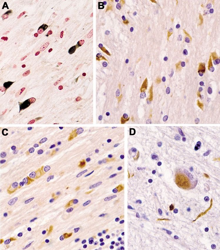
Table 1. Diagnostic clinical markers for MSA. Modified from [240].
MSA, multiple system atrophy; MSA-C, MSA with cerebellar features; MSA-P, MSA with predominant parkinsonism.
function and poor L-dopa-responsive parkinsonism portant clues for a correct and early diagnosis. They
or cerebellar ataxia. A diagnosis of probable MSA is include orofacial dystonia; inspiratory signs, con-
based on clinical features and ancillary diagnostic tractures of hands and feet, jerky myoclonic pos-
tests. Possible MSA can be diagnosed when a spo- tural/action tremor, polyminimyoclonus, severe
radic progressive adult-onset disorder with parkin- dysphonia and dysarthria, pathological laughter
sonism or cerebellar ataxia is accompanied by at and crying, snoring, disproportional antecollis,
least one of the following additional features within camptocormia and/or Pisa syndrome, and cold
3 years of motor onset: dysphagia, gait ataxia and hands and feet [225, 229] (Table 2). In addition,
other cerebellar symptoms (Table 1). severe disability milestones include: frequent falls,
use of urinary catheters, wheelchair dependence,
"Red flag" diagnostic features unintelligible speech, cognitive impairment, severe
dysphagia, and residential care. In a recent clinico-
The presence of "red flag" (warning sign) fea- pathological study of 203 clinically diagnosed MSA
tures highly specific for MSA may provide im- patients, a lifetime recorded number of red flags inFree Neuropathology 1:17 (2020) Kurt A. Jellinger
doi: https://doi.org/10.17879/freeneuropathology-2020-2813 page 14 of 28
both MSA-P and MSA-C was compared to LBD and criteria [225, 286], while 25% of patients with the
PSP [225]. Recognition of patients with early or diagnosis of "possible" MSA had different patholog-
possible MSA may be supported by one or more ical diagnoses, including PD and PSP [225]. The
red flags, and two or more out of six had a specifici- most common misdiagnoses were DLB (13 and
ty of 98.3% and a sensitivity of 84.2% [228, 229], 14%, respectively), PSP (6 and 11%) and PD (6%).
while no differences were found in the frequencies Autonomic failure was the leading cause of misdi-
of red flags within 3 years from disease onset be- agnosis in PD and DLB, and cerebellar ataxia that of
tween MSA and MSA look-alikes [225]. Recent misdiagnosis in PSP [286]. Sporadic spinocerebellar
studies confirmed the validity of an eight-item pilot ataxia (SCA) with autonomic failure can masquer-
scale for the assessment of early MSA [285]. ade as MSA-C. A study reported that 7% of patients
with clinically diagnosed MSA had mutations in SCA
Due to the heterogeneity of clinical phenotypes genes [288]. Fragile X tremor-ataxia syndrome and
and lack of specific biomarkers, it is a challenge to X-linked adrenoleukodystrophy can also be misdi-
make a correct antemortem diagnosis of MSA agnosed as MSA-C [61]. The possible explanations
[286]. The sensitivity of the second consensus crite- for the suboptimal diagnostic accuracy of the cur-
ria was 41% for possible and 18% for probable MSA rent consensus criteria for MSA that saw a positive
at first clinical visit and 92% and 63% at last clinical predictive diagnosis even in later disease stages
visit, respectively [287]. In two recent brain bank from 60 to 90% [286, 287] have been recently dis-
studies, among patients diagnosed with MSA dur- cussed [289].
ing life, only 62% and 79% met the pathological
Table 2. Clinical features supporting and non-supporting a diagnosis of multiple system atrophy. Modified from [240].Free Neuropathology 1:17 (2020) Kurt A. Jellinger
doi: https://doi.org/10.17879/freeneuropathology-2020-2813 page 15 of 28
ebellar symptoms [305]. An atypical case of fronto-
Atypical MSA temporal lobar degeneration (FTLD)-TDP type A
with MSA phenocopy syndrome showed severe
Almost all cases of MSA display neuronal loss striatal degeneration and cerebellar involvement
in both striatonigral and OPC structures [24, 148], [306], while four cases with clinical features of
with only 11 of 42 cases assigned to the category of FTLD, but without autonomic dysfunction, showed
"pure" SND [155]. However, MSA has a wider range frontotemporal atrophy and severe limbic αSyn
of presentations, which expands the list of differen- neuronal pathology with Pick body-like, but tau-
tial diagnoses. Several subtypes of MSA do not fit negative, inclusions. These cases were suggested to
into the current classification [290]. "Minimal represent a novel subtype of FTLD associated with
change" MSA is a rare aggressive form with GCIs αSyn (FTLD-αSyn) [307]. Rare cases in a family with
and neurodegeneration almost restricted to the SN, pathologic hexanucleotide repeat expansions in
putamen, and locus coeruleus, thus representing C9ORF72, a gene linked to amyotrophic lateral scle-
"pure" SND [291-294], suggesting that GCI for- rosis, demonstrated clinical and neuroimaging fea-
mation is an early event and may precede neuronal tures indistinguishable from MSA [308], and a cer-
loss. One patient with "minimal" MSA-C showed ebello-brainstem dominant form of X-linked adre-
abundant GCIs in pontine nuclei, middle cerebellar noleukodystrophy presented as MSA [61]. Recently,
peduncle and cerebellar white matter, with NCIs rare cases of MSA with transitional or diffuse DLB
and neuronal nuclear inclusions restricted to the developing clinical features of PDD or DLB have
pontine basis, cerebellar vermis, and inferior oli- been reported. Those with neuronal loss in SN but
vary nuclei, which were associated with neuronal not in striatal or OPC systems with widespread GCIs
loss indicating a link between both lesions in early were considered "minimal change" MSA, in which
disease [295]. Neurologically normal individuals are LBD was considered the primary pathology and
rarely found to have GCIs at autopsy as coincidental MSA as coincidental. APOE allele frequency was not
or incidental findings limited to the pons and infe- different between these forms [309]. These and
rior olivary nuclei with mild neuronal loss restricted other subtypes should be considered in establishing
to the SN, suggesting that these regions may be a correct diagnosis of MSA.
afflicted first in MSA-P [296, 297]. The presence of
GCIs may represent an age-related phenomenon
not necessarily progressing to overt clinical disease, Cognitive impairment in MSA
classifying these cases as "incidental" or "prodro-
mal/preclinical" MSA, similar to incidental LBD Unlike other synucleinopathies, MSA has not
[298]. Young-onset MSA with a mean age of 36.4 been associated with significant cognitive impair-
years shows more L-dopa-induced dyskinesia but ment (CI), which has been considered an exclusion
less common myoclonus and pyramidal signs com- criterion for the diagnosis of MSA [3]. However, a
pared to late-onset cases. On post mortem analysis, recent position statement by the Neuropathology
the "minimal change" variant was more common in Task Force of the Movement Disorder Society indi-
young-onset MSA [299]. cated that CI may be an under recognized feature
in MSA occurring in 17-47% of MSA patients, while
The other extreme are "benign" MSA cases severe dementia is rare [310]. Because CI has been
with prolonged survival up to 15 years in about 2- underestimated in MSA, not all patients have un-
3% of patients [217, 300]. Most of them showed dergone formal cognitive assessments and, there-
slowly progressing parkinsonism with subsequent fore, the frequency could be higher than reported
rapid deterioration after development of autonom- in several studies. The degree of CI in MSA patients
ic failure [301]. Many of them developed motor ranges from mild to moderate decline and affect
fluctuation and L-dopa-induced choreiform dyski- executive, attentional and visuospatial functions,
nesias [302, 303]. Other cases of survival up to 18 while memory is less often impaired [197, 310-
years revealed extensive distribution of GCIs in the 312]. CI may occur in early stages of MSA, but it is
CNS [304]. Another variant of pathological con- generally common in advanced cases [313] and
firmed MSA showed neither parkinsonism nor cer- often correlates with disease duration [314]. MildFree Neuropathology 1:17 (2020) Kurt A. Jellinger
doi: https://doi.org/10.17879/freeneuropathology-2020-2813 page 16 of 28
cognitive impairment (MCI) has been reported in the control groups (8.4%) [329]. In view of the lim-
up to 40% of MSA-P patients, mainly characterized ited data on the molecular basis of CI (and other
by frontal dysfunction [310, 315]. Mild or moderate neuropsychiatric symptoms) in MSA, further stud-
CI has been reported in 14-37% of pathologically ies on the pathological basis of CI in MSA are need-
proven MSA cases [134, 286, 302, 316]. More se- ed.
vere and widespread cognitive dysfunction was
seen in MSA-P than in MSA-C patients [317], prob- MSA - a prion-like or prion disease?
ably due to prefrontal impairment [315], whereas
others saw no differences in cognitive variables The spread of αSyn pathology from one cell to
between the two groups [318] or more severe cog- another and even from one nervous structure to
nitive dysfunctions in MSA-C [319]. CI has been another has been demonstrated in vivo [11, 330-
regarded as a result of cortical and subcortical 335]. This pattern, resembling prion spreading, has
structural changes [320], frontal lobe dysfunction led to the concept of prion-like propagation of αSyn
[321, 322], cortical dysfunction driven by focal fron- and tau [110]. Self-propagation of αSyn oligomers,
tostriatal degeneration [162], alterations in the however, is not sufficient to declare them as pri-
corpus callosum [323], the dorsolateral prefrontal ons, because they show "seeding" activity rather
cortex network [324], or neocortical neuronal loss than infectivity of αSyn [336]. However, the ap-
[159], while others have not found any differences plicability of the prion hypothesis in α-
in the severity of pathological findings between synucleinopathies and, in particular, MSA remains
cases with and without CI [325]. controversial, since injections of brain lysated from
Recent studies indicated that NCI burden in MSA patients failed to replicate the oligodendrogli-
the hippocampus and parahippocampal gyrus is al αSyn pathology that is typical for MSA. While
associated with memory impairment in MSA [326]. studies in wild type (wt) mice provided insights into
Alzheimer’s disease neuropathological changes the mechanisms of oligodendroglial αSyn aggrega-
(ADNC), cerebral amyloid angiopathy (CAA), and tions in MSA, intracerebral inoculation studies in
cerebrovascular lesions did not differ between cas- non-human primates to the best of our knowledge
es with and without CI [325], whereas others have not been performed yet.
showed a greater burden of NCIs in medial tem- There are other challenges to the hypothesis
poral regions, the hippocampus or perirhinal re- that MSA is a prion disease. First, endogenous wt
gions [77, 197, 316, 326, 327]. ADNC has been re- αSyn is insufficient to propagate αSyn pathology;
ported in only 2/35 (7%) autopsy-proven cases of mutant αSyn is needed as a template. The trans-
MSA [134], whereas two cases of combined MSA mission of αSyn "prions" to a second synucleinopa-
and AD (Braak stages III and VI) have been report- thy model and their ability to propagate between
ed, in which only a few neurons shared αSyn and two distinct mouse cell lines while retaining strain-
tau [328]. A recent retrospective clinicopathological specific properties was suggested to provide evi-
study of 48 MSA patients (33 MSA-P and 15 MSA-C) dence that MSA is a prion disease [337]. However,
with a mean age at death of 60.5±7.8 (range 46-82) these and other mouse experiments have not yet
years, reported MCI in 10 cases (20.8%), in which explained why in MSA αSyn pathology predomi-
three had associated moderate cortical tau pathol- nantly accumulates in oligodendroglia, as MSA-
ogy (Braak I-II), and moderate CI in seven patients derived αSyn does not appear to have the ability to
(14.5%), for which six had associated cortical amy- induce strain-like cell-specific aggregates. This
loid plaques and moderate cortical tau pathology demonstrates that the intrinsic properties of A53T
(Braak II-III), one had probable primary age-related αSyn in the M83 mouse model dominate over any
tauopathy (PART), and one female aged 82 years strain features harbored by misfolded αSyn in MSA
with severe dementia showed fully developed AD. brains [9].
Cortical Lewy pathology, observed in four cases,
was not associated with clinical CI. 77.1% of the Furthermore, GCIs have never been identified
MSA cases were free of ADNC, compared to 42% in in wt mouse brains inoculated with MSA-derived
controls, while Lewy pathology was higher than in αSyn [338]. Hence, αSyn aggregates ("prionoids")You can also read