A new threat from an old enemy: Re emergence of coronavirus (Review) - Spandidos Publications
←
→
Page content transcription
If your browser does not render page correctly, please read the page content below
INTERNATIONAL JOURNAL OF MOlecular medicine 45: 1631-1643, 2020

                      A new threat from an old enemy: Re‑emergence
                                 of coronavirus (Review)
      ANCA OANA DOCEA1*, ARISTIDIS TSATSAKIS2-5*, DANA ALBULESCU6*, OANA CRISTEA7*,
    OVIDIU ZLATIAN7, MARCO VINCETI8,9, STERGHIOS A. MOSCHOS10,11, DIMITRIS TSOUKALAS12,
   MARINA GOUMENOU2, NIKOLAOS DRAKOULIS13, JOSEF M. DUMANOV14, VICTOR A. TUTELYAN3,15,
GENNADII G. ONISCHENKO3,4, MICHAEL ASCHNER4,5, DEMETRIOS A. SPANDIDOS16 and DANIELA CALINA17

              1
               Department of Toxicology, University of Medicine and Pharmacy of Craiova, 200349 Craiova, Romania;
       2
       Department of Forensic Sciences and Toxicology, Faculty of Medicine, University of Crete, 71003 Heraklion, Greece;
         3
           Russian Academy of Sciences, 119991 Moscow; 4The State Education Institution of Higher Professional Training,
    The First Sechenov Moscow State Medical University under Ministry of Health of the Russian Federation, 119992 Moscow,
       Russia; 5Department of Molecular Pharmacology, Albert Einstein College of Medicine, New York, NY 10461, USA;
         Departments of 6Radiology and 7Microbiology, University of Medicine and Pharmacy of Craiova, 200349 Craiova,
          Romania; 8Department of Biomedical, Metabolic and Neural Sciences, University of Modena and Reggio Emilia,
             I‑41125 Modena, Italy; 9Department of Epidemiology, Boston University School of Public Health, Boston,
         MA 02118, USA; 10Department of Applied Sciences, Faculty of Health and Life Sciences, Northumbria University;
      11
         PulmoBioMed Ltd., Newcastle‑Upon‑Tyne NE1 8ST, UK; 12Metabolomic Medicine, Health Clinics for Autoimmune
       and Chronic Diseases, 10674 Athens; 13Research Group of Clinical Pharmacology and Pharmacogenomics, Faculty of
            Pharmacy, School of Health Sciences, National and Kapodistrian University of Athens, 15771 Athens, Greece;
        14
           Mycological Institute US EU, Subclinical Research Group, Sparta, NJ 07871, USA; 15Federal Research Centre of
            Nutrition and Biotechnology, 109240 Moscow, Russia; 16Laboratory of Clinical Virology, School of Medicine,
                          University of Crete, 71003 Heraklion, Greece; 17Department of Clinical Pharmacy,
                             University of Medicine and Pharmacy of Craiova, 200349 Craiova, Romania

                                      Received March 23, 2020; Accepted March 27, 2020

                                                  DOI: 10.3892/ijmm.2020.4555

Abstract. The new outbreak of coronavirus from December             In the present review, we address current knowledge on coro-
2019 has brought attention to an old viral enemy and has            naviruses from a short history to epidemiology, pathogenesis,
raised concerns as to the ability of current protection             clinical manifestation of the disease, as well as treatment and
measures and the healthcare system to handle such a threat. It      prevention strategies. Although a great amount of research
has been known since the 1960s that coronaviruses can cause         and efforts have been made worldwide to prevent further
respiratory infections in humans; however, their epidemic           outbreaks of coronavirus‑associated disease, the spread and
potential was understood only during the past two decades.          lethality of the 2019 outbreak (COVID‑19) is proving to be
                                                                    higher than previous epidemics on account of international
                                                                    travel density and immune naivety of the population. Only
                                                                    strong, joint and coordinated efforts of worldwide healthcare
                                                                    systems, researchers, and pharmaceutical companies and
Correspondence to: Professor Aristidis Tsatsakis, Department of
Forensic Sciences and Toxicology, Faculty of Medicine, University
                                                                    receptive national leaders will succeed in suppressing an
of Crete, 71003 Heraklion, Greece                                   outbreak of this scale.
E‑mail: tsatsaka@uoc.gr
Professor Anca Oana Docea, Department of Toxicology, University
                                                                    Contents
of Medicine and Pharmacy of Craiova, 2 Petru Rareş Street,
200349 Craiova, Romania
E‑mail: ancadocea@gmail.com                                         1. Introduction
                                                                    2.	A brief overview of coronavirus infections in human
*
    Contributed equally                                                 history
                                                                    3. Epidemiology of coronavirus infections
Key words: coronavirus, SARS‑CoV, MERS‑CoV, COVID‑19,               4. The transmission model
epidemiology, virulence strains, pathogeny, diagnostics             5. Pathogenesis of coronavirus infection
                                                                    6.	Clinical manifestations of coronavirus‑associated diseases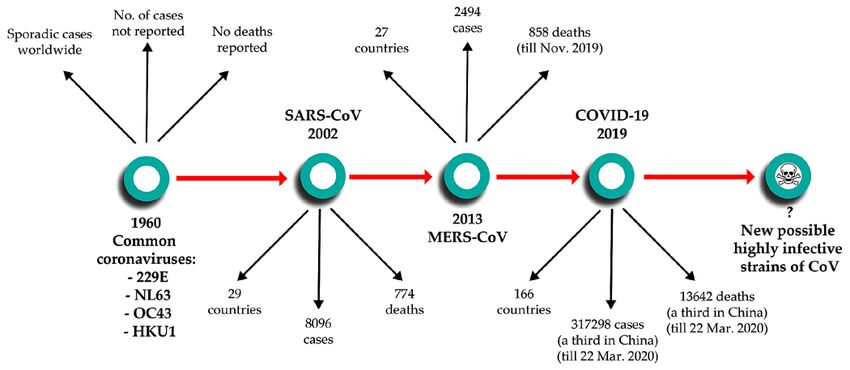
1632                  Docea et al: A NEW THREAT FROM AN OLD ENEMY: RE-EMERGENCE OF CORONAVIRUS

7. Treatment of coronavirus‑associated diseases                     succumb 774 patients (6). In 2012, a novel β‑coronavirus that
8. Prevention strategies                                            had not previously been observed in humans was detected
9.	Conclusions                                                      for the first time in a patient in Saudi Arabia. Since then, the
                                                                    new coronavirus, which causes Middle Eastern Respiratory
                                                                    Syndrome and is now known as MERS‑CoV, has infected
1. Introduction                                                     >2,494 individuals across 27 countries and led to the death
                                                                    of at least 858 individuals as of November 2019, through
Respiratory infections are common in the cold seasons world-        a series of emergences and re‑emergences from camelid
wide, and consequently considered trivial and mild. Affected        hosts (7). On December 8, 2019, in Wuhan, Hubei Province,
individuals rarely consult medical professionals, instead           China, the first case was reported of a new coronavirus that
treating themselves with symptomatic medications. Droplet           produces pneumonia. Since then, the new virus first named
and aerosol transmission further facilitates rapid dissemi-         2019‑nCoV and subsequently renamed SAS‑CoV‑2 was identi-
nation to numerous individuals at once, which amplifies             fied as a member of the β‑coronavirus subtype, spread rapidly
socioeconomic impact even with minimal increases to fatality        via human‑to‑human transmission. At the time of authoring
rate, particularly among patients with comorbidities.               (March 23, 2020), over 317,298 cases have been recorded
    The latest and contemporary outbreak of a respiratory           worldwide across 166 countries, with over 13,642 deaths
pathogen, namely severe acute respiratory syndrome coro-            attributed to the virus (8) (Fig. 1).
navirus 2 (SARS‑CoV‑2) responsible for coronavirus disease
(COVID‑19), has brought to attention a hidden threat from           3. Epidemiology of coronavirus infections
an old enemy. This is the third major coronavirus outbreak
over the past 20 years that has had substantial socioeconomic       The emergence of virulence strains of coronavirus over the
impact, but the first in the 21st century to affect countries       past 20 years has increased the interest of scientists regarding
across all continents except Antarctica. The general panic and      coronavirus strain variability, distribution, and the ability
insecurity expressed across all sociopolitical and economic         to be transmitted between animals to humans and further
tiers has dramatically disrupted day‑to‑day life, international     from humans‑to‑humans. The strains have slightly different
travel and trade. Notwithstanding severe lifestyle disrup-          distributions across sex, age and other demographic attributes.
tions, depression‑associated disease has been reported due to       For example, whilst SARS‑CoV‑2 affects males more than
extreme measures of isolation.                                      females with substantial age‑associated mortality increases
    Here we emphasize current knowledge regarding coro-             in frail elderly subjects, the four societally non‑disruptive
naviruses from a short history to epidemiology, diagnosis,          coronaviruses affect more females and children between
pathogenesis, clinical manifestation, as well as treatment          5 and 14 years (9).
and prevention strategies. Forward lessons informing future             In contrast, SARS‑CoV‑1 affected more females, and rela-
strategies to improve surveillance and prevent recurrence are       tively young people with the median age of SARS infected
highlighted.                                                        patients in Guangzhou province, China at 28.6 years) (10), and
                                                                    mortality rate at ~12% in people over 65 years (11). In compar-
2. A brief overview of coronavirus infections in human              ison, the MERS coronavirus was more frequently detected
history                                                             among males and middle‑aged adults aged 50‑59 years (12).
                                                                    Emerging data from hospitalized COVID‑19 cases in Wuhan
SARS‑CoV‑2 belongs to the Orthocoronavirinae subfamily,             (Hubei province, China) indicate males (68%) and middle‑age
family Coronaviridae, order Nidovirales (1). It comprises of        adults in the age group of 50‑59 years to be predominantly
four subtypes, α‑ and β‑coronaviruses that can infect humans,       affected, with a prevalence under 10% in people aged
and gamma‑ and delta‑coronaviruses, which are found only            90 years
and NL63 and 2 β coronaviruses ‑ OC43 and HKU1, which               old: 22.7%) (14). Whilst the older population demographics of
routinely produce noncomplicated infections of the upper            Italy is believed to drive the higher case fatality ratio differ-
and/or lower respiratory tract (3). Then, 2002 marked a key         ences (7.9%) vs. China (4.0%) (15), the Italian healthcare
moment in our understanding of coronavirus‑induced disease          system is experiencing increased requirement for intensive
with the emergence of the first lethal severe acute respira-        care unit (ICU) admission (~10% of cases) (16). Anecdotal
tory syndrome (SARS)‑causing coronavirus. Similarly to              reports from frontline clinicians in the principally affected
SARS‑CoV‑2, the original SARS‑CoV emerged in Guangdong              regions in Italy indicate all ICU beds dedicated to COVID‑19
Province in China, spreading through human transmission             and patient admission to ICU/mechanic ventilation restricted
chains to infect at least 8,096 individuals in 29 countries and     to age groups with a higher likelihood of survival. Of note,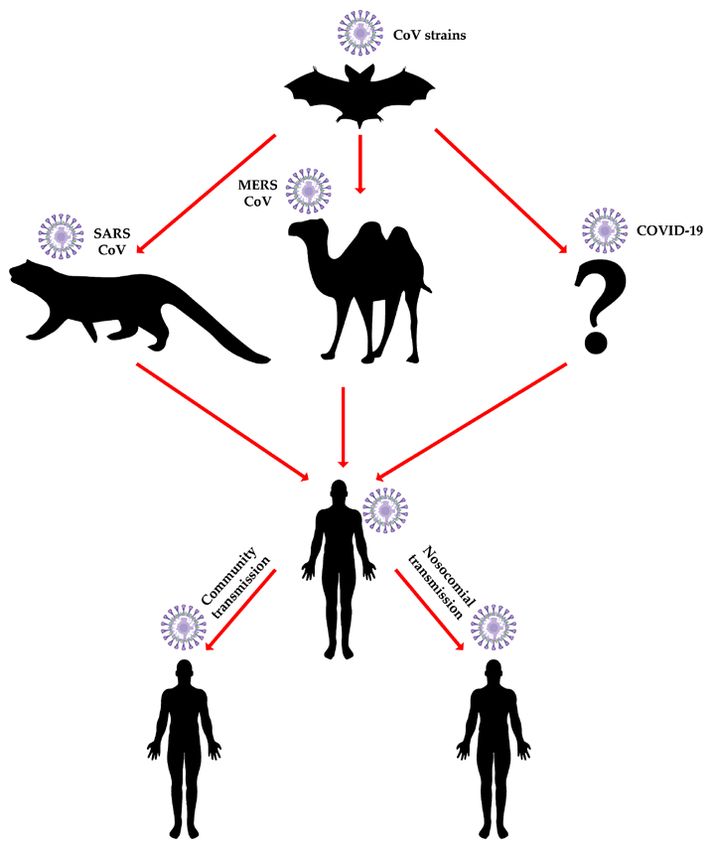
INTERNATIONAL JOURNAL OF MOlecular medicine 45: 1631-1643, 2020                                        1633

Table I. The comparative clinical evolution of coronavirus infections.

Coronavirus strain    Incubation period	Death period                                                Symptoms

SARS CoV 4‑10 days 20‑25 days                                   Fever, dry cough, myalgia, dyspnea, headache, sore throat, sputum
			                                                             production, rhinorrhea watery diarrhea confusion, poor appetite
MERS CoV  5‑6 days 11‑13 days                                   Myalgia, fever, chills, malaise associated with confusion, cough,
			                                                             shortness of breath, dyspnea pneumonia
COVID‑19  3‑7 days 17‑24 days                                   Fever, cough, dyspnea, muscle ache confusion, headache, sore throat,
			                                                             rhinorrhea, chest pain, diarrhea, nausea, vomiting, anosmia, dysgeusia

                                           Figure 1. History of coronavirus infections in humans.

these reports indicate 20% of ICU cases to involve individuals          protein (18). Considering that these inserts appear in hyper-
under the age of 65 and as young as 20 years of age. The reader         variable regions of the protein and are as short as 6 residues in
is strongly advised to consider carefully the territory‑specific        length, it is most probable that they arose naturally: as a conse-
diversity of case severity definition as well as the diagnostic         quence the article describing these similarities was recently
triage/definition criteria before making direct epidemiological         withdrawn from publication. To date, no evidence supports
comparisons.                                                            that SARS‑CoV‑2 is man‑made: COVID‑19 closely resembles
    One additional aspect complicating case epidemiological             two other coronaviruses that have triggered outbreaks in
oversight pertains to the highly mutagenic nature of corona-            recent decades, SARS‑CoV‑1 (79% sequence identity) and
viruses, which is common across RNA viruses, and has led                MERS‑CoV (51.8% identity) (19), and all three viruses are
to inappropriate comparative evolution interpretations and              most likely to have originated in bats (17,20): SARS‑CoV‑2
even the incorrect assertion of mild and sever COVID‑19                 has a 96% sequence identity with a recently sequenced bat
arising from separate strains of SARS‑CoV‑2. By comparing               coronavirus recovered from the wild by random sampling (21).
genomes of sequenced COVID‑19 strains, it was shown that
synonymous mutations in spike genes between COVID‑19 and                4. The transmission model
rat or bat coronaviruses (RaTG13 and Bat‑SL‑CoVZC45) were
quite different. The ratio of nucleotide substitutions to amino         A major question regarding coronavirus epidemiology is
acid substitutions was 9.07 in COVID‑19 compared with                   ‘Why does the bat of all animals play such a central role in
RaTG13, which was significantly higher than the 3.91 ratio              coronavirus epidemiology?’. Studies in bats have identified
from COVID‑19 to Bat‑SL‑CoVZC45 (17). Additionally, there               viruses originating in this species, as the potential vector to
were numerous concerns that COVID‑19 was an engineered                  human infections. Additionally, as bats live in colonies, they
bioweapon, hypotheses fed by speculation that sequences from            present a high risk of transmitting the viruses horizontally
COVID‑19 were identical to those in HIV. Alignments showed              (intra‑species) which contributes to the vertical (cross‑species)
that they are not present in any other coronavirus strains, but         spreading ability. This hypothesis is strongly favored by data
show identity/similarity with sequences in HIV‑1 gp120 and              on another high socioeconomic impact bat‑derived virus, the
Gag, the former being also a cellular receptor recognition              Ebola virus, which was shown to efficiently infect both bat and
1634 Docea et al: A NEW THREAT FROM AN OLD ENEMY: RE-EMERGENCE OF CORONAVIRUS
Figure 2. The transmission model of coronaviruses.
human cells (22) and to date remains the favored hypothesis for similarity (whole genome level) with a coronavirus isolated
index case infection in the 2014‑16 West African Ebolavirus from Chinese bat species (21). Another important question is
disease outbreak. ‘Why China?’ China is the third‑largest country by surface
Additional evidence of bat to human indirect transmission area worldwide but the most populated country. Its varied
comes from the COVID‑19's Spike protein and ACE2 receptor land characteristics and diverse climate range supports huge
interactions (23). Indeed, the two highly pathogenic strains of biodiversity, which contributes to enabling transmission of
coronaviruses ‑ SARS‑CoV and MERS‑CoV were identified viruses between animal populations. Principally, however,
both in bat species and in animals involved in transmission co‑habitation of animals and humans in close proximity, and
to humans (20). It is interesting to note that the first highly gastronomic customs involving the consumption of a variety
pathogenic strain of coronavirus, SARS‑CoV‑1, has a low of exotic animals, including wild fauna such as bats, increases
genetic similarity with other known coronaviruses (39% with the chance of vertical transmission throughout the food supply
bovine coronavirus and 46% with porcine epidemic diarrhea chain. Officially, live‑bird markets have been closed; however,
virus) (24). Recently, three comparison studies were made with black‑market vendors run illegal slaughterhouses, that are
coronavirus strains from pangolins. The first (February 18, crowded places with poor ventilation, where a number of
2020) compared the sequences of COVID‑19 with the coro- species are hoarded together: These create ideal conditions
naviruses in illegally trafficked pangolins to show a sequence for the spreading of the virus through airborne droplets of
similarity between 85.5 and 92.4% (25), with the subsequent blood and other secretions, or shared cages, trade tools, and
papers (February 20, 2020), reporting sequence similarities utensils, allowing for an ‘amplification’ of vertical transmis-
with pangolin coronaviruses at 90.23% (26) and 91.02% (27), sion risk. The prevalence of such culinary and wild animal
respectively. However, the α‑coronavirus strains that cause handling practices in Southern China have been causally
human disease were also originally identified in bats (28); it linked to both the 2002 SARS‑CoV‑1 outbreak starting from
is therefore not surprising that COVID‑19 shares 96% genetic a market in Shenzen (Guangdong, China) (29) and the entryINTERNATIONAL JOURNAL OF MOlecular medicine 45: 1631-1643, 2020 1635
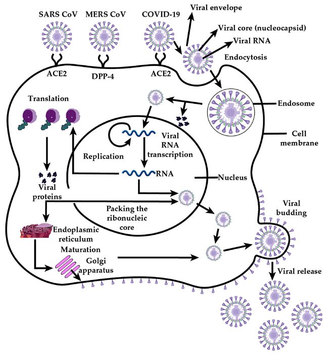
Figure 3. Pathogenesis of coronavirus infections.
of SARS‑CoV‑2 into the human population. Of the original 5. Pathogenesis of coronavirus infection
41 cases presenting with pneumonia of unknown origin, two
thirds had links to Huanan Seafood Wholesale Market that The genome of coronaviruses contains genes for the 4 struc-
also sold live animals (30). tural proteins: Envelope (E), membrane (M), nucleocapsid (N)
Such a species barrier bridge has been also documented and spike (S). Coronavirus virions are lipid bilayer‑enveloped
with filovirus disease outbreaks in Africa, wherein high risk particles variable in size (80‑160 nm), characterized by multiple
infectious agents are routinely detected in bushmeat markets 20‑nm spike‑like extensions on the surface in the form of
and the associated human population both by PCR and ‘corolla’ or ‘flower petals’. At the virion core, a nucleocapsid
immunoprecipitation methods (31). As with China, human and with icosahedral symmetry contains an electron‑dense layer
animal close contact and often co‑habitation remains common with a center that is clear. Its genomic nucleic acid consists
in rural African areas with practically no barriers to wild of is single‑stranded positive RNA which requires a negative
environments (e.g., tropical forests). Such exposure data have RNA replication cycle intermediate that generates subgenomic
fed debates on the role of so‑called ‘herd immunity’ through protein coding RNAs as well as genomic RNA for virion
natural exposure to emerging tropical, or perhaps rural, assembly. The core also features accessory proteins that differ
zoonoses. It is noteworthy that, comparable risks have been considerably between various types of coronaviruses.
documented with viruses not typically associated with highly The lipidic envelope features the trimerically‑organised
biodiverse geographies or the consumption of wild meat. For Type I transmembrane spike glycoproteins which consist of
example, the 2012 outbreak of MERS‑CoV in the Middle East the ectodomain subunits S1 and S2 protruding externally, a
and the Arab peninsula specifically, was shown to involve both transmembrane anchor, and a tail extending towards the viral
local and international city dwellers with no previous exposure core (34). Cell attachment involves the S1 subunit interacting
to the natural camelid host that continues to be in close contact with host cell surface receptors driving endocytosis and, after
with rural populations (Fig. 2) (32,33). The direct impact on membrane fusion with the involvement of subunit S2 (35),
individual health of zoonoses, and indirect impact on health- release of the virion core into the cytosolic milieu. The glyco-
care systems as evidenced through the COVID‑19 outbreak, proteins of coronaviruses mediate attachment, fusion, and
underscore how increasingly urban population organisation entry in the host cells, but different parts of those glycoproteins
even among developing nations amplifies the risk of onward are involved in each of these processes. These class I viral
human transmission, despite past exposure and indeed immu- membrane‑fusion proteins undergo structural rearrangements
nity in settings with blurred wild‑human habitat barriers. that produce fusion between the viral and cellular membranes.1636 Docea et al: A NEW THREAT FROM AN OLD ENEMY: RE-EMERGENCE OF CORONAVIRUS
Glycoprotein conformational changes and cathepsin L prote- type I and II cells), trachea and bronchial serous glands, as
olysis within endosomes are also involved in the pathogenesis well as in macrophages and alveolar monocytes. Notably, the
of coronavirus (Fig. 3). Thus, whilst cleavage of S protein is expression in the lung cells is much higher than in trachea (46).
required to expose the hydrophobic fusion peptide, it seems that In line with the expression profile of ACE2, viral genome load
receptor interaction is required for the cleavage to occur (36). has been consistently reported to be both more elevated in the
Historically, immunotherapies and many exploratory small lower than in the upper respiratory tract, with lower respira-
molecule treatments have targeted such host‑pathogen interac- tory specimens being additionally less prone to false negative
tions to disrupt infection. However, experience across multiple results (47,48). ACE2 is also diffusely located on other cells,
virus strains indicates such monotherapy to be inefficient due such as mucosal cells of the intestines, endothelial cells of
to mutational escape. veins and arteries including heart cells, epithelial cells of the
Accessory proteins play a definitive role in infectivity, renal tubules, epithelial cells of the kidneys, immune cells
however, their functions are not yet fully understood. It is and cerebral neuronal cells, which may also be susceptible to
speculated that some proteins play important roles in coun- coronavirus infections. The observation of COVID‑19 patient
tering the immune response; thus, viruses that lack these demise on account of severe heart failure brought about by
have a lower infectivity. For example, SARS‑CoV‑1 has two SARS‑CoV‑2 infection and the higher risk among patients with
accessory proteins, 3a and 3b, that play potential roles in the previous cardiovascular and hypertensive disease has driven
virulence of this strain (37). On the other hand, sometimes a multiple hypotheses regarding potential direct mechanisms of
complete infectious particle can be assembled without spike viral action on the circulatory system. Thus, ACE2 expression
proteins, indicating that accessory proteins can substitute can be increased as a result of using drugs such as ACE inhibi-
these (38). Therefore, there is a potential risk that a vaccine tors and angiotensin II type‑I receptor blockers (ARBs). Indeed,
targeting structural proteins alone, such as the spike protein, it was shown that expression of ACE2 is increased in diabetes
might be inefficient, driving evolutionary escape of the virus, patients (another high risk COVID‑19 group) treated with ACE
either through target protein mutation, or by favouring virions inhibitors and hypertension patients, treated with ARBs. ACE2
utilizing other accessory proteins for attachment. A some- expression can also be increased by thiazolidinediones and
what more viral escape‑proof approach involves targeting ibuprofen. Therefore, in these categories of patients, the risk
of the receptor‑binding domain (RBD) which is a conserved of infection with COVID‑19 is proposed to be higher (49). Yet,
domain of S protein (39). Notably, other respiratory viruses whilst the NL63 coronavirus strain binds to the same ACE2
that efficiently cross species barriers (e.g., influenza) require receptor as SARS‑CoV‑2, it produces only upper respiratory
close monitoring and annual adjustment of epitope targeting tract disease. This indicates that there are other unknown
to minimize spread and impact. factors, apart from the presence of receptors that influence the
The primary receptor used by coronaviruses to enter susceptibility of cells to coronavirus infection. Insights into the
into target cells is the angiotensin‑converting‑enzyme II differentiation factors influencing coronavirus strain‑specific
(ACE2) receptor (21,40), although some strains also use other outcomes come from recent data on host co‑factors mediating
alternative receptors, such as CD209L, for which they have SARS‑CoV‑2 fusion. Thus, SARS‑CoV‑2 uses the serine
a lower affinity (41). Whilst there is no evidence in pre‑ or protease TMPRSS2 for S protein priming; camostat mesylate,
post‑publication literature currently that SARS‑CoV‑2 also an inhibitor of TMPRSS2, blocked COVID‑19 infection
utilizes CD209L for cell entry this potential attachment of lung cells, but inhibition was more substantial when the
mechanism cannot be excluded until experimental data thereto endosomal cysteine protease cathepsin B/L (CatB/L) was
are produced. Likewise, other, as yet unidentified receptors, also inhibited e.g., with E‑64d (40). These results point to a
facilitate coronavirus cellular entry in the absence of ACE2 complex interplay of host factors in the endosome with the
in hepatocytes and some enterocytes (42). SARS‑CoV‑2 in spike protein in mediating virion fusion. It is thus speculated
particular, also appears to use the SARS‑CoV receptor ACE2 that in infected cells furin‑mediated precleavage at the S1/S2
to enter cells. Although the spike protein between SARS‑CoV‑1 site can promote TMPRSS2‑dependent entry, as in the case of
and ‑2 vary in sequence, Xu et al (43) suggested that the Spike MERS‑CoV (40). MERS‑CoV however is an exception to the
protein binding affinity for ACE2 is conserved as the 3D struc- SARS coronavirus set, as it uses at hedipeptidyl peptidase 4
ture of the receptor binding domain is identical with that of (DPP‑4) surface antigen as a receptor, and not ACE2. The
SARS‑CoV‑1, which would translates to equal infectivity. The relative expression of these endosomal factors and similarly
analysis of protein‑protein interaction using bioinformatics active proteins across cell types, as well as alternative recep-
showed that SARS nucleocapsidal proteins bind to human tors that mediate cellular attachment require urgent scrutiny
cyclophilin A (hCypA) and this binding was demonstrated to understand cell type tropism, and, more crucially, extra-
by surface plasmon resonance (SPR) technology. The 3D pulmonary reservoirs and sites of replication. Some of these
modelling detected the probable binding sites and allowed extrapulmonary reservoirs have been suggested by CT scans
deduction of important interaction between residue pairs (44). showing shadows and interstitial changes in tissues separate
Wan and colleagues observed that several amino acids in to the lungs (50), which require molecular and cellular studies
the receptor‑binding motif of SARS‑CoV‑2 allow binding to to confirm direct involvement in virus replication as opposed
human ACE2, even though with suboptimal strength (45). A to pathological findings secondary to SARS‑CoV‑2 infection.
recent study demonstrated that a mutation in the Spike protein After coronaviruses infect primary cells, the mature
(N501T) of SARS‑CoV‑2 enhanced the affinity for ACE2 (45). virions can be released and infect other target cells (51).
ACE2 is highly expressed in the respiratory tract, Infective viral particles can be found in sweat, stool, urine and
particularly in epithelial cells of the bronchi, alveoli (both respiratory secretions from patients with other coronaviruses,INTERNATIONAL JOURNAL OF MOlecular medicine 45: 1631-1643, 2020 1637
with viable SARS‑CoV‑2 so far documented in respiratory Hong Kong, Taiwan, Italy, and indeed China shows that mass
droplets, saliva, mechanically generated aerosols, and faeces. testing, case isolation and contact tracing can reduce human
After excretion environmental contamination can be substan- transmission even to zero. Indeed, presently the risk to these
tial (fomites) presenting what is currently believed to be the countries appears restricted to new case introduction from
primary mode of human‑to‑human transmission for up to abroad, justifying measures seeking to halt international travel.
7 days after surface deposition of the virus. The development Diagnostic process notwithstanding, a major drawback
of atypical pneumonia with rapid respiratory deterioration and lies with the diagnostic sensitivity of preferred as opposed to
failure determined by coronavirus infection is associated with available samples. Thus, data from the Chinese Centres for
increased levels of activated pro‑inflammatory chemokines Disease Control and Prevention, as well as clinical centres at
and cytokines (52). Thus, it has been proposed that Vitamin D the epicenter of the outbreak, have reinforced World Health
plays a role in the modulation of the immune response to Organisation technical diagnostic guidelines (60) that require
infectious agents, based on laboratory findings and obser- a lower respiratory sample as well as an upper respiratory
vational studies. However, randomized clinical trials have sample to rule out SARS‑CoV‑19 as the causal agent of
returned inconclusive, often controversial results. Therefore, viral pneumonia and COVID‑19‑like disease. The simplest
it has been suggested that cholecalciferol at elevated doses upper respiratory sample is presented in the form of an oral
might be useful for the prevention and therapy of infection and/or nasal swab (or combination of both, given the handful
with COVID‑19 (53,54). of reported nasal swab negatives in the presence of oral swab
In the pathogeny of coronavirus infection, an important positives). Lower respiratory specimens are difficult to obtain
role is played by the amplitude of host immunity; for example, as they require either intubated patients (tracheal aspirates),
canonical interferon levels terminate protein synthesis or or anesthesia (bronchoalveolar lavage, bronchial brushings).
even induce cell death. However, the intensity of the immune Notwithstanding risk of injury to the patient, these invasive
response can vary, depending on other comorbidities of the procedures create infectious aerosol generation risks and are
patient, explaining the role of these in the evolution of the contraindicated. The only suitable surrogate sample is lung
disease. Indeed, the majority of deaths have occurred among sputum, but only if this is naturally available, to minimize
individuals with comorbidities (55). In a nationwide study aerosol generation risk. Unfortunately, the Chinese experience
from China, 2.1% of patients with COVID‑19 had cancer, has been that1638 Docea et al: A NEW THREAT FROM AN OLD ENEMY: RE-EMERGENCE OF CORONAVIRUS
cost and 6‑12x faster). In addition, antibody tests can offer a alveolar damage, squamous metaplasia, giant‑cell infiltrates
clear answer as to the spread of exposure that may involve and increased macrophage levels in the interstitium and the
asymptomatic or low‑grade respiratory disease, supporting alveoli (46).
so called ‘herd immunity’. However, evidence from multiple Unlike SARS‑CoV‑1, Middle East Respiratory Syndrome
Asian countries and Italy show transmission containment was coronavirus (MERS‑CoV) has a wide variety of clinical mani-
achieved and can be achieved using RT‑qPCR which docu- festations from asymptomatic to severe respiratory symptoms.
ments infectious cases: antibody responses do not initiate until The mean incubation period is 5‑6 days (70‑73). In severe
at least a week after the onset of symptoms, meaning that the cases, death can occur within 11‑13 days after the onset of
deployment of antibody tests for mass screening will have an the disease (70,73). The clinical manifestations are similar
enhanced risk of false negatives as compared to oronasal swab to flu‑like symptoms, such as general myalgia, fever, chills,
RT‑qPCR by missing pre‑symptomatic, asymptomatic, and malaise associated with confusion, pulmonary symptoms,
newly symptomatic cases. Whilst the research and academic such as cough, shortness of breath, dyspnea and pneumonia.
value in understanding asymptomatic spread is undisputed, Extra‑pulmonary clinical manifestations include abdominal
the magnitude of the socioeconomic damage by COVID‑19 in disorders, nausea, diarrhea, vomiting, acute renal failure and
redirecting resources away from confirmation of active infec- neurological complications (74). The severity of the disease is
tion in mildly symptomatic cases and asymptomatic contacts associated with an increasing age, and with individuals with
onto exposure numbers, is likely to extend transmission long comorbidities, such as diabetes mellitus, renal and pulmonary
beyond the period of a few weeks documented in China and diseases. Death can occur after the development of acute
Korea into months, extending the impact onto healthcare respiratory distress syndrome (ARDS), multiorgan failure or
systems. Furthermore, such an extension of spread risks the septic shock (7). The neurological complications of the infec-
development of cycles of infection and socioeconomic damage tion with MERS‑CoV are associated with the in vitro ability
with the re‑introduction of new index cases within country of the virus to invade the central nervous system (75), although
regions and into countries free of the disease. The increasing it was not detected in the CSF. Among the neurological mani-
risk of virus evolution, and its establishment as a seasonal, festations, confusion has been reported in 25.7% and seizures
endemic respiratory disease agent, crucially, does not take in 8.6% of cases (76). Severe neurological complications
into account how immunity to other coronaviruses appears are encephalitis, stroke, polyneuropathy, acute disseminated
to be short‑lived: we simply do not know yet if COVID‑19 encephalomyelitis, Guillain Barré syndrome and Bickerstaff's
convalescence will protect from re‑infection, and if it does, encephalitis. Usually, the neurological complications appear at
for how long. a delayed time point from the respiratory symptoms, namely
2‑3 weeks from the onset of the clinical manifestation of the
6. Clinical manifestations of coronavirus‑associated disease and can be underdiagnosed (74,77‑79). Children are
diseases rarely affected (74). In laboratory tests, thrombocytopenia,
lymphopenia, neutrophilia, leucocytosis, reduced renal
The predominant symptoms of SARS‑CoV‑1 are respiratory functions and increased inflammatory markers have been
in nature, associated with fever and diarrhea. Clinical mani- observed. Similar to SARS‑CoV‑1, the pathogenesis has not
festations among patients are diverse, from mild‑moderate yet been fully elucidated. Viral‑mediated lung damage is
to severe‑life threatening symptoms. The virus has a mean characterized by diffuse alveolar damage. The virus has been
incubation period of 4.6 days (63), however in the majority of determined in multinucleated epithelial cells, pneumocytes
cases, symptoms appear after 10 days (64). In severe cases, the and bronchial submucosal glands. Apart from the respiratory
median time from the appearance of symptoms until artificial system, the virus has also been identified in the epithelial cells
ventilation is 11 days, and 23.7 days until the patient's death (65). of the proximal renal tubules (58). Acute tubular sclerosis and
The clinical manifestations are similar to flu‑like symptoms, tubulointerstitial nephritis have been observed in renal biop-
such as fever, a dry cough, myalgia, dyspnea, headache, sore sies collected from persons infected with MERS‑CoV (80).
throat, sputum production, rhinorrhea (66,67). In 40‑70% of The latest high clinical burden coronavirus, SARS‑CoV‑2,
cases, watery diarrhea appears within 1 week after the first presents a wide variety of clinical manifestations which are not
manifestations (42). In the elderly, the symptoms can also be yet fully characterized (81,82). The virus has a mean incubation
associated with confusion, a poor appetite and a decrease in period of 3‑7 days, and no more than 14 days (30) ‑ although
general well‑being. In is noteworthy that the symptomatology anecdotal reports likely related to poor contact tracing have
in children below 12 years is mild; no mortality has been suggested potentially up to 24 days of incubation. Outside
registered among children and teenagers. Asymptomatic cases Hubei province, the median incubation period has been
are reported to be rare (63,68). Increased levels of C‑reactive estimated to be 5.1 days (95% CI: 4.5 to 5.8), with 97.% of
protein, lymphopenia, thrombocytopenia, and increased levels infected subjects developing symptoms within 11.5 days of
of lactate dehydrogenase have been observed in laboratory infection (83). In a retrospective study on 99 patients that
tests. The infection manifests in two different stages, with the have confirmed COVID‑19 pneumonia treated in an infec-
first‑week characterized by flu‑like symptoms that improve tious diseases hospital in Wuhan, China, it was shown that the
even if the viral loads persist, and a recurrent period during the main clinical manifestations were flu‑like symptoms, such as
second week in which respiratory failure can appear and more fever and cough (83 and 81% of the cases), shortness of breath
than 20% of patients may require mechanic ventilation (69). (31% of cases), muscle ache (11% of cases), and less frequently
The respiratory system is the main affected area in the human confusion (9%), headache (8%), sore throat, rhinorrhea, and
body where the infection determines the appearance of diffuse chest pain. In less than 2% of patients, extra‑pulmonary symp-INTERNATIONAL JOURNAL OF MOlecular medicine 45: 1631-1643, 2020 1639
toms and diarrhea, nausea and vomiting were also observed. efficacy in monotherapy on a small number of patients; however
As complications, ARDS was reported in 17% of cases further studies are required to confirm these effects (98,99).
followed by acute respiratory injury (8%), septic shock (4%), In the case of the new COVID‑19‑associated pneumonia,
acute renal injury (3%), and ventilator‑associated pneumonia treatment also focuses on supportive care measures and symp-
(1%). Death occurred in 11% of participants in the study tomatic treatment. Following the example of SARS‑CoV‑1
determined by multiple organ failure. The main finding in treatment, the administration of systemic corticosteroids was
laboratory tests was lymphopenia. The severity of the disease attempted, although this did not exhibit any benefits. In the
was associated with co‑infections, smoking history, hyperten- case of patients with severe evolution, immunoglobulin G was
sion and age (13). Another retrospective study on 137 patients administered (50). The application of early respiratory support
infected with COVID‑19 revealed the same pattern of clinical improved the prognosis and the recovery of the patients (50).
manifestations with fever, cough and muscle pain, and less In a 54‑year‑old Korean man infected with SARS‑CoV‑2,
frequently diarrhea, headache and even heart palpitations the administration of the combination lopinavir/ritonavir
in some cases (50). Lymphopenia was observed in 72.3% of from day 10 of illness determined the decrease in viral load
cases associated with normal or decreased white blood cell till no detectable levels (100). The question of whether the
counts (50). The lung is the main organ affected. In the imaging decrease in viral load was associated with the natural course
examination by computer tomography or X‑ray, lesions were of the healing process or with antiviral treatment or both, is
identified in multiple lung lobes presented as patchy/punctate being currently investigated in order to confirm the efficacy of
ground‑glass opacities (GGO), patchy consolidation, irregular antiviral treatment for COVID‑19. The efficacy of remdesivir
solid nodules and fibrous stripes (50,84‑86). Studies that have (Gilead Sciences), a nucleotide analog, in inhibiting in vitro
investigated the pulmonary pattern of COVID‑19 pneumonia and in vivo SARS‑CoV‑1, MERS‑CoV and bat CoV strains
have demonstrated that the disease progresses rapidly, and CT that are capable to replicate in human airway epithelial cells
re‑examination after 3‑14 days has revealed significant changes render it a good choice for testing on newly affected COVID‑19
in lung structure. The single GGO observed in the early stages patients (85,101‑103). Taking into consideration that COVID‑19
can increase significantly in short‑term re‑examination. The binds to human ACE2 in order to penetrate human cells and to
same has also occurred with fibrous stripes observed in the induce severe pneumonia, the renin‑angiotensin system, ACEI
early stages that consolidate in short‑term re‑examination. In (angiotensin‑converting enzyme inhibitors) and AT1R (angio-
some cases, the irregular solid nodules observed in the early tensin II type‑I receptor) inhibitors have been suggested to
stages increased and merged in short‑term re‑examination. modify individual susceptibility to COVID‑19 by influencing
Short‑term repeated imaging scans should be carried out in SARS‑Cov‑2 virulence. However, further studies are needed
patients with COVID‑19 pneumonia for monitoring the evolu- to confirm the mechanisms involved (including the role of high
tion and for specific management of the patient as the disease ACE2 expression) and to consider possible changes in adminis-
can rapidly progress or indeed resolve (86,87). The peak of tration and dosage of antihypertensive drugs (46,49,104). Thus
lung abnormalities was reached on day 10 following the onset far, the Professional Association for Hypertension Therapy, USA,
of symptoms, followed by a decrease in symptomatology (87). has warned against changes in hypertensive drugs following
It was documented that in one third of the severely ill concerns about a possible increased susceptibility to COVID‑19
patients, death occurs because the COVID‑19 coronavirus (https://ish‑world.com/news/a/A‑statement‑from‑the‑Interna-
produce acute myocardial injury and damage to the cardiovas- tional‑Society‑of‑Hypertension‑on‑COVID‑19/). Interest has
cular system in general, that further degrade the state of these arisen also on the ‘old’, off‑patent antimalarial drug chloroquine
already very ill patients (88). and its less toxic hydroxychloroquine analog, both of which
are quinine derivatives (cinchoca tree bark, South America).
7. Treatment of coronavirus‑associated diseases Preliminary support from observations of potential inhibitory
activity against SARS‑CoV‑2 (105,106), clearly needs to be
The treatment for all types of coronaviruses is mainly confirmed through randomized controlled trials. There are
supportive, as no specific treatment has been discovered to also ongoing trials using monoclonal antibodies. One of them
date. For the treatment of SARS‑CoV‑1, apart from supportive is tocilizumab, an anti‑IL6 antibody, currently in phase III
measures, several therapeutic schemes have been implemented, clinical trials, with commercial name Actemra produced by
with varying success rates such as antiviral therapy with riba- Roche (107). Sarilumab, a drug used in rheumatoid arthritis is
virin (89), protease inhibitors ‑ lopinavir and ritonavir, alone about to enter clinical trials for COVID‑19 infection, according
or associated with ribavirin (67,90), interferon‑alfacon‑1 (91), to Regeneron Pharmaceuticals and Sanofi, respectively (108).
systemic corticosteroids (89,92), and convalescent plasma for In order to facilitate COVID‑19 virus replication, certain
passive immunotherapy (93). Apart from convalescent plasma, in vivo conditions, such as the pH value, the temperature,
none of these treatments have exhibited significant benefits humidity, and the sufficiency of oxygen supply should be
compared to the side‑effects produced. ensured. Consequently, in order to hinder the reproduction
For MERS‑CoV infections, taking as an example the procedures of the virus, it would be expedient to disturb the
early SARS‑CoV‑1 epidemic, supportive care was primarily environment of the virus, by interfering with the temperature or
used, and in some instances broad‑spectrum antibacterials, the pH values or both. SARS‑CoV‑2 has been speculated to be
antivirals such as ribavirin alone or in combination with a seasonal virus, in which case a hindrance of the rapid spread
interferon‑2α2b (94,95) and antifungals have been used to that characterizes it at ambient temperatures over 25˚‑30˚
prevent the co‑infection with other opportunistic patho- Celsius would be observed. Unfortunately, the sup‑tropical/arid
gens (96,97). Mycophenolic acid is another drug that exhibited environments favored by SARS‑CoV‑1 and MERS‑CoV, as1640 Docea et al: A NEW THREAT FROM AN OLD ENEMY: RE-EMERGENCE OF CORONAVIRUS
well as the comparatively higher temperatures in Israel, Egypt, cally all G20 nations. The lack of local implementation of
the Arab peninsula and now sub‑Saharan Africa compared what are demonstrably effective measures, as learned since
to Hubei province indicate that temperature probably does the SARS‑CoV‑1 outbreak due to scientific chauvinism
not affect COVID‑19 replication. Likewise, the ambient over healthcare standards between East and West, popula-
humidity levels thought to impact aerosol and droplet suspen- tion anthropology between Africa and Europe in terms of
sion time/travel distance, and by extension virus transmission, communication strategy and behaviour, or indeed local vs
do not seem to affect onward transmission chains across international experience between e.g., the UK and Italy with
geographical regions. Alteration of the pH value inside the emerging threats is lamentable. Going forward, the identifica-
human body within tolerable levels however is achievable; such tion of natural reservoirs and intermediate hosts for existing
an intervention would not only suppress replication of the virus, and future risks as identifiable through random sampling and
but also may have ‑ although possibly weak ‑ virucidal effect. monitoring (21) will become essential in preventing future
A pH value change with no negative impact on the health of the zoonotic virus outbreaks. Unfortunately, these alarms were
patient could be achieved by administering a drug that would raised repeatedly over the past two decades with minimal
lead to urine acidosis and endocellular alkalosis. This drug heed: perhaps the damage brought by COVID‑19 onto global
could be a simple small molecule with hydrophobic weak base economy and healthcare systems will transform the field of
properties, which would accumulate in lysosomes after having tropical emergent disease.
crossed both plasma and lysosomal membranes by diffusion. A good example of positive action are the actions of
Perhaps such a medicine may express both a therapeutic and currently more than 40 teams developing vaccines against
a prophylactic effect by increasing the environmental barriers SARS‑CoV‑2 infection (115); interest is largely spurred by the
to effective virus attachment in early endosomal pathways, and unprecedented success and rapid development of Ebolavirus
preventing spike proteolysis required for capsid release and vaccines during the West African outbreak. COVID‑19
cell membrane fusion within lysosomes, in line with cathepsin vaccines can be based on the viral RNA or derivatives:
protease‑targeted therapies. Given the success of combination Examples include Innovio and Moderna in USA, as well as
therapy strategies in other antiviral diseases centered on virus CureVac (Germany), and another team at Imperial College in
fusion and/or replication, promoting endocellular alkalosis London. RNA and DNA vaccines have the advantage of being
during antiviral drug therapy (e.g., replication inhibitors) could quick to develop and high likelihood of safety, though no RNA
amplify antiviral effect and thereby contribute to the reduction vaccine or therapeutic currently enjoys regulatory approval.
of the treatment period (109‑113). The Institute Pasteur in France is working on a traditional
COVID‑19 vaccine based on a SARS vaccine. CanSino
8. Prevention strategies Biologics developed a recombinant coronavirus vaccine incor-
porating adenovirus type 5 vector (Ad5). To date clinical trials
Although since 2004, the World Health Organization have been initiated by Moderna Therapeutics and Cansino
issued guidelines for the prevention of re‑emergence of Biologics (115) with further trials shortly anticipated in the
new coronavirus virulent strains (114), implementation has UK and other European countries.
evidently been inadequate. Key recommendations include: A sobering note, however, is that despite the ample amounts
the availability of the early detection of infected individuals of research performed on coronaviruses over the past 20 years,
to prevent the spread of emergent and re‑emergent infection, particularly following the SARS‑CoV‑1 outbreak, no vaccine
particularly to prevent international spread; the development is unfortunately yet available for either SARS‑CoV‑1 or
of contingency plans by each country for the management MERS, although some potential vaccines have reached as far
and detection of coronavirus, individualized by the risks that as phase I clinical trials (116‑118). Some of the strategies used
exist locally; preparing global and assisting national risk for vaccines are eliciting neutralizing‑antibody for S proteins
assessments; updating new discoveries; assisting countries and T‑cell responses (119) which seem to be jointly required
in their efforts to improve protection against infections; for convalescence, rather than simply neutralizing antibody
assessing the availability of all necessary resources in the titers. The quest for protective immunity to coronavirus is but
case of an outbreak; and, supporting the international scien- nascent, and virology offers a plethora of high socioeconomic
tific collaboration for SARS study. Unfortunately, all of these impact, high prevalence RNA viruses for whom decades of
strategies could not prevent the current outbreak, and after vaccine research have yet to deliver success.
17 years, the medical, scientific and governmental structures
have demonstrated that they are not adequately prepared 9. Conclusions
to effectively contain highly pathogenic viruses. The main
measures that should be taken for preventing the dissemina- The Guangdong region of China was considered a high‑risk
tion of pathogens in the general population are associated area for a coronavirus outbreak after the SARS‑CoV‑1
with infection control measures both in healthcare facilities epidemic in 2002. Nevertheless, although a large amount of
and in the community. Contact tracing and quarantine or research and efforts have been made worldwide to prevent
the isolation of those suspected of infection along with the further outbreaks, a coronavirus outbreak occurred again
education of the population has proven essential in preventing in 2019. Unfortunately, healthcare systems locally, nationally,
community transmission of the viruses both in developing and now internationally have again been overcome, as with
nations (West African and Democratic Republic of Congo the other two major coronavirus outbreaks. The evidence
Ebola virus outbreaks), developed nations (e.g., Hong Kong, from COVID‑19 is clear that highly aggressive measures
South Korea SARS and MERS outbreaks), and now practi- are necessary to break the transmission chain in the regionsINTERNATIONAL JOURNAL OF MOlecular medicine 45: 1631-1643, 2020 1641
at risk ‑ practically most countries globally at the time of 3. Geller C, Varbanov M and Duval RE: Human coronaviruses:
Insights into environmental resistance and its influence on the
authoring. Unfortunately, there is no specific prevention development of new antiseptic strategies. Viruses 4: 3044‑3068,
strategy against the new coronavirus, and its pandemic 2012.
status restrict the global community to two options: effective 4. Anthony SJ, Johnson CK, Greig DJ, Kramer S, Che X, Wells H,
Hicks AL, Joly DO, Wolfe ND, Daszak P, et al; PREDICT
measures for the immediate prevention of respiratory infections Consortium: Global patterns in coronavirus diversity. Virus
with predictable and measurable consequences, or ambitions Evol 3: vex012, 2017.
of ‘herd’ or vaccine‑derived immunity at a non‑specific time 5. Goumenou M, Spandidos DA and Tsatsakis A: [Editorial]
Possibility of transmission through dogs being a contributing
in the future. We can only hope that after this third significant factor to the extreme Covid 19 outbreak in North Italy. Mol Med
outbreak, sufficient experience has been gained; a joint Rep (In Press).
effort of the worldwide health authorities will be needed in 6. World Health Organization: Summary of probable SARS cases with
onset of illness from 1 November 2002 to 31 July 2003. https://www.
extinguishing the current epidemic. It can only be trusted that who.int/csr/sars/country/table2004_04_21/en/. Accesed July 24,
this experience will re‑emphasize the need for prevention and 2015.
readiness and for further studies to limit the transmission of 7. World Health Organization: Middle East respiratory syndrome
coronavirus (MERS‑CoV). MERS Monthly Summary, 2013.
animal viruses to humans and mitigation should it arise again https://www.who.int/emergencies/mers‑cov/en/. Accessed July 9,
in the future. 2013.
8. Wo rld H e a lt h O rga n i z a t io n: C o r o n av i r u s d i s e a s e
(COVID‑2019) situation reports. https://www.who.int/emer-
Acknowledgements gencies/diseases/novel‑coronavirus‑2019/situation‑reports.
9. Movert E, Wu Y, Lambeau G, Kahn F, Touqui L and Areschoug T:
Not applicable. Using Patient Pathways to Accelerate the Drive to Ending
Tuberculosis. J Infect Dis 208: 2025‑2035, 2013.
10. Chan‑Yeung M and Xu RH: SARS: Epidemiology. Respirology 8
Funding (Suppl): S9‑S14, 2003.
11. Xu RH, He JF, Evans MR, Peng GW, Field HE, Yu DW, Lee CK,
Luo HM, Lin WS, Lin P, et al: Epidemiologic clues to SARS
No funding was received. origin in China. Emerg Infect Dis 10: 1030‑1037, 2004.
12. Mobaraki K and Ahmadzadeh J: Current epidemiological status
Availability of data and materials of Middle East respiratory syndrome coronavirus in the world
from 1.1.2017 to 17.1.2018: A cross‑sectional study. BMC Infect
Dis 19: 351, 2019.
Not applicable. 13. Chen N, Zhou M, Dong X, et al: Epidemiological and clinical
characteristics of 99 cases of 2019 novel coronavirus pneumonia
in Wuhan, China: a descriptive study. Lancet 395: 507-513, 2020.
Authors' contributions 14. Livingston E and Bucher K: Coronavirus Disease 2019
(COVID‑19) in Italy. JAMA: Mar 17, 2020 (Epub ahead of print).
Conceptualization, AOD, DC, DT, MV, MA, JMD, MG, 15. Novel coronavirus (COVID‑19) situation. https://experience.
arcgis.com/experience/685d0ace521648f8a5beeeee1b9125cd.
VAT, GGO and DAS; validation, research, resources, data 16. Remuzzi A and Remuzzi G: COVID‑19 and Italy: what next?
reviewing, and writing AOD, DA, OZ, OC, DT, ND, SAM, Lancet: Mar 13, 2020 (Epub ahead of print).
MG, VAT, GGO; review and editing, AOD, AT, DAS, MV, 17. Lv L, Li G, Chen J, Liang X and Li Y: Comparative genomic
analysis revealed specific mutation pattern between human coro-
SAM, JMD, MA, ND and DC. All authors read and approved navirus SARS‑CoV‑2 and Bat‑SARSr‑CoV RaTG13. bioRxiv
the final manuscript. (In Press).
18. Pradhan P, Pandey AK, Mishra A, et al: Uncanny similarity of
unique inserts in the 2019‑nCoV spike protein to HIV‑1 gp120
Ethics approval and consent to participate and Gag. bioRxiv (In Press).
19. Ren LL, Wang YM, Wu ZQ, et al: Identification of a novel coro-
Not applicable. navirus causing severe pneumonia in human: a descriptive study.
Chin Med J: Feb 11, 2020 (Epub ahead of print).
20. Fan Y, Zhao K, Shi ZL and Zhou P: Bat Coronaviruses in China.
Patient consent for publication Viruses 11: 210, 2019.
21. Zhou P, Yang XL, Wang XG, Hu B, Zhang L, Zhang W, Si HR,
Zhu Y, Li B, Huang CL, et al: A pneumonia outbreak associated
Not applicable. with a new coronavirus of probable bat origin. Nature 579:
270‑273, 2020.
Competing interests 22. Leroy EM, Rouquet P, Formenty P, et al: Multiple Ebola Virus
Transmission Events and Rapid Decline of Central African
Wildlife. Science 303: 387‑390, 2004.
DAS is the Editor‑in‑Chief for the journal, but had no 23. Li W, Wong SK, Li F, Kuhn JH, Huang IC, Choe H and Farzan M:
personal involvement in the reviewing process, or any influ- Animal origins of the severe acute respiratory syndrome coro-
navirus: Insight from ACE2‑S‑protein interactions. J Virol 80:
ence in terms of adjudicating on the final decision, for this 4211‑4219, 2006.
article. The other authors declare that they have no competing 24. Drosten C, Günther S, Preiser W, et al: Identification of a novel
interests. coronavirus in patients with severe acute respiratory syndrome.
N Engl J Med 348: 1967‑1976, 2003.
25. Lam TTY, Shum MHH, Zhu HC, et al: Identification of
References 2019‑nCoV related coronaviruses in Malayan pangolins in
southern China. bioRxiv (In Press).
1. Groot RJ, Baker SC, Baric R, et al: Family - Coronaviridae. In: 26. Liu P, Jiang JZ, Hua Y, et al: Are pangolins the intermediate host
Virus Taxonomy. Ninth Report of the International Committee of the 2019 novel coronavirus (2019‑nCoV)? bioRxiv (In Press).
on Taxonomy of Viruses. King AMQ, Lefkowitz EJ, Adams MJ 27. Zhang T, Wu Q and Zhang Z: Pangolin homology associated
and Carstens EB (eds). Elsevier, pp806‑828, 2011. with 2019‑nCoV. bioRxiv (In Press).
2. van Doremalen N, Bushmaker T, Morris DH, et al: Aerosol 28. Wang LF and Cowled C (eds): Bats and Viruses: A New Frontier
and Surface Stability of SARS‑CoV‑2 as Compared with of Emerging Infectious Diseases. John Wiley & Sons Inc., 2015.
SARS‑CoV‑1. N Engl J Med: Mar 17, 2020 (Epub ahead of print). https://doi.org/10.1002/9781118818824.1642 Docea et al: A NEW THREAT FROM AN OLD ENEMY: RE-EMERGENCE OF CORONAVIRUS
29. Guan Y, Zheng BJ, He YQ, et al: Isolation and characterization 54. Gruber‑Bzura BM: Vitamin D and Influenza‑Prevention or
of viruses related to the SARS coronavirus from animals in Therapy? Int J Mol Sci 19: 2419, 2018.
Southern China. Science 302: 276‑278, 2003. 55. Lau EH, Hsiung CA, Cowling BJ, Chen CH, Ho LM, Tsang T,
30. Huang C, Wang Y, Li X, et al: Clinical features of patients Chang CW, Donnelly CA and Leung GM: A comparative epide-
infected with 2019 novel coronavirus in Wuhan, China. miologic analysis of SARS in Hong Kong, Beijing and Taiwan.
Lancet 395: 497‑506, 2020. BMC Infect Dis 10: 50, 2010.
31. Smiley Evans T, Tutaryebwa L, Gilardi KV, Barry PA, 56. Guan W, Ni Z, Hu Y, et al: Clinical Characteristics of Coronavirus
Marzi A, Eberhardt M, Ssebide B, Cranfield MR, Mugisha O, Disease 2019 in China. N Engl J Med: Feb 28, 2020 (Epub ahead
Mugisha E, et al: Suspected Exposure to Filoviruses Among of print).
People Contacting Wildlife in Southwestern Uganda. J Infect Dis 57. Ng DL, Al Hosani F, Keating MK, et al: Clinicopathologic,
218 (Suppl 5): S277‑S286, 2018. Immunohistochemical, and Ultrastructural Findings of a Fatal
32. Azhar EI, El‑Kafrawy SA, Farraj SA, Hassan AM, Al‑Saeed MS, Case of Middle East Respiratory Syndrome Coronavirus Infection
Hashem AM and Madani TA: Evidence for camel‑to‑human in the United Arab Emirates. Am J Pathol 186: 652‑658, 2016.
transmission of MERS coronavirus. N Engl J Med 370: 58. Alsaad KO, Hajeer AH, Al Balwi M, et al: Histopathology of
2499‑2505, 2014. Middle East respiratory syndrome coronovirus (MERS‑CoV)
33. Haagmans BL, Al Dhahiry SH, Reusken CB, et al: Middle East infection ‑ clinicopathological and ultrastructural study.
respiratory syndrome coronavirus in dromedary camels: an Histopathology 72: 516‑524, 2018.
outbreak investigation. Lancet Infect Dis 14: 140‑145, 2014. 59. Shah K, Bentley E, Tyler A, Richards KSR, Wright E,
34. Du L, He Y, Zhou Y, Liu S, Zheng BJ and Jiang S: The spike Easterbrook L, Lee D, Cleaver C, Usher L, Burton JE, et al:
protein of SARS‑CoV ‑ a target for vaccine and therapeutic Field‑deployable, quantitative, rapid identification of active
development. Nat Rev Microbiol 7: 226‑236, 2009. Ebola virus infection in unprocessed blood. Chem Sci (Camb) 8:
35. Li F: Evidence for a common evolutionary origin of coronavirus 7780‑7797, 2017.
spike protein receptor‑binding subunits. J Virol 86: 2856‑2858, 2012. 60. World Health Organization: Country & Technical Guidance
36. Simmons G, Gosalia DN, Rennekamp AJ, Reeves JD, - Coronavirus disease (COVID-19). https://www.who.int/emer-
Diamond SL and Bates P: Inhibitors of cathepsin L prevent gencies/diseases/novel‑coronavirus‑2019/technical‑guidance.
severe acute respiratory syndrome coronavirus entry. Proc Natl 61. Du Z, Xu X, Wu Y, Wang L, Cowling BJ and Meyers LA: Serial
Acad Sci U S A 102: 11876‑11881, 2005. Interval of COVID-19 Among Publicly Reported Confirmed
37. Narayanan K, Huang C and Makino S: SARS coronavirus Cases. Emerg Infect Dis: Mar 19, 2020 (Epub ahead of print).
accessory proteins. Virus Res 133: 113‑121, 2008. 62. Crisanti A and Cassone A: In one Italian town, we showed
38. Schoeman D and Fielding BC: Coronavirus envelope protein: mass testing could eradicate the coronavirus. Opinion. The
Current knowledge. Virol J 16: 69, 2019. Guardian, 2020. https://www.theguardian.com/comment-
39. Liu C, Zhou Q, Li Y, et al: Research and Development on isfree/2020/mar/20/eradicated‑coronavirus‑mass‑testing‑covid‑1
Therapeutic Agents and Vaccines for COVID‑19 and Related 9‑italy‑vo. Accessed March 20, 2020.
Human Coronavirus Diseases. ACS Cent Sci 6: 315-331, 2020. 63. Chiu WK, Cheung PC, Ng KL, et al: Severe acute respiratory
40. Hoffmann M, Kleine‑Weber H, Schroeder S, et al: SARS‑CoV‑2 syndrome in children: experience in a regional hospital in Hong
Cell Entry Depends on ACE2 and TMPRSS2 and Is Blocked by Kong. Pediatr Crit Care Med 4: 279‑283, 2003.
a Clinically Proven Protease Inhibitor Article SARS‑CoV‑2 Cell 64. Donnelly CA, Ghani AC, Leung GM, et al: Epidemiological
Entry Depends on ACE2 and TMPRSS2 and Is Blocked by a determinants of spread of causal agent of severe acute respiratory
Clinically Proven Protease Inhibitor. Cell 181: 1‑10, 2020. syndrome in Hong Kong. Lancet 361: 1761‑1766, 2003.
41. Jeffers SA, Tusell SM, Gillim‑Ross L, et al: CD209L (L‑SIGN) 65. Leung GM, Hedley AJ, Ho LM, et al: The epidemiology of severe
is a receptor for severe acute respiratory syndrome coronavirus. acute respiratory syndrome in the 2003 Hong Kong epidemic: an
Proc Natl Acad Sci USA 101: 15748‑15753, 2004. analysis of all 1755 patients. Ann Intern Med 141: 662‑673, 2004.
42. Leung WK, To KF, Chan PK, et al: Enteric involvement of severe 66. Booth CM, Matukas LM, Tomlinson GA, et al: Clinical features
acute respiratory syndrome‑associated coronavirus infection. and short‑term outcomes of 144 patients with SARS in the
Gastroenterology 125: 1011‑1017, 2003. greater Toronto area. JAMA 289: 2801‑2809, 2003.
43. Xu X, Chen P, Wang J, Feng J, Zhou H, Li X, Zhong W and 67. Chan JW, Ng CK, Chan YH, et al: Short term outcome and risk
Hao P: Evolution of the novel coronavirus from the ongoing factors for adverse clinical outcomes in adults with severe acute
Wuhan outbreak and modeling of its spike protein for risk of respiratory syndrome (SARS). Thorax 58: 686‑689, 2003.
human transmission. Sci China Life Sci 63: 457‑460, 2020. 68. Hon KL, Leung CW, Cheng WT, et al: Clinical presentations
44. Luo C, Luo H, Zheng S, Gui C, Yue L, Yu C, Sun T, He P, and outcome of severe acute respiratory syndrome in children.
Chen J, Shen J, et al: Nucleocapsid protein of SARS coronavirus Lancet 361: 1701‑1703, 2003.
tightly binds to human cyclophilin A. Biochem Biophys Res 69. Hui DS and Sung JJ: Severe acute respiratory syndrome.
Commun 321: 557‑565, 2004. Chest 124: 12‑15, 2003.
45. Wang C, Horby PW, Hayden FG and Gao GF: A novel coronavirus 70. Assiri A, McGeer A, Perl TM, et al: KSA MERS‑CoV
outbreak of global health concern. Lancet 395: 470‑473, 2020. Investigation Team, 2013b. Hospital outbreak of Middle East
46. Kuba K, Imai Y, Rao S, et al: A crucial role of angiotensin respiratory syndrome coronavirus. N Engl J Med 369: 407‑416,
converting enzyme 2 (ACE2) in SARS coronavirus‑induced lung 2013.
injury. Nat Med 11: 875‑879, 2005. 71. Assiri A, Al‑Tawfiq JA, Al‑Rabeeah AA, et al: Epidemiological,
47. Fred Hutchinson Cancer Research Center: INTERIM guidelines demographic, and clinical characteristics of 47 cases of Middle
for COVID‑19 management in hematopoietic cell transplant and East respiratory syndrome coronavirus disease from Saudi
cellular therapy patients, Version 1, 2020. https://www.fredhutch. Arabia: a descriptive study. Lancet Infect Dis 13: 752‑761, 2013.
org/content/dam/www/coronavirus/COVID-19_Interim_Patient_ 72. Memish ZA, Zumla AI, Al‑Hakeem RF, Al‑Rabeeah AA
Guidelines_3_9_20.pdf. Accessed March 8, 2020. and Stephens GM: Family cluster of Middle East respiratory
48. Wang W, Xu Y, Gao R, Lu R, Han K, Wu G and Tan W: Detection syndrome coronavirus infections. N Engl J Med 368: 2487‑2494,
of SARS‑CoV‑2 in Different Types of Clinical Specimens. 2013.
JAMA: Mar 11, 2020 (Epub ahead of print). 73. Ki M: 2015 MERS Outbreak in Korea: Hospital-To-Hospital
49. Fang L, Karakiulakis G and Roth M: Are patients with hyper- Transmission. Epidemiol Heal 37: e2015033, 2015.
tension and diabetes mellitus at increased risk for COVID‑19 74. Arabi YM, Balkhy HH, Hayden FG, et al: Middle East
infection? Lancet Respir Med 2600: 30116, 2020. Respiratory Syndrome. N Engl J Med 376: 584‑594, 2017.
50. Kui L, Fang YY, Deng Y, et al: Clinical characteristics of novel 75. Desforges M, Le Coupanec A, Stodola JK, Meessen‑Pinard M
coronavirus cases in tertiary hospitals in Hubei Province. Chin and Talbot PJ: Human coronaviruses: viral and cellular factors
Med J (Engl): Feb 7, 2020 (Epub ahead of print). involved in neuroinvasiveness and neuropathogenesis. Virus Res
51. Qinfen Z, Jinming C, Xiaojun H, et al: The life cycle of SARS 194: 145‑158, 2014.
coronavirus in Vero E6 cells. J Med Virol 73: 332‑7‑332‑7, 2004. 76. Saad M, Omrani AS, Baig K, et al: Clinical aspects and outcomes
52. Ding Y, He L, Zhang Q, et al: Organ distribution of severe of 70 patients with Middle East respiratory syndrome coronavirus
acute respiratory syndrome (SARS) associated coronavirus infection: a single‑center experience in Saudi Arabia. Int J Infect
(SARS‑CoV) in SARS patients: implications for pathogenesis Dis 29: 301‑306, 2014.
and virus transmission pathways. J Pathol 203: 622‑630, 2004. 77. Al‑Hameed FM: Spontaneous intracranial hemorrhage in a
53. Watkins J: Preventing a covid‑19 pandemic. BMJ 368: m810, patient with Middle East respiratory syndrome corona virus.
2020. Saudi Med J 38: 196‑200, 2017.You can also read