Cockayne syndrome group A and ferrochelatase finely tune ribosomal gene transcription and its response to UV irradiation - Oxford Academic Journals
←
→
Page content transcription
If your browser does not render page correctly, please read the page content below
Nucleic Acids Research, 2021 1
                                                                                                                     https://doi.org/10.1093/nar/gkab819

Cockayne syndrome group A and ferrochelatase finely
tune ribosomal gene transcription and its response to
UV irradiation
Manuela Lanzafame 1,† , Giulia Branca1,† , Claudia Landi2 , Mingyue Qiang3 , Bruno Vaz1 ,
Tiziana Nardo1 , Debora Ferri1 , Manuela Mura1 , Sebastian Iben 3 , Miria Stefanini1 ,

                                                                                                                                                                  Downloaded from https://academic.oup.com/nar/advance-article/doi/10.1093/nar/gkab819/6377400 by guest on 22 October 2021
Fiorenzo A. Peverali1 , Luca Bini2 and Donata Orioli 1,*
1
 Institute of Molecular Genetics -L.L. Cavalli Sforza, CNR, 27100 Pavia, Italy, 2 Department of Life Sciences,
University of Siena, 53100 Siena, Italy and 3 Department of Dermatology and Allergic Diseases, Ulm University,
Albert-Einstein Allee 23, 89081 Ulm, Germany

Received January 05, 2021; Revised August 10, 2021; Editorial Decision September 03, 2021; Accepted September 12, 2021

ABSTRACT                                                                              GRAPHICAL ABSTRACT
CSA and CSB proteins are key players in
transcription-coupled nucleotide excision repair (TC-
NER) pathway that removes UV-induced DNA lesions
from the transcribed strands of expressed genes.
Additionally, CS proteins play relevant but still elu-
sive roles in other cellular pathways whose alter-
ation may explain neurodegeneration and progeroid
features in Cockayne syndrome (CS). Here we iden-
tify a CS-containing chromatin-associated protein
complex that modulates rRNA transcription. Be-
sides RNA polymerase I (RNAP1) and specific ri-
bosomal proteins (RPs), the complex includes fer-
rochelatase (FECH), a well-known mitochondrial en-
zyme whose deficiency causes erythropoietic proto-
porphyria (EPP). Impairment of either CSA or FECH
                                                                                      INTRODUCTION
functionality leads to reduced RNAP1 occupancy on
rDNA promoter that is associated to reduced 47S pre-                                  Biallelic mutations in either ERCC8/CSA or ERCC6/CSB
rRNA transcription. In addition, reduced FECH ex-                                     gene are causative of Cockayne syndrome (CS), a segmen-
pression leads to an abnormal accumulation of 18S                                     tal progeroid disorder characterized by pre- or post-natal
rRNA that in primary dermal fibroblasts from CS and                                   growth failure and progressive neurological degeneration.
                                                                                      The disease is clinically heterogeneous with a wide range
EPP patients results in opposed rRNA amounts. Af-
                                                                                      in type and severity of symptoms that include sensorineu-
ter cell irradiation with UV light, CSA triggers the dis-                             ral hearing loss, cataracts, dental caries, muscle weakness,
sociation of the CSA–FECH–CSB–RNAP1–RPs com-                                          decreased subcutaneous adipose tissue, early atherosclero-
plex from the chromatin while it stabilizes its bind-                                 sis, skeletal abnormalities and cutaneous photosensitivity
ing to FECH. Besides disclosing a function for FECH                                   (1–3). The complexity of the pathological clinical presen-
within nucleoli, this study sheds light on the still un-                              tation in CS is likely linked to the multiple cellular func-
known mechanisms through which CSA modulates                                          tions of CS proteins, which are not yet fully elucidated.
rRNA transcription.                                                                   CSA and CSB play a key role in the sub-pathway of nu-
                                                                                      cleotide excision repair (NER), termed transcription cou-

* To   whom correspondence should be addressed. Tel: +39 0382 546330; Email: orioli@igm.cnr.it
†
 The authors wish it to be known that, in their opinion, the first two authors should be regarded as Joint First Authors.
Present addresses:
Bruno Vaz, The European Center for Brain Research, Santa Lucia Foundation, Roma, Italy.
Manuela Mura, Fondazione IRCCS Policlinico San Matteo, Pavia, Italy.

C The Author(s) 2021. Published by Oxford University Press on behalf of Nucleic Acids Research.
This is an Open Access article distributed under the terms of the Creative Commons Attribution-NonCommercial License
(http://creativecommons.org/licenses/by-nc/4.0/), which permits non-commercial re-use, distribution, and reproduction in any medium, provided the original work
is properly cited. For commercial re-use, please contact journals.permissions@oup.com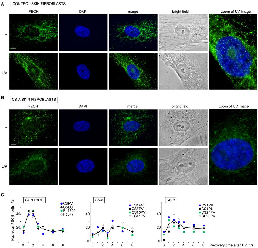
2 Nucleic Acids Research, 2021
ple repair (TC-NER), that rapidly removes the DNA helix- MATERIALS AND METHODS
distorting lesions and the ultraviolet (UV)-induced DNA
Cell culture conditions and treatments
photoproducts blocking the progression of the transcrip-
tion machinery in active genes (4). In particular, CSB is an The study was performed on primary dermal fibroblasts
ATP-dependent chromatin-remodelling factor that recruits established from biopsies taken from sun-unexposed areas
NER proteins at the site of DNA damage through the inter- of the skin of CS4PV, CS7PV, CS11PV, CS15PV (CS-A),
action with the stalled RNA polymerase II (RNAP2) (5). By CS1PV, CS26PV, CS27PV, CS1PL (CS-B) and GM05008
binding to DDB1, CUL4A and ROC1 proteins, CSA forms (EPP) patients and from four healthy donors C3PV, C5BO,
an E3–ubiquitin ligase complex (CSA-core complex) that Fb1609 and Fb377.
Downloaded from https://academic.oup.com/nar/advance-article/doi/10.1093/nar/gkab819/6377400 by guest on 22 October 2021
is required for lesion removal and proteasome-dependent In addition, the SV40-transformed MRC5 (normal) and
degradation of CSB and subsequent transcription recov- CS3BE.S3.G1 (CS-A) cell lines were used. MRC5 cells
ery (6–8). Defective TC–NER pathway accounts for the were a gift from A. Lehmann (University of Sussex, U.K.)
increased cellular sensitivity to the lethal effects of UV whereas CS3BE.S3.G1 fibroblasts were from the Coriell
light and inability to recover DNA and RNA synthesis Cell Repository. CS3BE.S3.G1 cells were used to gener-
at late times after irradiation in CS cells ((9) and refer- ate the isogenic cell lines CS3BE-cassette1 and CS3BE-
ences therein). Besides TC-NER, CS proteins are involved wtCSAFlag-HA . Cells were routinely cultured at 37◦ C in
in other cellular pathways. It was shown that CSB acts as a humidified atmosphere conditioned with 5% CO2 and
an auxiliary factor in base excision repair (BER), the ma- grown in DMEM (Gibco) supplemented with 10% fetal
jor pathway that removes nuclear and mitochondrial oxida- bovine serum (FBS, South America Origin, Gibco), 2 mM
tive DNA lesions, such as 8-hydroxyguanine (8-OH-Gua) L-glutamine (EuroClone), 0.1 mg/ml Streptomycin (Eu-
(10–13). It is involved in mitochondrial impairments and roClone) and 100 U/ml Penicillin (EuroClone). CS3BE-
cellular senescence via regulation of p21 and HTRA3 pro- cassette1 and CS3BE-wtCSAFlag-HA cell lines were routinely
tease expression (14). Moreover, CSB plays a role in nu- cultured in medium supplemented with 0.25 mg/ml G418
clear transcription by interacting with RNAP1 and RNAP2 (geneticin, Gibco) or 0.15 g/ml puromycin (Thermo Fis-
(15–21) and mediates the transcriptional programs follow- cher Scientific), respectively.
ing exposure to cellular stressors such as UV, oxidative dam- Where indicated, cells were exposed to UV-C light and
age, inflammation and hypoxia (22). CSB repositions nu- analysed after recovery in DMEM complete medium for the
cleosomes by interacting with NAP1-like histone chaper- indicated times. Alternatively, cells were treated with potas-
ones (23) and solves the G-quadruplex structures of ribo- sium bromate 20 mM (Sigma-Aldrich) or menadione 100
somal DNA (24). Less detailed information is available on M (Sigma-Aldrich) in DMEM for 1 h at 37◦ C.
CSA functioning outside TC-NER but, similarly to CS-B,
fibroblasts from CSA-defective (CS-A) patients are hyper-
sensitive to oxidative agents, contain high levels of reactive Cell survival assay
oxygen species (ROS) and are characterized by mitochon- Cell survival was determined by standard clonogenic assays.
drial dysfunction (25–30). Moreover, CSA acts as transcrip- Briefly, cells were incubated overnight in standard medium
tion factor of RNAP1 inside the nucleoli and stimulates supplemented with 10% FBS, then exposed to increasing
RNAP1 transcription re-initiation by recruiting CSB and energy levels of UV-C light (5, 10, 15 or 5, 10, 20, 25, 30
the TFIIH transcription factor at ribosomal gene promot- and 35 J/m2 ) or increasing menadione concentrations (25,
ers 1 (31). Both CSA and CSB favour the RNAP1 recruit- 50, 100 and 200 M), which was maintained for 1 h and
ment at the rDNA coding region by interacting with the then replaced with fresh standard medium supplemented
nucleolar protein Nucleolin (32) while the relevance of CS with 10% FBS. Cells were fixed after 10–15 days and stained
proteins in RNAP1 transcription is highlighted by the re- with Coomassie. The number of clones was counted and ex-
cent observations that ribosomal transcription defects are pressed as a percentage of the number of clones in the cor-
responsible for a loss of proteostasis in CS cells (33–35). responding untreated cultures.
To better understand the molecular mechanisms and the
signalling pathways by which CSA operates, we developed
a cell system that allowed the identification of a chromatin- UDS and RRS
bound complex containing RNAP1, CSA, CSB, ribosomal
protein (RP) 10, RP15 and ferrochelatase (FECH). FECH DNA repair capacity in responses to UV irradiation in pri-
is a well-known enzyme (heme-synthase) that catalyses the mary fibroblasts was determined by unscheduled DNA syn-
last step of the heme biosynthesis pathway in mitochondria. thesis (UDS) and recovery repair synthesis (RRS) as previ-
Pathogenic mutations in the FECH gene cause erythropoi- ously described (40–43).
etic protoporphyria (EPP, OMIM 177000), an inherited dis-
order characterized by painful cutaneous photosensitivity
Plasmids
as well as severe hepatic disease in 1–5% of patients, which
may require treatment by orthotopic liver transplantation The pL1Neo-Tk-L2 plasmid (cassette1) was generated by
(reviewed in (36–39). Besides demonstrating that FECH is cloning the Herpes virus simplex 1 Thymidine kinase (HSV-
a novel interactor of CSA, we find that this heme-synthase tk) transcription unit from the pTK2A vector (44) into the
protein also localizes in the nucleolus where, in combination pEGFP-N1 plasmid (BD Biosciences Clontech, GenBank
with CS proteins, it binds to RNAP1 and regulates riboso- acc, N, U55762), which contains the selection marker con-
mal biogenesis. ferring resistance to G418 (Neo gene). The DNA fragment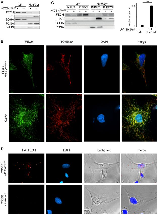
Nucleic Acids Research, 2021 3
containing the two selection markers was flanked by the het- g/ml) was added to the medium in order to counter-select
erotypic Cre-recombinase target sites, loxP and lox2272. against random integration of cassette2. Parallel aliquots of
The pRMCE2-wtCSAFlag-HA plasmid (cassette2) was ob- positive clones were also checked for G418 sensitivity.
tained by transferring the wtCSAFlag-HA cDNA sequence
into the expression vector pRMCE-OriC-ECT-RfA (patent
Polymerase chain reaction (PCR)
ITMI20120489A1). The wtCSAFlag-HA sequence was ob-
tained from the pcDNA3-CSAFlag-HA (kindly given by K. To identify clones that stably integrated a single-copy of cas-
Tanaka, Osaka University, Japan), cloned into the Gate- sette1 (CS3BE-cassette1) or that successfully recombined
way entry vector pENTR™11 (Thermo Fischer Scientific) cassette2 (CS3BE-wtCSAFlag-HA ) by Cre-mediated RMCE,
Downloaded from https://academic.oup.com/nar/advance-article/doi/10.1093/nar/gkab819/6377400 by guest on 22 October 2021
and then transferred to the pRMCE-OriC-ECT-RfA vector PCR-screening was carried out on genomic DNA isolated
by LR recombination reaction (Gateway system, Thermo from single colonies by lysing cells in H2 O, freezing and
Fischer Scientific) following manufacturer instructions. Re- thawing them in dry ice and at 95◦ C for 10 min, respectively.
combination reaction was set up with 65 ng pENTR11- After proteinase K treatment, PCR reactions were carried
CSAFlag-HA , 150 ng pRMCE–OriC–ECT–RfA, 2 l 5× LR out in 50 l containing 1× GoTaq Master Mix Reaction
Clonase reaction buffer in TE buffer (10 mM Tris–Cl pH mix (Promega), 2.5 mM each dNTPs, 20 pmol each primer,
7.5, 1 mM EDTA). either 1 or 3 mM MgCl2 (see Supplementary Table S1) and 1
The pRMCE–OriC–ECT–RfA plasmid derives from the U GoTaq DNA polymerase (Promega). Amplification was
pBKS-eLamB2-ori, which carries the ectopic 1.2 kb Lam- performed with one cycle of initial denaturation at 94◦ C for
inB2 origin of DNA replication (here indicated as ECT se- 3 min, forty cycles at 94◦ C for 30 s, annealing temperature
quence) (45). The SV40-driven pac transcription unit iso- for 30 s and 72◦ C for 30 s, and a final extension at 72◦ C for
lated from the pPur vector (BD Biosciences Clontech) and 5 min. Primer sequences are listed in Supplementary Table
the CMV-driven RfA transcription unit (Thermo Fischer S1.
Scientific) were cloned in opposite direction at each side of
the ECT sequence. The DNA fragment containing the pac,
RNA extraction and real time RT–PCR analysis
ECT and RfA sequences were flanked by loxP and lox2272
sites. Analyses were performed according to standard protocols
The pCre express the NLS-Cre recombinase under the (47). Total RNA was extracted from cells using the RNeasy
HSV-tk promoter. It was isolated from the pCre/Flp, gifted kit (Qiagen) according to the manufacturer’s instructions.
by Dr Lauth M (46), by digestion with HindIII and SalI, Contaminant DNA was removed by treatment with DNase
and cloned into pBluescript KS (+) (Stratagene). I (Qiagen), and in a single RT reaction, 1 g RNA was
reverse transcribed using the iScript cDNA synthesis kit
(BioRad). The cDNAs were used as template in real-time
Generation of the CS3BE-derivative cell lines
PCR reactions containing 10 pmol each forward and re-
The CS3BE-cassette1 cell line was obtained by transfecting verse primers (Supplementary Table S2) and the SYBR
CS3BE.S3.G1 cells with 2.5 g of the BamH1-linearized Green I fluorescent dye (Biorad). The PCR reactions were
pL1Neo-Tk-L2 plasmid (cassette1) by using the Lipofec- performed in the LightCycler 480 Real-Time PCR system
tamine LTX and Plus reagent (Thermo Fischer Scientific), (Roche Diagnostics).
according to manufacturer instructions. After G418 selec-
tion, resistant clones were isolated and PCR-screened for
Cloning of the genomic integration site (SpacerC3-PCR as-
the presence of both loxP and lox2272 recombination sites.
say)
Single copy integration of cassette1 was verified by
Southern blot analysis. Briefly, genomic DNA was purified High resolution mapping of the genomic integra-
by phenol:chloroform:isoamyl alcohol (25:24:1) extraction tion site of the transgenic vector was obtained
in Eppendorf Phase-Lock Gel™ Light and sodium acetate by applying a modified-splinkerette assay (48).
(1/10 volume)/ethanol 100% (2.5 volume) precipitation. Briefly, the splinkerette Top oligonucleotide was re-
About 12 mg of DNA were then digested with 72 IU EcoRI placed by the SPL-TOP-GATC-5 P-spC3 (5 [PHO]
or BglII restriction enzymes, fractionated on 0.8% TBE- GATCCCACTAGTGTCGACACCAGTCTC [SpC3] 3 )
agarose electrophoresis, blotted onto a Hybond N+ mem- oligonucleotide and annealed with the SPLNK-BOT2 (5 -
brane (Amersham Pharmacia) and probed with 32 P-labelled CGAAGAGTAACCGTTGCTAGGAGAGACCGTGG
PvuI-linearized pTk2A. Filters were exposed to phospho- CTGAATGAGACTGGTGTCGACACTAGTGG-3 )
imager cassettes and the autoradiographic signal was ac- oligonucleotide. Annealed double strand linker, spC3-
quired by Typhoon Trio scanner (GE Healthcare). GATC-linker, was obtained by mixing the above
CS3BE-cassette1 clone 68 cells were used to generate the (40M/each) oligonucleotides in the annealing buffer
CS3BE-wtCSAFlag-HA cell line by Recombinase-Mediated (10 mM Tris pH 7.5, 50 mM NaCl, 10 mM MgCl2 ), heating
Cassette Exchange. Briefly, CS3BE-cassette1 cells at 90– at 95◦ C for 2 min and slowly cooling at room temperature.
95% of confluence were co-transfected with 2 g pCre and 2 The linker was then diluted in water to the 4 M final stock
g pRMCE2-wtCSAFlag-HA (cassette2) by using the Lipo- concentration. In parallel, CS3BE-wtCSAFlag-HA ge-
fectamine™ LTX and Plus™ reagent (Thermo Fischer Sci- nomic DNA (gDNA) was purified with standard procedure
entific) according to manufacturer instructions. Forty-eight based on sodium dodecyl-sulphate lysis supplemented with
hours later, recombined cells were selected in 0.3 g/ml proteinase K followed by rounds of phenol-chloroform
puromycin containing medium and two days later, GCV (2 extractions. 200 ng of CS3BE-wtCSAFlag-HA gDNA was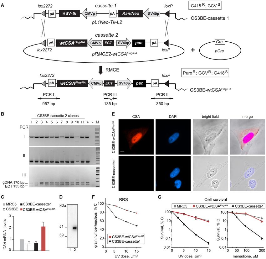
4 Nucleic Acids Research, 2021
digested by BglII (10 U/g) and ligated to a (0.5 M) Proximity ligation assay (PLA)
molar excess spC3-GATC-linker with 3 l T4 DNA
CS3BE-cassette1 or CS3BE-wtCSAFlag-HA cells were fixed
Ligase (400 U/ul) NEB in a 50 l final volume. Ligation
with 3% PFA for 12 min at RT and subsequently perme-
was precipitated in 0.3 M sodium acetate pH 5.2 and 3
abilized with 0.1% Triton X-100 in PBS at the indicated
volume of 95% ethanol. The pellet was resuspended in
time points after UV-C irradiation (10 J/m2 ). PLA was per-
10 l 0.1× TE, and 0.5l used as template in the first
formed by using the Duolink in situ Detection Kit (Sigma-
reaction of the three serial PCR (nested-PCR) with FDM-
Aldrich) following manufacturer’s instructions. Briefly, cells
003-fw/ SPLNK#1 S1 (PCR525) primer set. The second
were incubated with provided blocking buffer for 30 min
and third serial PCRs were then performed with FDM-
at RT, then incubated with anti-HA antibody (Cell Signal-
Downloaded from https://academic.oup.com/nar/advance-article/doi/10.1093/nar/gkab819/6377400 by guest on 22 October 2021
005 fw/ SPLNK#1 S2 (PCR534) and FDM-006 fw/
ing, 3724; 1:1000) and anti-FECH antibodies (S. Cruz, sc-
SPLNK#1 S2 (PCR535) primer sets on 1:20 dilutions of
377377; 1:40) or anti-RNAP1 (RPA194, S. Cruz, sc-48385;
the PCR525 and PCR534 templates, respectively. Primer
1:50) and anti-FECH (Abcam, ab55965; 1:50) and subse-
sequences are listed in Supplementary Table S3.
quently with anti-mouse PLUS and anti-rabbit MINUS
The plasmid-genomic-linker chimeric PCR product was
PLA probes. Interaction foci were imaged using an Olym-
then sequenced by standard procedure. To confirm the
pus IX71 inverted microscope equipped with a CCD cam-
cloning of the integration site and exclude artefactual
era (Robert Scientific Photometrics) and analyzed using the
chimeric DNA generated by the ligation step, PCR reac-
ImageJ software (49).
tions performed with primer sets targeting the 5 (Supple-
mentary Table S4) or 3 (Supplementary Table S5) arms of
the vector or the flanking gDNA sequences (Supplementary Run-on transcription assay
Table S6) were assayed on undigested gDNA.
BrUTP incorporation in permeabilized cells (Run-on Tran-
scription) was performed as previously described (50).
RNA interference Briefly, cells were seeded on coverslips, washed once with
Primary fibroblasts were transfected with 75mM control TBS and once with glycerol buffer (20 mM Tris–HCl, pH
(AllStars Negative Control siRNA, 1027280, Qiagen), 7.4, 5 mM MgCl2 , 25% glycerol, 0.5 mM PMSF, 0.5 mM
FECH siRNA (Hs FECH 1 SI00002898, Hs FECH 2 EGTA). Cells were then permeabilized with glycerol buffer
SI00002905, Hs FECH 3 SI00002912, Hs FECH 5 containing 0.05% Triton X-100 for 3 min at RT and incu-
SI03023090 FlexiTube siRNA, Qiagen), ERCC8/CSA bated with transcription buffer (100 mM KC1, 50 mM Tris–
siRNA (Hs ERCC8 9 SI03089492, Hs ERCC8 8 HCl, pH 7.4, 5 mM MgC12 , 0.5 mM EGTA, 25% glycerol, 5
SI03051713, Hs ERCC8 7 SI03025456, Hs ERCC8 3 U/ml RNase inhibitor from human placenta (Boehringer),
SI00001617 FlexiTube siRNA, Qiagen), RPS10 siRNA 1 mM PMSF, 0.5 mM ATP, 0.5 mM of ATP, CTP, GTP
(Hs RPS10 8 SI04270196, Hs RPS10 7 SI04193588, (GE Healthcare) and 0.2 mM BrUTP (Sigma-Aldrich)) at
Hs RPS10 5 SI02731071, Hs RPS10 2 SI00707126 Flex- RT for 1 h. Samples in which RNAP2 was inhibited were in-
iTube siRNA, Qiagen) or RPS15 siRNA (Hs RPS15 10 cubated with 200 M DRB (Sigma-Aldrich) for 1 h at RT,
SI04326140, Hs RPS10 9 SI04183256, Hs RPS10 8 concurrently with transcription reaction. Coverslips were
SI03208730, Hs RPS10 3 SI00707273 FlexiTube siRNA, then washed once for 3 min with TBS containing 0.5% Tri-
Qiagen) using the HiPerFect Transfection Reagent (301705, ton X-100 and 5 U/ml RNase inhibitor, and once with TBS
Qiagen). After 60 h, a second transfection was performed containing 5 U/ml RNase inhibitor. Afterwards, cells were
with the same siRNA and procedure. Cells were processed fixed in 2% PFA for 15 min and hybridized with fluores-
after 120 h of gene silencing. cent anti-BrdU antibodies (GeneTex, GTX128091; 1:100).
The intensity profiles were achieved by using the Meta-
Morph Microscopy Automation and Image Analysis Soft-
Immunofluorescence ware (Molecular Devices). Nuclei were segmented based on
Cells were fixed with 3.7% paraformaldehyde (PFA) for DAPI staining and the signal-integrated density of BrdU
12 min, permeabilized in PBS containing 0.1% Triton X- staining was quantified for each nucleolar region using Im-
100, hybridized with anti-HA (Roche, 11867423001; 1:250), ageJ software (49).
anti-FECH (S. Cruz, sc-377377; 1:40) or anti-TOMM 20
(Sigma) antibodies and afterwards with secondary antibod-
Whole cell extract and subcellular fractionations
ies conjugated to Alexa Fluor 448 or Alexa Fluor 555 (Jack-
son Immunoresearch). Nuclei were counterstained with Adherent cells were lysed 15 min at RT in Cell Lytic
DAPI. Images were acquired using an Olympus IX71 in- M buffer (Sigma-Aldrich) supplemented with protease in-
verted microscope equipped with a CCD camera (Robert hibitor cocktail (Roche). Whole cell lysate was collected
Scientific Photometrics) and analysed using the Meta- with a cell scraper, centrifuged at 13 000 rpm for 15 min
Morph Microscopy Automation and Image Analysis Soft- and stored at −20◦ C.
ware (Molecular Devices). The quantification of FECH Nuclear (Nuc) and Cytoplasmic (Cyt) fractions were pre-
accumulation inside the nucleolus was performed on im- pared by using the Nuclear complex Co-IP kit (Active Mo-
munostained cells 1, 2, 3 or 4 h after UV-C irradiation (10 tif) according to manufacturer’s instructions. Briefly, cells
J/m2 ). For each cell strain, at least 200 nuclei were anal- were collected by scraping in ice-cold PBS supplemented
ysed. Exposure time, binning, microscope settings and light- with phosphate inhibitors (Active Motif), centrifuged, re-
source intensity were kept constant for all the samples. suspended in hypotonic buffer, incubated for 15 min on ice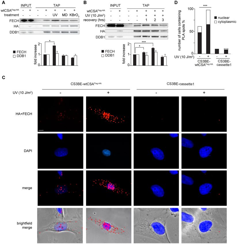
Nucleic Acids Research, 2021 5
and homogenized by 30 strokes with a dounce homoge- incubated at 70◦ C for 10 min and analyzed by SDS-
nizer. The homogenate was diluted with detergent buffer PAGE. For gel electrophoresis the 4–12% NuPAGE Bis-
and centrifuged at 12 000 rpm for 30 s to recover the su- Tris Gels (Thermo Fischer Scientific), 4–20% Mini-Protean
pernatant that included both cytosolic and mitochondrial TGX Gels (Bio-Rad) or 12% SDS-polyacrilamide gels
fractions. The nuclear pellet was resuspended in Complete were used. Samples were transferred onto a nitrocellulose
Digestion Buffer (Digestion Buffer supplemented with pro- membrane by using the Trans-Blot Turbo transfer system
tease inhibitor cocktail and 0.5 mM PMSF), supplemented (Bio-Rad). Immunoblotting was performed using antibod-
with the Enzymatic Shearing Cocktail and incubated at 4◦ C ies specific for CSA (Genetex, GTX100145; 1:500), CSB
for 3 h. The reaction was stopped by adding 0.5M EDTA (S. Cruz, sc-10459; 1:300), Cul4A (Genetex, GTX113876;
Downloaded from https://academic.oup.com/nar/advance-article/doi/10.1093/nar/gkab819/6377400 by guest on 22 October 2021
for 5 min on ice. Finally, the suspension was centrifuged at 1:500), c-Jun (S. Cruz, sc-1694; 1:500), DDB1 (a gift from
12 000 rpm for 30 s to recover the supernatant containing Dr R. Groisman; 1:1000), FECH (S. Cruz, sc-377377;
the nuclear fraction. 1:250), GAPDH (S. Cruz, sc-32233; 1:1000), HA (Roche,
Pure mitochondrial fractions (Mit) were obtained using 11867423001; 1:1000), HA (Cell Signaling, 3724; 1:1000),
the Mitochondrial fractionation kit (Active Motif) or the H3 (Abcam, ab1791; 1:10 000), MEK2 (BD Biosciences,
Mitochondria Isolation Kit (Sigma-Aldrich) according to 610235; 1:2000), ORC2 (S. Cruz, sc-32734; 1:2000), PCNA
the manufacturer’s instructions. Pure nuclear/cytoplasmic (S. Cruz, sc-10807; 1:1000), RNAP1 (S. Cruz, sc-48385;
fractions (Nuc/Cyt) free from mitochondria were obtained 1:500), ROC1 (Abcam, ab133565; 1:1000), RPSA (Ab-
as described in reference (51), steps 13–23. cam, ab133645; 1:1000), RPS10 (Genetex, GTX33475;
For nucleolar fractionation, previously described proto- 1:1000), RPS15 (Abcam, ab157193; 1:1000), RPS28 (Ab-
cols have been adapted in order to guarantee the absence of cam, ab133963; 1:500), SDHA (Abcam, ab14715; 1:10 000).
mitochondrial contamination (51,52). Briefly, 4 × 10⁷ pri- Chemiluminescent signals were detected using the Chemi-
mary dermal fibroblasts from healthy donors were washed Doc XRS system (Bio-Rad) and quantified using the Im-
in cold PBS, centrifuged and resuspended in five volumes ageJ software (49).
of extraction buffer A (20 mM Tris pH 7.6, 0.1 mM EDTA, For gel silver staining, proteins were resolved on a 4–
2 mM MgCl2 , 0.5 mM NaF and 0.5 mM Na3 VO4 ) supple- 12% NuPAGE Bis–Tris Mini gel (Thermo Fischer Sci-
mented with Proteinase Inhibitor Cocktail (PIC). Cell pel- entific) and stained with the SilverQuest Silver Staining
let was incubated 10 mins on ice, added 10% (vol/vol) so- kit (Thermo Fischer Scientific) following manufacturer
lution of Nonidet P-40 and mixed by tube inversion. Cell instructions.
membranes were fragmented by passing the lysate through
a G-20 needle and intact nuclei were collected by centrifu-
gation at 500g for 3 min at 4◦ C. Supernatant (cytoplasm) Single and two-step co-immunoprecipitations
was stored at −80◦ C. The pellet containing nuclei was re-
Whole cell lysates or subcellular fractions from 0.3–1 × 10⁷
suspended in 2 ml of S1 buffer (0.25 M sucrose, 10 mM
cells, were immunoprecipitated with anti-FECH (S. Cruz,
MgCl2 ), layered over 2 ml of S2 buffer (0.35 M sucrose, 0.5
sc-377377ac), anti-RNAP1 (S. Cruz, sc-48385ac) or anti-
mM MgCl2 ) and centrifuged at 1430g for 5 min at 4◦ C. Pel-
CSB (A301-345A Bethyl) antibodies conjugated with A/G
let was again resuspended in 2 ml of S2 buffer and sonicated
agarose beads according to standard protocols (54). Briefly,
(6 times alternating 10 s bursts and 10 s rest). After check-
extracts were incubated overnight at 4◦ C with 15 g of
ing the breakup of nuclei with a phase contrast microscope,
antibody-conjugated beads. Beads were washed three times
the sample was layered over 2 ml of S3 buffer (0.88 M su-
with 1X IP high buffer (Active Motif) supplemented with
crose, 0.5 mM MgCl2 ) and centrifuged at 3000g. The nu-
1 mg/ml BSA and three times with 1× IP high buffer. Im-
cleolar pellet was collected while the supernatant (nucleo-
munoprecipitated proteins were eluted for 10 min at 70◦ C
plasm) was added to the cytoplasm fraction. Nucleoli were
with NuPAGE-LDS Sample Buffer 2× (Thermo Fischer
resuspended in 1 ml of S2 buffer, centrifuged at 1430g for 5
Scientific), supplemented with 50 mM DTT and further in-
min at 4◦ C and finally resuspended in 50 l of RIPA buffer
vestigated by immunoblotting.
supplemented with PIC. Nucleolar extract was used for im-
For the two-step co-immunoprecipitation (TIP), chro-
munoblotting.
matin enriched fractions were first immunoprecipitated
Chromatin-enriched fractions were prepared as previ-
with anti-FECH antibody (S. Cruz, sc-377377ac) follow-
ously described (53). Cells were lysed in Triton Lysis Buffer
ing the conditions described above. The immunoprecip-
(10 mM Tris–HCl pH 7.4, 1% Triton X-100, 2.5 mM
itated proteins were eluted three times at RT with Fer-
MgCl2 , 0.025 mM Na3 VO4 , 12.5 mM NaF, 6.25 mM
rochelatase (A-3) blocking peptide (S.Cruz, sc-377377 P)
-glycerophosphate, protease and phosphatase inhibitors)
and subsequently immunoprecipitated with anti-RPA194
and centrifuged (13 400 rpm) for 5 min at 4◦ C. The super-
antibody (S. Cruz, sc-48385ac). Final elution was per-
natant was collected (soluble fraction) whereas the pellet
formed for 10 min at 70◦ C with NuPAGE-LDS Sample
(chromatin-enriched fraction) was dissolved in WCE buffer
Buffer 2× (Thermo Fischer Scientific) supplemented with
(25 mM HEPES pH 7.9, 130 mM NaCl, 1.5 mM MgCl, 0.2
50 mM DTT.
mM EDTA, 0.1% TritonX-100, 0.5 mM DTT, protease and
phosphatase inhibitors) and used for immunoblotting.
Chromatin-immunoprecipitation
Immunoblot analysis and silver staining
ChIP experiments were performed as described (55). Briefly,
Whole cell extracts or cellular fractions were supplemented DNA co-immunoprecipitated with the anti-RNAP1 anti-
with NuPage LDS sample buffer 1× and 50mM DTT, bodies (RPA194, S. Cruz, sc-48385) was quantified by real
6 Nucleic Acids Research, 2021
time PCR using the primers listed in Supplementary Table of matched peptides (>4) and the MASCOT algorithm as-
S7 and the LightCycler 480 (Roche). signed probabilistic score (>80 or P < 0.001).
Tandem affinity purification (TAP) Northern blot
Total extracts from 5 × 10 cells were incubated overnight at
7 Norther blot was performed according to previously de-
4◦ C with anti-Flag M2 Affinity Gel (Sigma-Aldrich). Beads scribed protocols (35,58). Briefly, 5 g of total RNA was
were washed five times with 1× IP low buffer (Active Mo- denatured at 65◦ C for 15 min, placed on ice for 5 min and
tif) and the immunoprecipitated proteins were eluted with run on a 0.9% agarose gel for 3 h at 80 V. RNAs were then
transferred to Amersham Hybond membrane overnight,
Downloaded from https://academic.oup.com/nar/advance-article/doi/10.1093/nar/gkab819/6377400 by guest on 22 October 2021
the Flag peptide (Sigma-Aldrich) in TBS (Tris 10 mM pH
7.4, NaCl 150 mM). Eluates were further immunoprecip- crosslinked with 1200 J UV and pre-hybridized for 2 h at
itated with anti-HA agarose affinity resin (Sigma-Aldrich) 65◦ C with pre-hybridization buffer (50% formamide, 0.1%
overnight at 4◦ C. Beads were washed 5 times with 1× IP low SDS, 8× Denhardt’s solution, 5× SSC buffer, 50 mM NaP
buffer and the immunoprecipitated proteins were eluted by buffer, 0.5 mg/ml t-RNA). The 32 P-labeled oligonucleotide
incubating the beads for 10 min at RT with 2× NuPAGE- probe was denatured at 95◦ C for 10 min and added to the
LDS Sample Buffer (Thermo Fischer Scientific). Eluates membrane. After 1 h at 65◦ C, the membrane was incubated
were supplemented with 50 mM DTT and further investi- at 37◦ C overnight, washed in 2× SSC and exposed to an
gated by immunoblotting. X-ray film. Bands were quantified using ImageQuant Soft-
ware.
Mass spectrometry
Statistics
Total cell extracts from 1 × 10⁹ of CS3BE-cassette1 or
CS3BE-wtCSAFlag-HA cells were subjected to TAP, as de- All the experiments were repeated at least two-three times.
scribed. All samples were loaded and resolved on 9–16% P-values were obtained by the unpaired two-tailed student
home-made polyacrylamide linear gradient gels (18 × 20 t-test. Fisher F-ratio at a probability level (P-value) of 0.05
cm × 1.5 mm) at 40 mA/gel constant current, at 9◦ C was used to compare variances among the analysed groups.
(56). Gels were stained according to a MS-compatible silver Data are reported as mean ± standard error of the mean
staining protocol (57) and digitized with Image Scanner III (SEM). P-values 15%), the number DNA plasmid containing the wtCSAFlag-HA cDNA flanked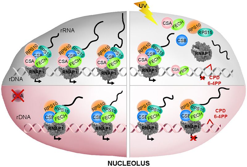
Nucleic Acids Research, 2021 7
Downloaded from https://academic.oup.com/nar/advance-article/doi/10.1093/nar/gkab819/6377400 by guest on 22 October 2021
Figure 1. Generation of the CS3BE-wtCSAFlag-HA cells. (A) A single copy of a recipient DNA cassette (cassette1) flanked by the heterotypic loxP/lox2272
Cre target sites and carrying positive (Kan/Neo) and negative (HSV-tk) selectable markers was randomly integrated in the genome of the CS-A cell
line CS3BE (CS3BE-cassette1). Co-transfection of CS3BE-cassette1 cells with the plasmids pRMCE-Ori-ECT-wtCSA (containing cassette2) and pCre
(expressing the Cre recombinase), allowed the Recombinase-Mediated Exchange of cassette1 with cassette2 (RMCE). Cassette2 contains the wtCSAFlag-HA
cDNA, a positive selectable marker (pac) and the ECT DNA sequence for stable expression of the flanking genes. Positions of the primers for PCR screening
and size of the corresponding products are indicated. ECT: ectopic sequence; G418R : resistance to G418; HSV-tk: Herpes simplex Virus 1 thymidine kinase;
Kan: kanamycin; Neo: neomycin; p: promoter; pA: polyadenylation site; PuroR : puromycin resistance. (B) PCR amplifications of amplicon I (loxP) and
III (lox2272) verify the correct RMCE event whereas amplicon II (ECT region) distinguishes the single integration event of cassette2. ECT corresponds
to a deleted form of an endogenous LaminB2 origin of replication (45) present on both homologous chromosomes. In case of a single integration of
cassette2, the PCR assay amplifies the endogenous (170 bp) and the deleted sequence (135 bp) in a 2:1 ratio, as observed in clones number 3, 4, 8 and
10. Positive (+) and negative (−) controls of the PCR reactions are shown. M: DNA ladder. (C) CSA expression levels in MRC5 (normal), CS3BE (CS-
A) and CS3BE-derivative cell lines. Transcript levels were first normalized to GAPDH expression levels and afterwards to the values in MRC5 cells.
Reported values represent the mean ± SEM. (D) Immunoblot analysis with anti-HA antibodies of whole cell extracts from the CS3BE-cassette1 (1) and
CS3BE-wtCSAFlag-HA (2) cells. (E) Immunofluorescence analysis with anti-HA antibodies in CS3BE-wtCSAFlag-HA and CS3BE-cassette1 cells. Nuclei
were counterstained with DAPI and cells visualized in bright field. (F) Recovery of RNA synthesis (RRS) in CS3BE-wtCSAFlag-HA and CS3BE-cassette1
cells. The mean numbers of autoradiographic grains per nucleus in irradiated samples are expressed as percentages of those in unirradiated cells. (G)
Sensitivity to UV-C irradiation or menadione treatment of MRC5 and CS3BE-derivative cell lines. Values in irradiated/treated samples are expressed as
percentages of those in unirradiated/control cells. Reported values represent the mean ± SEM (n = 3). Scale bar: 5 m.8 Nucleic Acids Research, 2021
by the loxP and lox2272 sites (cassette2) (Figure 1A). The
Cre recombinase mediated the swapping of cassette1 with
cassette2 by a site-specific recombination event between the
two identical loxP sites. Cells containing cassette2 were se-
lected for their resistance to puromycin (PuroR ) and gan-
ciclovir (GCVR ) and for their sensitivity to G418. The re-
combination process and the single-integration event of cas-
sette2 in chr9q21.11 were carefully characterized by PCR
analysis (Figure 1B) whereas the integration breakpoint was
Downloaded from https://academic.oup.com/nar/advance-article/doi/10.1093/nar/gkab819/6377400 by guest on 22 October 2021
mapped at the nucleotide level (Homo Sapiens chr9: 68 385
454–68 385 486 GRCh38.p12) by a modified version of the
splinkerette assay (Supplementary Figure S2). One of the
clones showing the proper single copy integration of cas-
sette2 (clone 4 thereby termed CS3BE-wtCSAFlag-HA ) was
further investigated for the expression level of CSA mRNA.
By using primer pairs specific for the CSA coding sequence,
we evaluated the total amount of CSA transcript in CS3BE-
wtCSAFlag-HA , CS3BE-cassette1, CS3BE cells and the nor-
mal human fibroblasts MRC5 (Figure 1C). The amount of
CSA transcripts in CS3BE-cassette1 is comparable to that
of the parental cell line CS3BE, indicating that the integra-
tion of cassette1 in the genome of CS3BE cells does not Figure 2. FECH is a novel interactor of CSA. (A) Silver staining analysis
of CSA-interacting proteins purified from total cell extracts of CS3BE-
alter the expression of the mutated endogenous alleles. In cassette1 (−) or CS3BE-wtCSAFlag-HA cells (+) by Tandem Affinity Pu-
CS3BE-wtCSAFlag-HA cells, the amount of CSA mRNA is rification (TAP). The identity of CSA-interacting proteins was determined
twice that of MRC5 cells, indicating similar expression lev- by mass spectrometry. (B) Immunoblot analysis with antibodies specific
els for the recombinant wtCSAFlag-HA and the endogenous for FECH, DDB1 or HA of the CSAFlag-HA -interacting proteins isolated
CSA gene. In addition, anti-HA antibodies recognize a sin- by TAP experiments in CS3BE-wtCSAFlag-HA (+) or CS3BE-cassette1
(−) cell lysates. (C) Immunoblot analysis with antibodies specific for
gle band of ∼48 kDa corresponding to the molecular weight FECH or HA of immunoprecipitation (IP) samples performed with an-
of the wtCSAFlag-HA protein (Figure 1D). By immunofluo- tibodies raised against FECH protein from whole cell lysates of CS3BE-
rescence, we found that the recombinant wtCSAFlag-HA pro- wtCSAFlag-HA (+) or CS3BE-cassette1 (−) cells. (D) Immunoblot analysis
tein is localized mainly in the nucleus with a weaker but still with antibodies specific for FECH, CSA, CSB, DDB1, CUL4A and ROC1
of immunoprecipitation (IP) samples performed with control IgG or anti-
detectable signal in the nucleolus (Figure 1E), as previously FECH antibodies in whole cell lysate of primary dermal fibroblasts from
reported for the endogenous CSA (18,31). the healthy donor C3PV.
The functionality of wtCSAFlag-HA in TC-NER was
demonstrated by the higher Recovery of UV-inhibited RNA
synthesis (RRS) in CS3BE-wtCSAFlag-HA cells compared sertion of the Fe2+ iron into protoporphyrin IX (61,62).
to CS3BE-cassette1 (Figure 1F) and by the higher level of Mutations in the FECH gene are associated with ery-
cell viability after UV irradiation or oxidative stress (Fig- thropoietic protoporphyria (EPP), an inherited disease of
ure 1G). While a drastically reduced survival was found in porphyrin-biosynthesis, characterized by severely painful
CS3BE-cassette1 cells, an overlapping survival curve was phototoxic reactions due to the accumulation of protopor-
observed between CS3BE-wtCSAFlag-HA and the control phyrin in the blood. Further investigations by TAP assay
MRC5 cells, indicative that wtCSAFlag-HA expression fully showed that wtCSAFlag-HA interacts with FECH as well
recovers the hypersensitivity of CS3BE-cassette1 cells to as with its well-known partner DDB1 (Figure 2B). Ac-
UV as well as oxidative stress. cordingly, by using anti-FECH antibodies we found that
wtCSAFlag-HA co-immunoprecipitates with the endogenous
FECH whereas no interaction was observed in CS3BE-
CSA interacts with the ferrochelatase (FECH) protein inside
cassette1 cells (Figure 2C), as expected. Remarkably, in pri-
the nucleus
mary dermal fibroblasts from healthy donors, we repeatedly
The sequential use of anti-Flag and anti-HA antibodies al- found that not only the endogenous CSA protein but also
lowed the purification of CSA-interacting proteins by Tan- a barely detectable amount of CSB co-immunoprecipitates
dem Affinity Purification (TAP) while the identity of the with FECH (Figure 2D). Conversely, we were unable to
CSA-binding proteins was determined by mass spectrom- provide reliable evidence of interaction between FECH and
etry. Besides the well-known interactors of CSA implicated components of CSA-core complex, such as DDB1, Cul4A
in TC-NER (namely, DDB1, Cul4A, CSN1, CSN2, CSN3, and Roc1, suggesting additional functions of CSA outside
CSN4, CSN5, CSN6, CSN7A and 7B, CSN8, Roc1), we the CSA-core complex.
isolated several other proteins among which the mitochon- FECH is always described as a mitochondrial protein
drial inner membrane protein ferrochelatase (FECH) (Fig- whereas CSA has been shown to localize both in the nu-
ure 2A and Supplementary Table S8). Notably, none of cleus and in mitochondria (10). To elucidate where CSA-
the CSA-interacting proteins was found in the parental FECH interaction takes place, we deeply investigated the
cell line CS3BE-cassette1. FECH is the terminal enzyme subcellular localization of both proteins by immunoblotting
in the heme biosynthesis pathway where it catalyses the in- distinct cellular fractions obtained by separating mitochon-Nucleic Acids Research, 2021 9
dria (Mit) from all other cellular compartments (Nuc/Cyt) In situ PLA experiments on fixed cells reveal that the nu-
(Figure 3A). The quality of the cell fractionation was at- clear red fluorescent signal for FECH-wtCSAFlag-HA inter-
tested by the distribution of the mitochondrial SDHA and action increases in number and size at 2 h post-UV recov-
the nuclear/cytoplasmic PCNA and c-Jun proteins. A small ery, while it remains at background level in UV irradiated
amount of FECH was unexpectedly found in the Nuc/Cyt CS3BE-cassette1 cells (Figure 4C–D). These findings are
fraction, although the majority of FECH is located within indicative of a dynamic CSA-FECH nuclear interaction in
the mitochondria. Conversely, the wtCSAFlag-HA protein is response to UV-irradiation that prompted us to investigate
present mainly in the Nuc/Cyt fraction with a weaker but whether FECH contributes to the removal of UV-induced
clearly detectable signal in mitochondria. By immunofluo- DNA damage via NER. To this purpose, we took advan-
Downloaded from https://academic.oup.com/nar/advance-article/doi/10.1093/nar/gkab819/6377400 by guest on 22 October 2021
rescence, we found that FECH co-localizes with the outer tage of commercially available primary fibroblasts from a
membrane mitochondrial protein TOMM20 in CS3BE- patient (GM05008) affected by EPP and whose deficiency
wtCSAFlag-HA cells and in normal C3PV primary fibroblasts of FECH activity is due to a missense mutation predicted to
(Figure 3B). Nevertheless, a weaker but still detectable sig- generate the Phe417Ser change (63) and resulting in reduced
nal is present also in the nucleus. This localization pattern amount of FECH protein (Supplementary Figure S5A).
is further supported by bioinformatic analysis of FECH The response of EPP cells to UV irradiation is compared to
protein sequence. cNLS Mapper indicates the presence of that of normal or CS primary fibroblasts by evaluating the
a monopartite nuclear localization signal (NLS) and five cell post-UV survival and the efficiency of GG-NER and
bipartite NLS with scores between 3 and 4.7 (Supplemen- TC-NER by unscheduled DNA synthesis (UDS) and re-
tary Figure S3A), predictive of protein localization to both covery of RNA synthesis (RRS), respectively (Supplemen-
nucleus and cytoplasm. Moreover, NucPred predicts that tary Figure S5B–D). As expected, CS-A and CS-B fibrob-
FECH spends at least some time in the nucleus (Supplemen- lasts show normal UDS but impaired RRS and survival lev-
tary Figure S3B). Therefore, FECH is both a mitochondrial els. Conversely, the UDS, RRS and survival levels of EPP
and nuclear protein. cells fall in the normal range, indicating a proper cellular
The physical interaction between FECH and response to UV irradiation. Similar results are obtained in
wtCSAFlag-HA in the distinct cellular fractions (Mit and normal fibroblasts after FECH expression knock-down by
Nuc/Cyt) was investigated by immunoprecipitating FECH RNA interference. To this purpose, cells were treated with
(Figure 3C). Despite the reduced levels of FECH outside FECH or control (CTR) short interfering RNA (siRNA)
mitochondria, a considerable amount of wtCSAFlag-HA is molecules for 120 h, a time point at which FECH levels
bound to FECH in the Nuc/Cyt fraction whereas a just are minimal (Supplementary Figure S5E). Upon UV ir-
detectable amount of wtCSAFlag-HA co-immunoprecipitates radiation, fibroblasts treated with FECH or CTR siRNA
with FECH in mitochondria. By using anti-HA and anti- show similar RRS values, demonstrating that impaired or
FECH antibodies, we applied the in situ proximity ligation reduced FECH activity does not affect the cellular re-
assay (PLA) to visualize the CSA-FECH interaction in sponse to UV exposure, including TC-NER (Supplemen-
fixed cells. As shown by the red fluorescent dots, the two tary Figure S5F). Therefore, the presence of CSA-FECH
proteins interact also inside the nucleus and, apparently, in interaction in the nucleus must concern a role outside
proximity of the nucleoli (Figure 3D). TC-NER.
Immunofluorescence studies were then applied to inves-
tigate the nuclear localization of FECH, its response to UV
irradiation and the relationship with CSA (Figure 5). No-
UV irradiation induces a transient FECH signal at the nucle-
tably, upon UV exposure a strong FECH positive signal is
oli that requires functional CSA
observed in the nucleoli of normal fibroblasts but not, or
Taking into account the well-established roles of CSA in less extensively, of CS-A cells (Figure 5A-B). Time-course
UV-induced DNA damage repair and oxidative stress re- analysis up to 8 h post-UV recovery in four normal fibrob-
sponse, we investigated the effect of different types of last strains showed that the percentage of cells with nucle-
stress on the FECH-wtCSAFlag-HA interaction by TAP ex- olar FECH signal (nucleolar FECH+ cells) drastically in-
periments. In whole cell extracts, the levels of both pro- creases within the first 2 h after UV irradiation, with mean
teins do not change following the various treatments (Sup- values ranging from 18% in unirradiated samples to about
plementary Figure S4). However, the amount of FECH 43% in irradiated samples (Figure 5C). The number of nu-
bound to wtCSAFlag-HA notably increases upon UV irradi- cleolar FECH+ cells decreases to the basal level at 4 h af-
ation (10 J/m2 ) but not after oxidative stress induced by ter UV and thereafter remains constant. A different pattern
either menadione (MD), a form of vitamin K that gen- is observed in fibroblasts from four CS-A donors carrying
erates reactive oxygen species (ROS), or potassium bro- different truncating mutations. Already in basal condition
mate (KBrO3 ), which mainly induces 8-OH-Gua (Figure the number of cells with detectable nucleolar FECH signal
4A). Conversely, none of the genotoxic treatments influ- is lower than that of normal cultures and it gradually in-
ences the level of wtCSAFlag-HA binding to DDB1. Thus, creases up to only 20% at 2 h after UV irradiation. A sec-
FECH-wtCSAFlag-HA interaction seems to be stabilized in ond wave of increase of nucleolar FECH+ cells is observed
response to UV irradiation. A time course analysis demon- at 4 h after UV (up to 24%), followed by a gradual decline
strates that the FECH-wtCSAFlag-HA interaction gradually (15%). Therefore, a functional CSA is critical for the tran-
increases (up to about two-fold) within the first 2 h of post- sient UV-induced appearance of FECH inside the nucleoli.
UV recovery and then rapidly returns to its basal levels (Fig- Because this event occurs in the time frame in which TC-
ure 4B). NER takes place, we investigated the relationship between10 Nucleic Acids Research, 2021
Downloaded from https://academic.oup.com/nar/advance-article/doi/10.1093/nar/gkab819/6377400 by guest on 22 October 2021
Figure 3. CSA interaction with FECH occurs mainly in the nucleus. (A) Immunoblot analysis of the mitochondrial fraction (Mit) separated from the
remaining portion of cell lysate (Nuc/Cyt) in CS3BE-cassette1 (−) or CS3BE-wtCSAFlag-HA cells (+) with antibodies raised against FECH, HA, the
inner membrane mitochondrial marker SDHA and the nuclear/cytosolic markers PCNA or c-JUN. (B) Co-immunofluorescence analysis with antibodies
raised against FECH and the mitochondrial protein TOMM20 in CS3BE-wtCSAFlag-HA cells or in primary dermal fibroblasts from the healthy donor
C3PV. Nuclei were counterstained with DAPI. Scale bar: 5 m. (C) Immunoblot analysis with antibodies specific for FECH, HA, the mitochondrial
marker SDHA or the nuclear/cytosolic marker PCNA in immunoprecipitations (IP) performed with anti-FECH antibodies in the mitochondrial (Mit)
and nuclear/cytosolic (Nuc/Cyt) fractions (input) of CS3BE-wtCSAFlag-HA (+) or CS3BE-cassette1 (−) cells. The intensity of co-immunoprecipitated
wtCSAFlag-HA band was quantified and normalized to the amount of immunoprecipitated FECH. Value are reported in the diagram on the right. Reported
values represent the mean ± SEM (n = 3), ***P < 0.001 (Student’s t-test). (D) Subcellular localization of wtCSAFlag-HA /FECH interaction by proximity
ligation assay (PLA) in CS3BE-wtCSAFlag-HA and CS3BE-cassette1 cells by using anti-HA and anti-FECH antibodies. Red fluorescence signal visualizes
the wtCSAFlag-HA /FECH interaction. Nuclei were counterstained with DAPI and cells visualized in bright field. Scale bar: 5 m.Nucleic Acids Research, 2021 11
Downloaded from https://academic.oup.com/nar/advance-article/doi/10.1093/nar/gkab819/6377400 by guest on 22 October 2021
Figure 4. CSA interaction with FECH is stabilized upon UV exposure. (A, B) Immunoblot analysis with antibodies specific for FECH, HA or DDB1 of
CSAFlag-HA -interacting proteins isolated by TAP experiments in CS3BE-cassette1 (−) or CS3BE-wtCSAFlag-HA (+) cells before and after treatment with
different stressing agents. Cells were irradiated with UV-C light (10 J/m2 ) or treated with menadione (MD, 100 M) or potassium bromate (KBrO3 , 20
mM) and analysed after 1 hr of recovery time (A) or at the indicated hours following UV irradiation (B). The protein levels were quantified and normalized
to the corresponding HA amount and expressed as fold increase of the corresponding untreated cells. Proteins fold increase relative to the not treated
sample are reported in the diagram below the plots. Reported values represent the mean ± SEM (n = 3), *P < 0.05, ***P < 0.001 (Student’s t-test).
(C) Sub-cellular localization of wtCSAFlag-HA /FECH interaction by proximity ligation assay (PLA) with anti-HA and anti-FECH antibodies in CS3BE-
wtCSAFlag-HA and CS3BE-cassette1 cells before and at 2 h of UV irradiation (10 J/m2 ). Red fluorescence signal visualizes the CSA/FECH interaction.
Nuclei were counterstained with DAPI and cells visualized in bright field. Scale bar: 5 m. (D) Number of cells containing the PLA spots shown in C and
expressed as percentage. In each sample 100 cells have been analyzed. The percentage of nuclear and cytoplasmic spots is indicated.12 Nucleic Acids Research, 2021
Downloaded from https://academic.oup.com/nar/advance-article/doi/10.1093/nar/gkab819/6377400 by guest on 22 October 2021
Figure 5. CSA functionality allows FECH visibility in the nucleolus upon UV irradiation. (A, B) Immunofluorescence analysis with antibodies specific for
FECH protein in primary dermal fibroblasts from the healthy donor C3PV (A) or the CSA-defective CS15PV patient (B), cultured under basal conditions
or at 2 h after UV-C irradiation (10 J/m2 ). Nuclei were counterstained with DAPI and cells visualized in bright field. (C) Percentage of cells showing FECH
accumulation in nucleoli (nucleolar FECH+ cells) of primary dermal fibroblasts from C3PV, C5BO, Fb1609 and Fb377 healthy donors (Normal), CS4PV,
CS7PV, CS15PV, CS11PV patients (CS-A) and CS1PV, CS1PL, CS27PV, CS26PV patients (CS-B) before and at different time points (1, 2, 3, 4, 6 and 8 h)
after UV irradiation (10 J/m2 ). For each strain at least 200 nuclei were counted and the mean value of at least two independent experiments is shown at
each point. The black line represents the mean value of all strains and all experiments. SE is below 10% in all cases. Scale bar: 5 m.
FECH positive signal at the nucleolus and functional TC- of FECH at the nucleoli requires CSA. This transient sig-
NER by analysing four CS-B fibroblast strains that are also nal may reflect either a UV-induced recruitment of the pro-
TC-NER defective but express a functional CSA protein. tein itself within the nucleoli or changes in the pattern of
In CS-B cultures, the percentage of nucleolar FECH+ cells FECH-interacting proteins leading to epitope unmasking.
approaches the levels detected in normal fibroblasts, with a The separation of nucleolar extract from the nucleoplasm,
rapid increase in the mean value up to the 31% at 2 h af- mitochondria and remaining cellular components, demon-
ter UV irradiation. Overall, these findings indicate that in strates the presence of FECH inside nucleoli of normal skin
UV-irradiated human fibroblasts the transient appearance fibroblasts already in basal condition and denies any incre-Nucleic Acids Research, 2021 13
ment of the protein at 2 h after UV irradiation (Supplemen- detachment from the chromatin of a fraction of the afore-
tary Figure S6). Besides demonstrating that FECH is also a mentioned proteins.
nucleolar protein, these findings indicate that CSA may im- To understand whether FECH, CSAFlag-HA , RNAP1,
pact on the protein-protein interaction network of FECH CSB, RPS10 and RPS15 are part of a unique chromatin-
inside the nucleoli. bound protein complex, we performed a series of co-
immunoprecipitations from native chromatin before
and after exposing the cells to UV irradiation. Co-
CSAFlag-HA and FECH form a protein complex with RNAP1, immunoprecipitated proteins have been normalized to
CSB, RPS15 and RPS10, a fraction of which dissociates from the amount of immunoprecipitated FECH (Figure 7A
the chromatin after UV irradiation
Downloaded from https://academic.oup.com/nar/advance-article/doi/10.1093/nar/gkab819/6377400 by guest on 22 October 2021
and Supplementary S8B-C), immunoprecipitated RNAP1
In the mass spectrometry analysis of CSA-interacting pro- (Figure 7B) or immunoprecipitated CSB (Supplementary
teins we found four ribosomal proteins of the small 40S ri- Figure S8D). While no interactions are found in the soluble
bosome subunits (RPS10, RPS15, RPS28 and RPSA, see fractions, a significantly larger amount of RNAP1, RPS10
Supplementary Table S8), which are known to enter the and RPS15 interact with the chromatin-bound FECH
nucleolus in order to assemble with the ribosomal RNA as well as with the chromatin-bound CSB in CS3BE-
(rRNA). The ability of CSA to bind ribosomal proteins wtCSAFlag-HA compared to CS3BE-cassette1 cells (Figure
has also been recently reported (35). Thus, we investi- 7A and Supplementary Figure S8B, D). Thus, CSA favours
gated whether FECH also interacts with the other nucleo- the formation of this large chromatin-associated protein
lar CSA-binding proteins. By co-immunoprecipitation ex- complex. Differently from FECH and CSB, the amount
periments we found that RPS10 and RPS15 proteins in- of chromatin-bound RNAP1 interactors is similar (for
teract with FECH in the nuclear compartment but not in RPS15) or even higher (for FECH, CSB and RPS10)
the cytoplasm (Figure 6A). No interaction is observed be- in CS3BE-cassette1 compared to CS3BE-wtCSAFlag-HA
tween FECH and RPS28 or RPSA, suggesting the for- cells (Figure 7B), indicating once more that the lack of
mation of a nucleolar protein complex involving CSA, CSA impairs the composition of the RNAP1-containing
FECH and specific ribosomal proteins (RPs). Knowing complex.
that CSA, similarly to CSB, is a transcription factor of At 2 h after UV irradiation the binding of FECH to
RNAP1 (18,31), we investigated whether nuclear FECH RNAP1, CSB, RPS10 and RPS15 on the chromatin of
also interacts with RNAP1 and/or CSB. Indeed, both CSA-expressing cells is reduced while no significant re-
the catalytic subunit of RNAP1 (RPA194) and CSB co- ductions are observed in the absence of CSA (Figure 7A
immunoprecipitate with FECH in the nuclear compartment and Supplementary Figure S8B, C). Similar results were
(Figure 6A), indicating that CSA–FECH–RNAP1–CSB– achieved by immunoprecipitating the chromatin-bound
RPS15–RPS10 may belong to a unique protein complex. RNAP1 or CSB (Figure 7B and S8D), further support-
Similarly, RNAP1 co-immunoprecipitates FECH in total ing our previous observation that the presence of CSA
nuclear extracts (Supplementary Figure S7). Since rRNA favours the dissociation of a fraction of the complex from
synthesis and ribosomal biogenesis take place on the chro- the chromatin after UV exposure (Figure 6B). Meanwhile,
matin template, we investigated the complex composition FECH slightly increases its binding to the chromatin-bound
associated to the chromatin. Thus, native chromatin was wtCSAFlag-HA (Figure 7A and Supplementary Figure S8B,
separated from the TritonX100-soluble fraction of CS3BE- C).
cassette1 and CS3BE-wtCSAFlag-HA cells lysed with the Finally, a two-step co-immunoprecipitation (TIP) exper-
Triton Lysis Buffer. The same procedure was applied to iment performed by first immunoprecipitating FECH and
CS1AN cells that are compound heterozygous for muta- subsequently RNAP1 from the native chromatin of control
tions in the ERCC6/CSB gene leading to the altered forms primary dermal fibroblasts fully demonstrates that FECH–
p.Lys337* and p.Tyr834Cysfs*25 of CSB. In all cell lines RNAP1–CSA–CSB–RPS15–RPS10 are all part of a unique
we found considerable amounts of FECH, RNAP1, RPS10 chromatin-bound protein complex (Figure 7C). The di-
and RPS15 associated to the chromatin marked by the pres- rect interaction between FECH and RNAP1, the influ-
ence of the histone H3 (Figure 6B). CSAFlag-HA protein is ence of CSA and the partial dissociation of FECH-RNAP1
bound to the chromatin of CS3BE-wtCSAFlag-HA cells; wild binding at 2 h after UV irradiation have also been ob-
type CSB to the chromatin of CS3BE-cassette1 and CS3BE- served by PLA analysis in CS3BE-cassette1 and CS3BE-
wtCSAFlag-HA cells (Supplementary Figure S8A) while the wtCSAFlag-HA cells (Figure 7D and Supplementary Figure
mutated p.Tyr834Cysfs*25 form of CSB is found associated S9).
to the chromatin-enriched fraction of CS1AN cells (Fig- In summary, these findings demonstrate that in normal
ure 6B). As expected, the cytosolic MEK2 and mitochon- cells there is a protein complex on the nucleolar chro-
drial SDHA markers are predominantly found in the solu- matin that includes FECH, RNAP1, CSA, CSB, RPS10
ble fractions. At 2 h after UV irradiation the amount of the and RPS15. Upon UV irradiation a fraction of this protein
chromatin-associated FECH, CSAFlag-HA , RNAP1, CSB, complex dissociates from the chromatin and disaggregates.
RPS10 and RPS15 is significantly reduced in CSA express- The chromatin-bound FECH partially loses its interaction
ing cells (CS3BE-wtCSAFlag-HA and CS1AN) while most of with RNAP1 but retains its binding to wtCSAFlag-HA . The
them preserve their interaction with the chromatin in CSA- absence of CSA affects the composition of this RNAP1-
defective cells (CS3BE-cassette1). This indicates that after contining complex and impairs its release from the chro-
UV irradiation a functional CSA is required to trigger the matin upon UV irradiation.You can also read