The QIAGEN Guide to Template Purification and DNA Sequencing (2nd Edition)
←
→
Page content transcription
If your browser does not render page correctly, please read the page content below
8745_SEQCOVER_0498.qxd 3/6/98 16:31 Uhr Page 2

                          The QIAGEN Guide to Template Purification
                                 and DNA Sequencing (2nd Edition)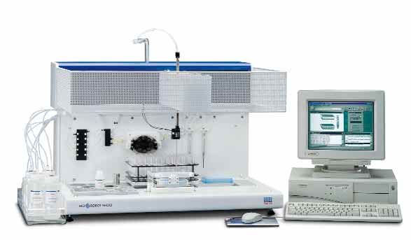
1008745_SEQ_0498.qxd 3/6/98 17:02 Uhr Page 2

        Trademarks
        The following are patented or patent-pending technologies and/or registered or registration-pending trademarks of QIAGEN: QIAGEN®, BioRobot™ 96OO, QIAsoft™,
        QIAfilter™, QIAprep®, QIAquick™, QIAwell™, R.E.A.L.™, TurboFilter™, QIAvac™, AirPore™.
        Adobe and Acrobat are trademarks of Adobe Systems Incorporated. Alconox is a registered trademark of Alconox, Inc. Bio-Rad is a registered trademark of Bio-Rad
        Laboratories. Empore is a registered trademark of 3M. Bluescript is a registered trademark of Stratagene Inc. DyeDeoxy, PRISM, BigDye, and AmpliTaq are trademarks of
        Applied Biosystems Division of Perkin-Elmer Corp. pGEM is a registered trademark of Promega Corp. Sequenase is a registered trademark of United States Biochemical Corp.
        Serdolit is a registered trademark of Serva. Perkin-Elmer is a trademark of the Perkin-Elmer Corp. DH5α and DH10B are trademarks of Life Technologies, Inc.
        ThermoSequenase is a trademark of Amersham International, Inc.
        * The PCR process is covered by U.S. Patents 4,683,195 and 4,683,202 and foreign equivalents owned by Hoffmann-La Roche AG.

        © 1998 QIAGEN, all rights reserved.

        Germany                 QIAGEN GmbH           Max-Volmer-Straße 4 • 40724 Hilden
                                                      Orders 02103-892-230 • Fax 02103-892-255 • Technical 02103-892-240

        USA                     QIAGEN Inc.           28159 Avenue Stanford • Valencia • CA 91355
                                                      Orders 800-426-8157 • Fax 800-718-2056 • Technical 800-DNA-PREP (800-362-7737)

        Australia               QIAGEN Pty Ltd        PO Box 25 • Clifton Hill • Victoria 3068
                                ACN 072 382 944       Orders 03-9489-3666 • Fax 03-9489-3888 • Technical 03-9489-3666

        Canada                  QIAGEN Inc.           151 Brunel Road • Unit 7 • Mississauga • Ontario • L4Z 2H6
                                                      Orders 800-572-9613 • Fax 905-501-0373 • Technical 800-DNA-PREP (800-362-7737)

        France                  QIAGEN S.A.           3 avenue du Canada • LP 809 • 91974 Courtaboeuf Cedex
                                                      Orders 01-60-920-920 • Fax 01-60-920-925 • Technical 01-60-920-930

        Japan                   QIAGEN K.K.           Hakusan Takayanagi Bldg. 8F • 7-6, Hakusan 1 Chome • Bunkyo-ku, Tokyo 113-0001
                                                      Telephone 03-5805-7261 • Fax 03-5805-7263 • Technical 03-5805-7261

        Switzerland             QIAGEN AG             Auf dem Wolf 39 • 4052 Basel
                                                      Orders 061-319-30-30 • Fax 061-319-30-33 • Technical 061-319-30-31

        UK                      QIAGEN Ltd.           Boundary Court • Gatwick Road • Crawley • West Sussex, RH10 2AX
                                                      Orders 01293-422-911 • Fax 01293-422-922 • Technical 01293-422-999
        http://www.qiagen.com
1008745_SEQ_0498.qxd 3/6/98 17:02 Uhr Page 3
Table of Contents
I. Preface 5
II. Sequencing Methods 6
Label location 6
Radioisotope labels 7
Chemiluminescent detection systems 7
Fluorescent dyes 7
Energy transfer dyes 8
Polymerases for DNA sequencing 8
III. Cloning Vectors for DNA Sequencing 9
Single-stranded phage vectors 9
Double-stranded plasmid vectors 9
Phagemid vectors 9
Vectors for large-insert DNA 9
IV. PCR-Amplified Template DNA 11
V. Sequencing Applications and Strategies 12
Primer walking 12
Nested deletion 13
Shotgun sequencing 14
Transposon-based strategies 15
VI. Automated DNA Sequencing 17
Effect of label location 17
Labeled primers 18
Labeled terminators 19
VII. DNA Template Purification 20
QIAGEN Anion-Exchange Resin 21
QIAGEN-tip columns 21
QIAwell Plasmid Purification System: ultrapure plasmid minipreps
for all purposes 22
QIAprep Plasmid Kits: high-purity plasmid minipreps for
general purpose use 23
R.E.A.L. Prep: standard-purity plasmid minipreps for
high-throughput applications 24
QIAprep M13: single-stranded template DNA from phage cultures 24
QIAquick: purification of PCR-amplified template DNA 25
The BioRobot 96OO: automated DNA template purification 25
VIII. Optimizing DNA Sequence Quality 26
Sequencing methods 26
The template-primer-polymerase complex 27
Template quantity 27
Template quality 29
Culture 29
Host strain 30
© 1998 QIAGEN Table of Contents 31008745_SEQ_0498.qxd 3/6/98 17:02 Uhr Page 4
Effect of specific contaminants 31
Nuclease 31
RNA 32
Salt 32
Ethanol 34
Phenol 35
Chloroform 35
Contaminants affecting PCR fragment templates 36
Primers 36
dNTPs 37
PCR mix 38
Priming artifacts 38
Mispriming 38
Poor-quality primer 40
Mixed templates 40
Template artifacts 41
Unbalanced base composition 41
GC-rich templates 41
Homopolymeric regions 42
Sequencing large templates 42
BACs, PACs, and P1s 42
Direct sequencing of genome-size DNA templates 44
IX. Conclusions 46
X. Web Addresses 46
XI. References 47
XII. Appendix: Protocols and Technical Information 50
A. Sequencing kits and reagents 50
B. Standard primers for Dye Terminator cycle sequencing 50
C. Cycle sequencing parameters 50
D. Recommended template DNA amounts 51
E. Sequencing reaction setup for resolving elongation stop structures 52
F. Direct genomic DNA sequencing protocols 52
G. Sequencing gel electrophoresis 53
H. Growth of E.coli cultures 54
i. Bacterial culture media 54
ii. Antibiotic selection 55
iii. Growth in 96-well blocks 55
iv. Origin of replication and copy number of various
plasmids and cosmids 55
v. Recommended culture volumes for plasmid
purification with QIAGEN-tips 56
I. QIAGEN sequencing template purification guide 57
i. Manual sequencing (radioactive or chemiluminescent) 57
ii. Automated cycle sequencing 57
J. Plasmid Miniprep Kit selection guide 58
XIII. Ordering Information 60
XIV. QIAGEN International Sales and Distributors 67
4 Table of Contents © 1998 QIAGEN1008745_SEQ_0498.qxd 3/6/98 17:02 Uhr Page 5
I. Preface
Since the publication of the first edition of The QIAGEN Guide to Template
Purification and DNA Sequencing, advances in sequencing chemistry and
instrumentation have significantly improved the capacity, sensitivity, and
reliability of DNA sequencing technologies. In this, the second edition of the
guide, much of the original text has been revised and new sections have
been added to accommodate recent developments1. The guide also contains
sequencing-related protocols and technical information, as well as helpful
technical and troubleshooting tips to improve your template purification and
DNA sequencing.
The DNA Sequencing and Genomics Group, in conjunction with the Scientific
Instruments Group at QIAGEN, is continually developing new products and
instrumentation to simplify template purification and DNA sequencing. These
products have enabled us to generate and assemble more than one
megabase of final DNA sequence data from various model organisms for
publicly funded genome projects. The DNA sequencing team at QIAGEN
also performs extensive in-house quality control, and offers contract QIAGEN
Sequencing Services for medium- to large-scale projects.
At QIAGEN we pride ourselves on the quality and availability of our technical
support. Our Technical Service Departments are staffed by experienced
scientists with extensive practical and theoretical expertise in molecular
biology and the use of QIAGEN products. If you have any questions or
experience any difficulties regarding sequencing, template purification, or
QIAGEN products in general, please do not hesitate to contact us.
Our customers are a major source of information regarding advanced or
specialized uses of our products. This information is helpful to other scientists
as well as to the researchers at QIAGEN. We therefore encourage you to
contact us if you have any suggestions about product performance or new
applications and techniques.
We hope you find this guide to be a useful tool for understanding and opti-
mizing your DNA sequencing results and look forward to working with you
in the years ahead to satisfy your DNA sequencing needs.
Hilden, May 1998
Dr. Andreas Düsterhöft
1 The scope of this guide is limited to those
Business Unit Manager methods currently in common use. We
DNA Sequencing and Genomics welcome comments from our customers on
the use of QIAGEN products in emerging
QIAGEN GmbH DNA sequencing-related technologies.
© 1998 QIAGEN I. Preface 51008745_SEQ_0498.qxd 3/6/98 17:02 Uhr Page 6
II. Sequencing Methods
Most DNA sequencing methods presently in use are variations of the chain-
termination method developed by Sanger and coworkers in the late 1970s (1).
In this method, the DNA to be sequenced acts as a template for the enzymatic
synthesis of new DNA starting at a defined primer binding site. A mixture of
both deoxy- and dideoxynucleotides is used in the reaction, at concentrations
which create a finite probability that a dideoxynucleotide will be incorporated
in place of the usual deoxynucleotide at each nucleotide position in the growing
chain. Incorporation of a dideoxynucleotide blocks further chain elongation,
resulting in a population of truncated fragments of varying length. The identity
of the chain-terminating nucleotide at each position can be specified either
by running four separate reactions, each of which contains a single
dideoxynucleotide (ddATP, ddCTP, ddGTP, ddTTP), or by running one
combined reaction but using labels specific for each dideoxynucleotide.
The resulting populations of molecules are separated by size on a denaturing
high-resolution polyacrylamide gel. The sequence is subsequently specified
by correlating the order of the bands on the gel with the dideoxynucleotide
used to generate each band.
Label location
In order to detect the picogram quantities of DNA produced in a
sequencing reaction, a label that can be readily identified in very small
amounts must be introduced into the DNA when it is synthesized (Figure 1).
The label can be incorporated into:
i) the oligonucleotide primer used to initiate the reaction
ii) the deoxynucleotides used for chain elongation
iii) the dideoxynucleotides used for chain termination
Labeling Strategies for Sanger DNA Sequencing
P
OH NT
OH dd
Primer OH OH
OH
A
Template
Polymerase
Primer
ddNTP
B
Template
Primer
C
Figure 1. Labeling strategies for Sanger chain Template
䊏
termination sequencing methods. A a new Primer
DNA strand is synthesized as the
polymerase moves down the template DNA, D
䊏
away from the priming site. B nucleotides Template
are added by the polymerase until a Primer
䊏䊏
dideoxynucleotide is incorporated. C , D ,
䊏
and E show different label locations. E
䊏C labeled primer. Template
䊏D labeled deoxynucleotides.
䊏
OH OH OH OH unlabeled nucleotides labeled nucleotides
E labeled dideoxynucleotide.
6 II. Sequencing Methods © 1998 QIAGEN1008745_SEQ_0498.qxd 3/6/98 17:02 Uhr Page 7
Radioisotope labels
For manual sequencing, the most common labels are the radioisotopes, 32P,
33
P, and 35S (2). The label can be incorporated throughout chain elongation by
including a labeled deoxynucleotide in the reaction, or during an initial
“labeling reaction” in which the radioactive nucleotide is limiting. Additional
unlabeled deoxy- and dideoxynucleotides are then added to complete the
reaction. The two-stage labeling procedure significantly increases the specific
activity of the label in the newly synthesized DNA relative to that obtained by
uniform labeling. Sequencing ladders incorporating radioactive labels are
typically visualized using X-ray film.
Chemiluminescent detection sytems
An alternative approach to radioisotope-based manual sequencing uses
nucleotides tagged with small molecules as labels to detect bands in a
sequence ladder (3,4). The DNA fragments are separated on a standard
polyacrylamide sequencing gel and transferred to a nylon membrane, either
by blotting or through direct transfer as the gel is running. The molecular tag
in the bound DNA is recognized by an enzyme-conjugate that catalyzes the
chemiluminescent reaction, and DNA bands can be visualized by exposure to
X-ray film. A useful feature of this technology is that the reagents used to detect
the bands in one tagged sequence ladder can be removed and additional
reactions can be performed to detect ladders with different tags run at the same
location (5,6). This approach, called multiplexing, can considerably increase
the amount of data obtained from a single gel run.
Fluorescent dyes
In automated sequencing, fluorescent dye labels allow the DNA bands to be
detected in the gel during electrophoresis (Figure 2, Refs. 7,8,9). When
different dye labels are used to tag each of the four dideoxynucleotides, the
reactions can be performed in a single tube and analyzed on a single gel
lane, further increasing the capacity and throughput of each sequencing gel.
The ability to collect data during electrophoresis is a major contributing
Radioactive Label and Fluorescent Dye Label Sequencing Methods
G A T C
3'
GAATTTCAAAACTATGCATTATATCAGTAACTTATAATAAGTTACTTATTGAGTC
GAATTTCAAAACTATGCATTATATCAGTAACTTATAATAAGTTACTTATTGAGTC
Figure 2. Comparison of radioactive label
and fluorescent dye label sequencing
methods. Left panel: the resulting X-ray film
of the conventional radioactive DNA
sequencing method. In semiautomated
fluorescent DNA sequencing (right panel),
“lane tracking” (represented by the grey
arrow) through the laser-detected fluorescent
bands of the electronic gel image is
performed by software which measures
the signal strength, and extracts an
electropherogram to which an automated
5'
“base calling” is applied.
© 1998 QIAGEN II. Sequencing Methods 71008745_SEQ_0498.qxd 3/6/98 17:02 Uhr Page 8
factor to the greater overall speed of automated sequencing, while the
ability to run several samples in a single lane significantly increases the
amount of sequence information per gel.
Energy transfer dyes
Recently, there has been a significant increase in new dye system development
both for conventional dyes and for an alternative arrangement where two
dyes are linked together in an energy transfer system. Energy transfer (ET) dyes
were originally developed by Richard Mathies (10), and were commercialized
▼ Tip by Amersham as ET primers. The Applied Biosystems Division of Perkin-Elmer
The additional sensitivity then introduced BigDyes™, which can be used with either dye-primer or
conferred by energy transfer dye-terminator chemistry (11,12). BigDyes consist of a fluorescein energy donor
dyes means that less template dye directly linked to an energy acceptor dichlorhodamine dye. Both dyes
is needed for a sequencing make up an energy-transfer system that provides significantly greater sensitivity
reaction, a feature that is compared to single dyes. BigDyes also exhibit improved color resolution, a
valuable for sequencing very property that allows better base calling. The additional sensitivity conferred
large templates and even for by energy transfer dyes means that less template is needed for a sequencing
direct sequencing of genomic reaction, a feature that is valuable for sequencing very large templates and
DNA. even for direct sequencing of genomic DNA (see Section VIII, page 44).
Polymerases for DNA sequencing
In the original Sanger protocol, the Klenow Fragment of E. coli DNA Polymerase I
was used. A significant improvement in the quality of the DNA sequence
obtained by the Sanger method occurred with the development of a modified
T7 DNA Polymerase with enhanced processivity and nucleotide incorporation
capabilities (13,14). Additional modifications to the basic protocol, such as
the use of modified nucleotides (e.g. 7-deaza-G) to avoid problems with DNA
secondary structure, have resulted in highly reliable manual sequencing
methods that work well with most DNA templates (15,16).
In addition, sequencing methods that use the thermostable Taq DNA
Polymerase and other thermostable polymerases have been developed (17).
Unlike the Klenow- and T7 DNA Polymerase-based methods, which involve
a single pass of the polymerase along each template, the use of a thermo-
stable polymerase allows the template DNA to be cycled in a series of
denaturation/renaturation steps. The input DNA serves as a template many
times in a single reaction, reducing the amount of input DNA required by
a factor of five or more.
More recently, thermostable DNA polymerases that have a high affinity for
dideoxynucleotides have been introduced (13,18–20). These enzymes,
AmpliTaq™ FS from ABI and ThermoSequenase™ from Amersham, have a
modified active site that accomodates dideoxynucleotides more easily than
the unmodified form of the enzyme. The advantage of these enzymes is that
much less dye terminator is required and incorporation of the terminators is
less affected by sequence context, resulting in more uniform band intensities.
The sequence data are therefore more reliable and require less manual
editing than data prepared with the original form of the enzyme.
8 II. Sequencing Methods © 1998 QIAGEN1008745_SEQ_0498.qxd 3/6/98 17:02 Uhr Page 9
III. Cloning Vectors for DNA Sequencing
Single-stranded phage vectors
The first vectors designed for DNA sequencing were derivatives of the single-
stranded bacteriophage M13 (21). Here, the DNA to be sequenced is cloned
into the double-stranded replicative form of the phage, transformed into E.
coli, and harvested from the culture supernatant in the form of phage
particles containing single-stranded DNA. Single-stranded template DNA is
ideal for sequencing and bypasses problems associated with strand
separation. However, single-stranded template DNA can only be sequenced
in one direction (without prior PCR amplification) and is of limited use for
other molecular biology applications. Larger M13 inserts (>1 kb) also tend
to be unstable, leading to difficulties with clone propagation and data
reliability. M13 is still used extensively by high-throughput sequencing
laboratories, but its use for smaller sequencing efforts has declined
dramatically since cycle sequencing methods and thermostable polymerases
eliminate the need for a separate template denaturation step.
Double-stranded plasmid vectors ▼ Tip
Double-stranded plasmid vectors are suitable for sequencing but require prior Plasmid templates sequenced
separation of the two DNA strands by denaturation. Plasmids to be sequenced with thermostable DNA
with modified T7 DNA Polymerase are typically denatured with sodium polymerases are subjected to
hydroxide, then neutralized in a way that maintains the denatured state until a successive cycles of heat
primer can be hybridized to the start site for the sequencing reaction (22,23). denaturation and cooling
Plasmid templates sequenced with thermostable DNA polymerases are (primer annealing) and a
subjected to successive cycles of heat denaturation and cooling (primer separate denaturation step
annealing) and a separate denaturation step is therefore not required (17,24). is therefore not required.
Phagemid vectors
Phagemids are hybrid vectors that consist of a double-stranded plasmid carrying
an origin of replication from a single-stranded bacteriophage (25,26). When
the host strain carrying the plasmid is infected with a helper phage virus, the
plasmid is replicated as a single-stranded molecule and packaged into phage
particles, from which template DNA can be purified. Mutations in the helper
phage limit its replication and ensure that most of the DNA obtained from the
mature phage particles is single-stranded phagemid DNA. Many common
vectors (pBluescript, pTZ18/19, pGEM18/19) have the ability to function
as phagemids but are much more typically used as simple plasmid vectors.
Vectors for large-insert DNA
Vectors capable of replicating large segments of foreign DNA have been
developed for use in genome mapping and large sequencing projects. The
vectors initially created for this purpose were cosmids (plasmids with lambda
phage packaging sites) (27) and YACs (yeast artificial chromosomes) (28).
Cosmids have an insert capacity of 35–45 kb, while YAC inserts can reach
up to 1 megabase. Since the number of clones that must be analyzed to
© 1998 QIAGEN III. Cloning Vectors for DNA Sequencing 91008745_SEQ_0498.qxd 3/6/98 17:02 Uhr Page 10
create a physical map depends on the insert size, larger inserts are
preferable to smaller inserts. The large inserts of YACs, however, are prone to
chimerism and instability and, since they are propagated in yeast, YACs are
unfortunately difficult to purify in sufficient quantity from the yeast chromosome
background. This limits their utility for subsequent procedures such as large-
▼ Tip scale sequencing.
Substantially higher yields These limitations have been addressed by a new generation of vectors called
can be obtained from P1 and P1s (29), PACs (phage artificial chromosomes) (30) and BACs (bacterial
PAC clones by inducing them artificial chromosomes) (31). P1s and PACs are derived from the E. coli
prior to purification. bacteriophage P1, while BACs are based on the E. coli F-plasmid. P1 and
PAC insert sizes are typically 90–145 kb, while BAC inserts range in size
from 75–350 kb. Insert instability is minimized because the clones are
propagated as low-copy plasmids (1–2 copies per cell) in E. coli. However,
P1 and PAC clones have the added advantage that they can be induced
prior to purification for substantially higher yields.
10 III. Cloning Vectors for DNA Sequencing © 1998 QIAGEN1008745_SEQ_0498.qxd 3/6/98 17:02 Uhr Page 11
IV. PCR-Amplified Template DNA
The polymerase chain reaction (PCR*) allows a segment of DNA located
between two defined priming sites to be amplified (32,33). If equal amounts
of the two primers are used, then the amplified DNA will be double-stranded.
Alternatively, if one primer is in excess, the final product will contain a
proportional amount of single-stranded DNA. Either double-stranded or
single-stranded amplified DNA can serve as a template for DNA
sequencing (34–36).
PCR has been used to amplify segments of genomic DNA for sequence-
based mutational analyses, and to amplify inserts directly from plaques or
colonies (37,38). Direct amplification from cells or plaques circumvents the
need to grow individual cultures of each clone to obtain enough DNA for
sequencing. However, it is essential to ensure that only one fragment is
amplified from each clone, and that all PCR components that can interfere
with the subsequent sequencing reaction such as primers, nucleotides and
salts, are removed.
The reliability of sequence data obtained from PCR-derived templates depends
on the number of molecules that served as templates for the original
amplification (see Appendix). If the amplification is performed from a small
number of template molecules, errors that occur in the early cycles of
replication can be propagated through subsequent cycles such that they
are detected in the final sequence. These problems can be avoided by
sequencing each region at least twice from independent amplification
reactions. Problems with PCR fidelity are most often detected when PCR-
amplified fragments are cloned, however. This is because fragments with ▼ Tip
errors that are present at a low level in the amplified DNA will not be PCR-amplified fragments
detected by direct sequencing but can be selected in the cloning process. containing low-level errors
No significant differences in error rates were found when data obtained by will not be detected by direct
Taq cycle sequencing were compared with data obtained using modified sequencing, but can be
T7 DNA Polymerase (39). selected in cloning processes.
© 1998 QIAGEN IV. PCR-Amplified Template DNA 111008745_SEQ_0498.qxd 3/6/98 17:02 Uhr Page 12
V. Sequencing Applications and Strategies
DNA sequencing applications in molecular biology fall into three broad classes:
● de novo sequencing of unknown DNA
● verification of known DNA, such as when a clone junction is
sequenced to confirm correct insertion
● diagnostic sequencing to identify variations in an otherwise known
sequence (e.g. mutation analysis)
Depending on the application, the DNA to be sequenced is carried on a
viral or plasmid cloning vector, or is part of an amplified fragment. The
approach used to sequence the target DNA (the sequencing strategy) is
designed to obtain the optimum balance between the time required for the
project and its cost. The strategy selected is usually determined by the size
of the segment to be sequenced, with larger segments requiring additional
processing before sequencing can begin.
Primer walking
If the segment of DNA to be sequenced is small, a primer that binds to a
known sequence in the flanking DNA can be used to initiate a sequence
run across the region of interest. A second sequencing run over the same
region from the opposite direction is highly recommended, since factors that
affect the sequence quality on one strand are often not present in the
opposite strand.
If the length of the DNA to be sequenced exceeds the length of a single
sequencing run, sequence data from the far end of the first run can be used
to prepare the primer for a second run into the region of interest. This
process, called primer walking, can be repeated many times to sequence
extensive tracts of DNA (Figure 3). The major advantages of primer walking
Primer Walking Strategy
UP RP
A
PBS PBS
1 3
2 4
B
Figure 3. Primer walking strategy. The bar 5
represents a segment of target DNA (shaded) 7
flanked by vector sequences (solid) containing 6
䊏
primer binding sites (PBS). A sequencing
C
8
runs are initiated from priming sites in
the vector DNA. (UP: –21M13 primer;
UP RP
䊏
RP: reverse primer). B reliable sequence
data from the far end of each first run is 1 3
used to make primers for sequence runs in the
䊏
forward and reverse directions. C the 2 5 4
priming cycle is repeated to provide 7
6
complete sequence data for both strands. 8
䊏 D overall sequencing strategy. D
12 V. Sequencing Applications and Strategies © 1998 QIAGEN1008745_SEQ_0498.qxd 3/6/98 17:02 Uhr Page 13
are that no subcloning is required, and the location and direction of each
sequencing run is known. Primer walking minimizes the degree of
redundancy needed to obtain the final sequence, and avoids assembly
problems caused by repetitive DNA. In theory, this approach can be used
to sequence a DNA segment of any size. However, its use is usually limited
to projects smaller than 5 kb because the successive cycles of sequencing,
primer design, and synthesis for the next walking step are too time-consuming.
The efficiency of primer walking is highly dependent on the length of
sequence read from each sequencing run. The priming site for a new
sequencing run is selected from the most distant reliable sequence obtained
in the previous step. Consequently, the data acquisition phase of a primer
walking project with an average accurate read length of 600 bases will be
completed in approximately half the time and with half the expense of a
project with an average read length of 300 bases.
Nested deletion
Nested deletion strategies work best for DNA segments in the 5–10 kb range
and require some prior knowledge of the restriction sites within the target
DNA. Nested deletion with exonuclease III requires a unique blunt-end or
5’-overhang restriction site adjacent to the region to be deleted, and a unique
3’- overhang site distal to the deleted region to protect the vector DNA
(Figure 4). When these sites are exposed after cleavage, exonuclease III
will enter the DNA at the 5’-overhang site and begin to digest one strand in
the 3’→5’ direction. The enzyme does not recognize the 3’-overhang and
therefore cannot initiate digestion on the opposite strand. At timed intervals,
aliquots of the reaction mix are removed, the DNA blunt-ended, and ligated
Nested Deletion Strategy
A
B
t
s er Primer binding site (PBS)
In
Ve c t o r
Digest with enzymes that cleave at A and B
A Insert Vector PBS B
5'-overhang 3'-overhang
or blunt end
Exonuclease III treatment
Remove aliquots at timed intervals
S1/Mung bean nuclease
Deletion direction
Fill-in with Klenow
DNA ligation
Figure 4. Generation of clones for nested
Transformation deletion sequencing. A and B represent
restriction endonuclease sites with 5’- and
Purification
3’-overhangs, respectively. The arrows on
the deleted plasmids at the bottom of the
figure represent sequence runs originating
from a universal priming site in the flanking
DNA of the vector.
© 1998 QIAGEN V. Sequencing Applications and Strategies 131008745_SEQ_0498.qxd 3/6/98 17:02 Uhr Page 14
to recircularize the molecules. Ideally, the time interval between each aliquot
corresponds to the amount of time required by the enzyme to digest just
under the equivalent of one sequence run (450–500 bases).
This process places a vector priming site next to the 5’-deletion terminus of
each molecule — sites that were previously located deep within the insert
DNA. The nested deletion approach has the advantage that the sequencing
runs are directed, that fewer sequencing runs are required to obtain a
complete sequence, and that the sequence data can be obtained from the
entire region of interest at one time using standard primers only.
As average read lengths increase, and primer costs decline, this method is
being used less frequently. An alternative approach that achieves much of
the benefit of the complete procedure is to subclone one or more internal
segments of the original clone and sequence these fragments in parallel by
primer walking.
Shotgun sequencing
In shotgun sequencing, the target DNA is fragmented by enzymatic
digestion or physical shearing to fragment sizes in the range of 0.5–5 kb.
Subfractions with a narrower size range (eg. 0.8–1.5 kb) are subcloned
into either a plasmid or M13 vector (Figure 5). The subclones can then be
sequenced from standard primer binding sites in the flanking vector DNA.
Shotgun sequencing requires no prior knowledge of the insert sequence,
and puts no limitations on the size of the starting DNA to be sequenced.
Shotgun Sequencing Strategy
Target DNA
Digest with enzymes
or shear
Clone fragments in vector
Sequence
Vector DNA
Plasmid M13
Assemble sequences
into contigs
Figure 5. Shotgun sequencing strategy.
Plasmid and M13 vectors are shown as
alternatives. Arrows on the vectors indicate
sequence runs into subcloned segments of
target DNA using standard primers binding
Close gaps with primer
in the vector. The arrows above and below
the bar (consensus sequence) represent walking or PCR ( )
individual sequence runs on different
subclones. The red arrows indicate primer
walking runs to eventually close the gaps
in the consensus sequence.
14 V. Sequencing Applications and Strategies © 1998 QIAGEN1008745_SEQ_0498.qxd 3/6/98 17:02 Uhr Page 15
Large projects taken to completion with the shotgun sequencing approach
have averaged a seven- to ten-fold level of redundancy. This means that the
entire segment has been sequenced the equivalent of seven to ten times to
guarantee full double-stranded coverage. 400–600 subclones are required to
sequence a cosmid-sized segment of DNA (40 kb) and 1500–1800 may
be required to sequence a medium-sized BAC clone (100–120 kb). As with
primer walking projects, the number of clones required to complete the
project depends on the read lengths obtained. Nested deletion and shotgun
sequencing both require a significant initial investment, but once subclones
are available, sequence data can be obtained rapidly from the entire target
DNA.
In theory it should be possible to complete a sequencing project using just
nested deletions or shotgun sequencing. In practice, however, it is often more
efficient to fill in gaps in the sequence by primer walking after most of the
sequence has been determined (Ref. 40, Figure 5). Recently, projects encom-
passing entire bacterial genomes have been completed using a whole
genome shotgun sequencing approach (41).
Transposon-based strategies
An alternative approach that avoids subcloning uses mobile segments of
DNA to introduce priming sites into a large segment of DNA (42,43). A clone
containing the target DNA is grown in a bacterial host strain carrying the
appropriate element and a gene encoding an enzyme (transposase) that
catalyzes transposition of the element into the target DNA (Figure 6).
In addition to sequences required for transposition, the transposable element
carries an antibiotic resistance gene or suppressor tRNA gene that can be
used as a selectable marker. Clones containing transposable elements are
selected by transforming plasmid DNA isolated from the transposase-
containing strain into a non-transposase-containing strain, with coselection
for markers carried by the plasmid vector and the inserted transposable
element. An ordered array of insertions can be obtained by using PCR or
restriction sites within the element to identify insertions at regular intervals in
Transposon-Based Sequencing Strategy
Figure 6. Transposon-based sequencing
strategy. Insertion of primer binding sites.
Tn Left: schematic diagram of a transposition
Donor Insert event. The segment on the far left
Site represents a segment of the host cell DNA.
Vector
The adjacent circle represents the target
DNA in a plasmid vector. Right:
Transposase transposon insertions in the target DNA of
individual plasmid molecules are indicated
in red. The arrows represent DNA
Transposase sequencing runs from priming sites at the
Gene ends of each transposon with transposon-
specific primers.
© 1998 QIAGEN V. Sequencing Applications and Strategies 151008745_SEQ_0498.qxd 3/6/98 17:03 Uhr Page 16
the target DNA. Priming sites at the ends of each element are then used for
sequencing runs extending away from the element into the target DNA.
Transposon-based strategies are being used less and less frequently as the
prequalification work to identify a good set of clones to be sequenced is
relatively high. The alternative strategies described above are faster and
easier to perform.
16 V. Sequencing Applications and Strategies © 1998 QIAGEN1008745_SEQ_0498.qxd 3/6/98 17:03 Uhr Page 17
VI. Automated DNA Sequencing
The development of instruments for automated DNA sequencing has
dramatically increased the throughput of individual laboratories. Unlike
manual sequencing methods, which generally use a radioactive label and
visualize the banding pattern by autoradiography, automated sequencers
use a laser to detect DNA fragments labeled with fluorescent dyes (7–9).
For many laboratories, the health risks and inconvenience of radioisotope
disposal are sufficient incentive to automate much or all of their DNA
sequence analysis. Additionally, considerable time and labor savings can
be obtained with automated sequencing through increased gel capacity
(one lane per reaction instead of four), immediate data acquisition, and
automatic base calling and data entry.
Effect of label location
As discussed previously, the label used for detection of the DNA synthesized
in a sequencing reaction can be incorporated into the primer, the
deoxynucleotides, or the dideoxynucleotides. For manual sequencing, the
label is generally supplied in the form of labeled deoxynucleotides, which
are incorporated at multiple sites along the newly synthesized DNA. For
automated sequencing, the labels are supplied as fluorescent dyes linked
to either the primer (dye primer: Ref. 44, Figure 7), the deoxynucleotides
(internal labeling: 45) or the dideoxynucleotides (dye terminator: Refs. 46, 47,
Figure 7).
Automated Dye Primer Sequencing
Taq polymerase
DNA template
dNTPs
Dye primers ddNTPs
A A
A
ACCG T A
C AC
C
ACC
Ethanol
A precipitation
G
G ACCG
Completed
sequencing
T ACCGT reaction
T
Annealing Extension ACCG T A T
Figure 7. Automated sequencing chemistries.
Automated Dye Terminator Sequencing 䊏A sequencing with dye-labeled primers.
Note the correlation between the dye on
DNA template Taq polymerase A the labeled primer and the specific
dNTPs AC dideoxynucleotide in each reaction.
ACC 䊏B sequencing with dye-labeled terminators.
B ACCG The colored circles represent different types
Annealing Extension
ACCGT of fluorescent dye. The extended primers
ACCG T A represent the shortest molecules in a
Primer Dye Deoxy™ Terminators Dye terminator Completed
ACCG T A T
A C G T removal sequencing population with lengths of up to 1400 bases.
reaction (Reproduced by kind permission of Applied
Biosystems Division of Perkin-Elmer
Corporation.)
© 1998 QIAGEN VI. Automated DNA Sequencing 171008745_SEQ_0498.qxd 18/6/98 9:25 Uhr Page 18
Labeled primers
Primer labeling with fluorescent dyes is generally limited to commonly used
primers. The reason for this is that dye primer synthesis is an expensive process,
and not one, but four derivatized primers must be synthesized. The four dye-
labeled primers are used in separate sequencing reactions, each of which
contains one of the four dideoxynucleotide terminators. Since each of the
four dideoxynucleotides is matched with a specific dye primer, the reactions
can be pooled before electrophoresis on a sequencing gel (Figure 7A).
Sequencing reactions run with dye-labeled primers typically exhibit uniform
peak intensities and high signal levels, but are more likely to show higher
background levels since nucleotide chains that terminate prematurely will add
to the signal.
Sequencing reactions requiring the use of non-standard (custom) primers are
usually done with dye-labeled dideoxynucleotides as chain terminators. The
reactions are performed in a single tube, and run on a single gel lane
(Figure 7B). Prematurely terminated molecules are not detected, since only
those molecules that have incorporated a dideoxynucleotide will be labeled.
As a result, molecules that contain regions of secondary structure that impede
the progress of the polymerase (strong stops) and cause premature termination
can be sequenced more readily with dye-labeled terminators than with dye-
labeled primers. On the other hand, insertion of modified dideoxynucleotides
is more sensitive to context (sequence of adjacent nucleotides) than the
incorporation of unmodified nucleotides, so the peak heights of reactions run
with dye-labeled terminators are more variable than those prepared with
dye-labeled primers (Ref. 20, Figure 8).
A
B
!
rge
C c ha
of
ly! ee
on EN fr ing
ty nc
u ali I A G e
Figure 8. Sequences of the plasmid pTZ19R w
q Q sequ
e o m /
vi fr com
with different sequencing chemistries to
D Pre ition n.
demonstrate the peak height uniformity e d i a ge
t e d . q
obtained. Note the increase in peak height n w
pri ww
from A to F. Sequencing reaction: 400 ng the ttp://
䊏 䊏 䊏䊏䊏
( A and B ) or 200 ng ( C , D , E , and d er h
Or
䊏F ) ultrapure plasmid template were used E
according to the manufacturer’s protocol.
䊏A Taq polymerase and dye terminators.
䊏B Taq polymerase FS and dye terminators.
䊏C Taq polymerase FS dRhodamine dye
䊏
terminators. D Taq polymerase FS BigDye F
䊏
terminators. E Taq polymerase FS dye
䊏
primers. F Taq polymerase FS BigDye
primers.
18 VI. Automated DNA Sequencing © 1998 QIAGEN1008745_SEQ_0498.qxd 3/6/98 17:03 Uhr Page 19
Labeled terminators
In general, modified T7 DNA Polymerase is less sensitive to sequence context
than the thermostable Taq DNA polymerase. The active sites of the new DNA
polymerases, AmpliTaq FS and ThermoSequenase, have been modified to
resemble that of T7 DNA Polymerase (13) and consequently exhibit less context
sensitivity. The variation that remains is generally predictable, if the one or two
bases immediately preceding a particular nucleotide are known (20). This
variability can therefore be compensated for by manual editing methods.
A further advance towards improved peak uniformity occurred with the
introduction of dichlororhodamine dyes by ABI. These dyes exhibit enhanced
spectral resolution and produce larger G peaks after A. Small G peaks
after A were a particular problem with the previous dye-terminators (20).
© 1998 QIAGEN VI. Automated DNA Sequencing 191008745_SEQ_0498.qxd 3/6/98 17:03 Uhr Page 20
VII. DNA Template Purification
Template purity is an important factor in all types of DNA sequencing, but
is most critical for automated sequencing. Data from automated sequencing
runs are collected as the gel is running, and samples with low signals
cannot be “exposed” for longer periods of time as with radioactive sequencing.
Reactions with dye-labeled dideoxynucleotides are particularly susceptible to
template contaminants. This is in part because of the additional requirements
placed on the polymerase by the bulky fluorescent dye, and because the
small peaks that can occur with dye terminator sequencing may be lost if
the signal strength is too low. On the other hand, new DNA polymerases
such as AmpliTaq FS and ThermoSequenase show improved incorporation
of dye terminators and increased tolerance to contaminants relative to the
unmodified Taq DNA Polymerase enzyme. Therefore, DNA of lower quality
that would not have been acceptable before can now produce satisfactory
results for some sequencing applications.
QIAGEN has developed a range of product options for template
purification to help you achieve the best balance between cost and purity
▼ Tip for each DNA sequencing application. These products have been, and are
QIAGEN has developed a routinely used in the sequencing facility at QIAGEN throughout our
range of product options for participation in the European Yeast Genome Project (48–51), the Bacillus
template purification to help subtilis Genome Project (52), the Arabidopsis thaliana Genome Project
you achieve the best balance (53,54), the Schizosaccharomyces pombe Genome Project (55), and the
between cost and purity for Full Length Human cDNA Sequencing Project (56). Our products are also
each DNA sequencing used for in-house sequencing demands and contract DNA sequencing
application. services.
20 VII. DNA Template Purification © 1998 QIAGEN1008745_SEQ_0498.qxd 3/6/98 17:03 Uhr Page 21
Elution Profile for QIAGEN Resin Figure 9. Efficient separation of nucleic
acids from at neutral pH with QIAGEN
Anion-Exchange Resin. Elution profile for
dNTPs 10mer 30mer tRNA rRNA M13 Plasmid DNA cellular components in a continuous
linker oligo 5S RNA ssDNA Lambda DNA gradient. QIAGEN Plasmid Kit buffers are
Genomic DNA
designed to purify DNA from other cellular
components and not for resolution of
Normalized A260
individual component peaks.
pH 7.0
Proteins
Polysaccharides
QIAfilter Plasmid Kits
Metabolites
Dyes Pelleted bacteria
0.2 0.4 0.6 0.8 1.0 1.2 1.4 1.6 M NaCl
Alkaline lysate
QIAGEN Anion-Exchange Resin
Clear lysates by filtration
For the highest level of template purity, we recommend purification methods
based on patented QIAGEN Anion-Exchange Resin. This unique resin house
vacuum
contains a high density of positively charged DEAE groups that tightly bind the or
900
negatively-charged phosphates of the DNA backbone. The key advantage
800
700
600
500
400
300
200
100
of this material is that the DNA remains bound over a wide range of salt QIAfilter Midi QIAfilter
concentrations (Figure 9). Impurities such as RNA, protein, carbohydrates QIAfilter Maxi Mega-Giga
and small metabolites can be efficiently washed away with low-salt buffers,
while the template DNA remains bound until eluted with a high-salt buffer.
QIAGEN-tip columns
The amount of plasmid DNA needed for a DNA sequencing project depends
on both the scale of the project and the sequencing strategy selected. To meet
these different requirements, QIAGEN Resin is supplied as prepacked columns Bind DNA
with capacities ranging from 20 µg to 10 mg of plasmid DNA (Figure 10).
QIAGEN Plasmid Kits include all the buffers and components needed for
high-yield purification of ultrapure DNA (57–63).
When used together with QIAfilter for efficient lysate filtration, QIAGEN-tip Wash
columns provide a fast and effective way to purify ultrapure plasmid DNA
from crude lysates.
Elute
Isopropanol precipitate
Figure 10. Schematic representation of
ultrapure plasmid DNA purification using
QIAGEN-tips.
QIAfilter is available for Midi, Maxi,
Mega, and Giga Kits only.
© 1998 QIAGEN VII. DNA Template Purification 211008745_SEQ_0498.qxd 3/6/98 17:03 Uhr Page 22
A QIAwell 8* QIAwell 8 Plus QIAwell 8 Ultra QIAwell 96 Ultra
Cell pellets Cell pellets Cell pellets Cell pellets
Resuspension Resuspension Resuspension Resuspension
& lysis & lysis & lysis & lysis
QIAfilter QIAfilter
† †
Lysate clearing Lysate clearing
Filtration Filtration
QIAwell QIAwell QIAwell QIAwell
Purification Purification Purification Purification
QIAprep QIAprep QIAprep
Isopropanol Desalting & Desalting & Desalting &
precipitation concentration concentration concentration
Ultrapure Ultrapure Ultrapure Ultrapure
plasmid DNA plasmid DNA plasmid DNA plasmid DNA
Figure 11. Schematic representation of QIAwell Plasmid Purification System: ultrapure plasmid minipreps
plasmid DNA purification from multiple
䊏
samples in parallel using A QIAwell with for all purposes
anion-exchange technology for ultrapure
䊏
DNA, B QIAprep with specialized silica For projects requiring small amounts of ultrapure DNA from larger numbers
䊏
membrane technology and C R.E.A.L. of samples, the QIAwell plasmid purification system is available (64,65).
with high-efficiency lysate clearing in high-
throughput format. All systems are QIAwell couples QIAGEN Anion-Exchange Resin chemistry with Empore®
compatible with the BioRobot 96OO
(page 25).
membrane technology from 3M. QIAGEN Resin particles are enmeshed in an
* Recommended for cosmids and low-copy inert matrix, combining the high capacity and selectivity of anion-exchange
plasmids chromatography with the handling advantages of a membrane. The core of
†
QIAfilter 8 strips are available as an
accessory for lysate clearing without
the QIAwell System is an 8-well or 96-well module containing QIAwell
centrifugation. membrane (Figure 11). Each well contains sufficient resin to purify up to 20 µg
§
‡
Optional, depending on E.coli strain. of plasmid DNA per sample . The QIAwell module is designed to be used
with additional modules that filter precipitated debris from bacterial alkaline
lysates (QIAfilter 8 or 96), and desalt the DNA after QIAwell purification
(QIAprep 8 or 96). The purification steps are performed on a vacuum
manifold (QIAvac) to facilitate the flow of solutions through the different
membranes, and to allow direct transfer of solutions from one unit to the next.
§ Elution of large plasmids (cosmids, QIAvac 6S holds up to six 8-well strips, and QIAvac 96 holds one 96-well
BACs, etc) from QIAGEN Resin is not
efficient in the buffers used to bind plate. The total preparation time for 96 samples of ultrapure DNA is
DNA to the silica membrane of the approximately two hours (1.5 min/sample).
QIAprep module. We therefore
recommend QIAGEN columns or
QIAwell 8 Plasmid Kit for purifying
large plasmids.
22 VII. DNA Template Purification © 1998 QIAGEN1008745_SEQ_0498.qxd 3/6/98 17:03 Uhr Page 23
B QIAprep Spin QIAprep 8 Turbo QIAprep 96 Turbo C R.E.A.L. Prep 96
in microfuges on vacuum manifolds on QIAvac 6S on QIAvac 96 Plasmid Kit Procedure
Cell pellets Cell pellets Cell pellets Cell pellets
Resuspension Resuspension Resuspension Resuspension
& lysis & lysis & lysis & lysis
Heating‡
TurboFilter TurboFilter QIAfilter
Lysate clearing Filtration Filtration Filtration
QIAprep QIAprep QIAprep QIAprep
Purification Purification Purification Isopropanol
precipitation
High-purity High-purity High-purity Standard-purity
plasmid DNA plasmid DNA plasmid DNA plasmid DNA
QIAprep Plasmid Kits: high-purity plasmid minipreps for general purpose use
QIAprep Plasmid Kits use a silica gel-based membrane to selectively bind
plasmid DNA in the presence of high concentrations of chaotropic salts.
Once the DNA is bound, the salt is removed with an 80% ethanol wash,
and eluted in a low ionic strength Tris buffer, ready for use in a sequencing
reaction (66–68).
Along with the QIAprep 8- or 96-well purification modules, QIAprep Turbo
Kits also contain TurboFilter 8 and 96 filter modules for rapid in-line lysate
clearing. (Figure 11). Plasmid DNA prepared by this approach is readily
digested by restriction endonucleases and performs well in both manual
(Figure 12) and automated sequencing reactions. QIAprep Kits (without
TurboFilter) are available in a spin-column format for use on either a
centrifuge or vacuum manifold (Figure 11), and in 8-well strip format for use
with the QIAvac vacuum manifold (up to 48 samples at one time). QIAprep
Turbo Kits with TurboFilter are available in both 8-well strip and 96-well
plate format for high-throughput sample processing. The total preparation
time for 96 samples with QIAprep Turbo 96 is 45 min (1008745_SEQ_0498.qxd 3/6/98 17:03 Uhr Page 24
R.E.A.L. Prep: standard-purity plasmid minipreps for high-throughput applications
R.E.A.L. (for Rapid Extraction Alkaline Lysis) Prep 96 Kits provide high
throughput at low cost for projects involving large numbers of preps (69,70).
The R.E.A.L. Prep 96 procedure begins with cell growth in 96-well blocks,
followed by a modified alkaline lysis and rapid filtration of the lysates through
QIAfilter 96 to remove all precipitated material (Figure 11). The DNA is
transferred directly into another 96-well block where it is desalted and
concentrated by isopropanol precipitation. Use of a 96-well format throughout
avoids individual sample handling, while the ability of QIAfilter to efficiently
remove precipitates provides sufficient DNA quality for reliable automated
sequencing. With this procedure, a single person can easily process over
1000 preps per day.
QIAprep M13: single-stranded template DNA from phage cultures
QIAprep can also be used for purification of single-stranded viral DNA from
culture medium containing phage particles (71). The phage particles are
precipitated, collected on the surface of the QIAprep membrane by direct
filtration, and treated with a high salt buffer to release the phage DNA and
bind it to the membrane (Figure 13). The bound DNA is washed with 80%
ethanol to remove the salt, then eluted in water or Tris buffer. QIAprep M13
Kits are available in spin column, 8-well, and 96-well formats.
1–96 M13 phage
supernatants
Phage
precipitation
and transfer
or or
Figure 12. Radioactive DNA sequencing
results of pBluescript clones purified using Lysis,
the QIAprep 8 Miniprep Kit. Double-stranded adsorption,
purification,
DNA was sequenced using [35S]dATP and
elution
T7 DNA Polymerase.
or or
Figure 13. Schematic representation of
single-stranded phage DNA purification
using QIAprep M13 Kits. Pure ssDNA
24 VII. DNA Template Purification © 1998 QIAGEN1008745_SEQ_0498.qxd 3/6/98 17:03 Uhr Page 25
QIAquick: purification of PCR-amplified template DNA PCR reaction
or
QIAquick PCR Purification Kits and QIAquick Gel Extraction Kits are designed solubilized gel slice
to purify PCR-amplified fragments directly from PCR reaction mixes, agarose or
gels or enzymatic reactions (Figure 14) (72–74). The simple bind-wash-elute enzymatic reaction
procedure removes proteins, nucleotides and unincorporated primers that can
interfere with subsequent reactions. The QIAquick Gel Extraction protocol
works equally well with standard or low melting point agarose gels in TBE Bind
or TAE buffers. QIAquick PCR Purification and QIAquick Gel Extraction Kits
are available in a spin column format, while QIAquick PCR Purification Kits Vacuum
is also available in 8- and 96-well formats.
Wash
The BioRobot 96OO: automated DNA template purification
To further enhance the convenience of multiwell nucleic acid purification and Vacuum
processing, QIAGEN developed the BioRobot™ 96OO (Figure 15). The
Elute
BioRobot 96OO is an automated workstation designed to maximize the
performance of QIAGEN purification chemistries and to execute related
laboratory procedures such as sequencing and PCR reaction setup, restriction
digests, and sample pooling (75–79). The versatile QIAsoft™ Operating System
includes ready-to-run protocols for plasmid, M13, and PCR fragment purification
(plus the laboratory procedures listed above), and features easy-to-use icon- Pure DNA fragments
based programming for custom applications. The BioRobot 96OO is equipped
Figure 14. Schematic representation of PCR
with an on-board shaker, vacuum manifold, and multiple pipetting probes. product purification and DNA fragment
Options include a High-Speed Pipetting System for increased throughput, isolation using QIAquick PCR Purification
and QIAquick Gel Extraction Kits.
and a Tip Change system for cross-contamination sensitive applications such
as PCR setup. New applications, protocols and kits for the BioRobot 96OO
are constantly being developed.
Figure 15. The BioRobot 96OO workstation.
© 1998 QIAGEN VII. DNA Template Purification 251008745_SEQ_0498.qxd 18/6/98 9:25 Uhr Page 26
VIII. Optimizing DNA Sequence Quality
Sequencing methods
A detailed description of the equipment, reagents, and conditions used at
QIAGEN for automated sequencing is presented in the Appendix. Many of
these procedures were derived through comparative studies, some of which
are presented below. Others have been taken from the manufacturers’
recommendations without further modification. These recommendations do
not imply that other equipment, reagents, and conditions should be considered
unsuitable.
The automated sequencing results (electropherograms) in this guide have all
been obtained with the Applied Biosystems Model 377 or 377XL DNA
Sequencers. The general considerations outlined here for optimizing DNA
sequencing quality are likely to apply to other types of sequencers, such as
the LI-COR and Pharmacia ALF instruments, and their corresponding
sequencing chemistries, although this has not been directly confirmed.
A typical sequencing result obtained with a standard primer and
dichlororhodamine dye terminator chemistry using optimum template preps
is shown in Figure 16. Read lengths in excess of 700 bases were achieved
with >99% unedited accuracy.
!
rge
cha
of
n ly! f r ee
o EN ncing
ty
ali AG e
w
qu QI sequ
m
r e vie n fro om/
c
Figure 16. Typical sequencing result with P itio gen.
ed ia
a 36 cm well-to-read gel. Plasmid pTZ19R
i n ted w.q
was purified using the QIAGEN Plasmid e pr /ww
h :/
Midi Kit. Sequencing reaction: 0.2 µg r t http
O rde
plasmid DNA in a Taq FS dRhodamine
dye terminator cycle sequencing reaction
under standard conditions with the
–21M13 primer. Gel: 5% polyacrylamide,
36 cm well-to-read distance, data
collection time 9 hours
(see Appendix for conditions).
The quality of the DNA sequence data can be affected by the quality of any
of the components of the sequencing reaction, as well as by suboptimal
conditions in any one of the three distinct phases of the sequencing process: the
enzymatic reaction, electrophoresis, and data collection. The most common
factors which limit sequence quality are impure template DNA, incorrect
template or primer concentrations, suboptimal primer selection or annealing,
and inefficient removal of unincorporated labeled dideoxynucleotides.
26 VIII. Optimizing DNA Sequence Quality © 1998 QIAGEN1008745_SEQ_0498.qxd 18/6/98 9:25 Uhr Page 27
The template-primer-polymerase complex
A key factor in the relative success or failure of a sequencing reaction is the
number of template-primer-polymerase complexes that are formed in the
course of the reaction. The formation of this complex is necessary (but not
sufficient) to produce labeled extension products, and many problems with
sequencing reactions can be traced to one or more of these elements.
For example, the ability of an oligonucleotide primer to bind to the template
and interact with the polymerase is a major determinant of signal strength.
Primers should be selected which have one or more G or C residues at the
3’-end, a base composition of approximately 55% GC and no inverted
repeats or homopolymeric regions. These factors affect the stability of the
primer–template interaction, which in turn dictates the number of primer–
template complexes available to the polymerase under a given set of cond-
itions. It is particularly important to use appropriate annealing temperatures
when performing linear amplification (cycle) sequencing with thermostable poly-
merases, since the remaining steps in the reaction are performed at higher
temperatures. Primers with >50% AT content may require a lower annealing
temperature than the standard 50°C, while primers with >50% GC content may
exhibit nonspecific binding at a low annealing temperature.
Template quantity
The amount of template needs to be within an appropriate range: if the amount
is too low, few complexes form and the signal level is too low to be accurately
extracted from the background noise. With manual sequencing, a film can be
exposed longer to provide a stronger signal. This is not possible with automated
sequencing since the data are collected during the run; however, the signal
processing software of the automated sequencers appears to perform a
similar function by increasing the size of the peaks to fill the space available
in each panel. Unfortunately, any background is also amplified and appears
as a chaotic pattern underlying the genuine peaks.
To distinguish high background from low signal, check the signal level numbers
shown in the header for each sequence. If the average of these numbers is ▼ Tip
below a certain threshold (below 50 for ABI-type sequencers), ambiguities, Check the signal levels in
inaccuracies, and limited read length due to low signal level may be observed. each sequence to distinguish
Low signal levels can arise from any factor that affects the formation of the high background from low
template-primer-polymerase complex, but is often due to insufficient or low signal. If the average signal
template. level is below a certain
On the other hand, if the template concentration is too high, the nucleotides in threshold, ambiguities, inac-
the reaction will be distributed over too many growing chains and an over- curacies, and limited read
abundance of short fragments will result. In a dye primer reaction, this will be lengths may be observed.
observed as a higher peak intensity towards the beginning of the sequence. In a
dye terminator reaction, most of the fragments will result from premature termin-
ation. Consequently, no label will be added and the effect will be observed as
a loss of signal strength. In addition, a high concentration of DNA may mean
a high concentration of contaminant if the template preparation is impure.
© 1998 QIAGEN VIII. Optimizing DNA Sequence Quality 27You can also read


