Therapeutic Potential of Quercetin to Alleviate Endothelial Dysfunction in Age-Related Cardiovascular Diseases - Frontiers
←
→
Page content transcription
If your browser does not render page correctly, please read the page content below
REVIEW
                                                                                                                                          published: 30 March 2021
                                                                                                                                    doi: 10.3389/fcvm.2021.658400

                                            Therapeutic Potential of Quercetin to
                                            Alleviate Endothelial Dysfunction in
                                            Age-Related Cardiovascular
                                            Diseases
                                            Olina Dagher 1,2,3*, Pauline Mury 3 , Nathalie Thorin-Trescases 3 , Pierre Emmanuel Noly 2,3 ,
                                            Eric Thorin 2,3 and Michel Carrier 2,3
                                            1
                                              Department of Cardiac Sciences, Cumming School of Medicine, University of Calgary, Calgary, AB, Canada, 2 Department
                                            of Surgery, Faculty of Medicine, Université de Montréal, Montreal, QC, Canada, 3 Center for Research, Montreal Heart
                                            Institute, Montreal, QC, Canada

                                            The vascular endothelium occupies a catalog of functions that contribute to the
                           Edited by:       homeostasis of the cardiovascular system. It is a physically active barrier between
                    Lamiaa A. Ahmed,
                                            circulating blood and tissue, a regulator of the vascular tone, a biochemical processor
                Cairo University, Egypt
                                            and a modulator of coagulation, inflammation, and immunity. Given these essential roles,
                        Reviewed by:
                          Suowen Xu,        it comes to no surprise that endothelial dysfunction is prodromal to chronic age-related
 University of Science and Technology       diseases of the heart and arteries, globally termed cardiovascular diseases (CVD). An
                       of China, China
            Owen Llewellyn Woodman,         example would be ischemic heart disease (IHD), which is the main cause of death from
           Monash University, Australia     CVD. We have made phenomenal advances in treating CVD, but the aging endothelium,
                   *Correspondence:         as it senesces, always seems to out-run the benefits of medical and surgical therapies.
                          Olina Dagher
                                            Remarkably, many epidemiological studies have detected a correlation between a
                  olina.dagher@ahs.ca
                                            flavonoid-rich diet and a lower incidence of mortality from CVD. Quercetin, a member
                    Specialty section:      of the flavonoid class, is a natural compound ubiquitously found in various food sources
          This article was submitted to
                                            such as fruits, vegetables, seeds, nuts, and wine. It has been reported to have a wide
         Cardiovascular Therapeutics,
                a section of the journal    range of health promoting effects and has gained significant attention over the years. A
  Frontiers in Cardiovascular Medicine      growing body of evidence suggests quercetin could lower the risk of IHD by mitigating
          Received: 25 January 2021         endothelial dysfunction and its risk factors, such as hypertension, atherosclerosis,
           Accepted: 05 March 2021
          Published: 30 March 2021
                                            accumulation of senescent endothelial cells, and endothelial-mesenchymal transition
                              Citation:
                                            (EndoMT). In this review, we will explore these pathophysiological cascades and their
                    Dagher O, Mury P,       interrelation with endothelial dysfunction. We will then present the scientific evidence to
Thorin-Trescases N, Noly PE, Thorin E       quercetin’s anti-atherosclerotic, anti-hypertensive, senolytic, and anti-EndoMT effects.
     and Carrier M (2021) Therapeutic
     Potential of Quercetin to Alleviate    Finally, we will discuss the prospect for its clinical use in alleviating myocardial ischemic
             Endothelial Dysfunction in     injuries in IHD.
Age-Related Cardiovascular Diseases.
  Front. Cardiovasc. Med. 8:658400.         Keywords: endothelial (dys)function, flavonoids, quercetin, hypertension, atherosclerosis, senescence, aging,
      doi: 10.3389/fcvm.2021.658400         ischemia-reperfusion

Frontiers in Cardiovascular Medicine | www.frontiersin.org                       1                                           March 2021 | Volume 8 | Article 658400
Dagher et al.                                                                                        Alleviating Endothelial Dysfunction With Quercetin

INTRODUCTION                                                               additional, yet poorly explored, therapeutic avenue. Finally, we
                                                                           will discuss its potential use in secondary and tertiary prevention
It is impressive to think that one single layer of cells                   of endothelial dysfunction by taking the example of myocardial
tightly regulates homeostasis of the cardiovascular system.                ischemic injury in IHD.
With its enormous surface area and its key location at the
interface between circulating blood and tissue, the vascular
endothelium has multiple physiological functions, such as                  ENDOTHELIAL DYSFUNCTION AS A
modulation of the vascular tone and local regulation of                    TARGET FOR PREVENTING
coagulative, immune and inflammatory stimuli, in addition to               CARDIOVASCULAR DISEASES
providing a semipermeable barrier (1). A normally functioning
endothelium appropriately arbitrates between opposing states               Conceptually, the core feature of endothelial dysfunction is a
of vasodilatation and constriction, permeability and non-                  disrupted nitric oxide (NO) bioavailability as a consequence
permeability, adhesion and non-adhesion, as well as anti-                  of a reduced production by endothelial NO synthase (eNOS)
thrombotic and pro-thrombotic conditions (1). Therefore, it is             from L-arginine and in favor of free-radicals generation
intuitive to imagine that a distortion in this equilibrium can             (11). Different causal paths have been implicated, including
result in adverse effects (2). Indeed, many cardiovascular diseases        shear stress, dyslipidemia, hyperglycemia, insulin resistance,
(CVD) are either a direct or indirect result of a dysfunction              hyperhomocysteinemia and, more recently, senescence and
of the endothelium that fails to maintain moment-to-moment                 EndoMT. The mechanisms by which they can lead to endothelial
homeostasis, ultimately creating maladaptation in meeting organ            dysfunction and CVD pathogenesis are broad and complex. Most
metabolic demand and chronic damages (3). An example would                 often, many of these factors accumulate in one person where they
be ischemic heart disease (IHD), which is the main cause of                cross talk and synergistically enhance dysfunction of the arterial
death from CVD (1). IHD itself represents an umbrella term                 wall. Treatment of these cardiovascular risk factors was shown
for a group of clinical syndromes characterized by myocardial              to reverse endothelial dysfunction and simultaneously improve
ischemia such as stable angina and acute coronary syndromes.               the incidence of cardiac events (12). Here, we will focus our
Risk factors for endothelial dysfunction, and, by extension,               attention on the mechanistic connections between hypertension,
IHD, include smoking, obesity, insulin resistance, diabetes,               atherosclerosis, senescence, and endothelial dysfunction.
hypercholesterolemia, and physical inactivity (4). Phenomenal
advances in pharmacology have enabled us to therapeutically                Endothelial Dysfunction in Hypertension
target many of these risk factors, resulting in a significant              and Atherosclerosis
decline in cardiovascular mortality over the last four decades (5).        Endothelial dysfunction is seen as an early step in the
However, the use of drugs remains hampered by their toxicity,              development of hypertension and atherosclerosis (13, 14).
patients’ tolerance and the limits of their clinical efficacy. In          Indeed, the functional characteristics of endothelial dysfunction
addition, endothelial dysfunction inevitably occurs with normal            include an impairment of endothelium-dependent vasodilation
aging, fuelled by a process of irreversible cell cycle arrest termed       and endothelial activation marked by pro-inflammatory,
senescence (6). For these reasons, there has been a burgeoning             proliferative, and procoagulatory states (14).
interest in introducing complementary therapies, such as dietary               Upon activation, endothelial cells switch from a predominant
components, in the prevention of CVD (7).                                  NO signaling to an oxidative stress signaling mediated by reactive
   Among promising nutraceuticals, a group of naturally                    oxygen species (ROS) (15). While NO promotes inhibition
occurring compounds found in plants, called flavonoids, have               of pro-inflammatory cytokine secretion, thrombosis, smooth
become increasingly popular. As early as in the 1990s, data from           muscle cell proliferation and immune cell extravasation, ROS
epidemiological studies have established a connection between              induce nuclear transcription factor kappa B (NFκB) signaling, the
a higher intake of flavonoid rich diets and a lower incidence              main regulator of inflammation (15). In addition, the diseased
of CVD (8). Quercetin has been singled out among flavonoids                endothelium acquires a pro-inflammatory state and becomes
mainly because of its ubiquitous presence in our diets. It was also        more permeable, allowing the avid accumulation of oxidized
the first flavonoid to be discovered, precisely in the context of a        low-density lipoproteins (ox-LDLs) and macrophages in the
vascular pathology. Indeed, in 1936, Albert Szent-Gyorgyi and his          subintimal layer, culminating in foam cell formation and fatty
collaborators published the case of a patient who recovered from           streaks which are hallmarks of atherosclerosis development (15).
a bleeding disorder after receiving an infusion of a substance             On the other hand, a defective L-arginine/NO pathway, impaired
extracted from a Hungarian red pepper, which they called                   responsiveness to exogenous NO and reduced generation of
vitamin P, for “permeability” (9). Quercetin has since gained              platelet NO result in a state of predominant vasoconstriction and
significant attention for its wide range of biological activities,         higher resting blood pressure (14). Furthermore, atherosclerotic
some of which can mediate cardioprotective effects (10). In this           lesions develop preferentially at arterial bifurcations, branching
review, we will examine quercetin’s potential to alleviate CVD             points and vessel curvatures, where the blood flow is disturbed
by protecting endothelial function. We will focus on three core            (16). This suggests the importance of hemodynamic forces and
pathophysiological mechanisms: atherosclerosis, hypertension               mechanical stress, hence of hypertension, in the initiation of
and endothelial senescence. We will also cover quercetin’s effects         atherosclerosis. When considering the role of atherosclerosis in
against endothelial-mesenchymal transition (EndoMT), as an                 hypertension, a number of studies reported that atherosclerotic

Frontiers in Cardiovascular Medicine | www.frontiersin.org             2                                      March 2021 | Volume 8 | Article 658400
Dagher et al. Alleviating Endothelial Dysfunction With Quercetin
segments were accompanied by an altered function of eNOS (TGF-β)] and hundreds of signaling molecules such as damage-
in which it produces superoxide instead of NO (17). NADPH associated molecular patterns, proteases, extracellular matrix
oxidase (NOX), which is induced by ox-LDLs, was shown to lie (ECM) components [matrix metalloproteinases (MMPs)],
upstream to this eNOS alteration (17). Referred to as “eNOS serine/cysteine proteinase inhibitors (SERPINs), tissue inhibitor
uncoupling,” this oxidative pathway is also present in aged of metalloproteinases and cathepsins), proteases, bradykinins,
microvessels (18). It goes without saying that oxidative stress and hemostatic factors (27–29). Without a doubt, the SASP
plays a critical role in endothelial dysfunction, and, as we will plays an essential role in normal tissue development (30), wound
next, in stress-induced senescence. healing (31), and cardiac repair (32). Transient expression of
This interconnection between endothelial dysfunction, SASP during the acute phase of a tissue injury assists with
atherosclerosis and hypertension has been confirmed clinically: repair and remodeling by recruiting the immune system to clear
using arterial dilatation as a non-invasive measure for damaged cells and by stimulating progenitor cells to repopulate
assessing endothelial function, endothelial dysfunction has the damaged tissue (33). However, senescence becomes a double-
been documented in both hypertensive and atherosclerotic edged phenomenon when ineffective clearance of SCs prolongs
patients (12, 19–22). Using acetylcholine to induce endothelium- their residency. In the concept of “inflamm-aging,” aberrant focal
dependent dilation, a reduction in arterial dilation was observed accumulation of SCs creates a pro-inflammatory environment
in the forearm and coronary beds of patients with essential favorable for the onset of various pathological conditions,
hypertension (12). Furthermore, the response to acetylcholine including endothelial dysfunction (34). Indeed, a growing body
and adenosine was significantly decreased in patients with of evidence shows that SCs are prominent in diseased vascular
hypertension and left ventricular hypertrophy, indicating an walls (35), including in intact arteries from IHD patients (36, 37).
impairment in both endothelium-dependent and endothelium- Furthermore, although vascular cells have a finite replicative
independent vasodilation (19). Ludmer et al. provided the first capacity, a combination of both damage-dependent replicative
evidence of compromised endothelium-dependent vasodilation senescence and stress-induced senescence might be especially
in the presence of atherosclerosis in humans (20). Using the relevant to premature vascular aging and endothelial dysfunction
acetylcholine test, they reported a paradoxical constriction in (3, 38).
the coronary arteries of patients with both mild and advanced
coronary artery disease (20). Endothelial dysfunction was
also present in the vasculature of patients with coronary The Vicious Circle of Endothelial
risk factors but no angiographic or ultrasound evidence of Dysfunction
structural coronary artery disease (21). These studies suggest Senescence of a vascular wall leads to two immediate
that endothelial dysfunction is detectable from the early consequences: induction of a pro-inflammatory environment
stages of atherosclerosis and that it might even be a trigger by the SASP and a reduction in the turnover of vascular cells
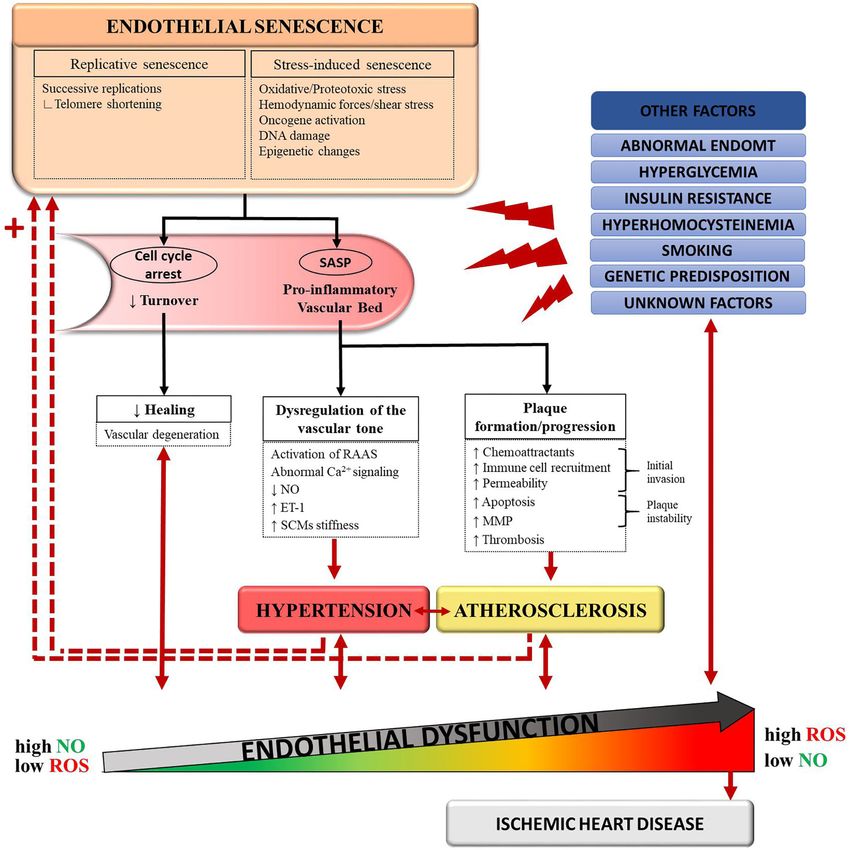
mechanism (22). (Figure 1). In atherogenesis, plaque initiation could be driven
Now endothelial dysfunction can be extended beyond the by senescent endothelial cells through their increased secretion
concept of a damaged conduit vessel to that of a defective vascular of chemoattractant factors and adhesion molecules, which allow
wall composed of layers of cells that are prone to aging. If for the initial invasion of circulating monocytes into the vessel
endothelial dysfunction is the primum movens of hypertension wall (39). Conversely, clearance of senescent vascular cells
and atherosclerosis, an upstream connection between the three lowered the pathogenesis of atherosclerosis in a mouse model of
could be linked to senescence. severe dyslipidemia (36). In addition, a senescent endothelium
presents an altered cellular lining, causing a break in selective
permeability (40). This can facilitate migration of ox-LDLs to
Senescence: The Natural Fate of Aging the subendothelial layers. The SASP can also stimulate vascular
Cells smooth muscle cells to secrete elastase and MMPs, which can
Successive replication (23) and harmful stimuli such as DNA digest components of the extracellular matrix (41). An amplified
damage, oxidative stress, and induction of mitochondrial degradation of the extracellular matrix could create a rupture-
dysfunction eventually impose a state of permanent proliferative prone vulnerable plaque. Thus, senescence of vascular cells leads
arrest on cells (24, 25). This phenomenon, termed “senescence,” to vascular inflammation and plaque progression. This vascular
is well-recognized as one of the nine hallmarks of aging (26). inflammation also raises the possibility of a multistep role of
Despite being in cell cycle arrest, senescent cells (SCs) undergo senescence in hypertension, although the link between the two
profound phenotypic changes and remain metabolically active. is less clearly established. The SASP could play a role in the
In response to stress, they secrete a set of proteins collectively dysregulation of the vascular tone: as an example, it was found to
termed the senescence-associated secretory phenotype activate the renin–angiotensin–aldosterone system (35).
(SASP) (27, 28). These include pro-inflammatory cytokines With aging, the clearance of SCs by the immune system
(interleukin (IL)-6, IL-8, membrane cofactor proteins (MCPs) is decreased, contributing to the accumulation of SCs
and macrophage inflammatory proteins) and chemokines, (33, 42). Senescence therefore begets senescence (Figure 1),
immune modulators, growth factors [hepatocyte growth a phenomenon that has been validated in mice (43). As
factor, fibroblast growth factors, granulocyte-macrophage senescence induces more senescence, the processes involved
colony-stimulating factor, or transforming growth factor beta in the pathogenesis of atherosclerosis and hypertension are
Frontiers in Cardiovascular Medicine | www.frontiersin.org 3 March 2021 | Volume 8 | Article 658400Dagher et al. Alleviating Endothelial Dysfunction With Quercetin FIGURE 1 | Schematic representation of the proposed connections between senescence, hypertension, atherosclerosis, and endothelial dysfunction. Normal aging and deleterious stimuli induce senescence in endothelial cells (ECs), vascular smooth muscle cells (VSMCs), and foam cells. Accumulation of these senescent cells favors a pro-inflammatory state of the vascular bed through the senescence-associated secretory pathway (SASP). In turn, the SASP promotes pathological changes leading to the development of hypertension and atherosclerosis. In a feedback manner, hypertension and atherosclerosis induce more stressors to an already dysfunctional and senescent vessel wall. This vicious circle translates into endothelial dysfunction and, eventually, ischemic heart disease. Other causal pathways of endothelial dysfunction include hyperglycemia, insulin resistance, abnormal endothelial-to-mesenchymal transition (EndoMT), genetic predisposition and detrimental lifestyle habits such as smoking. ET-1, endothelin-1; MMP, matrix metalloproteases; NO, nitric-oxide; RAAS, renin–angiotensin–aldosterone system; ROS, reactive oxygen species. further amplified. In parallel, important changes in the extra- atherosclerosis through shear stress (3). As mentioned before, cellular matrix (ECM) protein composition occur with aging other cardiovascular risk factors often coexist, such as metabolic and promote arterial stiffening (44). A rigid arterial wall disturbances, obesity, smoking or genetic predisposition, causes systolic hypertension, which in turn, contributes to accelerating this deleterious process (12). The vascular Frontiers in Cardiovascular Medicine | www.frontiersin.org 4 March 2021 | Volume 8 | Article 658400
Dagher et al.                                                                                        Alleviating Endothelial Dysfunction With Quercetin

wall eventually gets caught into a vicious circle where it                 Bioavailability and Pharmacology
must face more stressors with less protective capacities (3).              The chemical structure of aglycone quercetin makes it
Therefore, it is possible to acknowledge a cyclical, rather than           hydrophobic (52). Its solubility in water is 2.1 mg/L at
sequential, relationship between senescence, hypertension and              25◦ C, while it is up to 2 g/L in ethanol (52). This physical
atherosclerosis, all contributing to endothelial dysfunction. In           property limits its absorption and practical use in preparation
the next sections, we will explore quercetin’s potential to target         forms as a dietary supplement. Initial investigations on
this triad of endothelial dysfunction.                                     the pharmacokinetics of quercetin in humans suggested
                                                                           very poor oral bioavailability after a single oral dose
                                                                           (∼2%) (53). The absorption was found to increase to 3–
QUERCETIN
                                                                           17% when quercetin was consumed in a glycosidic bond
Classification and Structure                                               compared to its aglycone form (53). Different delivery
Quercetin is part of a larger family of molecular compounds,               systems using nanotechnology have since been developed
named flavonoids, which share a common hydroxylated 3-                     to further improve its water solubility and bioavailability, for
ringed skeleton with attached hydroxyl groups (45) (Figure 2).             example, by binding it to solid lipid carriers or nanosized
Combined with the pyrocatechol, a benzene ring, this chemical              polymeric micelles (54). A pharmacokinetic study in beagle
structure allows them to act as radical scavengers, explaining,            dogs showed that quercetin encapsulated in polymeric
in part, flavonoids’ antioxidant property (45). Flavonoids are             micelles induces a 2.19-fold longer half-life and a relative
themselves part of a large class of plant-derived substances               oral bioavailability increased by 286% as compared to free
named polyphenols (46). Flavonoids include several subclasses              quercetin (55).
such as flavonols, flavones, flavanols, flavanones, isoflavones, and          Since dietary quercetin is usually present in its glycosylated
anthocyanins (46). They exist in most of the plants and play               form, it can be rapidly hydrolyzed by β-glucosidases in the
a variety of biological activities involved in vegetative growth           digestive tract, which makes it easier for absorption by the colonic
(46). Being phytochemicals, flavonoids cannot be synthesized by            mucosa (56). It is then transferred to the liver through the
humans or animals, but they are ubiquitously present in our diet           portal circulation where it undergoes first-pass metabolism and is
(46). They are found in virtually all fruits and vegetables, as well       almost completely metabolized by glucuronidation, methylation,
as in seeds, nuts, tea and red wine (46). The mean daily intake of         or sulfonylation (56, 57). Peak plasma concentration following
flavonoids in Australian, European and US adult populations has            an oral quercetin dose is reached anywhere from 0.6 to
been estimated at 435 mg/day (47).                                         4 h (58–60). Quercetin glucuronides are the main circulating
    Structurally, quercetin is not only found in its free (aglycone)       metabolites and are rapidly eliminated in the urines (57, 60).
form, but also in various conjugated forms with glycosides or              This short elimination half-life is another limit to quercetin’s
methyl ethers attached to the hydroxyl groups. Glycosylation               medical use. Furthermore, quercetin’s metabolism seems to
preferentially occurs at the 3-hydroxyl position, such as quercetin        be dependent on individual characteristics. A correlation
3-O-β-D-glucoside (isoquercetrin) or quercetin 3-O-galactoside             between β-glucuronidase activity and the apolipoprotein (apo)
(hyperoside), whereas methylation usually occurs at the 3’, 4’, or         E phenotype may explain the efficacy of quercetin in patients
7-hydroxyl positions, such as 3-methylquercetin (isorhamnetin)             with apoE3 phenotype as opposed to those expressing apoE4
(48). Some quercetin derivatives even contain both glycosyl                (61, 62). On the other hand, an increased expression of
and ethyl groups. For example, tamarixetin has a glucose                   β-glucuronidase was correlated with inflammation, raising
residue at the 3’ position and a methyl group at the 4’                    the hypothesis that quercetin may be more effective under
position (48). Extensive studies of the biological activities              inflammatory conditions (63). This is especially favorable
of quercetin have shown that the various derivatives have                  as endothelial dysfunction is often associated with a pro-
different levels of efficacy. For example, free quercetin was              inflammatory state.
found to have the strongest antioxidant activity, confirming                  When it comes to pharmacokinetic interactions, conclusions
the important contribution of unbound hydroxyl groups (49).                are still open to debate. Some studies investigated the effects
Among its metabolites, free quercetin was also the most effective          of quercetin on the cytochrome P450 system and have noted a
recombinant human angiotensin-converting enzyme (ACE) 2                    potential inhibitory effect of quercetin on the activity of selected
inhibitor (50). Tamarixetin and isorhamnetin demonstrated                  enzymes (64). Studies in pigs have shown that quercetin can
a stronger inhibition of lipid peroxidation compared to                    decrease bioavailability of cyclosporine and increase that of
quercetin (49, 51). Tamarixetin also exhibited the highest anti-           digoxin, verapamil and various chemotherapeutic agents (65–
inflammatory activity, suggesting that unlike the antioxidant              67). However, conflicting results between in vitro and in vivo
activity, anti-inflammatory activity is not correlated with                studies have been found (67). Quercetin was also reported to bind
the number of free hydroxyl groups (49). These disparities                 to DNA gyrase enzyme in bacteria, which could competitively
in biological activities prompt the synthesis of particular                inhibit fluoroquinolone antibiotics’ activity (68). One case
metabolites that present the highest efficacy of a desired effect.         report of a clinically relevant warfarin interaction resulting in
This can be achieved by inducing glycosylation or methylation              supratherapeutic international normalized ratio values has been
using purified biocatalysts in vitro and native or metabolically           documented in an elderly patient who ate large quantities of
engineered microorganisms (48).                                            scuppernongs, a quercetin-containing muscadine grape (69).

Frontiers in Cardiovascular Medicine | www.frontiersin.org             5                                      March 2021 | Volume 8 | Article 658400
Dagher et al. Alleviating Endothelial Dysfunction With Quercetin
FIGURE 2 | Classification and chemical structure of quercetin, a family member of flavonoids. Quercetin is a pentahydroxyflavone, having five hydroxyl groups placed
at the 3-, 3’-, 4’-, 5-, and 7-positions. Combined with the pyrocatechol, a benzene ring, this chemical structure allows them to act as radical scavengers, explaining, in
part, quercetin’s strong antioxidant properties.
Safety Profile Nephrotoxicity has been reported with high intravenous doses in
In the 1970s, in vitro mutagenicity of quercetin in the Ames cancer patients (67). Quercetin was not found to cause critical
test was reported, leading to concerns about its safety (70). adverse effects on fetal growth in rats, but human studies are
Later, in vivo studies contradicted these findings and showed not available (67). Therefore, dosages above those found in foods
that quercetin may be protective against carcinogens (70). Since should be avoided by pregnant women and nursing mothers (67).
1999, it is classified as a group 3 agent (“not classifiable as to
its carcinogenicity” to humans) by the International Agency for
Research on Cancer (70). In 2010, QU995, a highly pure form of A Recent Resurgence in Interest
quercetin, was granted a “generally recognized as safe” (GRAS) While it was previously known as “vitamin P,” the National
status by the U.S. Food and Drug Administration (59). Many Nutrition Institute withdrew its status in 1950 when it was found
other quercetin formulas have since been developed and made to be a non-essential nutrient (71). Added to a mislabeling of
widely available over the counter as oral dietary supplements or genotoxicity, altogether this contributed to a loss of interest
added ingredient to numerous multivitamin preparations. in the molecule. In 1993, however, the Zutphen Elderly Study
Quercetin is generally well-tolerated. Some minor side-effects first reported a 50% reduction of mortality from IHD in Dutch
such as mild headache, nausea, and tingling of the extremities men who consumed >29 mg flavonoids/day compared with
were observed in long-term supplementation at 1,000 mg/day those who consumedDagher et al. Alleviating Endothelial Dysfunction With Quercetin
FIGURE 3 | Timeline of the cumulative number of published results, from 1980 to 2020, of an online PubMed literature search using “Quercetin” (dotted line) and
“Quercetin [and] Cardiovascular” (solid line) as the search term. Note the progressive increase from the mid-1990s, coinciding with publication of observational studies
associating flavonoid consumption with lower cardiovascular risks. Search performed January 10, 2021 (www.ncbi.nlm.nih.gov/pubmed).
Most debates have focused on high consumption of red wine, topic of interest (158). Considerable evidence from experimental
which contains a variety of polyphenols, including flavonoids data indicates that quercetin may protect against atherosclerosis
(73). Other epidemiological studies soon followed and showed by interfering with multiple pathways involved in disease
a positive correlation between dietary intake of flavonoids and a progression (Table 1). Several high-fat animal models exhibited
reduced incidence of stroke, myocardial infarction and mortality reduced atherosclerotic plaque areas when exposed to quercetin
from IHD (74). (76–84). This observation was associated with a prevention
Over the years, quercetin was found to have a diverse array of of atherosclerosis-related acute aortic syndromes: in a mouse
biological properties, such as anti-inflammatory, anti-oxidative, model with an exaggerated degeneration of the elastic lamina:
anti-platelet, anti-diabetic, anti-histaminic, anti-carcinogenic, administration of quercetin 2 weeks before inducing aortic
anti-bacterial, immunomodulating, and neuroprotective (75). diseases was found to reduce the incidence of aneurysms,
These prominent effects have sparked attention and hope among dissections and aortic ruptures (109).
the scientific community. As of the end of 2020, there are more First, quercetin could positively regulate the metabolism of
than 20,000 published articles on quercetin, and this number lipids. A recent systematic and meta-analysis of 16 randomized
exceeds 120,000 when including all flavonoids (Figure 3). Despite controlled trials (RCTs) published between 2007 and 2017 looked
quercetin being discovered for its role in treating capillary wall at the effects of quercetin on lipid profiles of patients with
dysfunction, it has gained more popularity in oncology and sports metabolic syndrome traits (112). A pooled analysis revealed
medicine, each counting 50% more publications than the field of that quercetin leads to a significant reduction in total and
cardiovascular research. However, its promising benefits for the LDL cholesterol, without affecting triglyceride levels (112). The
endothelium cannot be ignored (Table 1). daily doses and treatment durations used in the trials varied
greatly, from 3.12 to 3,000 mg/ day and from 3 to 12 weeks
(112). Another meta-analysis of 9 RCTs done in overweight and
CARDIOVASCULAR PROTECTIVE obese subjects found that quercetin supplementation significantly
PROPERTIES OF QUERCETIN reduces LDL cholesterol levels at doses of ≥250 mg/day and
Anti-Atherosclerotic Effects of Quercetin for a total dose ≥14,000 mg (159). Similar findings were
With the increasing epidemic of the metabolic syndrome, observed in metabolically healthy non-obese adults after an
the burden of atherosclerosis-related disorders persists despite 8-week regimen, with comparable effects among men and
the current pharmacologic treatment of dyslipidemia (64). women (113). Recent studies have highlighted the influence
Therefore, finding additional anti-atherogenic drugs remains a of the gut microbiota on host metabolic health through its
Frontiers in Cardiovascular Medicine | www.frontiersin.org 7 March 2021 | Volume 8 | Article 658400Dagher et al. Alleviating Endothelial Dysfunction With Quercetin
TABLE 1 | Summary of the main in vitro and in vivo cardiovascular effects of quercetin.
Effects Subjects Evidence and possible mechanisms References
Anti- Animals Reduced atherosclerotic plaque areas (76–84)
atherosclerotic
Increased concentration of SCFAs in the intestinal tract of ApoE−/− mice (85)
Promoted cholesterol-to-bile acid conversion and cholesterol efflux (78, 82, 86–92)
Downregulated PCSK9 expression in RAW264.7 cells and in ApoE−/− mice (90–92)
Normalized plasmatic/hepatic activities of HMG-CoA reductase in Wistar (93)
rats
Downregulated MMP-1, MMP-2, MMP-9 (94–98)
Decreased platelet aggregation in a concentration-dependent manner (99)
Inhibited thrombus formation through intracellular Ca2+ mobilization, (100)
granule secretion, and integrin activation
Inhibited phosphorylation of signaling proteins downstream of glycoprotein (100–102)
VI
Decreased ox-LDLs accumulation and foam cell formation (92, 95, 103–106)
Attenuated LDL oxidation (107, 108)
Decreased expression of adhesion molecules (ICAM-1, VCAM-1) (109–111)
Inhibited LOX-1 in RAW264.7 cells (95)
Decreased inflammatory cytokines, MCP-1 and COX-2 in RAW264.7 cells (95)
Humans Reduced total and LDL-cholesterol in patients with metabolic syndrome (112)
traits*
Reduced total and LDL-cholesterol in metabolically healthy patients* (113)
Decreased platelet aggregation in citrated whole blood in a (114)
concentration-dependent manner
Increased cAMP levels, inhibition of ADP-induced platelet aggregation (115, 116)
Decreased expression of adhesion molecules (ICAM-1, VCAM-1) (117, 118)
Reduced plasma concentration of ox-LDLs (119, 120)
Vasodilating Animals Improved Ach-induced relaxation of aortic rings harvested from (121)
hypertensive rats
Reduced systolic, diastolic, and mean arterial blood pressure in (122–124)
hypertensive rats
Improved endothelium-dependent aortic vasodilatation and eNOS activity (125–129)
Reduced eNOS uncoupling (124, 130, 131)
Inhibited LTCCs and enhanced VGKCs in coronary artery rings (132)
Reduced ACE activity in Wistar rats (133)
Humans Decreased expression of ET-1 gene/protein, and production of ET-1 (134–136)
Reduced systemic blood pressures in both normotensive and hypertensive (137–139)
patients*
Senolytic Animals Reduced viability of senescent HUVECs (140)
Combined with dasatinib, reduced the number of p16-positive SCs in fat (140)
and liver from old mice
Combined with dasatinib, increased median lifespan in old mice (43)
Increased the density of Sirt1 in aorta of ApoE–/– mice (141)
Decreased expression of β-galactosidase and improved cell morphology of (141)
HAECs
Humans Decreased expression of AATK, CDKN2A, and IGFBP3 in HAECs (141)
Combined with dasatinib, reduced the number of adipose tissue SCs and (43, 142)
circulating SASP factors
Myocardial Animals Alleviated ischemia-induced reduction in LVSP (143–147)
protectant
Reduced the decline in LVEF and FS induced by ischemia (148)
Reduced myocardial infarct size (149–151)
Lowered levels of CK, CK-MB, cTnT, and LDH post infarction (144, 147–149,
151–155)
Decreased leukocytes’ infiltration and edema in infarcted myocardium (149, 152, 153)
(Continued)
Frontiers in Cardiovascular Medicine | www.frontiersin.org 8 March 2021 | Volume 8 | Article 658400Dagher et al. Alleviating Endothelial Dysfunction With Quercetin
TABLE 1 | Continued
Effects Subjects Evidence and possible mechanisms References
Inhibited HMGB1 and TLR4 in cardiomyocytes (151, 153)
Up-regulated PPAR-γ positive myocardial cells (148)
Protected against calcium overload by downregulating calpain 1 and 2 (152)
Humans Reduced levels of IL-1β and TNF-α in patients with stable angina (156)
Improved profile of cardiac biomarkers and LVEF in patients with acute (157)
myocardial infarction
* From a meta-analysis of randomized controlled trials. AATK, apoptosis-associated tyrosine kinase; ACE, angiotensin-converting enzyme; ADP, adenosine diphosphate; CDKN2A, p16,
cyclin-dependent kinase inhibitor 2A; COX-2, cyclooxygenase-2; cAMP, cyclic adenosine monophosphate; CK, creatine kinase; CK-MB, creatine kinase-MB; cTnT, cardiac troponin T;
eNOS, endothelial nitric oxide synthase; ET-1, endothelin-1; FS, fractional shortening; HAECs, human Aortic Endothelial Cells; HMGB1, high mobility group box protein 1; HUVECs, human
umbilical vein endothelial cells; ICAM-1, intercellular adhesion molecule 1; IGFBP3, insulin-like growth factor binding protein-3; IL-1β, interleukin-1β; LDH, lactate dehydrogenase; LOX-1,
lectin-like ox-LDL receptor-1; LTCCs, L-type Ca2+ channels; LVEF, left ventricular ejection fraction; LVSP, left ventricular systolic pressure; MCP-1, monocyte chemoattractant protein-1;
MMPs, matrix metalloproteases; NO, nitric oxide; ox-LDLs, oxidized low density lipoproteins; PCSK9, proprotein convertase subtilisin/kexin type 9; PPAR-γ, peroxisome proliferator-
activated receptor gamma; SCs, senescent cells; SCFAs, short-chain fatty acids; SASP, senescence-associated secretory phenotype; SIRT1, sirtuin-1; TLR4, Toll-like receptor 4; TNF-α,
Tumor necrosis factor alpha; VCAM-1, vascular cell adhesion protein 1; VGKCs, voltage-gated K+ channel.
metabolites, especially short-chain fatty acids, which have been to 3.32, 2.99, and 1.11 µM upon co-administration of 2.5, 5, and
linked with improved lipid metabolism (160, 161). Quercetin 10 µM quercetin, respectively (100). In addition, isorhamnetin
was shown to increase the concentration of short-chain fatty and tamarixetin, two methylated metabolites of quercetin, were
acids in the intestinal tract of ApoE knockout (ApoE−/− ) shown to inhibit platelet aggregation and thrombus formation in
mice (85). Experiments on both in vivo rodent models and vitro through effects on activation processes such as intracellular
murine cultured macrophages (RAW264.7 cells) have suggested Ca2+ mobilization, granule secretion, and integrin activation
that quercetin promotes cholesterol-to-bile acid conversion and (100). Their antithrombotic effect was confirmed with laser-
cholesterol efflux by upregulating activity of hepatic CYP7A1, induced thrombi in mouse cremaster arterioles (100). In human
liver X receptor α, ABCG1, ABCA1, and LDLR (78, 82, 86–92). platelets, quercetin significantly increases cyclic AMP levels and
Quercetin also downregulated PCSK9 expression in RAW264.7 inhibits arachidonic acid and adenosine diphosphate (ADP)-
cells and in ApoE−/− mice (90–92). HMG-CoA reductase plays induced platelet aggregation (115, 116). Antiplatelet effects
a major role in the regulation of cholesterol metabolism as a of quercetin and its metabolites have also been associated
rate limiting enzyme in the pathway of cholesterol biosynthesis to inhibition of the phosphorylation of signaling proteins
(162). Results relating the effects of quercetin on HMG-CoA downstream of glycoprotein VI, namely the Src family tyrosine
reductase activity have been inconsistent (86, 93). Wistar rats kinases Fyn and Syk, the phospholipase Cγ2 and the linker for
fed with a diet containing 0.4% quercetin for 5 weeks did not activation of T cells (100–102).
express a change in the enzyme’s activity (86). However, in a Fourth, once oxidized in the intima, LDLs transform into
model of isoproterenol (ISO)-induced myocardial infarction in an antigenic factor, ox-LDLs, which attract monocyte-derived
Wistar rats, a 2 week oral quercetin pre-treatment at a dose of macrophages to the vascular wall, thereby initiating a phagocytic
10 mg/kg normalized plasmatic and hepatic activities of HMG- process leading to foam cell formation (163). Accumulation of
CoA reductase (93). Another protective mechanism of quercetin foam cells is an early step in the pathogenesis of atherosclerosis
involving enhancement of autophagy by aortic macrophages was (163). In their study, Kawai et al. used mAb14A2, a novel
highlighted in ApoE−/− mice (84). monoclonal antibody binding quercetin, to stain aortic samples
Second, quercetin has been suggested to downregulate in Japanese subjects (164). Their results revealed that quercetin
the expression of MMP-1, MMP-2, and MMP-9 in studies metabolites accumulate in atherosclerotic lesions, but not in
using molecular modeling techniques, cultured endothelial cells, normal-appearing aorta (164). In addition, intense staining
murine macrophage cells and in hypertensive rats, an effect that was primarily localized with foam cells, suggesting a potential
translates in the prevention of plaque instability (94–98). cellular target of quercetin (164). Several studies done on
Third, platelet aggregation at the site of an unstable plaque also cultured cells showed that quercetin can attenuate ox-LDLs
contributes to acute complications of atherosclerosis. Quercetin accumulation, foam cell formation, as well as ox-LDLs induced
was found to have an antiaggregatory effect on rat platelet-rich cytotoxicity and calcification (92, 95, 103–106). Interestingly,
plasma in a concentration-dependent manner (99). This was also quercetin significantly reduced plasma concentrations of ox-
observed in human citrated whole blood: using samples from 100 LDLs in two RCTs (119, 120). A retrospective comparison of
healthy volunteers, the minimal antiaggregatory concentration the participants’ apoE genotypes revealed no significant inter-
of quercetin was estimated at 15.26 µM (114). A synergistic group difference in the reduction of ox-LDLs between the
enhancement of antiplatelet effect was noted when quercetin apoE3 and apoE4 subgroups (62). This lowering effect on ox-
was added to aspirin (100). The half maximal inhibitory LDLs might be achieved through direct attenuation of LDL
concentrations (IC50 ) values for the inhibition of platelet oxidation: the lag time of LDL oxidation was increased by 3-
aggregation decreased from 10.83 µM when using aspirin alone, to 4-fold after administration of quercetin in vitro and in rats
Frontiers in Cardiovascular Medicine | www.frontiersin.org 9 March 2021 | Volume 8 | Article 658400Dagher et al. Alleviating Endothelial Dysfunction With Quercetin
(107, 108). The authors proposed two mechanisms contributing quercetin induced phosphorylation of eNOS at serine 1179 in a
to this attenuation of LDL oxidation: inhibition of copper- concentration, time-dependent manner; this effect was abolished
induced LDL oxidation, as well as up-regulation of Paraoxonase by H-89, an inhibitor of protein kinase A (128). Using the
1 (PON1) and its protective capacity against LDL oxidation same primary cell cultures, Khoo et al. proposed that quercetin
(107, 108). Lectin-like ox-LDL receptor-1 (LOX-1) is a scavenger stimulates eNOS phosphorylation at serine 1179 by causing a
receptor that mediates uptake of ox-LDLs by macrophages rapid increase in intracellular Ca2+ (129).
(165). Administration of anti-LOX-1 antibodies was shown to Other calcium-mediated vasoactive effects of quercetin have
inhibit atherosclerosis by decreasing these cellular events (165). been proposed. L-type Ca2+ channels (LTCCs) and voltage-gated
Quercetin was shown to block LOX-1 in RAW264.7 cells (95). K+ channels (VGKCs) play a tonic role in the regulation of
Moreover, ox-LDLs activate endothelial cells by inducing cell arterial vasomotricity and are commonly expressed in vascular
adhesion molecules, especially vascular cell adhesion molecule-1 smooth muscle cells (172, 173). LTCCs are involved in excitation-
(VCAM-1) and intracellular cell adhesion molecule-1 (ICAM-1) contraction coupling while VGKCs are critical for restoring the
(166). Quercetin was found to downregulate ICAM-1 expression resting membrane potential (172, 173). Large (big)-conductance
in diabetic rats and human endothelial cells (110, 117). Quercetin Ca2+ -sensitive potassium channels (BK) and VGKCs are closely
and isoquercetin were shown to attenuate VCAM-1 expression in associated with coronary arterial smooth muscle vasodilatation
mice, HUVECs and rat intestinal microvascular endothelial cells (174). Of note, aging is associated with a reduced expression
by suppressing multiple pathways including caveolin-1 (CAV- of BK channels in coronary arteries, which is consistent with a
1), Toll-like receptor 4 (TLR4) and NFκB (109, 111, 118). As higher frequency of spontaneous vasospasmic activity in elderly
previously mentioned, ox-LDLs also stimulate eNOS uncoupling people (174). Hou et al. showed that quercetin can inhibit LTCCs
and ROS overproduction by macrophages and endothelial cells and enhance VGKCs in rat coronary artery rings, resulting
via activation of NOX (167, 168). In ApoE−/− mice, quercetin in a decrease of the vasocontractions induced by high-K+
partially reversed NOX expression and inhibited ox-LDL induced depolarizing solution (132). Moreover, coronary vasodilation
ROS formation in macrophages (83). induced by quercetin was not lost after denuding the arterial rings
Finally, atherosclerosis is also a chronic inflammatory of their endothelium, suggesting that quercetin can also promote
disease mediated by a network of pro-inflammatory cytokines. its vasodilatory effect through VSMC-mediated mechanisms
Quercetin’s administration was associated with a decrease in (132). Cogolludo et al. noted that quercetin could activate BK
multiple inflammatory cytokines, such as IL-1α, IL-1β, IL- channels in coronary artery myocytes while generating hydrogen
2, IL-10, TNF-α, macrophage chemoattractant protein-1 and peroxide (H2 O2 ) (175). Although H2 O2 is considered as a
cyclooxygenase-2 (95). The impacts of quercetin on such a wide relaxing endothelium-derived hyperpolarizing factor (176) and
range of inflammatory markers are in favor of a multi-target effect can also activate the soluble guanylate cyclase as does NO (177,
of signal transduction. 178), the data of Cogolludo et al. may nonetheless represent an
instance where quercetin behaves as a pro-oxidant rather than
Vasodilating Effects of Quercetin a vasodilator.
Several ex-vivo reactivity studies have shown a vasodilating ability Endothelin (ET) is one of the most potent vasoconstrictors
of quercetin in rat aorta, portal vein, mesenteric arteries and and is mainly produced by the vascular endothelium (179). ET-
coronary arteries (169–171) (Table 1). In addition, Choi et al. 1 plays a major role in the homeostasis of the cardiovascular
reported that quercetin acutely improved acetylcholine-induced system. ET-1 has been associated with increased oxidative
relaxation of aortic rings harvested from two-kidney, one-clip stress and endothelial dysfunction in humans (179). It was
(2K1C) hypertensive rats (121). shown to stimulate eNOS uncoupling, therefore superoxide
Quercetin’s BP lowering effects were first documented in production, and promote vasoconstriction via activation of
vivo in spontaneously hypertensive rats (122). Rats exposed NOX (130, 180). ET-1 can further reduce NO bioavailability
to quercetin had a significant lower systolic (−18%), diastolic by interfering with eNOS expression through protein kinase C
(−23%), and mean (−21%) arterial BP (122). In normotensive (PKC)-mediated activation of STAT3 (181). These data indicate
rats, endothelial dysfunction induced by a high-fat high-sucrose that diminished ET-1 concentrations may be accompanied by
diet was prevented by the supplementation with quercetin for elevated NO bioavailability (181). Lodi et al. showed that
28 days: both endothelium-dependent aortic vasodilatation and quercetin significantly decreased expression of ET-1 in human
eNOS activity were improved by quercetin (125). The vasomotor umbilical artery smooth muscle cells and human vein endothelial
protective effects of quercetin were also demonstrated in mice cells (HUVECs) co-culture model exposed to TNF-α-induced
exposed to lipopolysaccharide-induced endotoxemia. Whether change in vasomodulatory molecules (134). Zhao et al. also
given before or after lipopolysaccharide injection, quercetin showed that quercetin decreases ET-1 production in thrombin-
dose-dependently restored eNOS expression while abolishing stimulated HUVECs in a concentration-dependent manner, with
inducible NO synthase (iNOS) (126). an IC50 of 1.54 µmol/L (135). In rat aortic rings, quercetin
Several hypotheses have been formulated regarding the prevented ET-1-induced PKC activation, with a subsequent
up regulation of eNOS activity induced by quercetin. Some decrease in superoxide production (130). Moreover, chronic
authors have suggested that quercetin phosphorylates eNOS by treatment with quercetin reduced blood pressure and improved
an AMP-activated protein kinase-dependent mechanism (127). endothelial function in deoxycorticosterone acetate (DOCA)-
Li et al. observed, in bovine aortic endothelial cells, that salt rats, a low renin model of hypertension in which ET-1
Frontiers in Cardiovascular Medicine | www.frontiersin.org 10 March 2021 | Volume 8 | Article 658400Dagher et al. Alleviating Endothelial Dysfunction With Quercetin is overexpressed (123, 124). These effects of quercetin were these results indicate a significant anti-hypertensive effect of associated with a reduction in both vascular and systemic quercetin supplementation only when doses ≥500 mg/day are oxidative stress (124). Quercetin protective effects against taken for ≥8weeks (137). Another meta-analysis which included eNOS uncoupling were even maintained under glucotoxic 896 participants across 17 RCTS mirrored the results obtained by conditions (131). Serban et al., which indeed is the meta-analysis done by Huang A RCT studied the acute effects of administering 200 mg of et al. (138). More recently, a meta-analysis of 8 RCTs conducted quercetin in 12 healthy men (136). Blood and urine samples among patients with metabolic syndrome traits showed that taken, respectively, 2 and 5 h after oral ingestion of quercetin quercetin supplementation significantly reduced systolic BP, yet revealed a significant acute reduction in both the plasma did not affect diastolic BP (139). Clearly, trials directly comparing and urinary concentrations of ET-1, translated into a reduced different doses and regimen durations are needed. ET-1 production (136). This effect is substantial given the relatively small dose used and the low bioavailability of quercetin. Senolytic Properties of Quercetin Interestingly, it was reported that NO inhibits ET-1 production The accumulation of SCs in the aging and diseased vessel through the suppression of NFκB (182). A second mechanism wall raises the possibility that reducing senescence might delay involves the renin–angiotensin system: ACE inhibitors neutralize deterioration of vascular structure and function. In an elegant ACE by binding a zinc atom at the active site of the enzyme, and seminal experiment, Baker et al. demonstrated that the which slows conversion of angiotensin I to angiotensin II, a health span in progeroid mice can be enhanced by killing SCs powerful vasoconstrictor (183), including in human coronary using a transgenic suicide gene (188). Elimination of SCs also arteries (184). Quercetin can chelate metal ions, including zinc delayed progression of multiple age-related phenotypes, such (185), and it is tempting to presume it can act as an ACE as cancer, cataract, sarcopenia, lordokyphosis, loss of adipose inhibitor. However, available results have been discordant. In tissue and skeletal muscle fibers, as well as improved exercise vitro, quercetin inhibited ACE activity in a concentration- capacity (188). Translating that same effect into a druggable dependent manner, with an IC50 of 310 µM (186). This value compound sparked research interest and led to the recent concept was significantly higher than that of captopril (0.02 µM) (186). of “senolytic therapy” (140). Formed by the words “senescence” In Wistar rats receiving an angiotensin-1 infusion, Hackl et al. and “lytic” (destroying), a senolytic represents a molecule that showed that an attenuation of the BP was obtained with both could specifically induce cell death in SCs (189). Based on the oral and intravenous administration of quercetin (133). They knowledge that SCs survive despite their harsh internal state, also reported a 31% reduction in ACE activity in the quercetin the hypothesis was that this would be achieved by targeting group compared to the control group (133). In contrast, one their survival pathways and anti-apoptotic mechanisms (189). double-blind placebo-controlled RCT did not find ACE activity An alternative strategy to interfere with senescence would be inhibition after a single-dose of quercetin (187). In this study, five to reduce the burden of SASP. The advent of antibody-based normotensive men and twelve hypertensive men ingested a total techniques such as sandwich enzyme-linked immunosorbent of 1,095 mg quercetin and 10 h later, the mean BP was reduced assay, and large-scale molecular biology techniques such as among the hypertensive patients by 5 mm Hg compared to the mRNA profiling, antibody arrays, proteomics or multiplex assays placebo group (187). Plasma ACE activity, ET-1, and brachial have made the detection and measurement of several SASP artery flow-mediated dilation were unaffected by quercetin, factors possible (190). These powerful tools therefore serve to test suggesting that the reduction in BP in hypertensive men was pharmaceutical efficacy of drugs that target SASP (190). In the independent of the changes in ACE and ET-1 activity, or NO following sections, we will see evidence suggesting that quercetin bioavailability (187). eliminates SCs and reduces the SASP. Serban et al. conducted a systematic review of 7 RCTs In 2008, quercetin was reported to increase longevity of published between 1998 and 2014, looking at the effects of worms (191), but it was not until 2015 that its potential quercetin on BP (137). Their meta-analysis revealed a significant as a senolytic was highlighted in Kirkland’s laboratory (140) reduction in systemic BPs associated with oral supplementation (Table 1). First, the investigators identified a series of senolytic of quercetin (137). The weighed mean differences for the transcripts on pre-adipocytes. These included components of systolic and diastolic BPs were 3.04 mm Hg (p = 0.028) and the ephrin regulating system, ephrin ligands B (EFNB), as 2.63 mm Hg (p < 0.001), respectively (137). These values are well as the plasminogen-activated inhibitor-1 (PAI-1) and a appreciable considering that the cohorts were largely made up member of the phosphatidylinositol-4,5-bisphosphate 3 kinase of normotensive subjects. The doses of quercetin ranged from (PI3 K) family, involved in regulating multiple cellular functions 100 to 1,000 mg/day. Interestingly, when using a meta-regression including survival (140). Then, they tested whether drugs that analysis, the systolic BP-lowering effect was only associated with target any of these gene products would effectively induce the duration of supplementation, and not the administered dose, apoptosis in radiation-induced senescent human pre-adipocytes contrarily to the diastolic BP-lowering effects (137). Furthermore, and HUVECs. Of the 46 agents tested, quercetin and dasatinib, when the RCTs were stratified according to the duration of a non-specific tyrosine kinase inhibitor used for cancer therapy, supplementation, quercetin had no significant benefit in the were noticeably promising (140). Dasatinib is known to block subsets of studies lasting
Dagher et al.                                                                                       Alleviating Endothelial Dysfunction With Quercetin

effective on pre-adipocytes, quercetin preferentially reduced             human aortic endothelial cells (HAECs) (141). In their study,
viability of senescent HUVECs (140). Parallel cultures of non-            senescence was induced by ox-LDLs. Their results revealed that
senescent HUVECs proliferated 2- to 3-fold in the presence                quercetin decreased the expression of senescence-associated β-
of quercetin over the same period of 3 days, indicating that              galactosidase and improved cell morphology of HAECs (141).
quercetin’s induction of apoptosis is selective to SCs (140). In          The senolytic effect was dose dependent, as 0.3, 1, and 3
addition, the combination of dasatinib and quercetin achieved             µmol/L of quercetin improved cells viability by 10.8, 40.9, and
a synergistic effect by selectively killing both senescent pre-           48.9%, respectively (141). Quercetin simultaneously decreased
adipocytes and HUVECs, whom viability was, respectively,                  ROS generation, also in a concentration-dependent manner.
reduced by ∼70% and ∼50% (140). This suggests that using                  In addition, transcriptome microarray assays were performed
a mix of senolytics to target a broader range of anti-apoptotic           and identified differentially expressed genes in the mRNAs
networks may be a strategy to follow in developing future                 profile of senescent HAECs treated with quercetin (141). Among
senolytic therapies (189). Used in vivo, the senolytic cocktail           them, several were involved in p53 and mammalian target
also reduced the number of p16-positive SCs in fat and liver              of rapamycin (mTOR) signaling pathways, NO metabolism,
from old mice (140). After a single 5 day treatment course                maintenance of the cytoskeleton, extracellular matrix-receptor
of dasatinib+quercetin, the rodents exhibited an improved left            interaction, as well as complement and coagulation cascades,
ventricular ejection fraction (LVEF) and fractional shortening            suggesting the potential mechanisms by which quercetin was
with no alteration of cardiac mass, as well as increased smooth           effective against ox-LDLs (141). Quercetin also decreased the
muscle vascular reactivity to nitroprusside (140). Similar results        genetic expression of AATK, CDKN2A, and IGFBP3 (141). AATK
were obtained by Xu et al.: 20 month-old mice who were                    is induced during apoptosis, while CDKN2A (p16) is one of the
fed dasatinib+quercetin intermittently for 4 months performed             most important senescence markers (141). Interestingly, a high
better at physical endurance tests compared to the control                circulating concentration of IGFBP3 was found to be a predictor
group (43). Next, they administered biweekly oral doses of                of IHD (196). One can therefore wonder if quercetin could
dasatinib+quercetin to 24 to 27 month-old mice, equivalent to             alleviate the risks of IHD in patients by decreasing IGFBP3.
a human age of 75–90 years; compared to the controls, these                   Clinical trials studying the senolytic effects of quercetin
mice had a 36% higher median post-treatment lifespan and a 65%            remain scarce (Table 1). In the first clinical trial of senolytics,
lower mortality hazard (43). This was neither associated with an          an intermittent regimen of dasatinib+quercetin (dasatinib: 100
increased physical morbidity nor an increased age-related disease         mg/day, quercetin: 1,250 mg/day, 3 days/week over 3 weeks)
burden (43). In addition, in ApoE−/− mice fed with a high-                improved physical tolerance, but not pulmonary function, in
fat diet, dasatinib+quercetin given once monthly for 3 months             patients with idiopathic pulmonary fibrosis, a fatal senescence-
was shown to decrease aortic calcifications and increase vascular         associated disease (197). Another open label pilot study was
reactivity (193). When used alone, quercetin increased the                conducted by Hickson et al. in 9 adults aged 50–80 years with
density of sirtuin 1 (Sirt1) in aorta of ApoE−/− mice (141). Sirt1        diabetic kidney disease (142). The patients received a 3 day oral
functions as a nicotinamide adenosine dinucleotide (NAD+)-                treatment regimen with dasatinib 100 mg daily and quercetin
dependent deacetylase and is involved in genomic stability, basal         500 mg bid. Eleven days after treatment completion, there was
level autophagy and cell survival (194). Sirt1 was found to delay         a significant reduction in the number of adipose tissue SCs and
both replicative and stress-induced senescence (194).                     circulating SASP factors, including IL-1α, IL-6, MMP-9, and
    Hwang et al. conducted in vitro experiments with adult                MMP-12, accompanied by an increase of adipocyte progenitors,
human coronary artery endothelial cells (HCAEC) from three                suggesting a selective cytotoxic effect for SCs (142). These results
deceased female donors using replicative senescence as a relevant         are in agreement with a previous in vitro study performed on
model for human arterial aging (195). Contrary to the previous            human omental tissue resected during gastric bypass surgery
results reported in HUVECs (140), their findings showed that              (43). The surgical explants treated with a dasatinib+quercetin
quercetin induces death in both early (non-senescent) and late-           medium for 48 h had significantly less SCs and a lower secretion
passage (senescent) HCAECs, without any selectivity for the               of SASP components compared to the explants treated with a
latter (195). Quercetin’s cytotoxicity was evident in all three           vehicle (43). To the best of our knowledge, no clinical trial has
donors at a concentration of 10 µM, which was half the amount             yet examined the senolytic effects of quercetin on endothelial
used with HUVECs (140). Late-passage cells were more sensitive            dysfunction in humans, in the context of CVD.
to quercetin’s toxic effects as their relative cell abundance
was already significantly decreased at a concentration of 6 µM            Myocardial Protective Effects of Quercetin
(195). Their study also investigated hyperoside, also known as            The beneficial effects of quercetin on dyslipidemia, hypertension,
quercetin 3-D-galactoside, as an alternative to quercetin (195).          senescence and other risk factors can be seen as a primary
Hyperoside is a natural derivative of quercetin produced by St.           prevention measure against endothelial dysfunction. Once the
John’s Wort and structurally identical except for a galactoside           endothalial dysfunction has resulted in an adverse cardiac event,
group attached in position 3 (Figure 2) (195). In contrast to             secondary and tertiary prevention strategies become crucial in
quercetin, hyperoside had no significant cytotoxicity to either           order to reduce the progression of the disease and its impacts
proliferating or late-passage HCAECs but was unable to display            on patients’ quality of life. One of the most striking examples
any senolytic activity (195). A second in vitro model of an adult         is myocardial ischemia. In the latter, dysfunctional endothelial
human vasculature model was investigated by Jiang et al. using            cells of the coronary arteries induce a local disturbance

Frontiers in Cardiovascular Medicine | www.frontiersin.org           12                                      March 2021 | Volume 8 | Article 658400
You can also read


