Elucidating mechano-pathology of osteoarthritis: transcriptome-wide differences in mechanically stressed aged human cartilage explants - Arthritis ...
←
→
Page content transcription
If your browser does not render page correctly, please read the page content below
Houtman et al. Arthritis Research & Therapy          (2021) 23:215
https://doi.org/10.1186/s13075-021-02595-8

 RESEARCH ARTICLE                                                                                                                                  Open Access

Elucidating mechano-pathology of
osteoarthritis: transcriptome-wide
differences in mechanically stressed aged
human cartilage explants
Evelyn Houtman1, Margo Tuerlings1, Janne Riechelman1, Eka H. E. D. Suchiman1, Robert J. P. van der Wal2,
Rob G. H. H. Nelissen2, Hailiang Mei3, Yolande F. M. Ramos1, Rodrigo Coutinho de Almeida1 and
Ingrid Meulenbelt1*

  Abstract
  Background: Failing of intrinsic chondrocyte repair after mechanical stress is known as one of the most important
  initiators of osteoarthritis. Nonetheless, insight into these early mechano-pathophysiological processes in age-
  related human articular cartilage is still lacking. Such insights are needed to advance clinical development. To
  highlight important molecular processes of osteoarthritis mechano-pathology, the transcriptome-wide changes
  following injurious mechanical stress on human aged osteochondral explants were characterized.
  Methods: Following mechanical stress at a strain of 65% (65%MS) on human osteochondral explants (n65%MS = 14
  versus ncontrol = 14), RNA sequencing was performed. Differential expression analysis between control and 65%MS
  was performed to determine mechanical stress-specific changes. Enrichment for pathways and protein-protein
  interactions was analyzed with Enrichr and STRING.
  Results: We identified 156 genes significantly differentially expressed between control and 65%MS human
  osteochondral explants. Of note, IGFBP5 (FC = 6.01; FDR = 7.81 × 10⁻³) and MMP13 (FC = 5.19; FDR = 4.84 × 10⁻²)
  were the highest upregulated genes, while IGFBP6 (FC = 0.19; FDR = 3.07 × 10⁻⁴) was the most downregulated
  gene. Protein-protein interactions were significantly higher than expected by chance (P = 1.44 × 10⁻¹⁵ with
  connections between 116 out of 156 genes). Pathway analysis showed, among others, enrichment for cellular
  senescence, insulin-like growth factor (IGF) I and II binding, and focal adhesion.
  Conclusions: Our results faithfully represent transcriptomic wide consequences of mechanical stress in human
  aged articular cartilage with MMP13, IGF binding proteins, and cellular senescence as the most notable results.
  Acquired knowledge on the as such identified initial, osteoarthritis-related, detrimental responses of chondrocytes
  may eventually contribute to the development of effective disease-modifying osteoarthritis treatments.
  Keywords: Osteoarthritis, Cartilage, Chondrocytes, Mechanical stress, Mechano-pathology, RNA sequencing, Cellular
  senescence, IGF-1 signaling, MMP13

* Correspondence: i.meulenbelt@lumc.nl
1
 Section of Molecular Epidemiology, Department of Biomedical Data
Sciences, Leiden University Medical Center, Postzone S05-P, Einthovenweg
20, 2333, ZC, Leiden, The Netherlands
Full list of author information is available at the end of the article

                                       © The Author(s). 2021 Open Access This article is licensed under a Creative Commons Attribution 4.0 International License,
                                       which permits use, sharing, adaptation, distribution and reproduction in any medium or format, as long as you give
                                       appropriate credit to the original author(s) and the source, provide a link to the Creative Commons licence, and indicate if
                                       changes were made. The images or other third party material in this article are included in the article's Creative Commons
                                       licence, unless indicated otherwise in a credit line to the material. If material is not included in the article's Creative Commons
                                       licence and your intended use is not permitted by statutory regulation or exceeds the permitted use, you will need to obtain
                                       permission directly from the copyright holder. To view a copy of this licence, visit http://creativecommons.org/licenses/by/4.0/.
                                       The Creative Commons Public Domain Dedication waiver (http://creativecommons.org/publicdomain/zero/1.0/) applies to the
                                       data made available in this article, unless otherwise stated in a credit line to the data.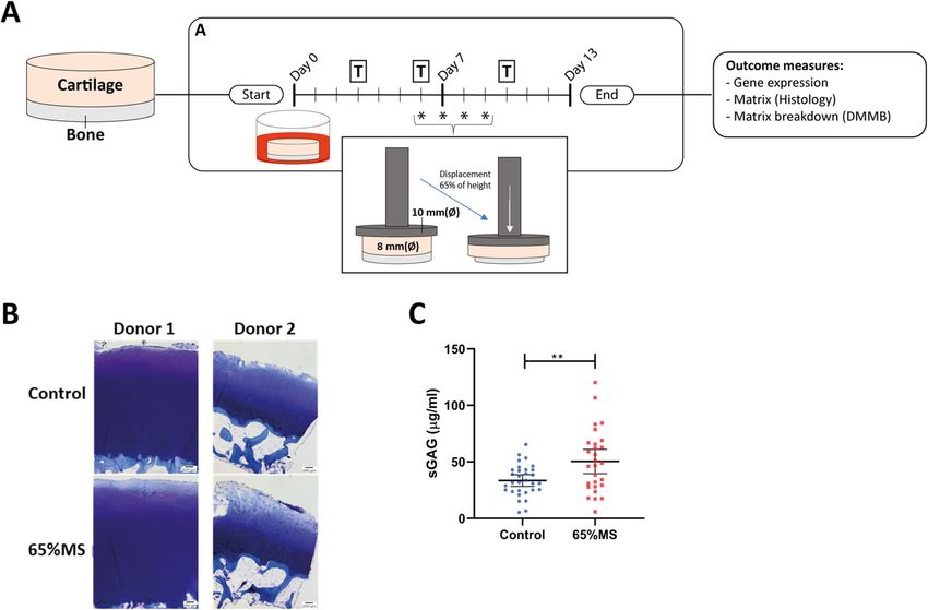
Houtman et al. Arthritis Research & Therapy   (2021) 23:215                                                   Page 2 of 12

Introduction                                                   to a trauma in adult (human) tissue [18]. More recently,
Osteoarthritis (OA) is an age-related joint disease, affect-   global gene expression profiling in 14-month-old mice
ing diarthrodial joints [1, 2]. Despite the fact that OA is    subjected to non-invasive injurious tibial compression
the most prevalent and disabling disease among elderly,        identified genes involved in inflammation and matrix
resulting in high social and economic burden, no effect-       regeneration to be involved in the response of aged
ive treatment exists except for lifestyle changes, pain        tissue [14].
medication, and eventually a joint replacement surgery            A more appropriate model to identify which molecular
at end-stage disease [3, 4].                                   processes are initiated in response to mechanical stress
  To characterize deregulated signaling pathways in OA         in humans would comprise aged human ex vivo osteo-
cartilage, comprehensive differential expression analyses      chondral explants. Injurious compression reaching
have been performed comparing preserved versus end-            strains above 50% induced catabolic processes in cartil-
stage lesioned OA cartilage [5]. These studies revealed        age and eventually led to cell death [19]. In aged human
that OA pathology is marked by recuperation of growth          osteochondral explants, injurious cyclic mechanical
plate signaling, wound healing, and skeletal system de-        stress at a strain of 65% (65%MS), mimicking trauma,
velopment, while also highlighting inherent differences        was previously shown to induce OA-like damage [20]. In
in OA pathophysiology between patient subtypes based           the current study, we therefore exploited our previously
on gene expression changes [5–7]. Nonetheless, the pre-        established ex vivo osteochondral explant model by per-
served versus lesioned study design by definition cap-         forming RNA sequencing on explants subjected to in-
tures end-stage pathophysiological OA disease processes        jurious mechanical stress in comparison to controls. The
and gives no information on early initial processes trig-      hypothesis-free, transcriptome-wide approach presented
gering cartilage to become diseased. In contrast, disease-     here contributes to further understanding the debilitat-
modifying OA drugs should preferably target early OA           ing response of aged chondrocytes to mechanical injury
disease triggers when irreversible damage of cartilage         and how this affects their propensity to enter an OA dis-
has not yet taken place. Therefore, more knowledge on          ease state.
the initial response of chondrocytes to OA-relevant
stresses, such as mechanical trauma, should be investi-        Material and methods
gated in an appropriate model.                                 Sample description
  In this regard, failing of intrinsic chondrocyte repair      To generate osteochondral explants, biopsies (diameter
after mechanical stress is known to impact the integrity       of 8 mm) were punched from the macroscopically pre-
of articular cartilage via cell apoptosis [8], increased       served load-bearing area of femoral condyles of human
catabolic gene expression [9], and reduced matrix pro-         knee joints obtained within the Research in Articular
duction [10] and is, as such, an important trigger to OA       Osteoarthritis Cartilage (RAAK) biobank containing
onset. Nonetheless, little knowledge exists on the inher-      patients that undergo a joint replacement surgery as a
ent dysregulation of signaling pathways initiating repair      consequence of OA [21]. For this study, a total of 60
responses in human aged articular cartilage upon mech-         osteochondral explants were investigated originating
anical stress. To gain some insight, several in vivo animal    from nineteen independent donors in which multiple ex-
studies have investigated the effect of joint overuse or       plants were taken from each donor. This difference be-
trauma on gene expression in cartilage [11–16]. Some           tween the amount of samples taken per donor was
examples of non-invasive in vivo mechanical loading            dependent on several factors. Among them were the size
studies are Bomer et al. [11], reporting on involvement        of knee condyle, size of the preserved area, surgical dam-
of metabolic processes and skeletal system development         age area, and other simultaneous experiments this donor
pathways upon physiological forced running in 6-month-         was used for. RNA sequencing was performed on sam-
old mice; Chang et al. [14], reporting on involvement of       ples from nine donors, while the remaining ten donors
cell proliferation and chondroitin sulfate proteoglycan        were used for replication purposes. All donor character-
metabolic process upon injurious tibial compression in         istics are given in Table S1 and were equal between
16-week-old mice; and Sebastian et al. [13], reporting on      mechanical stressed and control explant donors.
single-cell RNA-seq upon tibial compression in 10-week-
old mice. Thus far, one study has investigated genome-         Application of mechanical stress
wide expression consequences of an impact injury in por-       Explants of nineteen donors were equilibrated in serum-
cine explants and identified involvement of genes associ-      free chondrogenic differentiation medium (DMEM, sup-
ated with matrix molecules, protein biosynthesis, skeletal     plemented with ascorbic acid (50 μg/ml; Sigma-Aldrich;
development, and cell proliferation [17]. Nevertheless,        Zwijndrecht, The Netherlands), L-proline (40 μg/ml;
most studies were performed using relatively young ani-        Sigma-Aldrich), sodium pyruvate (100 μg/ml; Sigma-
mal tissues and likely do not cover the biological response    Aldrich), dexamethasone (0.1 μM; Sigma-Aldrich), ITS+,
Houtman et al. Arthritis Research & Therapy      (2021) 23:215                                                                          Page 3 of 12

and antibiotics (100 U/ml penicillin; 100 μg/ml strepto-                    Determining cartilage integrity
mycin)) in a 5% (v/v) CO2 incubator at 37 °C. As                            Histology
depicted in Fig. 1A, after a 6-day period, dynamic un-                      A sagittal section of the osteochondral explant was fixed
confined compression was applied to explants (diameter                      in 4% formaldehyde for 1 week and decalcified using
of 8 mm) using the Mach-1 mechanical testing system                         EDTA (12.5%, pH = 7.4) during 2 weeks, dehydrated
on 4 subsequent days (Biomomentum Inc., Laval, QC,                          with an automated tissue processing apparatus, and em-
Canada). In short, osteochondral explants were placed                       bedded in paraffin. Tissue sections were cut at a thick-
under an indenter (diameter of 10 mm) attached to a                         ness of 5 μm, deparaffinized, rehydrated, subsequently
250-N MACH-1 load cell (Fig. 1A) and unconfined                             stained for 1 min in a toluidine blue solution with a pH
cyclic compression was applied at a strain of 65% of                        of 2.5 (Sigma-Aldrich), and mounted with Pertex
cartilage height at a frequency of 1 Hz (1 compression                      (Sigma-Aldrich) to investigate cartilage integrity as
cycle per second), mimicking walking speed, during 10                       quantified by applying Mankin Score [23].
min, long enough to be injurious and short enough for
chondrocytes to survive, at strains suggested to be detri-                  Sulfated glycosaminoglycan (sGAG) measurement
mental [22]. Dynamic (cyclic) compression means that a                      Sulfated glycosaminoglycan (sGAG) concentrations in
force was applied that varied over time to simulate a                       conditioned media collected from osteochondral ex-
more cyclic compression such as walking. To investigate                     plants were measured with the photometric 1.9 dimethy-
lasting effects of mechanical stress, 4 days after mechan-                  lene blue (DMMB; Sigma-Aldrich) dye method [24].
ical stress, the cartilage and bone were separated, snap-                   Shark chondroitin sulfate (Sigma-Aldrich) was used as
frozen in liquid nitrogen, and stored at −80 °C.                            the reference standard. The concentration of sGAG was

 Fig. 1 Study setup of human osteochondral explants receiving 65% MS. A Osteochondral explants were punched from preserved areas of knee joints
 and the medium is refreshed on indicated days (T). B Damage in our mechanical stress model was confirmed by degradation of sGAG in cartilage by
 toluidine blue staining (histology of two independent donors) and measuring C sGAG release in conditioned media on day 13 (ncontrol = 31 versus
 n65%MS = 28). The average ± 95% CI are presented with each dot representing a sample. To adjust for donor variation, P-values were estimated by
 performing logistic generalized estimation equations, with sGAG concentration as dependent variable and treatment as covariate: sGAG concentration
 ∼ Treatment + (1|Donor). **P ≤ 0.01. Legend: 65%MS 65% mechanical stress, DMMB dimethylmethylene blue, sGAG sulfated glycosaminoglycans
Houtman et al. Arthritis Research & Therapy (2021) 23:215 Page 4 of 12
determined in conditioned media collected on day 13, by protein-protein interactions, analysis was performed
measuring absorbance at 525 nm and 595 nm in a mi- using the online tool STRING version 11.0 [30].
croplate reader (Synergy HT; BioTek, Winooski, USA).
RNA sequencing Real-time quantitative PCR (RT-qPCR) validation
RNA from cartilage was extracted by pulverizing the tis- 250 ng of RNA was processed into cDNA using the First
sue and subsequently homogenizing the powder in TRI- Strand cDNA Synthesis Kit (Roche Applied Science, Al-
zol reagent (Invitrogen, San Diego, CA) using a Mixer mere, The Netherlands). RT-qPCR was performed on 10
mill 200 (Retsch, Germany). RNA was extracted using paired 65%MS samples with matched controls included
chloroform, followed by precipitation using ethanol, and in the RNA sequencing (Technical validation) and 10
purified with the RNeasy Mini Kit (Qiagen, Chatsworth, novel paired 65%MS samples with matched controls
CA). Genomic DNA was removed by DNase digestion. (Biological validation) to determine the expression of six
Paired-end 2 × 150 base pair RNA sequencing (Illumina downregulated (IGFBP6, CNTFR, WISP2, FRZB,
TruSeq mRNA Library Prep Kit, Illumina HiSeq X ten) COL9A3, and GADD45A) and four upregulated genes
was performed. Strand-specific RNA-sequencing librar- (IGFBP5, PTGES, TNC, and IGFBP4). Primer sequences
ies were generated which yielded on average 14 million are listed in Table S2. The relative gene expression was
reads per sample. Data from the Illumina platform was normalized for two endogenous reference genes, SDHA
analyzed with an in-house pipeline as previously de- and YWHAZ, to determine −ΔCT values. To determine
scribed [5]. The adapters were clipped using Cutadapt effect sizes, fold changes (FC) were calculated according
v1.1. RNA-seq reads were then aligned using GSNAP to the 2−ΔΔCT method, in which expression of 65%MS
against GRCh38 [25]. Read abundances per sample were was extracted from controls (−ΔΔCT). These two en-
estimated using HTSeq count v0.11.1 [26] with Ensembl dogenous reference genes were chosen based on litera-
gene annotation version 94. Only uniquely mapping ture stating the stability of these genes in response to
reads were used for estimating expression. The quality mechanical stress, which was confirmed by our RNA se-
of the raw reads and initial processing for RNA sequen- quencing [31, 32].
cing was checked using MulitQC v1.7 [27]. Samples con-
taining > 50% genes with zero values and average read Statistical analysis
count < 10 were removed from further analysis. To iden- Analysis on RNA-sequencing data was performed in R
tify outliers, principal component analysis (PCA) was ap- as described above. Statistical analysis for RT-qPCR and
plied. For further analysis, samples not in the main sGAG concentrations were performed using IBM SPSS
cluster were removed, resulting in n = 28 samples from statistics 25. The P-values were determined by applying
9 unique donors. In total, 58,735 unique genes were de- a linear generalized estimating equation (GEE) to effect-
tected by RNA sequencing of which 6509 were protein- ively adjust for dependencies among donors of the ex-
coding genes that were included in further analyses. plants by adding a random effect for the sample donor
as we did not have perfect pairs for each analysis [33].
Differential expression analysis, protein-protein The following GEE was fitted in which gene expression
interactions, and pathway enrichment was the dependent variable and treatment the covariate:
Differential expression analysis was performed in Gene expression ~ Treatment + (1|donor). To determine
65%MS cartilage compared to control cartilage obtained differences in sGAG concentration on day 13, another
from osteochondral explants using DESeq2 R package linear GEE model was fitted with sGAG concentration
version 1.24 [28] on 6509 protein-coding genes. A gen- as dependent variable and treatment as covariate: sGAG
eral linear model assuming a negative binominal distri- concentration ~ Treatment + (1|donor).
bution was applied and followed by a Wald test between
control and 65%MS samples in which donor number
was added as a random effect to correct for inter- Results
individual differences. In all analyses, control samples Prior to RNA sequencing, cartilage tissue integrity of hu-
were set as reference. To correct for multiple testing, the man osteochondral explants was characterized by per-
Benjamini-Hochberg method was used, as indicated by forming histology and measuring sGAG concentrations
the false discovery rate (FDR) in which a significant cut- in conditioned media. Mechanical strains at 65% cause
off value of 0.05 was used. Furthermore, the comprehen- detrimental changes to cartilage integrity as previously
sive gene set enrichment analysis web tool Enrichr [29] shown [20] (Fig. 1B), and these effects were further ex-
was used to identify enrichment for gene ontologies plored in a larger samples size (ncontrol = 31; n65%MS =
(Cellular Component, Biological Process, Molecular 28), where an increased sGAG release was measured in
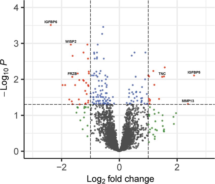
Function) and pathways (KEGG and Reactome). For 65%MS cartilage when compared to controls (Fig. 1C).Houtman et al. Arthritis Research & Therapy (2021) 23:215 Page 5 of 12 Differential expression of genes responsive to injurious were IGFBP6 (FC = 0.19; FDR = 3.07 × 10⁻⁴), CNTFR mechanical stress (FC = 0.27; FDR = 1.44 × 10⁻²), WISP2 (FC = 0.31; FDR To characterize the response of cartilage to mechanical = 1.08 × 10⁻³), and FRZB (FC = 0.32; FDR = 8.51 × stress at a strain of 65% indentation in aged articular 10−3). cartilage, we performed RNA sequencing on control (n = 14 samples) and 65% mechanically stressed (n = 14 Validation of differentially expressed genes with samples) articular cartilage samples obtained from mechanical stress macroscopically preserved osteochondral explants of hu- For validation and replication of the differentially man patients that underwent a knee replacement surgery expressed genes identified, a set of samples for technical due to OA. Baseline characteristics of donors of the (n = ten pairs) and biological (n = ten pairs) replication RNA-sequencing dataset are depicted in Table S1a. We was selected for RT-qPCR. Baseline characteristics of found 156 genes to be significantly differentially donors in the replication dataset are depicted in Table expressed (DE) (FDR < 0.05) with absolute fold changes S1b. Replication was performed for ten genes (Fig. 3), of (FC) ranging between 1.1 and 6.0 (Fig. 2, Table S3). which six were upregulated (IGFBP6, CNTFR, WISP2, Among these 156 DE genes, 46 (29%) were upregulated FRZB, COL9A3, and GADD45A) and four were down- and 110 (71%) were downregulated. The 20 genes with regulated (IGFBP5, PTGES, TNC, and IGFBP4). Tech- the highest absolute FC, and their respective direction of nical replication showed a significant difference for all effect previously identified in OA cartilage [5], are ten genes between controls and 65%MS cartilage, with shown in Table 1. Notable among the upregulated genes similar direction and size of effects. Biological replication were IGFBP5 (FC = 6.01; FDR = 7.81 × 10⁻³), MMP13 also showed the same direction of effects and similar ef- (FC = 5.19; FDR = 4.84 × 10⁻²), TNC (FC = 2.80; FDR = fect sizes as identified in the RNA-sequencing data. For 8.51 × 10⁻³), and PTGES (FC = 2.92; FDR = 8.29 × GADD45A, however, the difference was not significant 10−3). Notable genes among the downregulated genes (P-value = 0.12). Taken together, technical and biological Fig. 2 Volcano plot of differentially expressed genes. Dots represent genes expressed in mechanically stressed cartilage in comparison to control osteochondral explant cartilage. Red dots represent significantly differentially expressed (DE) genes that have an absolute fold change (FC) of ≥2, blue dots represent significantly DE genes, green dots represent genes that have an absolute FC of ≥2 but are not significantly DE, and gray dots represent genes not DE expressed between controls and 65% mechanically stressed cartilage. The FC presented here is the gene expression of 65% mechanically stressed relative to control cartilage
Houtman et al. Arthritis Research & Therapy         (2021) 23:215                                                                              Page 6 of 12

Table 1 Top 20 genes with the highest absolute FC in 65% mechanically stressed cartilage compared to controls
Ensemble ID                             Gene name                        FC                        FDRa                            Differential expression
                                                                                                                                   in OA cartilage [5]b
ENSG00000115461                         IGFBP5                           6.01                      7.81 × 10⁻³
ENSG00000137745                         MMP13                            5.19                      4.84 × 10⁻²
ENSG00000204103                         MAFB                             2.97                      4.66 × 10⁻³                     ↓
                                                                                                            −3
ENSG00000148344                         PTGES                            2.92                      8.29 × 10                       ↑
ENSG00000041982                         TNC                              2.80                      8.51 × 10⁻³                     ↑
                                                                                                            −2
ENSG00000141753                         IGFBP4                           2.59                      3.50 × 10                       ↑
ENSG00000160111                         CPAMD8                           0.39                      4.98 × 10⁻²                     ↓
                                                                                                            −2
ENSG00000166165                         CKB                              0.38                      1.04 × 10                       ↑
ENSG00000106258                         CYP3A5                           0.38                      3.62 × 10⁻²
ENSG00000107736                         CDH23                            0.37                      6.88 × 10⁻³
ENSG00000187720                         THSD4                            0.35                      1.44 × 10⁻²
ENSG00000144908                         ALDH1L1                          0.33                      2.22 × 10⁻³                     ↓
ENSG00000092758                         COL9A3                           0.32                      4.89 × 10⁻²
ENSG00000162998                         FRZB                             0.32                      8.51 × 10⁻³                     ↓
ENSG00000170891                         CYTL1                            0.32                      4.11 × 10⁻²
ENSG00000064205                         WISP2                            0.31                      1.08 × 10⁻³                     ↓
ENSG00000082196                         C1QTNF3                          0.30                      3.64 × 10⁻²                     ↑
                                                                                                            −2
ENSG00000122756                         CNTFR                            0.27                      1.44 × 10                       ↓
ENSG00000165966                         PDZRN4                           0.26                      1.44 × 10⁻²                     ↓
                                                                                                            −4
ENSG00000167779                         IGFBP6                           0.19                      3.07 × 10
a
 To correct for multiple testing, the Benjamini-Hochberg method was applied to P-values and reported as the false discovery rate (FDR). bGene expression
changes measured in RNA-sequencing data between preserved and lesioned OA articular cartilage, with preserved as reference [5]. Legend: FC fold change, FDR
false discovery rate

    Fig. 3 Technical and biological validation of the highest up- and downregulated genes was performed using RT-qPCR. Expression of six
    downregulated (IGFBP6, CNTFR, WISP2, FRZB, and GADD45A) and four upregulated (IGFBP5, PTGES, TNC, and IGFBP4) genes was measured in n = 10
    paired technical and n = 10 paired biological osteochondral explants. Figures show connected paired samples and −ΔCT of each independent
    sample is depicted by black dots (control) or squares (65%MS) in the graphs. Statistical differences between gene expression in control and 65%
    mechanically stressed were determined with a linear generalized estimation equation (GEE) with mRNA level as the dependent variable. *P ≤
    0.05; ***P ≤ 0.001. Legend: 65%MS 65% mechanical stress, RT-qPCR reverse transcriptase-quantitative PCR
Houtman et al. Arthritis Research & Therapy (2021) 23:215 Page 7 of 12
replication confirmed the robustness of our RNA- the same direction of effect are the highly downregulated
sequencing results. FRZB, WISP2, and CNTFR and the upregulated PTGES
and CRLF1.
In silico exploration of differentially expressed genes Next, we selected for exclusive mechanical stress-
To explore whether significant DE genes (N = 156 responsive genes, i.e., DEMS genes, not overlapping with
genes) were involved in particular pathways, they were previously identified DEOA genes [5]. This resulted in 92
further analyzed using Enrichr. Gene enrichment was genes that were differentially expressed exclusively in
observed, among others, for insulin-like growth factor I response to mechanical stress (DEExclusiveMS; Table S6;
and II binding (GO:0031995; GO:0031994, Padj = 1.83 × Figure S1). Notable DEExclusiveMS genes are the downreg-
10−2; Padj = 2.89 × 10⁻², involving IGFBP4, IGFBP5, and ulated IGFBP6, ITGA10, and COL9A3 and the upregu-
IGFBP6), cellular senescence (hsa04218, Padj = 1.15 × lated IGFBP5, MMP13, and GAPDH. Subsequent
10−2, involving 8 genes, e.g., GADD45A, MYC, SER- pathway analyses showed gene enrichment among genes
PINE1, and FOXO1), and focal adhesion (GO:0005925; involved in focal adhesion (GO:0005925, Padj = 0.02, 9
hsa04510, Padj = 2.54 × 10⁻²; Padj = 1.33 × 10⁻², involv- genes, e.g., CD9, RPL10A, and ENAH) and kinase inhibi-
ing 11 and 6 genes, respectively, e.g., TNC, CAV1, and tor activity (GO:0019210, Padj = 0.01, 5 genes, e.g.,
TLN2) (Table 2; Table S4a). CDKN1C, SOCS3, and SOCS5) (Table S4b). Upon ex-
To visualize interacting proteins, the online tool ploring protein-protein interactions between the 92
STRING was used. Among the 156 genes, 116 of the DEExclusiveMS genes using STRING, a highly significant
encoded proteins showed significant protein-protein in- enrichment for PPI was identified (P = 1.07 × 10⁻⁴; Fig-
teractions (PPI) (P = 1.44 × 10⁻¹⁵; Fig. 4). Among these ure S2), indicating that these genes act together or re-
proteins, we found several that have many connections spond in concert to detrimental mechanical stress.
with other proteins in the DE gene network, such as
GAPDH with 35 connections, IGFBP5 with 12 connec- OA risk genes responding to mechanical stress
tions, and in the cellular senescence involved genes Finally, to investigate which OA risk genes are repre-
MYC and FOXO1 with respectively 26 and 13 connec- sented among the mechanically stress-responsive genes
tions to other DE genes. Moreover, two clusters of genes in cartilage, we checked N = 90 genes previously recog-
are observed that correspond with two of the pathways nized as strong OA risk genes [34] identified in recent
identified. One cluster corresponds with genes found genome-wide association studies (GWAS) [35, 36]. As
mainly in the cellular senescence pathway (Fig. 4, dotted shown in Table S7, two of our identified DEMS genes
circle), while the other cluster consists of proteins that were also shown to be an OA risk gene in previous stud-
are involved in IGF-1 signaling (Fig. 4, black circle). ies. These genes were TNC, encoding for tenascin C,
which was highly increased (FC = 2.80; FDR = 8.5 ×
Comparison between mechanical stress genes and OA- 10−3) upon 65%MS and SCUBE1, encoding for signal
responsive genes peptide, CUB domain, and EGF-like domain-containing
To investigate to what extend the genes DE with mech- 1, which was decreased (FC = 0.53; FDR = 0.04) upon
anical stress (DEMS) coincide with OA pathophysiology, 65%MS.
we next compared the DEMS genes (Table S3) to previ-
ously identified genes DE between preserved and le- Discussion
sioned OA cartilage (DEOA) [5]. Of the 156 DEMS genes, To our knowledge, we are the first to report genome-
64 were previously identified with OA pathophysiology wide differentially expressed mRNAs in articular cartil-
and their majority (48 genes, 75%) had the same direc- age following repeated exposure to 65% mechanical
tion of effect (Table 1 and Table S5a, Figure S1). Notable stress using a human ex vivo osteochondral explant
genes coinciding with OA pathophysiology and showing model. Since injurious loading is considered a major
Table 2 Gene ontology and pathway enrichment analysis of differentially expressed genes in mechanically stressed cartilage
Term Entry Overlap Adj P-valuea Odds ratio Genes
−2
Cellular senescence hsa04218 8/160 1.15 × 10 6.41 GADD45A, MYC, SERPINE1, AKT3, EIF4EBP1, SLC25A5, ETS1, FOXO1
Focal adhesion hsa04510 8/199 1.33 × 10⁻² 5.15 SHC4, CAV1, ITGA10, AKT3, LAMA3, TNC, COL9A3, TLN2
−2
Insulin-like growth GO:0031995 3/7 1.83 × 10 54.95 IGFBP5, IGFBP4, IGFBP6
factor II binding
Focal adhesion GO:0005925 11/356 2.54 × 10⁻² 3.96 ENAH, EHD3, GSN, CAV1, TNC, CD9, TLN2, RPL10A, DCAF6, RHOB, ENG
−2
Insulin-like growth GO:0031994 3/13 2.89 × 10 29.59 IGFBP5, IGFBP4, IGFBP6
factor I binding
a
Enrichr uses a modified Fisher’s exact test to compute enrichment and this is reported as the adjusted P-value [29]. Legend: Adj P-value, adjusted P-valueHoutman et al. Arthritis Research & Therapy (2021) 23:215 Page 8 of 12 Fig. 4 Protein-protein interaction network in STRING of proteins encoded by differentially expressed genes. Only connected (N = 116 genes) genes that were identified to be differentially expressed between mechanically stressed and control cartilage of osteochondral explants are shown. Two clusters with high interactions were identified upon studying connections within STRING. One cluster corresponds with genes found in the cellular senescence pathway (dotted circle), while the other cluster consists of proteins that are involved in IGF-1 signaling (black circle) trigger in the initiation of OA onset, the results pre- breakdown of extracellular matrix in articular cartilage sented in our manuscript contribute important insight by cleaving, among others, collagen type II. Despite the into how injurious stress affects the propensity of aged well-known role of MMP13 in collagen type II break- human articular chondrocytes to lose their steady state down, it should be noted that the MMP13 gene is not towards a debilitating OA disease state. Notable genes found to be responsive with end-stage OA pathophysi- identified were different members of the insulin-like ology, i.e., not consistent and not among the genes high- growth factor I and II binding family (IGFBP6, IGFBP5, est differentially expressed between preserved and and IGFBP4) and the catabolic gene MMP13. Gene en- lesioned OA cartilage (Table 1) [5, 21, 37]. We therefore richment analyses showed that cellular senescence advocate that MMP13 expression could specifically mark (GADD45A, MYC, SERPINE1, and FOXO1) and focal initial responses to cartilage damage and not that of a adhesion (ITGA10, TLN2, and CAV1) processes are sig- chronic degenerative OA disease state. Henceforth, abro- nificantly changing in articular cartilage with injurious gating the MMP13 signaling shortly after an injurious loading. Together, identified genes and pathways facili- cartilage event could prevent the detrimental down- tate clinical development by exploring ways to counter- stream enzymatic breakdown of extracellular matrix pro- act these initial unbeneficial responses to injurious teins. Moreover, and as indicated above, MMP13 may be loading by supplementing or inhibiting of key genes. a suitable candidate sensitively marking injurious loading Moreover, we advocate that here identified specific re- of aged human articular cartilage independent of other sponsive genes to injurious loading can function as sen- physiological factors such as OA disease state. sitive markers facilitating the development of Four out of seven members of the insulin growth fac- scientifically founded strategies with respect to prevent- tor binding proteins (IGFBP4, IGFBP5, IGFBP6, and ive or curative exercise OA therapy among elderly. IGFBP7; Table S8) were found to be FDR DE. IGFBP1-6 Among the highest FDR significantly upregulated have an equal or greater affinity for binding IGF-1 when genes with 65% mechanical stress, we identified MMP13, compared to IGF-1R; hence, most of IGF-1 in the body encoding matrix metallopeptidase 13 (FC = 5.19; FDR = is bound to IGFBPs, antagonizing IGF-1 signaling [38–41]. 4.84 × 10⁻²) [20]. MMP13 is involved in the detrimental On the other hand, IGFBP7 has a low affinity for IGF and
Houtman et al. Arthritis Research & Therapy   (2021) 23:215                                                        Page 9 of 12

therefore more likely affects cell metabolism via binding to      promising target to follow up on in future research. Next
activin A, influencing the growth-suppressing effects of          to genes in this pathway, lookup of our DEMS genes in a
TGF-β, and antagonizing bone morphogenetic protein                proteomic atlas of senescence-associated secretory
(BMP) signaling [42, 43]. IGFBP4 and IGFBP5 can also              phenotype (SASP) identified 35 of our DEMS genes to
function as a transporter and bring IGF-1 close to its recep-     have previously been found in different senescent cells
tor, where IGF-1 is released via cleavage by proteins such        (Figure S3) [56]. Taken together, the upregulation of
as pregnancy-associated plasma protein-A (PAPPA), HtrA            MYC in combination with upregulation of several im-
Serine Peptidase 1 (HTRA1), and disintegrin and                   portant SASP protein markers suggests increased cellu-
metalloproteinase      domain-containing         protein     12   lar damage is occurring upon mechanical stress likely
(ADAM12) [44–46]. Additionally, notable in this respect is        driving cells to go into senescence. As cellular senes-
that three genes, HTRA1, ADAM12, and STC2 [47], in-               cence is a factor that is thought to play a significant role
volved in IGF-1 cleavage were found among the FDR sig-            in the OA pathophysiology, our model could provide
nificant upregulated genes in our dataset (Table S3).             more knowledge on how this pathway is involved in the
IGFBPs can also affect cells via IGF-independent mecha-           onset of OA and how therapeutics could be used to
nisms. The most noteworthy IGF-independent mechanism              minimize this response [57].
is observed for the highly upregulated IGFBP5, being induc-          To investigate whether OA risk loci could confer risk
tion of cell proliferation and apoptosis [48, 49]. In             via modifying the response to mechanical stress, we
summary, our data showed that, despite the fact that the          compared DEMS genes to strong OA risk genes identi-
mechanical stress applied affected cartilage integrity (Fig.      fied in the most recent GWAS [35, 36]. This resulted in
1), the upregulation of IGFBP4 and IGFBP5 in combination          the identification of two OA risk genes, TNC and
with the upregulation of its cleaving proteins might reflect      SCUBE1, present in our dataset (Table S7). Based on al-
an anabolic response of chondrocytes to initiate repair by        lelic imbalanced expression and linkage disequilibrium,
increasing bio-availability of IGF-1. Two studies support         the TNC OA risk allele rs1330349-C, in high linkage
our suggestion that IGF-1 signaling might be a beneficial         disequilibrium with the transcript SNP rs2274836-T, ap-
anabolic response to mechanical stress. In an OA dog              peared to act via decreasing expression of TNC [58]. For
model, increasing intact IGFBP5 proteins resulted in in-          that matter, the observed high upregulation of TNC ex-
creased IGF-1 levels and reduced destruction of cartilage         pression with mechanical stress (FC = 2.80; FDR = 8.51
[50]. While in a human explant model, addition of IGF-1           × 10−3) as well as the previously observed upregulation
after mechanical stress increased COL2A1 gene expression          with OA pathophysiology (FC = 1.41; FDR = 1.09 ×
and slightly increased cell viability [51]. Our results in com-   10−2) [5] is likely a beneficial response to rescue or
bination with those previously found suggest that addition        maintain articular cartilage integrity. This is further con-
of IGFBP4 and/or IGFBP5 would be an interesting therapy           firmed by animal studies showing that the addition of
to further explore in combatting the catabolic response.          exogenous TNC reduced cartilage degeneration and
   To identify upstream processes and to put our results          repaired cartilage [59, 60]. In contrast, for the intronic
in a broader perspective, we investigated connections be-         OA risk SNP located in the vicinity of SCUBE1
tween genes on the protein level in STRING (Fig. 4) and           (rs528981060), we were not able to determine a tran-
determined pathway enrichment (Table 2) of the differ-            script proxy SNP; hence, potential AEI of SCUBE1 could
entially expressed genes. Based on this pathway analysis,         not be explored.
we identified enrichment for proteins involved in cellular           With regard to overlap with in vivo animal models, we
senescence. DE genes with mechanical stress in this               compare our DE genes to those found in physiological
pathway have already been linked to aging and OA, such            [11], surgical destabilization of the medial meniscus
as GADD45A, SERPINE1, MYC, and FOXO1. Notable                     (DMM) [18] and non-invasive tibial compression (TC)
are the two transcription factors, MYC and FOXO1,                 models [12, 14]. The most striking overlap in DE genes
showing many connections to other proteins (Fig. 4) and           (46 genes) was found between our model and the non-
previously shown to be dysregulated in OA chondro-                invasive TC model using gene expression data of 14-
cytes [52, 53]. FOXO1 is an essential mediator of                 month-old mice 1 week after injury. Among the
cartilage growth and homeostasis and its expression is            overlapping genes, we confirmed the involvement of all
decreased in aged and OA cartilage [52]. In addition,             IGFBPs, HTRA1, and ADAM12 and of several OA-
FOXO1 was shown to be an antagonist of MYC and pre-               associated genes such as FRZB, TNC, and SCUBE1 in both
vents, among others, ROS production [54, 55]. Our re-             models [14]. As also shown by other studies [14, 18], age of
sults suggest that reduced expression of FOXO1 could              animals used in these models can greatly influence results.
be one of the reasons for increased expression of MYC.            This could also, next to a difference in species, partially
As one of the known responses of chondrocytes to                  explain why there is little overlap with other injurious
mechanical stress is ROS production, this would be a              mechanical stress studies.
Houtman et al. Arthritis Research & Therapy   (2021) 23:215                                                                     Page 10 of 12

  A strength of our aged human ex vivo osteochondral           towards a debilitating OA disease state. Exploring ways to
model is that it allowed us to investigate the chondro-        counteract the initial unbeneficial responses to injurious
cyte response to an OA-relevant trigger in its natural         loading may facilitate clinical development prior to the
environment. In addition, our model comprises aged             onset of irreversible damage. Moreover, we advocate that
cartilage, which is likely more vulnerable to OA onset,        the here identified unique responsive genes to injurious
and hence, results are relevant to the population at risk.     loading, such as MMP13, can function as a sensitive
Another strong point in our model is that we measure           marker to strategically develop preventive and/or curative
the changes in gene expression that are measured 4 days        exercise therapy for OA independent of other physio-
post-injury as such reflecting representative lasting          logical factors. Preferably such an endeavor would exploit
changes in chondrocyte signaling rather than acute             our established ex vivo osteochondral model while apply-
stress responses only. On the other hand, our data could       ing variable mechanical loading regimes.
facilitate treatment strategies, prior to irreversible dam-
age of OA-affected cartilage. Some limitations of our          Supplementary Information
study are the relatively low sample size of 14 explants        The online version contains supplementary material available at https://doi.
                                                               org/10.1186/s13075-021-02595-8.
per condition, hence limiting our power. As a result, we
may have missed subtle gene expression changes in re-
                                                                Additional file 1. Supplementary Tables and Figures. List of content:
sponse to detrimental mechanical stress. Another point          Supplementary Table S1. [a] Donor characteristics of samples for which RNA
of our study to address is the heterogeneity of preserved       was sequenced. [b] Donor characteristics of independent samples used for
cartilage collected from OA patients with Mankin scores         replication of RNA-sequencing findings. Supplementary Table S2. Primer se-
                                                                quences used for replication and validation by RT-qPCR. Supplementary
ranging from 0 to 7. Although such heterogeneity may            Table S3. Genes differentially expressed in 65%MS (DEMS) cartilage compared
also have affected the power of our study, hence the            to control cartilage of human osteochondral explants. Supplementary Table
total number of differentially expressed genes with in-         S4. Gene enrichment found in Enrichr. Enrichment for [a] 156 DEMS genes in
                                                                and [b] 92 DEExclusiveMS for the gene ontology terms: biological process, mo-
jurious loading, we want to highlight that despite the dif-     lecular function and cellular component 2018, and pathways: KEGG 2019
ferences in Mankin scores, we were able to consistently         human and reactome. Supplementary Table S5. DEMS genes coinciding with
detect (at the genome-wide significant level) 156 differ-       previously reported DE genes in OA pathophysiology (DEOA). [a] DEMS genes
                                                                with same direction of effect as DEOA genes. [b] DEMS genes with opposite
entially expressed genes reflecting strong and/or very          direction of effect as DEOA genes. Supplementary Table S6. Exclusive mech-
consistent mechano-pathological processes triggered             anical response genes (DEExclusiveMS). Supplementary Table S7. Previously re-
after mechanical stress. Moreover, due to the heterogen-        ported OA risk loci present in our DE gene dataset. Supplementary Table S8.
                                                                All insulin growth factor binding proteins (IGFBPs) and related DE genes
eity in eligible waste articular cartilage after joint re-      identified in our analysis. Supplementary Figure S1. Venn diagram of coin-
placement surgery (i.e., osteochondral explants), we were       ciding genes between differentially expressed genes in mechanically
not able to generate a RNA-sequencing dataset of per-           stressed versus control cartilage from osteochondral explants (DEMS) and
                                                                previously identified differentially expressed genes in preserved versus le-
fect control—mechanically stressed sample pairs. Hence-         sioned OA cartilage (DEOA). Supplementary Figure S2. Protein-protein inter-
forth, to adjust for dependencies among control and/or          action network in STRING of proteins encoded by differentially expressed
mechanically stressed samples, we added donor as a              genes (N = 92 genes) not coinciding with OA pathophysiology (DEExclusi-
                                                                veMS). Supplementary Figure S3. Heat-map of proteins present in SASP.
random effect during differential expression analyses.
Adding to the validity of this approach was the fact that
                                                               Acknowledgements
we successfully replicated expression changes for ten          We thank all the participants of the RAAK study. The LUMC has and is
genes in ten novel independent perfectly paired samples.       supporting the RAAK study. We also thank Enrike van der Linden, Demiën
A final limitation of our study is that we have focused        Broekhuis, Peter van Schie, Anika Rabelink-Hoogenstraaten, Shaho Hasan,
                                                               Maartje Meijer, Daisy Latijnhouwers, and Geert Spierenburg for collecting sur-
on exploring gene expression changes following mech-           gical waste material and all members of our research group. Data is gener-
anical stress and have not studied changes at the protein      ated within the scope of the Medical Delta Programs Regenerative Medicine
level. However, we advocate that chondrocyte signaling         4D: Generating complex tissues with stem cells and printing technology and
                                                               Improving Mobility with Technology. We thank the Sequence Analysis Sup-
at the gene expression level is a more sensitive measure       port Core (SASC) of the Leiden University Medical Center for their support.
of underlying ongoing processes.
                                                               Authors’ contributions
                                                               All authors have made contributions to the completion of this study. Study
Conclusions                                                    concept and design: EH, YFM, RCA, and IM. Acquisition of material and data:
To conclude, our results faithfully represent transcrip-       EH, MT, JR, HED, RJPvdW, RGHHN, and HM. Data analysis: EH, MT, RCA, and
tomic wide consequences of injurious loading in human          IM. Preparation of the manuscript: EH and IM. Critical reviewing and approval
                                                               of the final manuscript: all authors.
aged articular cartilage with MMP13, IGF binding
proteins, and cellular senescence as the most notable          Funding
results. Since injurious loading is considered a major trig-   This work was supported by the Dutch Scientific Research council NWO/
ger of OA onset, these findings provide important insight      ZonMW VICI scheme [91816631/528] and the Dutch Arthritis Society/
                                                               ReumaNederland [DAF-15-4-401]. The funding body played no role in the
into how injurious stress affects the propensity of aged       design of the study; in the collection, analysis, and interpretation of the data;
human articular chondrocytes to lose their steady state        and in writing the manuscript.
Houtman et al. Arthritis Research & Therapy (2021) 23:215 Page 11 of 12
Availability of data and materials mice are less prone to develop OA-like cartilage damage upon excessive
The list of all significantly affected genes is included in the Supplementary mechanical stress. Ann Rheum Dis. 2016;75(3):571–7. https://doi.org/10.113
data (Table S3). 6/annrheumdis-2014-206608.
12. Chang JC, Sebastian A, Murugesh DK, Hatsell S, Economides AN,
Declarations Christiansen BA, et al. Global molecular changes in a tibial compression
induced ACL rupture model of post-traumatic osteoarthritis. J Orthop Res.
Ethics approval and consent to participate 2017;35(3):474–85. https://doi.org/10.1002/jor.23263.
Informed consent was obtained from participants of the RAAK biobank, and 13. Sebastian A, McCool JL, Hum NR, et al. Single-cell RNA-Seq reveals
ethical approval was given by the medical ethics committee of the Leiden transcriptomic heterogeneity and post-traumatic osteoarthritis-associated
University Medical Center (P08.239/P19.013). early molecular changes in mouse articular chondrocytes. Cells. 2021;10(6).
14. Sebastian A, Murugesh DK, Mendez ME, et al. Global gene expression
Consent for publication analysis identifies age-related differences in knee joint transcriptome during
Not applicable the development of post-traumatic osteoarthritis in mice. Int J Mol Sci.
2020;21(1).
Competing interests 15. Gardiner MD, Vincent TL, Driscoll C, Burleigh A, Bou-Gharios G, Saklatvala J,
The authors declare that they have no competing interests. et al. Transcriptional analysis of micro-dissected articular cartilage in post-
traumatic murine osteoarthritis. Osteoarthr Cartil. 2015;23(4):616–28. https://
Author details doi.org/10.1016/j.joca.2014.12.014.
1
Section of Molecular Epidemiology, Department of Biomedical Data 16. Appleton CT, Pitelka V, Henry J, Beier F. Global analyses of gene expression
Sciences, Leiden University Medical Center, Postzone S05-P, Einthovenweg in early experimental osteoarthritis. Arthritis Rheum. 2007;56(6):1854–68.
20, 2333, ZC, Leiden, The Netherlands. 2Department of Orthopaedics, Leiden https://doi.org/10.1002/art.22711.
University Medical Center, Leiden, The Netherlands. 3Sequencing Analysis 17. Ashwell MS, O'Nan AT, Gonda MG, Mente PL. Gene expression profiling of
Support Core, Leiden University Medical Centre, Leiden, The Netherlands . chondrocytes from a porcine impact injury model. Osteoarthr Cartil. 2008;
16(8):936–46. https://doi.org/10.1016/j.joca.2007.12.012.
Received: 15 June 2021 Accepted: 30 July 2021 18. Loeser RF, Olex AL, McNulty MA, et al. Microarray analysis reveals age-
related differences in gene expression during the development of
osteoarthritis in mice. Arthritis Rheum. 2012;64(3):705–17. https://doi.org/1
0.1002/art.33388.
References
1. Englund M, Guermazi A, Gale D, Hunter DJ, Aliabadi P, Clancy M, et al. 19. Loening AM, James IE, Levenston ME, Badger AM, Frank EH, Kurz B, et al.
Incidental meniscal findings on knee MRI in middle-aged and elderly Injurious mechanical compression of bovine articular cartilage induces
persons. N Engl J Med. 2008;359(11):1108–15. https://doi.org/10.1056/ chondrocyte apoptosis. Arch Biochem Biophys. 2000;381(2):205–12. https://
NEJMoa0800777. doi.org/10.1006/abbi.2000.1988.
2. Jordan JM, Helmick CG, Renner JB, Luta G, Dragomir AD, Woodard J, et al. 20. Houtman E, van Hoolwerff M, Lakenberg N, Suchiman EHD, van der Linden-
Prevalence of knee symptoms and radiographic and symptomatic knee van der Zwaag E, Nelissen RGHH, et al. Human osteochondral explants:
osteoarthritis in African Americans and Caucasians: the Johnston County reliable biomimetic models to investigate disease mechanisms and develop
Osteoarthritis Project. J Rheumatol. 2007;34(1):172–80. personalized treatments for osteoarthritis. Rheumatol Ther. 2021;8(1):499–
3. Woolf AD, Erwin J, March L. The need to address the burden of 515. https://doi.org/10.1007/s40744-021-00287-y.
musculoskeletal conditions. Best Pract Res Clin Rheumatol. 2012;26(2):183– 21. Ramos YF, den Hollander W, Bovee JV, et al. Genes involved in the
224. https://doi.org/10.1016/j.berh.2012.03.005. osteoarthritis process identified through genome wide expression analysis
4. Tonge DP, Pearson MJ, Jones SW. The hallmarks of osteoarthritis and the in articular cartilage; the RAAK study. PLoS One. 2014;9(7):e103056. https://
potential to develop personalised disease-modifying pharmacological doi.org/10.1371/journal.pone.0103056.
therapeutics. Osteoarthr Cartil. 2014;22(5):609–21. https://doi.org/10.1016/j. 22. Sanchez-Adams J, Leddy HA, McNulty AL, O’Conor CJ, Guilak F. The
joca.2014.03.004. mechanobiology of articular cartilage: bearing the burden of osteoarthritis. Curr
5. Coutinho de Almeida R, Ramos YFM, Mahfouz A, den Hollander W, Rheumatol Rep. 2014;16(10):451. https://doi.org/10.1007/s11926-014-0451-6.
Lakenberg N, Houtman E, et al. RNA sequencing data integration reveals an 23. Mankin HJ, Dorfman H, Lippiello L, Zarins A. Biochemical and metabolic
miRNA interactome of osteoarthritis cartilage. Ann Rheum Dis. 2019;78(2): abnormalities in articular cartilage from osteo-arthritic human hips. II.
270–7. https://doi.org/10.1136/annrheumdis-2018-213882. Correlation of morphology with biochemical and metabolic data. J Bone
6. Soul J, Dunn SL, Anand S, Serracino-Inglott F, Schwartz JM, Boot- Joint Surg Am. 1971;53(3):523–37. https://doi.org/10.2106/00004623-197153
Handford RP, et al. Stratification of knee osteoarthritis: two major 030-00009.
patient subgroups identified by genome-wide expression analysis of 24. Farndale RW, Buttle DJ, Barrett AJ. Improved quantitation and discrimination
articular cartilage. Ann Rheum Dis. 2018;77(3):423. https://doi.org/10.113 of sulphated glycosaminoglycans by use of dimethylmethylene blue.
6/annrheumdis-2017-212603. Biochim Biophys Acta. 1986;883(2):173–7. https://doi.org/10.1016/0304-41
7. Coutinho de Almeida R, Mahfouz A, Mei H, Houtman E, den Hollander W, 65(86)90306-5.
Soul J, et al. Identification and characterization of two consistent 25. Wu TD, Watanabe CK. GMAP: a genomic mapping and alignment program
osteoarthritis subtypes by transcriptome and clinical data integration. for mRNA and EST sequences. Bioinformatics. 2005;21(9):1859–75. https://
Rheumatology (Oxford). 2021;60(3):1166–75. https://doi.org/10.1093/rheuma doi.org/10.1093/bioinformatics/bti310.
tology/keaa391. 26. Anders S, Pyl PT, Huber W. HTSeq--a Python framework to work with high-
8. Chen CT, Burton-Wurster N, Borden C, Hueffer K, Bloom SE, Lust G. throughput sequencing data. Bioinformatics. 2015;31(2):166–9. https://doi.
Chondrocyte necrosis and apoptosis in impact damaged articular cartilage. org/10.1093/bioinformatics/btu638.
J Orthop Res. 2001;19(4):703–11. https://doi.org/10.1016/S0736-02 27. Ewels P, Magnusson M, Lundin S, Kaller M. MultiQC: summarize analysis
66(00)00066-8. results for multiple tools and samples in a single report. Bioinformatics.
9. Lee JH, Fitzgerald JB, Dimicco MA, Grodzinsky AJ. Mechanical injury of 2016;32(19):3047–8. https://doi.org/10.1093/bioinformatics/btw354.
cartilage explants causes specific time-dependent changes in chondrocyte 28. Love MI, Huber W, Anders S. Moderated estimation of fold change and
gene expression. Arthritis Rheum. 2005;52(8):2386–95. https://doi.org/10.1 dispersion for RNA-seq data with DESeq2. Genome Biol. 2014;15(12):550.
002/art.21215. https://doi.org/10.1186/s13059-014-0550-8.
10. Kurz B, Jin M, Patwari P, Cheng DM, Lark MW, Grodzinsky AJ. Biosynthetic 29. Kuleshov MV, Jones MR, Rouillard AD, Fernandez NF, Duan Q, Wang Z, et al.
response and mechanical properties of articular cartilage after injurious Enrichr: a comprehensive gene set enrichment analysis web server 2016
compression. J Orthop Res. 2001;19(6):1140–6. https://doi.org/10.1016/S073 update. Nucleic Acids Res. 2016;44(W1):W90–7. https://doi.org/10.1093/nar/
6-0266(01)00033-X. gkw377.
11. Bomer N, Cornelis FM, Ramos YF, et al. The effect of forced exercise on 30. Szklarczyk D, Gable AL, Lyon D, Junge A, Wyder S, Huerta-Cepas J, et al.
knee joints in Dio2(-/-) mice: type II iodothyronine deiodinase-deficient STRING v11: protein-protein association networks with increased coverage,Houtman et al. Arthritis Research & Therapy (2021) 23:215 Page 12 of 12
supporting functional discovery in genome-wide experimental datasets. 51. Riegger J, Joos H, Palm HG, Friemert B, Reichel H, Ignatius A, et al. Striking a
Nucleic Acids Res. 2019;47(D1):D607–D13. https://doi.org/10.1093/nar/ new path in reducing cartilage breakdown: combination of antioxidative
gky1131. therapy and chondroanabolic stimulation after blunt cartilage trauma. J Cell
31. Al-Sabah A, Stadnik P, Gilbert SJ, Duance VC, Blain EJ. Importance of reference Mol Med. 2018;22(1):77–88. https://doi.org/10.1111/jcmm.13295.
gene selection for articular cartilage mechanobiology studies. Osteoarthr Cartil. 52. Matsuzaki T, Alvarez-Garcia O, Mokuda S, et al. FoxO transcription factors
2016;24(4):719–30. https://doi.org/10.1016/j.joca.2015.11.007. modulate autophagy and proteoglycan 4 in cartilage homeostasis and
32. McCulloch RS, Ashwell MS, O’Nan AT, Mente PL. Identification of stable osteoarthritis. Sci Transl Med. 2018;10(428).
normalization genes for quantitative real-time PCR in porcine articular 53. Wu YH, Liu W, Zhang L, Liu XY, Wang Y, Xue B, et al. Effects of microRNA-24
cartilage. J Anim Sci Biotechnol. 2012;3(1):36. https://doi.org/10.1186/2049-1 targeting C-myc on apoptosis, proliferation, and cytokine expressions in
891-3-36. chondrocytes of rats with osteoarthritis via MAPK signaling pathway. J Cell
33. Diggle P, Liang K-Y, Zeger SL. Analysis of longitudinal data. Oxford New Biochem. 2018;119(10):7944–58. https://doi.org/10.1002/jcb.26514.
York: Clarendon Press; Oxford University Press; 1994. p. xi, 253. 54. Tan P, Guan H, Xie L, Mi B, Fang Z, Li J, et al. FOXO1 inhibits
34. Reynard LN, Barter MJ. Osteoarthritis year in review 2019: genetics, osteoclastogenesis partially by antagnozing MYC. Sci Rep. 2015;5(1):16835.
genomics and epigenetics. Osteoarthr Cartil. 2020;28(3):275–84. https://doi. https://doi.org/10.1038/srep16835.
org/10.1016/j.joca.2019.11.010. 55. Peck B, Ferber EC, Schulze A. Antagonism between FOXO and MYC
35. Styrkarsdottir U, Lund SH, Thorleifsson G, Zink F, Stefansson OA, Sigurdsson regulates cellular powerhouse. Front Oncol. 2013;3:96.
JK, et al. Meta-analysis of Icelandic and UK data sets identifies missense 56. Basisty N, Kale A, Jeon OH, Kuehnemann C, Payne T, Rao C, et al. A
variants in SMO, IL11, COL11A1 and 13 more new loci associated with proteomic atlas of senescence-associated secretomes for aging biomarker
osteoarthritis. Nat Genet. 2018;50(12):1681–7. https://doi.org/10.1038/s41588- development. PLoS Biol. 2020;18(1):e3000599. https://doi.org/10.1371/journa
018-0247-0. l.pbio.3000599.
36. Tachmazidou I, Hatzikotoulas K, Southam L, et al. Identification of new 57. McCulloch K, Litherland GJ, Rai TS. Cellular senescence in osteoarthritis
therapeutic targets for osteoarthritis through genome-wide analyses of UK pathology. Aging Cell. 2017;16(2):210–8. https://doi.org/10.1111/acel.12562.
Biobank data. Nat Genet. 2019;51(2):230–6. https://doi.org/10.1038/s41588- 58. den Hollander W, Pulyakhina I, Boer C, et al. Annotating transcriptional
018-0327-1. effects of genetic variants in disease-relevant tissue: transcriptome-wide
37. Dunn SL, Soul J, Anand S, Schwartz JM, Boot-Handford RP, Hardingham TE. allelic imbalance in osteoarthritic cartilage. Arthritis Rheum. 2019;71(4):561–
Gene expression changes in damaged osteoarthritic cartilage identify a 70. https://doi.org/10.1002/art.40748.
signature of non-chondrogenic and mechanical responses. Osteoarthr Cartil. 59. Matsui Y, Hasegawa M, Iino T, Imanaka-Yoshida K, Yoshida T, Sudo A.
2016;24(8):1431–40. https://doi.org/10.1016/j.joca.2016.03.007. Tenascin-C prevents articular cartilage degeneration in murine osteoarthritis
38. Firth SM, Baxter RC. Cellular actions of the insulin-like growth factor binding models. Cartilage. 2018;9(1):80–8. https://doi.org/10.1177/194760351
proteins. Endocr Rev. 2002;23(6):824–54. https://doi.org/10.1210/er.2001- 6681134.
0033. 60. Unno H, Hasegawa M, Suzuki Y, Iino T, Imanaka-Yoshida K, Yoshida T, et al.
39. Duan C, Xu Q. Roles of insulin-like growth factor (IGF) binding proteins in Tenascin-C promotes the repair of cartilage defects in mice. J Orthop Sci.
regulating IGF actions. Gen Comp Endocrinol. 2005;142(1-2):44–52. https:// 2020;25(2):324–30. https://doi.org/10.1016/j.jos.2019.03.013.
doi.org/10.1016/j.ygcen.2004.12.022.
40. Baxter RC. IGF binding proteins in cancer: mechanistic and clinical insights. Publisher’s Note
Nat Rev Cancer. 2014;14(5):329–41. https://doi.org/10.1038/nrc3720. Springer Nature remains neutral with regard to jurisdictional claims in
41. Clemmons DR. Role of IGF binding proteins in regulating metabolism. published maps and institutional affiliations.
Trends Endocrinol Metab. 2016;27(6):375–91. https://doi.org/10.1016/j.tem.2
016.03.019.
42. Kato MV. A secreted tumor-suppressor, mac25, with activin-binding activity.
Mol Med. 2000;6(2):126–35. https://doi.org/10.1007/BF03401780.
43. Esterberg R, Delalande JM, Fritz A. Tailbud-derived Bmp4 drives proliferation
and inhibits maturation of zebrafish chordamesoderm. Development. 2008;
135(23):3891–901. https://doi.org/10.1242/dev.029264.
44. Laursen LS, Overgaard MT, Soe R, et al. Pregnancy-associated plasma
protein-A (PAPP-A) cleaves insulin-like growth factor binding protein
(IGFBP)-5 independent of IGF: implications for the mechanism of IGFBP-4
proteolysis by PAPP-A. FEBS Lett. 2001;504(1-2):36–40. https://doi.org/10.101
6/S0014-5793(01)02760-0.
45. Hou J, Clemmons DR, Smeekens S. Expression and characterization of a
serine protease that preferentially cleaves insulin-like growth factor binding
protein-5. J Cell Biochem. 2005;94(3):470–84. https://doi.org/10.1002/jcb.2
0328.
46. Loechel F, Fox JW, Murphy G, Albrechtsen R, Wewer UM. ADAM 12-S
cleaves IGFBP-3 and IGFBP-5 and is inhibited by TIMP-3. Biochem Biophys
Res Commun. 2000;278(3):511–5. https://doi.org/10.1006/bbrc.2000.3835.
47. Jepsen MR, Kloverpris S, Mikkelsen JH, et al. Stanniocalcin-2 inhibits
mammalian growth by proteolytic inhibition of the insulin-like growth
factor axis. J Biol Chem. 2015;290(6):3430–9. https://doi.org/10.1074/jbc.
M114.611665.
48. Cobb LJ, Salih DA, Gonzalez I, et al. Partitioning of IGFBP-5 actions in
myogenesis: IGF-independent anti-apoptotic function. J Cell Sci. 2004;117(Pt
9):1737–46. https://doi.org/10.1242/jcs.01028.
49. Tripathi G, Salih DA, Drozd AC, Cosgrove RA, Cobb LJ, Pell JM. IGF-
independent effects of insulin-like growth factor binding protein-5 (Igfbp5)
in vivo. FASEB J. 2009;23(8):2616–26. https://doi.org/10.1096/fj.08-114124.
50. Clemmons DR, Busby WH Jr, Garmong A, et al. Inhibition of insulin-like
growth factor binding protein 5 proteolysis in articular cartilage and joint
fluid results in enhanced concentrations of insulin-like growth factor 1 and
is associated with improved osteoarthritis. Arthritis Rheum. 2002;46(3):694–
703. https://doi.org/10.1002/art.10222.You can also read