Multidisciplinary approach to orbital decompression. A review
←
→
Page content transcription
If your browser does not render page correctly, please read the page content below
ACTA OTORHINOLARYNGOLOGICA ITALICA 2021;41(SUPPL.1):S90-S101; doi: 10.14639/0392-100X-suppl.1-41-2021-09

Multidisciplinary approach to orbital decompression.
A review
L’approccio multidisciplinare alla decompressione orbitaria. Una review
Claudio Parrilla1, Dario Antonio Mele1, Silvia Gelli2, Lorenzo Zelano2, Francesco Bussu3, Mario Rigante1,
Gustavo Savino4, Emanuele Scarano5
1
  Otorhinolaryngology and Head-Neck Surgery Unit, “A. Gemelli” Hospital Foundation IRCCS, Catholic University of the Sacred
Heart, Rome, Italy; 2 Endocrinology Unit, “A. Gemelli” Hospital Foundation IRCCS, Catholic University of the Sacred Heart, Rome,
Italy; 3 Division of Otolaryngology, Università di Sassari, Italy; 4 Ophthalmology Unit, “A. Gemelli” Hospital Foundation IRCCS,
Catholic University of the Sacred Heart, Rome, Italy; 5 Division of Otolaryngology, Azienda Ospedaliera Pia Fondazione di Culto e
Religione Cardinale G. Panico, Tricase, Italy

SUMMARY
Endoscopic orbital surgery has become a highly evolving multidisciplinary surgical field
thanks to development in technical skills of ophthalmologists and otolaryngologists. These
advances expanded the clinical application of orbital decompression, with a growing body
of literature describing the multidisciplinary management of thyroid eye disease and com-
pressive optic neuropathy, since 1990. Although techniques have improved considerably,
only few Randomized Control Trials (RCT) provide evidence to support recommenda-
tions in clinical practice. This review provides an overview of the current knowledge of
orbital decompression to clarify which is the most standardized therapeutic strategy. In the        Received: October 27, 2020
literature, we observed several approaches with contradicting results and the comparison            Accepted: January 15, 2021
of different surgical techniques was biased by inclusion of patients at different stage of
disease (active or inactive), different surgical indications (dysthyroid neuropathy or dis-         Correspondence
figuring proptosis) and measures of outcomes (such as different system for ocular motility          Dario Antonio Mele
evaluation). The timing of surgical decompression is one of the debated issues. One RCT             Otorhinolaryngology, Head and Neck Surgery,
focusing on Graves’ orbitopathy showed how intravenous corticosteroids achieve better               “A. Gemelli” Hospital Foundation IRCCS, Catholic
visual recovery than surgical orbital decompression; but in case of absent or poor response         University of the Sacred Heart
to medical therapy the patient should undergo surgery within two weeks. There is slight             largo A. Gemelli 8, 00168 Rome, Italy
                                                                                                    E-mail: darioam90@gmail.com
evidence that the removal of the medial and lateral wall (so-called balanced decompression)
with or without fat removal could be the most effective surgical technique, with low com-
                                                                                                    Funding
plication rate, but an increasing number of authors are promoting, for selected cases, a pure
                                                                                                    None.
endoscopic surgical approach (with removal of medial and infero-medial orbital wall), less
invasive than the balanced one; the latter indicated to more severe proptosis or diplopia after
                                                                                                    Conflict of interest
endoscopic procedure. Three-wall decompression is chosen for high degrees of proptosis,             The Authors declare no conflict of interest.
but complications are more frequent. Timing of surgical orbital decompression, in particu-
lar when a concomitant optic neuropathy is present, is still to be determined. Additional
ophthalmological procedures are needed to restore normal eye function and cosmesis. Stra-             How to cite this article: Parrilla C, Mele DA,
bismus surgery to address diplopia and lowering the position of the upper eyelid represent            Gelli S, et al. Multidisciplinary approach to
                                                                                                      orbital decompression. A review. Acta Otorhi-
some of the additional steps for the final rehabilitation of Graves’ orbitopathy. The main
                                                                                                      nolaryngol Ital 2021;41(SUPPL.1):S90-S101.
clinical outcomes including visual acuity, proptosis, and new-onset diplopia are changing. Recent     https://doi.org/10.14639/0392-100X-sup-
studies focused on the development of imaging measurements in order to objectively evaluate the       pl.1-41-2021-09
surgical results and QOL questionnaires are gaining increasing importance.
                                                                                                    © Società Italiana di Otorinolaringoiatria
KEY WORDS: nasal endoscopy, orbital decompression, thyroid eye disease, compressive
                                                                                                    e Chirurgia Cervico-Facciale
optic neuropathy
                                                                                                                   OPEN ACCESS
RIASSUNTO                                                                                           This is an open access article distributed in accordance with
La chirurgia endoscopica orbitaria rappresenta un ambito multidisciplinare in costante              the CC-BY-NC-ND (Creative Commons Attribution-Non-
evoluzione grazie allo sviluppo delle tecniche sia in ambito oftalmologico che otorino-             Commercial-NoDerivatives 4.0 International) license. The
                                                                                                    article can be used by giving appropriate credit and mentio-
laringoiatrico. Tali progressi hanno esteso le applicazioni cliniche della decompressione
                                                                                                    ning the license, but only for non-commercial purposes and
orbitaria, con incremento del numero di lavori in letteratura, volti a descrivere la gestione       only in the original version. For further information: https://
multidisciplinare dell’orbitopatia tiroidea e della neuropatia ottica compressiva, a partire        creativecommons.org/licenses/by-nc-nd/4.0/deed.en

S90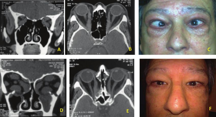
Multidisciplinary management in orbital decompression

dagli anni ’90. Sebbene le tecniche si siano evolute notevolmente, solo pochi studi controllati randomizzati mostrano evidenze a supporto
di indicazioni per la pratica clinica. Questa review fornisce una panoramica delle conoscenze attuali sulla decompressione orbitaria, per
chiarire quali siano le strategie terapeutiche maggiormente standardizzate. In letteratura, abbiamo osservato i diversi approcci con risultati
contrastanti e il confronto delle varie tecniche chirurgiche è influenzato dall’inclusione di pazienti in stadio di malattia differente (fase attiva
o inattiva), da differenti indicazioni alla chirurgia (neuropatia distiroidea o proptosi sfigurante) e metodi di misurazione dei risultati (come
ad esempio vari sistemi per la valutazione della motilità oculare). La tempistica della decompressione chirurgica è un altro dei temi ancora
oggi dibattuti. Uno studio clinico randomizzato ha mostrato come la terapia steroidea endovenosa consenta un miglior recupero visivo rispetto
alla decompressione orbitaria chirurgica; ma in caso di scarsa risposta alla terapia medica il paziente dovrebbe essere sottoposto a chirurgia
entro due settimane. C’è una lieve evidenza che la rimozione delle pareti mediale e laterale (cosiddetta decompressione bilanciata) con o
senza rimozione del grasso orbitario rappresenti la tecnica chirurgica più efficace, con un basso tasso di complicanze. La decompressione a
tre pareti è scelta in caso di proptosi di alto grado, ma le complicanze sono più frequenti. Un numero crescente di autori sta promuovendo, per
casi selezionati, un approccio chirurgico puramente endoscopico (con rimozione della parete orbitaria mediale e infero-mediale), meno inva-
sivo della decompressione bilanciata; quest’ultima appare indicata nei casi di proptosi più grave o di diplopia post-procedura endoscopica.
La corretta tempistica della decompressione orbitaria è ancora da determinare, soprattutto se presente una concomitante neuropatia ottica.
Procedure aggiuntive oftalmologiche sono necessarie per la normale funzionalità e cosmesi. La chirurgia dello strabismo per correggere la
diplopia e l’abbassamento della posizione della palpebra superiore rappresentano alcuni dei passaggi successivi per la riabilitazione finale
dell’orbitopatia di Graves. I principali outcome attualmente valutati includono acuità visiva, proptosi e diplopia di nuova insorgenza. Studi
recenti si focalizzano sullo sviluppo di misurazioni sull’imaging per valutare i risultati chirurgici e i questionari sulla qualità della vita stanno
acquisendo importanza sempre maggiore.
PAROLE CHIAVE: endoscopia nasosinusale, decompressione orbitaria, orbitopatia di Graves, neuropatia ottica compressiva

Introduction                                                                technique 2. However, several early reports of endoscopic
                                                                            decompression described new-onset diplopia in up to 45%
Orbital decompression may be indicated for patients with
                                                                            of cases 3. The consequent technical refinement was rep-
orbital abscess, periorbital or orbital hematoma, neoplasm,
                                                                            resented by the preservation of an inferior-medial orbital
cosmesis in patients with proptosis for other reasons, but
the most common indication for orbital decompression is                     bone strut in endoscopic orbital decompression that result-
represented by Thyroid Eye Disease (TED), also called                       ed in a considerable reduction in this complication 4. This
Graves’ Orbitopathy (GO).                                                   technical evolution was the result of a multidisciplinary
Unfortunately reaching the cure of GO, defined as both res-                 team-work, as described by Goldberg, Shorr and Cohen in
toration of normal quality of life and complete regression                  1992 5. In fact, the expertise of otolaryngologist and oph-
of aesthetic and functional ocular impairment, is a rare oc-                thalmologist permitted to understand the importance of the
currence in real-life patients, treated with medical therapy.               orbital strut and suspensory ligament complex to preserve
Although the effort of preventive, immunosuppressive and                    globe position after endoscopic surgery. The subsequent
novel targeted treatment, considering the complex patho-                    step has been represented by the preservation of a medial
genesis and natural history of GO, the rate of patients that                periorbital strip to reduce the medial rectus muscle pro-
need ocular surgery is still elevated, making surgery a valid               lapse into nasal cavity 6.
and often necessary therapeutic option in the multidiscipli-                What the ophthalmologists earned from endoscopic ap-
nary management of GO 1.                                                    proach, for example, was the improvement in visualization
In 1990 Kennedy et al. introduced the transnasal endoscop-                  of the posterior medial wall, limiting the risk of optic nerve
ic approach for orbital decompression.                                      injury and maximizing the extent of decompression at the
Since then, endoscopic orbital surgery has become a highly
                                                                            orbital apex: this is of paramount importance in case of
evolving multidisciplinary surgical field thanks to develop-
                                                                            rapidly progressive thyroid disease-related optic neuropa-
ment in technical skills and cooperation of ophthalmolo-
                                                                            thy. What the otolaryngologist learned from the oculoplas-
gists and otolaryngologists 2.
These advances expanded the clinical applications of en-                    tic surgeon was the direct visualization of the infra-orbital
doscopic transnasal orbital decompression technique in the                  nerve that permits extensive (both medial and lateral to
management of TED and compressive optic neuropathy                          the nerve) inferior wall decompression in case of external
(CON), leading to a growing body of literature in the oto-                  transconjunctival and lateral canthal approach. This ap-
laryngology and ophthalmology communities. In his first                     proach enables simultaneous three-wall decompression
study, Kennedy reported a mean improvement of 4.7 mm                        also, addressing a balancing effect because both the inferi-
in Hertel exophtalmometry measurement after inferior-                       or-medial and lateral wall are decompressed, reducing the
medial wall decompression with transnasal endoscopic                        incidence of new-onset postoperative diplopia.

                                                                                                                                               S91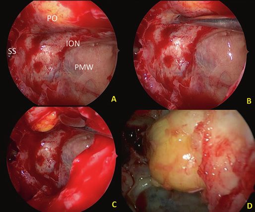
C. Parrilla et al.
Thyroid Eye Disease peutic cornerstone of Active GO. Indeed, in moderate-to-se-
vere active GO, the first line approach consists in intravenous
Orbital involvement in Graves’ disease has a complex methylprednisolone, administrated weekly at initial dose of
pathophysiology, resulting from the deposition of immune 0.5 g for 6 weeks, then 0.25 for other 6 weeks (4.5 g cu-
complexes that cause oedema and fibrosis of the extraocu- mulative dose). This therapeutic scheme appears to have
lar muscles and orbital fat. The retro-orbital pressure in- higher efficacy and fewer adverse effect then glucocorticoids
creases and causes proptosis and exophthalmos, threaten- oral administration. The optimal cumulative dose appears
ing the vision due to vascular impairment or stretching of to be 4.5-5 g of methylprednisolone, but higher doses (up
the optic nerve. to 8 g) can be used for more severe forms 13. Patients with
GO, which is the main extra-thyroidal manifestation of sight-threatening GO, due to Dysthyroid Optic Neuropathy
Graves’ disease, is a rare pathology and still represents a (DON) or severe corneal exposure, should be rapidly treat-
clinical and therapeutic challenge 7. ed with high doses of intravenous glucocorticoids as first-
Although the role of TSH receptor activating autoantibod- line treatment. A commonly used regimen consists of giving
ies (TSHRAb) on orbital adipogenesis has been widely 1 g of intravenous methylprednisolone for three consecutive
demonstrated in literature, the complex pathogenesis of GO days, that can be repeated on the subsequent week. It is im-
is not clear yet. A new promising pathogenetic mechanism portant to strictly control clinical conditions because in case
has been recently discovered, based on an active “cross- of absent or poor response, the patient should undergo orbit-
talk” between TSH-R and IGF-1R in thyrocytes and orbital al decompression within two weeks 9 (Fig. 1, Tab. II).
fibroblast, which lead to activation of an IGF-1R-depend- However, to achieve the reduction of relapse rates and the
ent downstream intracellular pathway. Although GO is pre- optimization of the final results, it seems necessary to re-
sent in about 25% of Graves’ Disease cases, fortunately, the sort to combinations of other current therapies, as the active
sight-threatening variant is a rare occurrence 8. disease could last 1-2 years and often a relapse occurs upon
withdrawal of glucocorticoids 14.
Therapeutic management of TED
According to the recent Guidelines, the therapy of GO has Radiotherapy
to be gauged on two main parameters, Severity and Activ- Orbital radiotherapy, usually with a cumulative dose of
ity, which can be evaluated through validated scores, such 10 to 20 Gy per orbit, can be used as second-line therapy,
as NOSPECS and Clinical Activity Score (CAS) 9. The when GO results to be still active after glucocorticoids
EUGOGO classification defines disease severity as sight- treatment, particularly in case of diplopia and motility
threatening, moderate-to-severe, and mild Graves’ Orbi- disorders. In randomized clinical trials, radiotherapy
topathy 10 (Tab. I). appeared to be as effective as oral glucocorticoid thera-
In case of mild GO, an oral daily dose of 200 mcg of Seleni- py and may have a synergic effect if combined with in-
um appears to be effective as maintenance therapy, although travenous steroids. Although data on safety seem to be
its role in preventing orbitopathy has still to be clarified 11. reassuring, further studies are needed to assess the real
Furthermore, local measures, such as teardrops, ointments long-term effectiveness and safety, because of controver-
or gel, appear to be useful, particularly in alleviating ocular sial for the theoretical concerns about carcinogenesis, es-
dryness or foreign body sensation. If diplopia impacts on the pecially for younger patients, and other side effects such
quality of life, using corrective prisms may be a valid option. as retinopathy and cataract 15-17.
Botulin injection has been tested too, with apparently tran-
sient benefits in reducing eyelid retraction 12. Antiproliferative agents
Intravenous high-dose glucocorticoids represent the thera- Mycophenolate mofetil (MMF) inhibits the proliferative
Table I. Severity classification in Graves’ Orbitopathy, recommendations and levels of evidence (from EUGOGO 10).
Classification Recommendation Level of
evidence
Sight-threatening GO: DON and/or corneal breakdown Immediate intervention IV, C
Moderate-to-severe GO: eye disease with sufficient impact on daily life (lid retraction > 2 Active: immunosuppression IV, C
mm, exophthalmos > 3 mm, moderate or severe soft tissue involvement) Inactive: surgical intervention
Mild GO: minor impact on daily life (minor lid retraction< 2 mm, exophthalmos < 3 mm, mild Local measures to alleviate symptoms IV, C
soft tissue involvement, transient or no diplopia, corneal exposure responsive to lubricants)
DON: Dysthyroid Optic Neuropathy; GO: Graves’ Orbitopathy.
S92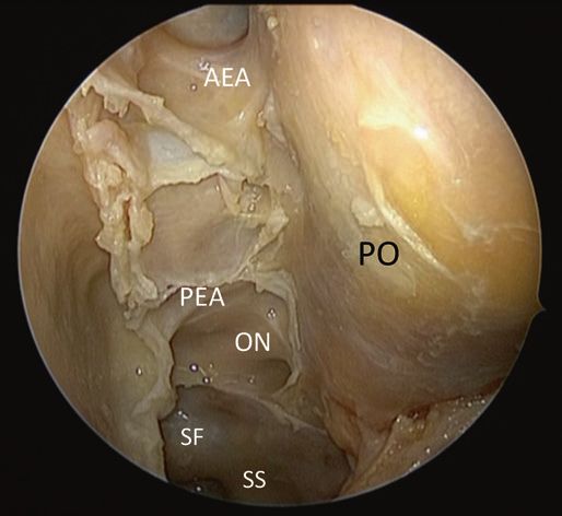
Multidisciplinary management in orbital decompression
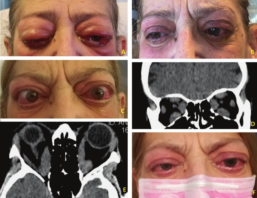
Figure 1. 63-year-old female patient, smoker, affected by active, sight-threatening GO, treated with administration of 1 g of intravenous methylprednisolone
for three consecutive days. (A) Patient before therapy. (B) Two weeks after therapy. After thyroidectomy, a better control of hormonal levels was achieved. (C) Two
months later, her visual acuity worsts to right eye: 4/20 left eye: 3/20 with anomalous colour vision test (dysthyroid optic neuropathy). (D-E) On CT-scans images,
all the EOMs are enlarged with apex crowding. (F) After endoscopic bilateral endonasal optic nerve and medial wall decompression the visual acuity has greatly
improved, and a 4 mm of proptosis reduction.
CT: computed tomography; EOMs: Extraocular muscles; GO: Graves’ Orbitopathy.
responses of T and B lymphocytes and has a mechanism of Tocilizumab, a humanized monoclonal antibody directed
a direct effect on orbital fibroblasts 18; Cyclosporin, inhibits against the IL-6 receptor, showed efficacy in terms of ac-
calcineurin pathway reducing IL-2, that is highly produced tivity reduction 21. A multicentre, randomized, trial versus
by T-cells activated in GO. Both MMF and Cyclosporin placebo, showed that the use of tocilizumab in glucocorti-
showed benefit in ocular symptoms and quality of life 19. coid-resistant GO showed a reduction in Clinical Activity
Methotrexate and Azathioprine have also been evaluated Score (CAS) of at least 2 points, improvement in EUGO-
for utility in GO management as steroid-sparring agents, GO ophthalmic score and a bigger reduction of exophthal-
but more data are needed 20. mos 22. Most adverse effects tend to be mild and transient;
however, a consistent risk of developing opportunistic in-
Targeted therapy fections has been noted during treatment for rheumatoid
Several monoclonal antibodies have been tested as novel arthritis and need to be monitored 23. Rituximab is a chime-
therapeutic options in second-line treatment for Active GO. ric mouse-human monoclonal antibody, which targets the
S93C. Parrilla et al.
Table II. Glucocorticoids and orbital decompression in Dysthyroid Optic Neuropathy (from EUGOGO 10).
Management of DON Level of evidence
GCs and surgical decompression are effective in patients with DON III, B
High-dose i.v. GCs is the preferred first-line treatment for DON III, B
If the response to GCs is absent after 1-2 weeks, prompt orbital decompression should be carried out IV, C
Orbital decompression should be performed promptly in case of DON o corneal breakdown in patients who cannot tolerate GCs III, B
DON: Dysthyroid Optic Neuropathy; GCs: Glucocorticoids; i.v.: intravenous.
B-lymphocyte antigen CD20. Two clinical trials reported restrict the vascular supply to the optic nerve: this is the
contradictory outcomes: no benefit in one trial, when com- most well-accepted mechanism of DON. The diagnosis of
pared with placebo; a significant improvement in CAS, DON remains still challenging and controversial, and pos-
when compared with intravenous steroids, in the other 24,25. sible clinical findings are represented by decreased visual
acuity, a relative afferent pupillary defect, altered colour
Indications for surgery vision, optic disc abnormalities and visual field defects. In
Generally speaking, it is recommended to avoid surgery in this case, the amount of orbital wall removal is particularly
Active TED, because it is evident that surgical procedures critical and should be achieved as complete as possible
during this phase may increase orbital inflammation 26. along the intraorbital portion of the optical nerve 29.
Urgent indications: surgical indication for orbital decom- Extreme eye proptosis can also determine vision loss: per-
pression can be considered in patients with active GO who sistent eyelid retraction caused by persistent inflammation
are nonresponsive or intolerant to glucocorticoids, if wait- and scarring of the eyelid retractors increasing corneal ex-
ing for spontaneous inactivation could be a risk for vision posure, predisposing patients to ulceration and subsequent
loss 27, in case of CON (estimated incidence 3-9% of TED vision compromise.
patients) 28. In fact, extraocular muscle enlargement could Nonurgent indications: orbital decompression for disfigur-
Figure 2. 65-years old, male patient affected by severe TED with proptosis and strabismus. (A-C) Pre-operative CT-scan images and appearance. (D-F) CT-scan
images and appearance after bilateral endoscopic orbital decompression.
CT: computed tomography; TED: Thyroid Eye Disease.
S94Multidisciplinary management in orbital decompression
Table III. Timing and the order for surgery in Graves’ Orbitopathy (from EUGOGO 10).
Timing and the order for surgery Level of evidence
Surgical management should proceed in the sequence: orbital decompression, squint surgery, lid lengthening with blepharoplasty III, B
Rehabilitative surgery should be performed in patients with inactive GO for at least 6 months III, B
GO: Graves’ Orbitopathy.
ing exophthalmos could be deferred until the orbitopathy has This approach started with a sublabial incision in the
been inactive for at least 6 months 27. Other nonurgent indica- oral vestibule mucosa, taking care to not injure the infra-
tions are represented by chronic retrobulbar pain or discom- orbital nerve during the exposition of the anterior wall of
fort, congestion, ocular hypertension and diplopia (Fig. 2). the maxillary sinus. Then an osteoplastic anterior max-
The majority of patients presents a good response to con- illotomy is performed, by the temporary removal of a
servative treatments, only in 5% of cases an orbital decom- bony gusset that is replaced with microplates at the end
pression surgery is demanded in the 1st year after diagnosis, of the procedure. The orbital floor and the lamina papy-
but in the ten years after diagnosis this rate rise up to 20% 28. racea are then exposed and resected, resulting in orbital
In relation to the staging of the surgical ophthalmic proce- decompression and subsequent immediate reduction in
dures for the rehabilitation of the patients affected by the proptosis.
TED, the decompression should be the first procedure per- It is important to preserve a bony bridge together with the
formed, as it can affect ocular motility 30. This is followed infra-orbital canal, avoiding downward displacement of the
by eyelid surgery, which can be affected by both decom- eyeball and subsequent diplopia.
pression and strabismus surgery 31. Some have challenged
this paradigm, suggesting that there is minimal change in Complications
upper eyelid position with decompression or strabismus • hypoesthesia of the cheek, the lateral nasal ala, and the
surgery and that simultaneous upper eyelid and decompres- anterior teeth, due to lesion or temporary stretching of
sion surgery may be performed in order to shorten rehabili- infra-orbital nerve in the bony canal inside the orbital
tation intervals 32 (Tab. III). floor or after its exit from the infraorbital foramen;
There are no absolute surgical contraindications to orbital • paresis of extraocular muscles (inferior rectus, inferior
decompression surgery; patients under anticoagulant treat- oblique, possibly medial and lateral rectus muscles) and
ment should discontinue the assumption preoperatively subsequent diplopia, due to lesions to nerve fibres that
because of the major risk of bleeding intraoperatively that enter the muscles;
could impact negatively on surgery and the risk of post- • haemorrhage due to injury to the infraorbital artery or
operative haemorrhage. When a concomitant paranasal si- infra-orbital branches of the ophthalmic artery;
nuses disease is present (sphenoidal and/or frontal rhinosi- • enophthalmos, if the entire orbital floor is resected.
nusitis), a parallel surgical opening of the involved sinuses
could be addressed even if not needed for decompression Transnasal (Figures 3 and 4)
surgery. Most of the studies show the efficacy and rela- The endoscopic transnasal technique starts performing an
tive safety of orbital decompression 33; however, the avail- uncinectomy and a maxillary antrostomy in order to obtain
able studies do not allow any meaningful comparison of a good exposure of the posterior maxillary wall and orbital
the available approaches 27. Orbital decompression can be floor, and also to avoid obstruction due to inferior dislocation
achieved with different surgical techniques: fat decompres- of orbital fat. Then a complete sphenoid-ethmoidectomy ex-
sion, orbital floor or medial or inferior-medial wall decom- poses the medial orbital wall from the sphenoid sinus down
pression, orbital lateral wall decompression isolated or as- to the crista ethmoidalis and superiorly to the skull base. In
sociated with the other wall decompression 31. some cases, it is possible to realize the resection of the mid-
dle turbinate for better visualization with subsequent cauteri-
Multidisciplinary surgical approaches zation of its postero-lateral remnant to avoid postoperative
bleeding. The second step of the procedure is the removal of
in orbital decompression the bone from the inferior and medial orbital wall.
Transantral The lamina papyracea is then fractured with a blind dis-
Before the endoscopic era, the Walsh-Ogura transantral ap- sector and elevated away from periorbita. Bone is thicker
proach was the gold standard for treatment of exophthal- in the region of the orbital apex in the proximity of the an-
mos in patients with Graves’disease 34. nulus of Zinn, through which the optic nerve passes and
S95C. Parrilla et al.
inferior-medial strut may reduce postoperative diplopia and
help in improving proptosis 35. The next step is the incision
of the periorbita to enable extraconal orbital fat herniation
into the ethmoid and maxillary cavities, avoiding lesions to
the orbital contents, in particular of the medial rectus mus-
cle. In order to decrease the risk of postoperative diplopia, it
could be preserved the periorbital sling that covers the me-
dial rectus muscle, as described by Metson and Samaha 6.
Complications
The more common complications of this kind of surgery
include epistaxis, nasal adhesions, sinusitis, worsening of
pre-existing diplopia or new-onset diplopia; pre-operative
diplopia frequently does not resolve with surgery. Rare
complications are represented by injury to the optic nerve
and ophthalmic artery; otherwise, cerebrospinal fluid leaks
rarely occur 36.
Bony removal, periosteum opening and fat resection
Figure 3. Anatomic dissection showing the periorbit (PO) after complete
spheno-ethmoidectomy with the exposure of the Skull base from the I fovea The techniques and extent of bony removal widely differ in
ethmoidalis, anterior ethmoidal artery (AEA), Posterior ethmoidal artery (PEA); the literature. Currently, for the floor the lateral extent re-
to the sphenoid sinus and the following structures: Optic nerve (ON), Interoptic moval has been limited to the space medial of the infraor-
carotid recess and sellar floor Sphenoid Sinus (SS).
bital nerve 2. Anteriorly, some suggest leaving 10mm of
bone under the globe to prevent hypoglobus 37. The limits
of medial wall removal differ depending on the surgeon and
the clinical features. Anteriorly, the maximal extent is classi-
cally thought to be the posterior lacrimal crest 2. Posteriorly,
most descriptions involve dissection to the anterior wall of
the sphenoid sinus; however, dissection can extend as far as
the optic canal 2. Many endonasal approaches describe canal
decompression as part of the management for DON 38.
Superiorly, most authors describe dissection to the fron-
toethmoidal suture. At this level, it is critical to be aware of
the skull base anatomy, as inadvertent entry into the cranial
cavity through the fovea ethmoidalis has been reported 39.
Inferiorly, the inferior-medial orbital strut separates the
medial and inferior walls and provides structure to the bony
orbital cone. Authors have argued that removing this strut
is important to allow for complete prolapse of the orbital
tissue and maximal decompression. Others have suggested
that leaving the strut (or part of it) intact is vital to avoid
Figure 4. (A-D) Intraoperative view of left orbital decompression with the hypoglobus and important strabismus postoperatively 31,35.
exposure of the sphenoid sinus (SS), Posterior maxillary wall (PMW), infraorbi- The orbital periostium is unelastic and will minimize pro-
tal nerve (ION), with incision of the inferior periorbit and the final fat ernjation. lapse of orbital fat in periorbital spaces. For this reason,
some suggest opening periostium completely after the
bony decompression.
where four extraocular muscles originate. Then, the perior- Other authors have suggested cross-hatching or linear inci-
bita can be elevated off the bony floor of the medial orbit. sions 31.
Only the portion of the floor that is medial to the infraor- Extensive opening of the periosteum has been associated
bital nerve is removed, preserving its canal as the lateral with greater rates of postoperative strabismus 37.
limit of dissection. Moreover, the preservation of a bony Medially, endoscopic surgeons have described the impor-
S96Multidisciplinary management in orbital decompression
tance of leaving a strip of periosteum overlying the medial • sinking of soft tissue above the zygomatic arch because
rectus and the inferior rectus to reduce the rate of eye infe- of incomplete repositioning of the temporalis muscle.
rior prolapse and strabismus 6.
The infraorbital nerve should be spared to avoid permanent Optic nerve decompression
anaesthesia dissecting the orbital floor only medial to the Many studies have reported the efficacy of medial wall de-
nerve or leaving a bony canal and dissecting on either side 31. compression in optic compressive neuropathy. Transcaruncu-
lar, transorbital, transantral and endonasal approaches seem
Lateral wall decompression similar in the rate of DON improvement reported, range 75-
Lateral orbitotomy is an external approach first described 90% 31. The periosteum was described not incised, incised
by Krönlein in 1888 and since then has undergone some in the anteroposterior direction, radially incised. Despite the
modifications. The original approach involved a curvilin- clinical efficacy of the medial wall optic nerve decompres-
ear incision running towards the lateral canthus from the sion, a few studies focus on pressure orbital changes 43.
hairline 31. Stallard and Wright modified the incision with
a more acceptable postoperative scar running through the Discussion
eyebrow 40. Berke in 1953 proposed a canthal splitting lat-
eral incision 41, while the first coronal flap to access lateral Most of the literature about orbital decompression consists
orbit was described in 1982 by Bonavolonta. Today, the of retrospective, cohort or case series studies. These papers
most used approach is the lateral eyelid creased incision provide useful descriptive information, but clarification is
that provides excellent access and hides well in the eyelid required to show the effectiveness of each operation related
crease 42. to different indications.
The skin incision could be performed in the area of the lat- Orbital decompression is achieved by removing bony
eral orbital rim, with possibility of injury to the ramus fron- walls, orbital fat or both. It is an established procedure to
talis and ramus ocularis of the facial nerve, or in alternative, correct exophthalmos for visual improvement in patients
a pterional incision into the hairline could be realized with- with optic neuropathy, corneal involvement and for the re-
out a cosmetic problem. habilitation of patients with marked anterior positioning of
With a pterional incision, a scalp flap is formed and pre- the eyes. Several approaches are described with contradict-
pared until the lateral orbital rim is exposed, preserving the ing results, and the comparison of different surgical tech-
lateral palpebral ligament; temporalis muscle that overlies niques was biased by inclusion of patients at a different
the lateral orbital wall is incised and bluntly pushed away. stage of disease (active or inactive), different surgical in-
Microplates are needed for the reconstruction of the lateral dications (dysthyroid neuropathy or disfiguring proptosis)
orbital rim. With a saw, the bone at the lateral orbital rim and assessment of outcomes (such as a different system for
is now separated. This is followed by the resection of the ocular motility evaluation).
lateral orbital wall including parts of the zygomatic bone The few Randomized Control Trials (RCTs) do not pro-
up to the greater wing of the sphenoid bone in the region of vide robust evidence to support recommendations for clini-
the inferior orbital fissure. cal practice. There is evidence only from available uncon-
For the reconstruction of the lateral orbital wall, the tem- trolled studies that the removal of the medial and lateral
porarily separated bone is repositioned and fixed in place wall (so-called balanced decompression) with or without
by microplates and the temporalis muscle is moved back fat removal could be the most effective surgical technique,
and fixed. It could be necessary to insert drainage before related to few complications 44.
closure layer by layer of the scalp. The removal of the inferior wall through the antrum and
transnasal removal of the medial wall had similar effects
Complications in reducing exophthalmos, but the second approach had a
• blunt shape of the lateral canthus due to detachment of lower rate of complications, such as diplopia and infraorbi-
the lateral palpebral ligament from the orbital rim; tal nerve damage 45.
• impairment of eye globe motility and diplopia because The timing of surgical decompression is another debated
of injury to the lateral rectus and the inferior rectus mus- issue and must be related to the failure of medical therapy.
cles; One of the few RCTs showed how intravenous corticos-
• anisocoria due to injury of the ciliary ganglion (laterally teroids achieve better visual recovery than surgical orbital
to optic nerve), during very extensive medial preparation; decompression (56 vs 17%) 46. Adverse outcomes were re-
• disorder of the mimetic musculature due to injury to the ported more frequently in the steroids group, i.e. weight
ramus frontalis or the ramus orbitalis of the facial nerve; gain and a Cushing-like syndrome, hypertension and tran-
S97C. Parrilla et al. sient diabetes; while side effects of surgery consisted of ment of the extraocular muscle paths, but no one of these transient numbness of the facial skin in 4/14 participants theories has been commonly accepted 49. It has been sug- or decrease in extraocular muscle motility. The beneficial gested that preservation of inferomedial strut and a bal- effect of I.V. steroids on visual rehabilitation would appear anced orbital decompression causes less diplopia and some to overcome the increased number of transient side effects studies show new onset or worsening of diplopia ranges in active TED with optic neuropathy. from 10 to 20% 50-52. In case of urgent orbital decompression, the main outcome Although, some endoscopic surgeons described lower rates is obviously represented by visual acuity: different studies of strabismus with modifications to the periosteal opening have demonstrated high success rates with improvements medially. Additionally, higher rates of over 30% have been in more than 82% of patients 28,33. reported for balanced decompression. These rates could be Medial and inferior-medial wall decompression is advis- technique dependant, both on the side of balanced decom- able in patients with severe posterior optic neuropathy, pression and medial decompression alone 31. caused by apical crowding of the enlarged muscles, in par- Finally, the comparison of induced diplopia rates after dif- ticular of medial rectus. ferent orbital approaches is difficult to perform because of For nonurgent orbital decompression, proptosis is the main many factors: type of surgical indications, different meas- outcome, orbital fat removal alone has been shown to re- urements, the timing of outcome assessment, different cri- duce proptosis up to 4.7 mm, while decompression of the teria to define the condition. medial, inferior, and/or lateral walls could reduce proptosis In the study of Mourits et al. 49, authors used two tools to up to 7.4 mm 28,33. assess diplopia: ophthalmologist and orthoptist and deter- Lateral decompression allows for exophthalmos reduction mined clinically whether or not there was diplopia in any causing less strabismus, especially with the incision of fas- direction of gaze, while patients self-assessed their diplopia cia temporalis. The literature suggests that three-wall de- using the Gorman score. Using these criteria seemed to be compression is chosen for high degrees of proptosis while a tendency for the swinging eyelid approaches to be associ- two-wall for patients with less exophthalmos; fat removal ated with less induced diplopia. In the group of three-wall in addition to bone removal could increase the effective- swinging eyelid decompression, the incidence of diplopia ness of the procedures 44. decreased, and the Gorman score improved. In some stud- The aim of orbital decompression surgery is not to create ies, the subjective response to treatment was measured us- the biggest space with disruption of periorbital structures ing Terwee’s GO-QOL, a validated disease-specific ques- for maximal decompression, but to realize adequate de- tionnaire to assess changes in visual function and changes compression for relief of optic neuropathy or keratopathy in appearance 53. caused by severe proptosis 47. Moreover, cosmesis, was one Various attempts have been made in the literature to study of the most common indications for surgery and represent- radiographic-based and QOL-based outcomes after orbital ed an important quality of life issue for patients 48. decompression, in addition to the evaluation of clinical fea- Several studies suggest that three-wall decompression tures. achieves the greatest reduction in proptosis but that compli- An objective evaluation could be performed by the imag- cations are more frequent; for this reason, more conserva- ing measurements of radiographic-based outcomes; recent tive approach, such as balanced medial and lateral wall or research has been focused on establishing the validity of endoscopic inferior-medial decompression may be prefer- an algorithm to determine various parameters, such as the able choices. The accuracy of measuring proptosis was also measurement of the angle of the orbital apex, diameter of questioned. Although postoperative Hertel measurements the extraocular muscles, exophthalmos and orbital vol- were widely reported, they were inaccurate in particular ume 54-56. when lateral canthotomy is performed, as these were made Unfortunately, the use of radiographic-based outcomes is with a Hertel exophthalmometer using altered reference not assessed, and its specific role in outcome evaluation points, which may result in overestimation of improvement needs to be determined by further studies aimed to stand- in proptosis 47. ardize analyzed parameters. Interestingly, diplopia can be considered both an outcome Probably, in the last years, the most important concept and a complication. In every kind of approach, new-onset about outcome evaluation is the patient’s point of view. For diplopia could be present at different rates 33. this reason, recently, many questionnaires about quality of Many theories have been proposed to understand this phe- life have been developed and validated. One of these is rep- nomenon, for example, removal of the posterior medial resented by Graves Ophthalmopathy Quality of Life (GO- wall, removal of the inferomedial strut, and the displace- QOL) scale that provides vision-related and appearance- S98
Multidisciplinary management in orbital decompression

related scores, which were assessed before, 6 weeks and 6       overlying the medial rectus muscle is an additional techni-
months after surgery 57.                                        cal trick to reduce diplopia.
Improvements in eyelid retraction and congested orbit
did not predict the change in the appearance-related qual-      References
ity of life. As reported in previous studies, the correlation   1
                                                                     Rao R, MacIntosh PW, Yoon MK, et al. Current trends in the manage-
between clinical changes and quality of life outcomes is             ment of thyroid eye disease. Curr Opin Ophthalmol 2015;26:484-490.
weak, also supporting the notion that subjective appraisals          https://doi.org/10.1097/ICU.0000000000000203
of appearance will predict psychological well-being better      2
                                                                     Kennedy DW, Goodstein ML, Miller NR, et al. Endoscop-
than clinical measures of severity. For example, patients            ic transnasal orbital decompression. Arch Otolaryngol - Head
                                                                     Neck Surg 1990;116:275-282. https://doi.org/10.1001/archo-
with strabismus evaluated appearance as more important               tol.1990.01870030039006
than other clinical factors 57.                                 3
                                                                     Yao WC, Sedaghat AR, Yadav P, et al. Orbital decompression in
A recent study has examined the impact of orbital decom-             the endoscopic age: the modified inferomedial orbital strut. Arch
pression surgery on sinonasal-specific QOL evaluated by              Otolaryngol - Head Neck Surg 2016;154:963-969. https://doi.
                                                                     org/10.1177/0194599816630722
SNOT-22 administered preoperatively and one year after          4
                                                                     Wehrmann D, Antisdel JL. An update on endoscopic orbital decom-
surgery, that showed a statistically significant improvement         pression. Curr Opin Otolaryngol Head Neck Surg 2016;25:73-78.
in sinonasal-specific QOL, in particular, improvements               https://doi.org/10.1097/MOO.0000000000000326
have been observed in the psychological domains of the          5
                                                                     Goldberg RA, Shorr N, Cohen MS. The medical orbital strut in the
test, confirming a preserved sino-nasal function even after          prevention of postdecompression dystopia in dysthyroid ophthal-
                                                                     mopathy. Ophthal Plastic Reconstruct Surg 1992;8:32-34. https://doi.
fat prolapse into nasal cavities 58.                                 org/10.1097/00002341-199203000-00005
                                                                6
                                                                     Metson R, Samaha M. Reduction of diplopia following endoscopic orbital
                                                                     decompression: the orbital sling technique. Laryngoscope 2002;112:1753-
Conclusions and key notes                                            1757. https://doi.org/10.1097/00005537-200210000-00008
TED is the most common indication for orbital decom-            7
                                                                     Bartalena L, Burch HB, Burman KD, et al. A 2013 European survey
                                                                     of clinical practice patterns in the management of Graves’ disease.
pression in case of failure of medical therapies. The ur-            Clin Endocrinol (Oxf) 2016;84:115-120. https://doi.org/10.1111/
gent surgical indications are represented by optic neu-              cen.12688
ropathy and severe corneal exposure, while non-urgent           8
                                                                     Perros P, Hegedüs L, Bartalena L, et al. Graves’ orbitopathy as a rare
surgical indications include diplopia, proptosis, orbital            disease in Europe: a European Group on Graves’ Orbitopathy (EU-
                                                                     GOGO) position statement. Orphanet J Rare Dis 2017;12:72. https://
and retrobulbar pain, and ocular hypertension. In the                doi.org/10.1186/s13023-017-0625-1
case of optic neuropathy, bony removal in the region of         9
                                                                     Bartalena L. Graves’ orbitopathy: imperfect treatments for
the orbital apex is mandatory, reducing pressure on the              a rare disease. Eur Thyroid J 2013;2:259-269. https://doi.
optic nerve and leading to improvement in vision in pa-              org/10.1159/000356042
tients with visual loss.                                        10
                                                                     Perros P, Žarković M, Azzolini C, et al. PREGO (presentation of
                                                                     Graves’ orbitopathy) study: changes in referral patterns to European
The main clinical outcomes include visual acuity, propto-            Group On Graves’ Orbitopathy (EUGOGO) centres over the period
sis, and new-onset diplopia.                                         from 2000 to 2012. Br J Ophthalmol 2015;99:1531-1535. https://doi.
Recent studies focused on the development of imaging                 org/10.1136/bjophthalmol-2015-306733
assessment in order to objectively evaluate the surgical        11
                                                                     Marcocci C, Kahaly GJ, Krassas GE, et al. European Group on Graves’
                                                                     Orbitopathy. Selenium and the course of mild Graves’ orbitopathy.
results. QoL questionnaires are gaining increasing impor-            N Engl J Med 2011;19;364:1920-1931 https://doi.org/10.1056/NEJ-
tance, based on patients’ point of view.                             Moa1012985
The goal of orbital decompression is the reduction of           12
                                                                     Grisolia ABD, Couso RC, Matayoshi S, et al. Non-surgical treatment
proptosis, but additional ophthalmological procedures are            for eyelid retraction in thyroid eye disease (TED). Br J Ophthalmol
                                                                     2017 Aug 9;bjophthalmol-2017-310695. https://doi.org/10.1136/
needed to normal eye function and cosmesis. Strabismus               bjophthalmol-2017-310695
surgery to address diplopia and lowering the position of the    13
                                                                     Bartalena L, Baldeschi L, Boboridis K, et al. European Group on
upper eyelid represent some of the additional steps for the          Graves’ orbitopathy (EUGOGO). The 2016 European Thyroid As-
final rehabilitation of GO.                                          sociation/European Group on Graves’ orbitopathy Guidelines for
                                                                     the Management of Graves’ orbitopathy. Eur Thyroid J 2016;5:9-26.
A balanced decompression (combined external and endo-                https://doi.org/10.1159/000443828
scopic or only external approach) or pure endoscopic ap-        14
                                                                     Marcocci C, Bartalena L, Tanda ML, et al. Comparison of the effec-
proach with the preservation of the anterior inferomedial            tiveness and tolerability of intravenous or oral glucocorticoids associ-
orbital strut represents the gold standard to reduce the in-         ated with orbital radiotherapy in the management of severe Graves’
                                                                     ophthalmopathy: results of a prospective, single-blind, randomized
cidence of new-onset diplopia, but RCTs are required to              study. J Clin Endocrinol Metabol 2001;86:3562-3567. https://doi.
confirm these data. Moreover, the orbital sling technique,           org/10.1210/jcem.86.8.7737
obtained by the preservation of a strip of the periorbita       15
                                                                     Tanda ML, Bartalena L. Efficacy and safety of orbital radiotherapy

                                                                                                                                       S99
C. Parrilla et al.
for graves’ orbitopathy. J Clin Endocrinol Metab 2012;97:3857-3865. tion after orbital decompression for thyroid orbitopathy. Ophthal-
https://doi.org/10.1210/jc.2012-2758 mic Plast Reconstr Surg 2017;33:289-293. https://doi.org/10.1097/
16
Marquez SD, Lum BL, McDougall IR, et al. Long-term results of IOP.0000000000000758
irradiation for patients with Graves’ ophthalmopathy. Int J Radiat 31
Rootman DB, Orbital decompression for Thyroid Eye Disease.Sur-
Oncol Biol Phys 2001;51:766-774. https://doi.org/10.1016/s0360- vey Ophthalmol 2018;63:86-104. https://doi.org/ 10.1016/j.survoph-
3016(01)01699-6 thal.2017.03.007
17
Wakelkamp IM, Tan H, Saeed P, et al. Orbital irradiation for Graves’ 32
Ben Simon GJ, Mansury AM, Schwarcz RM, et al. Simultaneous
ophthalmopathy: is it safe? A long-term follow-up study. Oph- orbital decompression and correction of upper eyelid retraction ver-
thalmology 2004;111:1557-1562. https://doi.org/10.1016/j.oph- sus staged procedures in thyroid-related orbitopathy. Ophthalmology
tha.2003.12.054 2005;112:923-932. https://doi.org/10.1016/j.ophtha.2004.12.02833
18
Mazumder AG, Patial V, Singh D. Mycophenolate mofetil contrib- 33
Kingdom TT, Davies BW, Durairaj VD. Orbital decompression for
utes to downregulation of the hippocampal interleukin type 2 and 1β the management of thyroid eye disease: an analysis of outcomes
mediated PI3K/AKT/mTOR pathway hyperactivation and attenuates and complications. Laryngoscope 2015;125:2034-2040. https://doi.
neurobehavioral comorbidities in a rat model of temporal lobe epi- org/10.1002/lary.25320
lepsy. Brain Behav Immun 2019;75:84-93. https://doi.org/10.1016/j. 34
Ogura JH, Walsh TE. The transantral orbital decompression operation
bbi.2018.09.020 for 1506 progressive exophthalmos. Laryngoscope 1962;72:1078-
19
Kahaly GJ, Riedl M, König J, et al. European Group on Graves’ orbit- 1097. https://doi.org/10.1288/00005537-196208000-00009
opathy (EUGOGO). Mycophenolate plus methylprednisolone versus 35
Finn AP, Bleier B, Cestari DM, et al. A retrospective review of or-
methylprednisolone alone in active, moderate-to-severe Graves’ orbit- bital decompression for thyroid orbitopathy with endoscopic pres-
opathy (MINGO): a randomised, observer-masked, multicentre trial. ervation of the inferomedial orbital bone strut. Ophthalmic Plas-
Lancet Diabetes Endocrinol 2018;6:287-298. https://doi.org/10.1016/ tic Reconstruct Surg 2017;33:334-339. https://doi.org/10.1097/
S2213-8587(18)30020-2 IOP.0000000000000782
20
Sipkova Z, Insull EA, David J, et al. Early use of steroid-sparing 36
Sellari-Franceschini S, Dallan I, Bajraktari A, et al. Surgical compli-
agents in the inactivation of moderate-to-severe active thyroid eye
cations in orbital decompression for Graves’ orbitopathy. Acta Otorhi-
disease: a step-down approach. Clin Endocrinol (Oxf) 2018;89:834-
nolaryngol Ital 201636:265-274. https://doi.org/10.14639/0392-
839. https://doi.org/10.1111/cen.13834
100X-1082
21
Pérez-Moreiras JV, Alvarez-López A, Gómez EC. Treatment of 37
Mainville NP, Jordan DR. Effect of orbital decompression on diplo-
active corticosteroid-resistant graves’ orbitopathy. Ophthalmic
pia in thyroid-related orbitopathy. Ophthal Plastic Reconstruct Surg
Plast Reconstr Surg 2014;30:162-167. https://doi.org/10.1097/
2014;30:137-140. https://doi.org/ 10.1097/IOP.0000000000000029
IOP.0000000000000037
38
Schaefer SD, Soliemanzadeh P, Della Rocca D, et al. Endoscopic
22
Perez-Moreiras JV, Gomez-Reino JJ, Maneiro JR, et al. Tocilizumab
and transconjunctival orbital decompression for thyroid-related or-
in Graves orbitopathy Study Group. Efficacy of tocilizumab in pa-
bital apex compression. Laryngoscope 2003;113:508-513. https://doi.
tients with moderate-to-severe corticosteroid-resistant graves orbit-
org/10.1097/00005537-200303000-00021
opathy: a randomized clinical trial. Am J Ophthalmol 2018;195:181-
190. https://doi.org/10.1016/j.ajo.2018.07.038
39
McCormick CD, Bearden WH, Hunts JH, et al. Cerebral vasospasm
and ischemia after orbital decompression for graves ophthalmopa-
23
Yamamoto K, Goto H, Hirao K, et al. Longterm safety of tocili-
thy. Ophthal Plastic Reconstruct Surg 2004;20:347-351. https://doi.
zumab: results from 3 years of follow-up postmarketing surveillance
org/10.1097/01.iop.0000134248.64325.c7
of 5573 patients with rheumatoid arthritis in Japan. J Rheumatol
2015;42:1368-1375. https://doi.org/10.3899/jrheum.141210 40
Stallard HB. The presidential address. The evolution of lateral orbit-
otomy. Trans Ophthalmol Soc UK 1973;93:3-17.
24
Salvi M, Vannucchi G, Currò N, et al. Efficacy of B-cell targeted ther-
apy with rituximab in patients with active moderate to severe Graves’ 41
Berke RN. A modified Kronlein operation. Trans Am Ophthalmol Soc
orbitopathy: a randomized controlled study. J Clin Endocrinol Metab 1953;51:193-231.
2015;100:422-431. https://doi.org/10.1210/jc.2014-3014 42
Sellari-Franceschini S, Lenzi R, Santoro A, et al. Lateral wall orbital
25
Stan MN, Garrity JA, Carranza Leon BG, et al. Randomized con- decompression in graves’ orbitopathy. Int J Oral Maxillofacial Surg
trolled trial of rituximab in patients with Graves’ orbitopathy. J 2010;39:16-20. https://doi.org/10.1016/j.ijom.2009.10.011
Clin Endocrinol Metab 2015;100:432-441. https://doi.org/10.1210/ 43
McCord CD, Putnam JR, Ugland DN. Pressure-volume orbital meas-
jc.2014-2572 urement comparing decompression approaches. Ophthal Plastic
26
Pletcher SD, Sindwani R, Metson R. Endoscopic orbital and optic Reconstruct Surg 1985;1:55-63. https://doi.org/10.1097/00002341-
nerve decompression. Otolaryngol Clini North Am 2006;39:943-958. 198501000-00009
https://doi.org/ 10.1016/j.otc.2006.06.003 44
Boboridis KG, Bunce C. Surgical orbital decompression for thyroid
27
Bartalena L, Baldeschi L, Dickinson A, et al. European Group on eye disease. Cochrane Database System Rev 2011;12:CD007630.
Graves’ Orbitopathy (EUGOGO). Consensus statement of the Eu- https://doi.org/10.1002/14651858.CD007630.pub2
ropean Group on Graves’ orbitopathy (EUGOGO) on management 45
Pliego-Maldonado A, Miranda-Ruiz R, Vargas-Aguayo A, et al. Or-
of GO. Eur J Endocrinol 2008;158:273-285. https://doi.org/10.1530/ bit decompression surgery in patients with exophthalmos caused by
EJE-07-0666 Graves-Basedow disease [Cirugía descompresiva de la órbita en pa-
28
Braun TL, Bhadkamkar MA, Jubbal KT, et al. Orbital decompression cientes con exoftalmos por enfermedad de Graves-Basedow]. Gaceta
for thyroid eye disease. Semin Plastic Surg 2017;31:40-45. https:// Médica de México 2000;136:11-5.
doi.org/10.1055/s-0037-1598192 46
Wakelkamp IM, Baldeschi L, Saeed P, et al. Surgical or medi-
29
Saeed P, Tavakoli Rad S, Bisschop P. Dysthyroid optic neuropathy. cal decompression as a first-line treatment of optic neuropathy
Ophthal Plastic Reconstruct Surg 2018;34:S60-S67. https://doi.org/ in Graves’ ophthalmopathy? A randomized controlled trial. Clin
10.1097/IOP.0000000000001146 Endocrinol 2005;63:323-328. https://doi.org/10.1111/j.1365-
30
Rootman DB, Golan S, Pavlovich P, et al. Postoperative changes 2265.2005.02345.x
in strabismus, ductions, exophthalmometry, and eyelid retrac- 47
Leong SC, Karkos PD, MacEwen CJ, et al. A systematic review of
S100Multidisciplinary management in orbital decompression
outcomes following surgical decompression for dysthyroid orbit- ophthalmopathy: the GO-QOL. Br J Ophthalmol 1998;82:773-779.
opathy. Laryngoscope 2009;119:1106-1115. https://doi.org/10.1002/ https://doi.org/10.1136/bjo.82.7.773
lary.20213 54
Kang EM, Yoon JS. Clinical and radiological characteristics of
48
Tehrani M, Krummenauer F, Mann WJ, et al. Disease-specific as- Graves’ orbitopathy patients showing spontaneous decompression. J
sessment of quality of life after decompression surgery for Graves’ Cranio-Maxillo-Facial Surg 2015;43:48-52. https://doi.org/10.1016/j.
ophthalmopathy. Eur J Ophthalmol 2004;14:193-199. https://doi. jcms.2014.10.008
org/10.5301/EJO.2008.2576 55
Thapa S, Gupta AK, Gupta A, et al. Proptosis reduction by clinical
49
Mourits MP, Bijl H, Altea MA, et al. (EUGOGO) outcome of orbit- vs radiological modalities and medial vs inferomedial approaches:
al decompression for disfiguring proptosis in patients with Graves’ comparison following endoscopic transnasal orbital decompression
orbitopathy using various surgical procedures. Br J Ophthalmol in patients with dysthyroid orbitopathy. JAMA Otolaryngology -
2009;93:1518-1523. https://doi.org/10.1136/bjo.2008.149302 Head Neck Surg 2015;141:329-334. https://doi.org/10.1001/jamao-
50
Kacker A, Kazim M, Murphy M, et al. ‘‘Balanced’’ orbital decom- to.2014.3659
pression for severe Graves’ orbitopathy: technique with treatment al- 56
Schiff BA, McMullen CP, Farinhas J, et al. Use of computed tomogra-
gorithm. Otolaryngol Head Neck Surg 2003;182:228-235. https://doi. phy to assess volume change after endoscopic orbital decompression
org/10.1067/mhn.2003.61 for Graves’ ophthalmopathy. Am J Otolaryngol 2015;36:729-735.
51
Sellari-Franceschini S, Berrettini S, Santoro A, et al. Orbital de- https://doi.org/10.1016/j.amjoto.2015.06.005
compression in Graves’ ophthalmopathy by medial and lateral wall 57
Wickwar S, McBain H, Ezra DG, et al. The psychosocial and clinical
decompression. Otolaryngol Head Neck Surg 2005;133:185-189. outcomes of orbital decompression surgery for thyroid eye disease and
https://doi.org/10.1016/j.otohns.2005.02.006 predictors of change in quality of life. Ophthalmology 2015;122:2568-
52
Sellari-Franceschini S, Berrettini S, Forli F, et al. Orbital decom- 2576.e1. https://doi.org/10.1016/j.ophtha.2015.08.030
pression in Grave’s disease: comparison of techniques. Acta Otorhi- 58
Mueller SK, Miyake MM, Lefebvre DR, et al. Long-term impact
nolaryngol Ital 1999;19:307-314. of endoscopic orbital decompression on sinonasal-specific quality
53
Terwee CB, Gerding MN, Dekker FW, et al. Development of a dis- of life. Laryngoscope 2018;128:785-788. https://doi.org/10.1002/
ease specific quality of life questionnaire for patients with Graves’ lary.26812
S101You can also read