AMERICAN VENOUS FORUM AND SOCIETY FOR VASCULAR SURGERY DOCUMENT
←
→
Page content transcription
If your browser does not render page correctly, please read the page content below
AMERICAN VENOUS FORUM AND SOCIETY FOR
                        VASCULAR SURGERY DOCUMENT

          Editors’ Choice

          Classification and treatment of endothermal heat-induced
          thrombosis: Recommendations from the American Venous
          Forum and the Society for Vascular Surgery
          Lowell S. Kabnick, MD,a Mikel Sadek, MD,b Haraldur Bjarnason, MD,c Dawn M. Coleman, MD,d
          Ellen D. Dillavou, MD,e Anil P. Hingorani, MD,f Brajesh K. Lal, MD,g Peter F. Lawrence, MD,h
          Rafael D. Malgor, MD,i and Alessandra Puggioni, MD,j Morristown, NJ; New York and Brooklyn, NY; Rochester, Minn;
          Ann Arbor, Mich; Durham, NC; Baltimore, Md; Los Angeles, Calif; Aurora, Colo; and Scottsdale, Ariz

          ABSTRACT
          The American Venous Forum (AVF) and the Society for Vascular Surgery set forth these guidelines for the management of
          endothermal heat-induced thrombosis (EHIT). The guidelines serve to compile the body of literature on EHIT and to put
          forth evidence-based recommendations. The guidelines are divided into the following categories: classification of EHIT,
          risk factors and prevention, and treatment of EHIT. One major feature is to standardize the reporting under one classi-
          fication system. The Kabnick and Lawrence classification systems are now combined into the AVF EHIT classification
          system. The novel classification system affords standardization in reporting but also allows continued combined evalu-
          ation with the current body of literature. Recommendations codify the use of duplex ultrasound for the diagnosis of EHIT.
          Risk factor assessments and methods of prevention including mechanical prophylaxis, chemical prophylaxis, and abla-
          tion distance are discussed. Treatment guidelines are tailored to the AVF EHIT class (ie, I, II, III, IV). Reference is made to
          the use of surveillance, antiplatelet therapy, and anticoagulants as deemed indicated, and the recommendations
          incorporate the use of the novel direct oral anticoagulants. Last, EHIT management as it relates to the great and small
          saphenous veins is discussed. (J Vasc Surg: Venous and Lym Dis 2021;9:6-22.)

SUMMARY                                                                                 Guideline 1.3: Kabnick classification system. We sug-
                                                                                      gest consideration of the Kabnick classification for
Classification of endothermal heat-induced thrombosis
                                                                                      reporting of EHIT at the saphenofemoral (great saphe-
(EHIT)
                                                                                      nous vein [GSV]) or saphenopopliteal (small saphenous
  Guideline 1.1: Classification system for EHIT. We sug-                               vein [SSV]) junction. [BEST PRACTICE]
gest the use of a classification system to standardize                                   Guideline 1.4: Lawrence classification system. We sug-
the diagnosis, reporting, and treatment of EHIT. [BEST                                gest consideration of the Lawrence classification for
PRACTICE]                                                                             reporting of EHIT at the saphenofemoral (GSV) or saphe-
  Guideline 1.2: Classification system based on duplex                                 nopopliteal (SSV) junction. [BEST PRACTICE]
ultrasound. We suggest that venous duplex ultrasound                                    Guideline 1.5: American Venous Forum EHIT classifi-
with the patient in the upright position, performed                                   cation system. We suggest preferential use of the uni-
within 1 week of the index procedure, forms the basis                                 fied American Venous Forum EHIT classification system
for the classification system. [BEST PRACTICE]                                         to standardize ongoing reporting, given that it maintains

From the Atlantic Health System, Morristown Medical Center, Kabnick Vein              This paper has been co-published in the Journal of Vascular Surgery: Venous
    Center, Morristowna; the Division of Vascular Surgery, NYU Langone Health,          and Lymphatic Disorders (DOI: 10.1016/j.jvsv.2020.06.008) and Phlebology
    New Yorkb; the Division of Vascular and Interventional Radiology, Mayo Clinic,      ([DOI: 10.1177/0268355520953759). The publications are identical except for
    Rochesterc; the Section of Vascular Surgery, Department of Surgery, Univer-         minor stylistic and spelling differences in keeping with each journal’s style.
    sity of Michigan, Ann Arbord; the Division of Vascular Surgery, Duke University   Correspondence: Lowell S. Kabnick, MD, Atlantic Health System, Morristown
    Medical Center, Durhame; the Division of Vascular Surgery, NYU Langone              Medical Center, 95 Madison Ave, Morristown, NJ 07960 (e-mail: lsk@
    HospitaldBrooklyn, Brooklynf; the Center for Vascular Research and Depart-          lowellkabnickmd.com).
    ment of Vascular Surgery, University of Maryland, and the Vascular Service,       The editors and reviewers of this article have no relevant financial relationships to
    Baltimore VA Medical Center, Baltimoreg; the Department of Surgery, Divi-           disclose per the Journal policy that requires reviewers to decline review of any
    sion of Vascular Surgery, David Geffen School of Medicine at UCLA, Los Ange-        manuscript for which they may have a conflict of interest.
    lesh; the Division of Vascular Surgery and Endovascular Therapy, The              2213-333X
    University of Colorado, Anschutz Medical Center, Aurorai; and the Yourveins       Copyright Ó 2020 by the Society for Vascular Surgery, the American Venous
    of AZ, Scottsdale.j                                                                 Forum and the Authors
Author conflict of interest: L.S.K. and E.D.D are consultants for AngioDynamics.       https://doi.org/10.1016/j.jvsv.2020.06.008

6
Journal of Vascular Surgery: Venous and Lymphatic Disorders Kabnick et al 7
Volume 9, Number 1
the essence of the Kabnick and Lawrence classification deep venous thrombosis. [GRADE - 1; LEVEL OF EVI-
systems, remains recognizable, and may be used for DENCE - A]
ongoing meta-analyses and systematic reviews. It is a
Management of SSV
four-tiered classification: I, junction; II, 50% lumen; IV, occlusive deep venous thrombosis.
suggest that management and treatment for EHIT as it re-
[BEST PRACTICE]
lates to the SSV parallel those for the GSV. [GRADE - 2;
Risk factors and prevention LEVEL OF EVIDENCE - C]
Guideline 2.1: Risk factors for EHIT. Some possible but
inconsistent predictors or risk factors for EHIT include
large GSV diameter, previous history of venous throm-
INTRODUCTION AND RATIONALE
Western data suggest that chronic venous insufficiency
boembolic disease, and male sex. These may be
has a significant impact on the population, both quanti-
considered in the preprocedure phase, but the evi-
tatively and qualitatively.1 Chronic venous insufficiency
dence is inconsistent. [GRADE - 2; LEVEL OF EVI-
ranges in presentation from the asymptomatic state to
DENCE - C]
varicose veins, edema, skin changes, and ulceration. Vari-
Guideline 2.2: Prevention of EHIT with chemical pro-
cose veins are found in upward of 20% to 30%, skin
phylaxis. The use of chemical prophylaxis for prevention
changes in up to 6%, and active venous ulcerations in
of EHIT should be tailored to the patient after an assess-
up to 0.5% of the population.2,3 Clinical presentation is
ment of the risks, benefits, and alternatives. [GRADE - 2;
also coupled with variable impacts on quality of life
LEVEL OF EVIDENCE - C]
ranging from cosmetic concerns to debilitating symp-
Guideline 2.3: Prevention of EHIT with mechanical
toms and limb- and life-threatening complications.4-7
prophylaxis. The use of mechanical prophylaxis for pre-
Endothermal ablation revolutionized the treatment of
vention of EHIT should be tailored to the patient after
clinically significant superficial venous reflux. The tech-
an assessment of the risks, benefits, and alternatives.
nologies that have undergone the most robust evalua-
[GRADE - 2; LEVEL OF EVIDENCE - C]
tion are endovenous laser ablation (EVLA) and
Guideline 2.4: Prevention of EHIT by increasing abla-
radiofrequency ablation (RFA). They have been proven
tion distance. There is a trend toward decreased EHIT
safe, efficacious, and durable.8-12 Performed with tumes-
when ablation is initiated >2.5 cm from the saphenofe-
cent anesthesia, RFA and EVLA allow a transition of care
moral (GSV) or saphenopopliteal (SSV) junction. [GRADE
to the ambulatory setting. Moreover, these techniques
- 2; LEVEL OF EVIDENCE - C]
demonstrate improved periprocedural outcomes as
Treatment of EHIT well as a more rapid return to work compared with sur-
Guideline 3.1: Classification system. We suggest the gical stripping.13-15
stratification of treatment based on an accepted EHIT In an early report, Hingorani et al16 observed that
classification system. [BEST PRACTICE] endovenous thermal ablations were associated with
Guideline 3.2: Treatment for EHIT I. We suggest no deep venous thrombosis (DVT) of the common femoral
treatment or surveillance for EHIT I. [GRADE - 2; LEVEL vein on postprocedure surveillance ultrasound. Other
OF EVIDENCE - C] reports from the early 2000s also indicated an
Guideline 3.3: Treatment for EHIT II. We suggest no increased risk of DVT that ranged between 0% and
treatment of EHIT II but do suggest weekly surveillance 8%.17-19 Later publications started referring to these
until thrombus resolution. In high-risk patients, consider- postoperative thrombi as thrombus extension rather
ation may be given to antiplatelet therapy vs prophylactic than DVT as it was believed that they represented a
or therapeutic anticoagulation with weekly surveillance. distinct phenomenon.20,21
Treatment would cease after thrombus retraction or res- Although the occurrence of superficial thrombus within
olution to the saphenofemoral (GSV) or saphenopopliteal the treated vein segment is considered to be a normal
(SSV) junction. [GRADE - 2; LEVEL OF EVIDENCE - C] ultrasound finding, its propagation into a deep vein
Guideline 3.4: Treatment for EHIT III. We suggest treat- may pose a risk for the development of symptomatic
ment with therapeutic anticoagulation for EHIT III, DVT and pulmonary embolism (PE).19,22
weekly surveillance, and cessation of treatment after In 2006, Kabnick first introduced the term endother-
thrombus retraction or resolution to the saphenofemoral mal-heat induced thrombosis (EHIT), defining it as the
(GSV) or saphenopopliteal (SSV) junction. [GRADE - 1; propagation of thrombus into the deep vein contiguous
LEVEL OF EVIDENCE - B] with the ablated superficial vein.23 This definition has
Guideline 3.5: Treatment for EHIT IV. We suggest that been widely adopted to describe this clinical entity.
treatment should be individualized, taking into account From a diagnostic and clinical standpoint, EHIT is an en-
the risks and benefits to the patient. Reference may be tity separate from classic DVT. EHIT, for the most part, has
made to the Chest guidelines for the treatment of a distinct sonographic appearance, behaves like a stable8 Kabnick et al Journal of Vascular Surgery: Venous and Lymphatic Disorders
January 2021
thrombus, and often regresses spontaneously after a few coagulation cascade during endothermal ablation at a
weeks of observation or a short course of remote location.33
anticoagulation.23 The sensitivity of ultrasound for diagnosis of DVT
Contemporary reported EHIT rates after endovenous varies widely, particularly for below-knee duplex ultra-
ablation range from 0% to 3%.24,25 Most EHITs are sound scans. It is possible that the incidence of calf
asymptomatic, and the diagnosis is usually made on DVT after endovenous ablations is higher than re-
routine duplex ultrasound follow-up; however, the pres- ported, and it may account for some cases of PE of un-
ence of a thrombus at the junction or a history of recent known source. Whereas a clear distinction between
endothermal venous ablation has been associated with EHIT and non-EHIT DVT should be made on the basis
rare cases of PE.18,19,22,26 Typically, these thrombi are of anatomic location as discussed before, it is unclear
detected by postprocedure duplex ultrasound examina- whether any pathologic differentiation can be estab-
tions performed anywhere from 24 to 72 hours to 1 to lished on the basis of ultrasound appearance of the
2 weeks after the procedure, depending on the local ul- thrombus.
trasound surveillance protocol. They appear as a hypere- In an animal study comparing histologic specimens of
chogenic, noncompressible area with abnormal venous veins with classic DVT and those with EHIT after RFA, it
flow and augmentation involving the saphenofemoral was demonstrated that EHIT displays a significantly
or saphenopopliteal junction after great saphenous vein higher hypercellular response, fibroblastic reaction, and
(GSV) or small saphenous vein (SSV) ablations, edema. Also, when authors examined the two groups,
respectively.23,27,28 thrombi in EHIT animals were more echogenic
Although the occurrence of EHIT is attributed to an compared with their DVT counterparts.33,34 Preliminary
actual thermomechanical event, that is, the presence of human studies have confirmed these ultrasound find-
a catheter delivering thermal energy in proximity to a ings as EHIT appears more echogenic and displays a
deep vein, the exact differences between RFA and mildly echoreflective thrombus that distinguishes EHIT
EVLA in terms of mechanism of excessive thrombus for- from the usual echolucent characteristics of classic acute
mation are unknown. Whereas EHIT is considered DVT.35
anatomically a form of DVT, its clinical course is more It is currently believed that most EHITs develop within
benign than an unprovoked DVT or one occurring in a 72 hours, but postprocedure surveillance ultrasound
remote vein segment. scans may occasionally identify an EHIT after 7 days
In reporting of thrombotic complications after venous and even up to 4 weeks after endovenous ablation.31,34-36
ablation, it is important to consider the full spectrum of As timing of occurrence is not fully understood, a contro-
findings captured by surveillance ultrasound. The major- versial point is whether an EHIT occurring more than
ity of EHIT reports aim to describe those thrombi pro- 1 week after ablation should be regarded and treated
truding into the common femoral vein or the popliteal as an EHIT or as a classic DVT.37,38
vein. However, when deep calf thrombi are identified In a prospective study by Lurie and Kistner31 of patients
on postprocedure venous ultrasound, they may still be undergoing RFA of the GSV, levels of C-reactive protein
considered EHIT if the thrombus extends into a calf and D-dimer were measured before and after treatment.
vein from a treated perforator, a treated SSV directly Both markers significantly increased at 24 to 36 hours
draining into a gastrocnemius vein, or a treated below- and returned to the baseline values at 1 month after
knee GSV through a perforator.29,30 the treatment, thus indicating that after venous surgical
Examples of non-EHIT DVT include a thrombus in a trauma, both inflammation and hemostatic activation
deep vein nonadjacent to the saphenofemoral junction are present for a prolonged time. Given this evidence,
after GSV ablation, a thrombus remote from the saphe- the practitioner can assume that any thrombus occur-
nopopliteal junction after SSV ablation, a remote calf ring at the site of endovenous ablation within 30 days
vein thrombus after GSV ablation, and a DVT in the of the procedure could be directly or indirectly related
contralateral limb. Both types of DVT, EHIT and non- to the procedure itself.
EHIT, may be present in the same patient.22,31 Some authors have introduced the broader term post-
Based on current literature, practitioners report that the ablation superficial thrombus extension to indicate a
overall rate of DVT after endovenous ablations isJournal of Vascular Surgery: Venous and Lymphatic Disorders Kabnick et al 9
Volume 9, Number 1
In an effort to provide clinical guidelines for the man- sonographic features and progression of all these thrombi
agement of thromboembolic events occurring after at follow-up ultrasound examinations should be reported.
endovenous thermal ablation and keeping in mind Non-EHIT thrombotic events that occur during thermal
that any of these events may potentially lead to serious ablation are likely to be triggered by systemic factors that
consequences, such as PE, we recommend the definition have more to do with an acquired prothrombotic state
of the following entities: than with the thermal energy itself. Therefore, the pres-
ence of thrombotic events other than EHIT must be
EHIT: any thrombus detected by ultrasound within
4 weeks of endovenous thermal ablation originating also recognized and reported.
from the treated vein and protruding into a deep vein.
Non-EHIT DVT: a DVT occurring in a venous segment not
contiguous with the thermally ablated vein. METHODOLOGY
Postablation superficial venous thrombosis: presence of The American Venous Forum (AVF) guidelines commit-
thrombus in a superficial vein other than the treated tee in collaboration with the Society for Vascular Surgery
vein. This vein may or may not be contiguous with the created a writing group to analyze the available literature
ablated vein. on EHIT to gauge the quality of clinical evidence and to
provide guidance on its diagnosis and treatment. A total
We recommend that future reports on thromboembolic of four subgroups were tasked to accomplish the
events after endovenous thermal ablation include following: to establish the EHIT definition, to discuss the
detailed data on anatomic location, clinical presentation, available EHIT classification systems, to evaluate preven-
and time of occurrence of these events to validate or to tion strategies and its risk factors, and to appraise treat-
update the current proposed definitions. Ideally, detailed ment options.
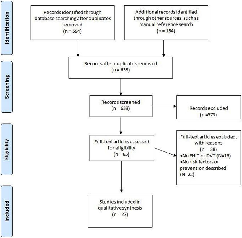
Fig. Preferred Reporting Items for Systematic Reviews and Meta-Analyses flow chart. DVT, Deep venous throm-
bosis; EHIT, endovenous heat-induced thrombosis.10 Kabnick et al Journal of Vascular Surgery: Venous and Lymphatic Disorders
January 2021
A systematic literature review of four scientific reposi- explicit association with endothermal ablation had not
tories was performed, including PubMed, Embase yet been made. In spite of the differences between clas-
(Excerpta Medica Database), Cochrane libraries, and sification systems, the similarities are significant, which
Web of Sciences, to identify potential publications may allow their unification into a single system. The suc-
related to EHIT. The terms used in this review were pri- cess of any proposed unified classification system is
marily related to the adverse outcome studied, EHIT in predicated on delineating clinically significant grada-
patients undergoing either laser or radiofrequency tions of the disease being reported. Ultimately, a unified
venous ablation. However, related terms, such as DVT EHIT classification will help standardize reporting of the
and superficial thrombophlebitis (STP), were also used disease in the literature as well as in clinical practice.
during our search, based on the lack of a clear definition The goals of the proposed EHIT classification system are
of EHIT before 2006. Procedures performed to ablate the as follows:
GSV, SSV, and accessory saphenous veins were included.
1. to provide a standardized classification for vascular
Endovenous ablation of perforating veins was excluded. laboratory reporting of EHIT;
There was no restriction regarding language or research 2. to provide a single classification system for EHIT in
design (Fig). developing practice guidelines regarding the timing
The Grading of Recommendations Assessment, Devel- of duplex ultrasound, technique of duplex ultrasound,
opment, and Evaluation (GRADE) system was chosen to and imaging characteristics;
gauge the quality of published evidence and to rank 3. to provide a uniform classification system for data
the strength of recommendations.40 This grading sys- reporting and research; and
tem comprises four categories of recommendations 4. to allow the possibility of the application of the classi-
paired with a classification of recommendations as fication system to be expanded to nonthermal abla-
tion modalities.
strong or weak to aid health care providers in recom-
mending a specific workup or treatment strategy. Classification system prerequisites
Grade 1 recommendations differ from grade 2 on the
basis of the balance between risks and benefits of a 1. Although different imaging systems (computed to-
mography, magnetic resonance venography) may be
practice. Grade 1 recommendations rely on outcomes
used for the classification of EHIT, duplex ultrasound
that show the benefits involved in a certain practice
should serve as the foundation. It is the “gold stan-
clearly outweigh its risks. Conversely, grade 2 recom- dard” for evaluating the peripheral venous anatomy,
mendations show proximity between risks and benefits and it is the most readily available in outpatient
of a practice that requires further discussion between venous treatment centers.34 The diagnostic ultra-
provider and patient regarding whether a test or treat- sound should be performed within 1 week of the in-
ment should be performed according to the patient’s dex procedure.41,42 The data suggest that most
specific clinical scenario. The grades of recommenda- EHITs develop within 72 hours, but postprocedure
tion rely on three distinct categories used to gauge level surveillance ultrasound scans have identified an
of clinical evidence (A, high quality; B, moderate quality; EHIT up to 4 weeks after endovenous ablation.31,34-36
and C, low quality). The GRADE system has been previ- The diagnostic duplex ultrasound examination can
be performed in either the supine or standing posi-
ously used by the Society for Vascular Surgery; further
tion, although there is a greater incidence of false-
information on this system has been published
positive results in the supine position. Therefore, all
elsewhere.40 identified EHITs should be confirmed in the standing
position, or supine on a tilt table, to ensure that the
CLASSIFICATION OF EHIT
thrombus does not retract peripherally into the super-
Guideline 1.1: Classification system for EHIT ficial vein lumen, thereby changing the diagnosis.
We suggest the use of a classification system to stan- Measurements should be taken with an electronic
cursor in transverse, axial, and orthogonal positions
dardize the diagnosis, reporting, and treatment of EHIT.
to determine the distance and relationship between
[BEST PRACTICE]
the EHIT thrombus and the vein wall as well as the
Guideline 1.2: Classification system based on duplex presence, absence, and extent of protrusion into the
ultrasound deep system lumen.
2. We recommend that the imaging study be conduct-
We suggest that venous duplex ultrasound with the pa-
ed in an accredited vascular laboratory (eg, Intersoci-
tient in the upright position, performed within 1 week of
etal Accreditation Commission, American College of
the index procedure, forms the basis for the classification Radiology Ultrasound Accreditation, and others) by a
system. [BEST PRACTICE] technologist who is trained in duplex ultrasound
Ultrasound-based classification systems have been and can obtain images that accurately identify the ef-
developed for EHIT, but there is a clear lack of standard- fect of the endovenous thermal procedure on the
ization among the systems. Moreover, the initial report- treated vein and vein wall at or near the junction of
ing of the entity was in the context of DVT, and the the superficial axial vein within the deep venousJournal of Vascular Surgery: Venous and Lymphatic Disorders Kabnick et al 11
Volume 9, Number 1
Table I. Kabnick endothermal heat-induced thrombosis EHIT I refers to a benign condition whereby management
(EHIT) classification is not altered. The thrombus propagation remains periph-
Class Definition eral to the associated deep vein, and no further treatment
I Thrombus extended up to and is required. In much of the early literature, this entity was
including the deep vein junction being combined with more significant propagation of the
II Thrombus propagation into the thrombus, thereby resulting in falsely elevated incidence
adjacent deep vein but of disease.16,20 Moreover, there have not been any reported
comprising 50% of the deep vein lumen
gulation until thrombus regression to antiplatelet treat-
IV Occlusive deep vein thrombus
contiguous with the treated
ment until thrombus regression and continued
superficial vein observation.20,23,43 This is an area that warrants ongoing
study and characterization.
EHIT III comprises a more severe form of nonocclusive
thrombosis, and most practitioners are in agreement to
system. This will typically occur at the GSV/common
femoral vein junction and the SSV/popliteal vein junc- treat with an antiplatelet or anticoagulant. Interestingly,
tion; however, EHIT can also occur at any junction be- thisisanexceedinglyraredesignation,giventhatmostEHITs
tween the superficial and deep venous systems after are small and may be classified as an EHIT II, or they present
an endovenous thermal ablation procedure. at the other extreme, which is an occlusive DVT or EHIT IV.
3. The key to the classification system’s being clinically The current consensus is that EHIT IV is treated as an acute
relevant is to determine whether a thrombus has occlusive DVT according to the Chest guidelines.44 Given
protruded into the deep venous system as well as the low-morbidity nature of the treatment, EHIT IVs are
the extent of the protrusion. For example, one of seldom identified in the contemporary literature.
the classification systems allows determination of The Lawrence classification system is as follows
the exact site of the thrombus and vein closure in
(Table II).26 Levels 1, 2, and 3 are encompassed by Kabnick
the superficial system relative to the superficial
EHIT I. In the stated reference, no further treatment was
epigastric vein. This may be used for future out-
comes studies of symptom relief or recurrence, for recommended for EHITs that progressed to level 1 or
example; however, there is no known clinical level 2. A level 3 EHIT was treated according to the discre-
outcome or treatment modification that correlates tion of the operator. Level 3 applied only to 4.3% of the
with this anatomic boundary. On the other hand, patient cohort, and treatment with anticoagulation vs
an occlusion of the adjacent deep vein lumen observation demonstrated no differences in outcomes,
should be treated as a DVT. nor was there any instance of further thrombus exten-
sion. No definitive conclusions could be made on the ba-
Current classification systems sis of the low sampling.
Guideline 1.3: Kabnick classification system. We sug- Levels 4, 5, and 6 roughly correlate to Kabnick EHIT II, III,
gest consideration of the Kabnick classification for and IV. Treatment with anticoagulation resulted in
reporting of EHIT at the saphenofemoral (GSV) or saphe- regression of thrombus in all cases of level 4 or level 5
nopopliteal (SSV) junction. [BEST PRACTICE] EHIT to a level 2 or level 3 EHIT, and this occurred within
Guideline 1.4: Lawrence classification system. We sug- an average of 16 days. As with most of the literature, there
gest consideration of the Lawrence classification for were no instances of an occlusive thrombus (level 6).
reporting of EHIT at the saphenofemoral (GSV) or saphe- Consistent with the Kabnick EHIT classification, clinically
nopopliteal (SSV) junction. [BEST PRACTICE] significant alterations in management occur when the
Guideline 1.5: AVF EHIT classification system. We sug- thrombus extends into the respective deep vein lumen.
gest preferential use of the unified AVF EHIT classification In this sense, levels 4, 5, and 6 serve the same purpose
system to standardize ongoing reporting, given that it as Kabnick EHIT II, III, and IV, with the lower gradations
maintains the essence of the Kabnick and Lawrence clas- being an anatomic characterization of benign disease
sification systems, remains recognizable, and may be used that may benefit from further research.
for ongoing meta-analyses and systematic reviews. It is a The Harlander-Locke classification system was devised
four-tiered classification: I, junction; II, 50% lumen; IV, occlusive DVT. [BEST PRACTICE] creating a supplemental scheme for the SSV relates to
The EHIT classification systems that have gained trac- the variability in anatomy associated with the sapheno-
tion in the literature are as follows. The first described popliteal junction.45 Much like the prior classification
classification system is the Kabnick classification schemes, a distinction is made between thrombus prop-
(Table I).23 agation into the popliteal vein and thrombus that12 Kabnick et al Journal of Vascular Surgery: Venous and Lymphatic Disorders
January 2021
Table II. Lawrence endothermal heat-induced throm- Specifically, EHIT I refers to a benign condition whereby
bosis (EHIT) classification management is not altered. It is unknown whether
Level Definition termination of the thrombus peripheral or central to
1 Thrombus extension that remains peripheral to the superficial epigastric vein bears any clinical signifi-
the epigastric vein cance with regard to symptoms or overall prognosis.
2 Thrombus extension that is flush with the orifice Therefore, to maintain this data point for research pur-
of the epigastric vein poses, there is an (a) and (b) subdivision, which allows
3 Thrombus extension that is flush with the future study and evaluation.
saphenofemoral junction EHIT II remains the most commonly identified of the
4 Thrombus bulging into the CFV various categories. Treatment recommendations have
5 Thrombus bulging into the CFV and adherent to varied from anticoagulation until thrombus regression to
the wall of the CFV past the saphenofemoral antiplatelet medication until thrombus regression and
junction even observation with serial duplex ultrasound examina-
6 Thrombus extension into the CFV consistent tions. This is an area that warrants continued study.
with a DVT EHIT III comprises a more severe form of nonocclusive
CFV, Common femoral vein; DVT, deep venous thrombosis. thrombosis, and most are in agreement to treat with
an antiplatelet or anticoagulant. The consensus currently
remains within the SSV. The cutoff in this instance is be- is that all EHIT IVs are treated as acute occlusive DVTs ac-
tween class B and class C, and there are no further grada- cording to the Chest guidelines.
tions with regard to DVT unless an occlusive thrombus is Conclusions
identified (class D). In this particular study, asymptomatic The reporting of the EHIT phenomenon in a consis-
patients were not evaluated by duplex ultrasound. More- tent way is essential to all other aspects of diagnosis,
over, classes C and D comprised only two patients, prevention, and treatment. To a great extent, this has
rendering it challenging to generalize any conclusions. occurred already with the classification schemes that
Use of the current classification schemes have been created, and there has been a commensu-
To date, these classification schemes have been used rate improvement in the consistency of the associ-
inconsistently across the literature. A sampling of the ated literature. With the increased volume of
literature with the respective classifications used illus- procedures being performed, the data being acquired
trates this (Table IV). (especially within national databases such as the
Vascular Quality Initiative), and the advent of wide-
Unified AVF EHIT classification spread use of the nonthermal ablation techniques,
Given the heterogeneity in reporting and outcomes, the the importance of a consistent classification will in-
authors propose to combine the classification systems crease accordingly.
accordingly (Table V). The new classification system is
based on previously published data, and therefore the RISK FACTORS AND PREVENTION OF EHIT
essence of the classification system has remained un-
Risk factors
changed. Having noted this, it includes definitions that
Guideline 2.1: Risk factors for EHIT. Some possible but
are broad enough to encompass the necessary disease
inconsistent predictors or risk factors for EHIT include
for both research and clinical purposes. Last, it remains
large GSV diameter, previous history of venous throm-
simple, recognizable, and consistent with the widely
boembolic disease, and male sex. These may be
accepted notion that thrombi propagating into the
considered in the preprocedure phase, but the evi-
deep vein should be treated differently compared with
dence is inconsistent. [GRADE - 2; LEVEL OF EVI-
thrombi that do not extend beyond the saphenofemoral
DENCE - C]
or saphenopopliteal junction.
Whereas these relatively new ablation techniques have
Table III. Harlander-Locke classification for endothermal improved the quality of care rendered to patients with
heat-induced thrombosis (EHIT), specific for small saphe- venous insufficiency, as with any new technique, there
nous vein (SSV) are unique complications. Early reports suggested that
Class Definition postprocedure thrombosis rates may be as high as
16%.16 The aim of this systematic review is to investigate
A Thrombus propagation peripheral to the SPJ
the risk factors of EHIT and to assess prevention strate-
B Thrombus propagation extending to the SPJ
gies used during endothermal ablation.
C Thrombus propagation into the popliteal vein
The correlation of some general and other venous
but nonocclusive
thromboembolism (VTE)-related risk factors with EHIT
D Occlusive DVT of the popliteal vein
has been investigated, such as age, sex, use of statins,
DVT, Deep venous thrombosis; SPJ, saphenopopliteal junction.
presence of venous stasis ulcers, history of thrombophilia,Journal of Vascular Surgery: Venous and Lymphatic Disorders Kabnick et al 13
Volume 9, Number 1
Table IV. Sampling of classification schemes used in the patients with GSV diameter >7.5 mm to be at a higher
literature risk for development of EHIT (adjusted OR, 2.83; 95%
Reference Kabnick Lawrence Other CI, 1.18-6.77; P < .02).48 Puggioni et al54 reported
Ahn, 46
Dermatol Surg 2016 X dilated proximal GSVs as a risk factor, but not a spe-
Chi,47 Vasc Med 2011 X
cific threshold (mean GSV diameter, 1.1 6 0.39 mm vs
0.93 6 0.27 mm; P < .01). Ryer et al42 found a
Jones,43 J Invasive Card X
2014 maximum GSV diameter of 11 mm to be associated
Kane,48 Ann Vasc Surg X
with increased risk for development of EHIT compared
2014 with maximum GSV diameter of 7.8 mm (OR, 4.18; 95%
Harlander-Locke,27 J Vasc X CI, 1.47-11.84; P < .007).
Surg 2013 Previous history of VTE (DVT or PE) or STP has also
Lurie,31 J Vasc Surg Venous X been investigated. In a study of 1000 vein ablations,
Lymphat Disord 2013 Harlander-Locke et al49 demonstrated that history of
Lin,33 Vasc Endovascular X X previous DVT is associated with EHIT (P ¼ .041). Howev-
Surg 2012 er, Jacobs et al52 analyzed 277 procedures and failed to
Monahan,50 Vasc X find a correlation between EHIT and history of previous
Endovascular Surg 2012 DVT. A previous history of STP was demonstrated by
Haqqani,35 J Vasc Surg X Puggioni et al54 (P ¼ .0135) and by Chi et al47 (OR, 3.6;
2011 P ¼ .002) to be an EHIT risk factor. Nonetheless, others
Lawrence,26 J Vasc Surg X have not found history of DVT or STP to be an EHIT risk
2010 factor. In a large study of 6707 vein ablations,
Marsh,22 Eur J Vasc X Sufian et al24 did not find history of DVT to correlate
Endovasc Surg 2010
with EHIT (EHIT, 3.98%; non-EHIT, 4.73%; P ¼ .065). In
a dedicated series of vein ablations performed in 73
selected patients with history of STP, Skeik et al58 did
not find history of DVT or STP to be associated with
diameter of saphenous vein, ablation modality, location
EHIT. The Caprini score system, which uses several
of the catheter tip, operative time, and concomitant
VTE risk factors, has been studied in patients undergo-
microphlebectomy. A description of cohort characteris-
ing thermal vein ablation to assess its EHIT develop-
tics of the references included is shown in Table VI.
ment predictability.61 In a series of 519 vein ablations,
The diameter of the GSV was found to be an impor-
this system was found to aid in identifying patients
tant predictor of EHIT in several series by multivariable
who are at higher risk for development of EHIT.55 A
analysis applied to retrospective findings.
mean Caprini score of 6.9 6 2.7 vs 5.0 6 2.1 was associ-
Sermsathanasawadi et al57 demonstrated higher risk
ated with higher risk of EHIT (OR, 1.58; 95% CI, 1.24-2.01;
for development of EHIT if the GSV diameter was
P ¼ .0002).55 However, another study of 97 vein abla-
>10 mm (odds ratio [OR], 5.97; 95% confidence inter-
tions failed to show that a Caprini score >6 was associ-
val [CI], 1.161-30.716; P < .05). Harlander-Locke
ated with increased odds of EHIT on multivariable
et al27,49 found a GSV diameter >8 mm (P ¼ .027;
analysis.57
95% CI, 3.66-9.89) and an SSV diameter >6 mm (P ¼
Male sex has also been reported as a risk factor by
.27) to increase the risk of EHIT. The lowest GSV diam-
Rhee et al55 (OR, 5.98; CI, 2.28-15.7l; P ¼ .0003) and
eter threshold involved in increased risk of EHIT was
Jacobs et al52 (OR, 4.91; P ¼ .027). However, female
demonstrated by Kane et al. 48 These authors found
sex was associated with EHIT in another study of 360
EVLAs by Chi et al47 (OR, 2.6; P ¼ .048). Nonetheless,
Table V. American Venous Forum (AVF) endothermal
heat-induced thrombosis (EHIT) classification sex was not found to be a significant EHIT risk factor
in other series.33,48,52 Age has also been disputed as
Class Definition
an EHIT risk factor. In a study of 360 consecutive
I Thrombus without propagation into the deep vein EVLAs, it was demonstrated that age >66 years in-
a. Peripheral to superficial epigastric vein creases the odds for development of EHIT (OR, 4.1;
b. Central to superficial epigastric vein, up to and P < .007).47 However, five other studies failed to prove
including the deep vein junction any correlation between age and EHIT.27,48,51,52,55 Laser
II Thrombus propagation into the adjacent deep vein catheter tip location, its wavelength and energy deliv-
but comprising 50% of the deep vein lumen
for development of EHIT.35,55,57,62 A list of risk factors
IV Occlusive deep vein thrombus contiguous with the reported in the literature selected is summarized in
treated superficial vein
Table VII.14 Kabnick et al Journal of Vascular Surgery: Venous and Lymphatic Disorders
January 2021
Table VI. Cohort characteristics of selected studies related to endothermal heat-induced thrombosis (EHIT) risk factors and
prevention
References Cohort, No. Technique Vein treated
46
Ahn et al, 2016 91 RF Only RF GSV, SSV; adjunct stab phlebectomies
(14%) and sclerotherapy (36%)
Benarroch-Gampel 2897 RFA, 977 EVLA EVLA vs RF GSV, SSV; no phlebectomy or
et al,51 2013 sclerotherapy
Chi et al,47 2011 360 EVLA Only EVLA GSV, SSV
Dzieciuchowicz 128 EVLA, 43 RF EVLA (810 nm, GSV, SSV, intersaphenous vein, anterior
et al,30 2011 980 nm, 1470 nm) vs accessory, large tributaries
RF
Haqqani et al,35 2011 73 RF Only RF GSV; some cases with phlebectomies
Harlander-Locke 1000 RF Only RF GSV and accessory (95%), SSV (5%); 355
et al,27 2013 concomitant stab phlebectomies
Harlander-Locke 76 RF Only RF SSV; 29 cases with phlebectomy
et al,27 2013
Jacobs et al,52 2014 277 RF Only RF GSV, SSV; no concomitant procedures
48
Kane et al, 2014 528 EVLA Only EVLA GSV, SSV; 388 (74%) done along with
stab phlebectomy
Knipp et al,32 2008 460 EVLA Only EVLA Phlebectomy, perforator treatment as
indicated
Lawrence et al,26 500 RF Only RF Phlebectomy as indicated
2010
Lin et al,33 2012 326, RF (169), EVLA (157) EVLA vs RF GSV, SSV; phlebectomy as indicated
Lomazzi et al,53 2018 512 RF Only RF GSV, SSV
Lurie and Kistner,31 120 RF Only RF GSV; phlebectomy, sclerotherapy as
2013 indicated
Marsh et al,22 2010 2470 RF, 350 EVLA EVLA vs RF GSV; phlebectomy, perforator
treatment as indicated
Puggioni et al,20 53 RF, 77 EVLA EVLA vs RF GSV, SSV; SEPS, phlebectomies, as
2005 indicated
Puggioni et al,54 293 RF Only RF GSV; SEPS, phlebectomies, as indicated
2009
Rhee et al,55 2013 482 EVLA, 396 RF EVLA (810-nm) vs. RF GSV or SSV 6 anterior saphenous,
duplicate saphenous vein, and
posterior thigh communicating/
extension veins
Ryer et al,42 2016 842 RF Only RF GSV
Sadek et al,56 2013 1267 EVLA, 2956 RF EVLA jacket-tipped GSV or SSV; no vein stripping,
fiber, wavelength of saphenofemoral disconnections, or
810 nm or 1470 nm endoscopic or open perforator
and power at 14 W operations performed during this
(810 nm) and at 6 W study
(1470 nm) vs RF
Sermsathanasawadi 97 RF Only RF GSV 6 microphlebectomy (23
et al,57 2016 procedures [23.7%]) or ultrasound-
guided foam sclerotherapy with 1%
or 3% polidocanol (18 procedures
[18.5%]) in the same setting of
endovenous ablation
Skeik et al,58 2013 146 RF or EVLA RF and EVLA (pa- GSV or SSV insufficiency with a history
tients with and of SVT
without history of
SVT)
Sufian et al,24 2013 6707 RF Only RF GSV, accessory GSV, or SSV 6 stab
phlebectomiesJournal of Vascular Surgery: Venous and Lymphatic Disorders Kabnick et al 15
Volume 9, Number 1
Table VI. Continued.
References Cohort, No. Technique Vein treated
Trip-Hoving et al,59 52 EVLA Only EVLA GSV or SSV
2009
Zuniga et al,60 2012 667 RF Only RF (312 first- GSV
generation
RF vs 355 second-
generation RF)
EVLA, Endovenous laser ablation; GSV, great saphenous vein; RF, radiofrequency; SEPS, subfascial endoscopic perforator surgery; SSV, small saphenous
vein; SVT, superficial venous thrombosis.
Prevention an assessment of the risks, benefits, and alternatives.
Guideline 2.2: Prevention of EHIT with chemical pro- [GRADE - 2; LEVEL OF EVIDENCE - C]
phylaxis. The use of chemical prophylaxis for prevention Guideline 2.4: Prevention of EHIT by increasing abla-
of EHIT should be tailored to the patient after an assess- tion distance. There is a trend toward decreased EHIT
ment of the risks, benefits, and alternatives. [GRADE - 2; when ablation is initiated >2.5 cm from the saphenofe-
LEVEL OF EVIDENCE - C] moral (GSV) or saphenopopliteal (SSV) junction. [GRADE
Guideline 2.3: Prevention of EHIT with mechanical - 2; LEVEL OF EVIDENCE - C]
prophylaxis. The use of mechanical prophylaxis for pre- Chemical and mechanical methods for prophylaxis of VTE
vention of EHIT should be tailored to the patient after before or after endovenous ablation have been scarcely
Table VII. Reported risk factors associated with endothermal heat-induced thrombosis (EHIT) in the selected literature
References EHIT risk factors
51
Benarroch-Gampel et al, 2013 Increased risk in patients with venous stasis ulcersb
Chi et al,47 2011 Age >66 years, female sex, and history of SVTb
35
Haqqani et al, 2011 Diameter of vein and position of the catheter tip did not correlate with risk of EHITa
Harlander-Locke et al, 27
2013 Prior history of DVT and >8-mm GSV diameterb
Harlander-Locke et al, 27
2013 Prior history of DVT and >6-mm SSV diameterb
Jacobs et al, 52
2014 Prior history of DVT,a tobacco use,a treated vein (SSV > GSV),a factor V Leiden,b male sexb
Kane et al,48 2014 GSV or SSV diameter $7.5 mmb
32
Knipp et el, 2008 Concomitant phlebectomy or perforator interruptiona
Lawrence et al, 26
2010 Prior history of DVT and >8-mm GSV diameterb
Lin et al, 33
2012 Valvular incompetence at the SFJ,a >8-mm GSV diametera
Lomazzi et al,53 2016 Long distance between the SFJ and the EV, large average and maximum GSV diameter,
and large SFJ diameter
Lurie and Kistner,31 2013 Increased D-dimer concentration with normal CRP level,a GSV diameter >7.3 mma
Marsh et al,22 2010 Concomitant SSV RF and incompetent PV occlusiona
20
Puggioni et al, 2005 Older patients (>50 years of age)a
Puggioni et al, 54
2009 Prior history of SVT,b larger GSV diameter (1.1 6 0.39 mm),b EVLA catheter temperature,a
concomitant venous operationsa
Rhee et al,55 2013 Female sex,a prior history of DVT or phlebitis, mean Caprini score (6.9 6 2.7)
42
Ryer et al, 2016 Maximum GSV diameter (7.8 mm)b
Sadek et al, 56
2013 Location of catheter tip >2.5 cm from SFJ (trends, P ¼ .066)a
Sermsathanasawadi et al,57 2016 GSV diameter >10 mm,a operative time >40 minutes
58
Skeik et al, 2013 Prior history of VTEa or history of thrombophiliaa was not associated with EHIT
24
Sufian et al, 2013 Large vein diameter (10 mm),a male sex,a older patients,a multiple phlebectomiesa
Zuniga et al, 60
2012 Type of RF generation catheter (increased risk with ClosurePlus, first generation, vs
ClosureFast, second generation)
CRP, C-reactive protein; DVT, deep venous thrombosis; EV, epigastric vein; EVLA, endovenous laser ablation; GSV, great saphenous vein; PV, perforator
vein; RF, radiofrequency; SFJ, saphenofemoral junction; SSV, small saphenous vein; SVT, superficial venous thrombosis; VTE, venous
thromboembolism.
a
Univariate analysis.
b
Multivariate analysis.16 Kabnick et al Journal of Vascular Surgery: Venous and Lymphatic Disorders
January 2021
described. All data on EHIT prevention are based on obser- reflux, there was a trend toward a decreased rate of
vational clinical studies subjected to retrospective review. EHIT when ablation was initiated >2.5 cm from the
Perioperative use of chemical venous thromboembolic deep vein junction.56 Additional techniques that may
prophylaxis was reported in four series.22,32,36,55 The use of prevent EHIT in large saphenous veins found to be bene-
low-molecular-weight heparin was used in two of ficial by the authors include an extreme Trendelenburg
them.32,33 A third series developed a prevention DVT pro- position as well as abundant tumescence, particularly
phylaxis protocol including unfractionated heparin or at the saphenofemoral junction. Data on such tech-
enoxaparin.32 Rhee et al55 used enoxaparin in patients niques remain forthcoming.
who were at higher risk of thrombosis, such as those
with prior thrombotic episodes including STP, family his- TREATMENT OF EHIT
tory, or known hypercoagulable state. Marsh et al22 The management of EHIT remains controversial in light
routinely used one dose of 4000 units of enoxaparin un- of its presumed benign natural history compared with
less the patient was already taking warfarin. For patients conventional DVT. Specifically, patients are often asymp-
who were chronically taking warfarin, enoxaparin was tomatic, and the progression to PE is rarely reported. In
administered immediately postoperatively for EVLA and addition, there is no conclusive evidence to support the
intraoperatively for RFA.22 In this study with 2820 pa- theory that treating EHIT reduces the incidence of PE.
tients undergoing RFA and EVLA, all 7 patients who As such, whereas early series recognizing EHIT as a
were diagnosed with EHIT received low-molecular- complication of thermal ablation reported on cases of
weight heparin.22 Knipp et al32 instituted a DVT prophy- inferior vena cava filter placement and saphenofemoral
laxis protocol based on a DVT risk factors predictive sys- thrombectomy with ligation, a far more conservative
tem. Patients with two risk factors did not receive any approach has since been widely adopted.16,18,22 The low
chemical prophylaxis. Patients with three or four risk fac- incidence of EHIT makes it challenging to conduct a pro-
tors received a single dose of 5000 units of unfractio- spective randomized trial. Therefore, treatment recom-
nated heparin or 30 mg of enoxaparin within mendations are based primarily on retrospective
60 minutes of the operation. Those with five or more institutional series, but they are also guided by the sur-
risk factors received a perioperative prophylactic dose geon’s preference and anecdotal experience. Two EHIT
of unfractionated heparin or enoxaparin along with classification schemes are present in the literature, the
enoxaparin for 1 week postoperatively. Despite the insti- Kabnick classification23 and the Lawrence classifica-
tution of a DVT prophylaxis protocol before endovenous tion.27 There is also a proposed modification for the
ablation, no difference on DVT rate after endovenous SSV. Also of note, a majority of the reports were pro-
ablation was demonstrated. duced before the widespread use of direct oral anticoag-
Similar rates of EHIT and DVT were demonstrated ulants, and this evolution in treatment should also be
despite the use of chemical DVT prophylaxis.32 taken into account in this consensus statement. Last, as
Haqqani et al35 reported the use of subcutaneous injec- a method of attempting to reduce the number of EHITs
tion of unfractionated heparin in 73 patients undergoing from the outset, Sadek et al56 demonstrated that it may
RFA varying from 3000 to 5000 units perioperatively. be beneficial to increase the ablation distance to
Neither of these series reported a lower incidence of >2.5 cm from the deep venous junction.
EHIT due to use of chemical prophylaxis.
The use of elastic compression or compression stock- Guideline 3.1: Classification system
ings after endovenous ablation was described in 15 series, We suggest the stratification of treatment based on an
and these data were analyzed.22,27,32,33,46-49,52,55-57,59,60,63 accepted EHIT classification system. [BEST PRACTICE]
Ten series reported compression bandages placed right Therefore, the recommendations for antiplatelet and
after the procedure.12,27,32,35,48,49,52,54,57,60 Of those, two re- anticoagulant therapies have been tempered for the
ported that compression bandages were left on for treatment of EHIT. This section on classification of EHIT
24 hours and four others for a total of 48 hours after delineates the combined AVF EHIT classification system
the procedure.12,32,35,54,57,60 No specific duration of post- that forms the basis for the treatment
operative elastic compression bandage was described recommendations.
by the other four series.27,48,49,52 Five studies prescribed
compression stockings immediately after the EHIT after ablation of the GSV
procedure.22,35,46,47,56 The compression grading pre- Guideline 3.2: Treatment for EHIT I. We suggest no
scribed included both 20 to 30 mm Hg and 30 to treatment or surveillance for EHIT I. [GRADE - 2; LEVEL
40 mm Hg. No correlation between the use of elastic OF EVIDENCE - C]
bandage or compression stockings postoperatively and Guideline 3.3: Treatment for EHIT II. We suggest no
EHIT was stated in any of the studies included. treatment of EHIT II but do suggest weekly surveillance
In an evaluation of endothermal ablation using laser until thrombus resolution. In high-risk patients, consid-
and radiofrequency for the treatment of GSV and SSV eration may be given to antiplatelet therapy vsJournal of Vascular Surgery: Venous and Lymphatic Disorders Kabnick et al 17 Volume 9, Number 1 prophylactic or therapeutic anticoagulation with weekly complication, most frequently in the form of low- surveillance. Treatment would cease after thrombus molecular-weight heparin, ultimately noting complete retraction or resolution to the saphenofemoral (GSV) or thrombus resolution.35,56 Proponents of anticoagulation saphenopopliteal (SSV) junction. [GRADE - 2; LEVEL OF suggest that treatment duration should be dictated by EVIDENCE - C] concurrent weekly surveillance venous duplex ultra- Guideline 3.4: Treatment for EHIT III. We suggest treat- sound such that anticoagulation may be discontinued ment with therapeutic anticoagulation for EHIT III, once the thrombus has retracted to the saphenofemoral weekly surveillance, and cessation of treatment after junction (flush with the ostium of the GSV). Kane et al48 thrombus retraction or resolution to the saphenofemoral anticoagulated 6 of 19 patients diagnosed with AVF class (GSV) or saphenopopliteal (SSV) junction. [GRADE - 1; II EHIT. All patients demonstrated complete thrombus LEVEL OF EVIDENCE - B] resolution by 7 weeks. A more contemporary report sup- Guideline 3.5: Treatment for EHIT IV. We suggest that ports the use of antiplatelet therapy with 7 to 10 days of treatment should be individualized, taking into account aspirin for class II EHIT, acknowledging a 3% incidence of the risks and benefits to the patient. Reference may be thrombus propagation with this approach that was clin- made to the Chest guidelines for the treatment of DVT. ically insignificant (thrombus remained class II). [GRADE - 1; LEVEL OF EVIDENCE - A] Sufian et al24 similarly reported on 61 cases of class II The suggested algorithm was compiled from the exist- EHIT complicating 4906 GSV thermal ablations treated ing literature as well as from expert consensus and anec- with either observation or antiplatelet therapy. These au- dotal experience. The following practice thors noted thrombus progression in three patients to recommendations for the treatment of EHIT after abla- class III EHIT, for which therapeutic anticoagulation was tion of the GSV, as classified by the AVF EHIT classifica- prescribed. These same authors also reported on the sin- tion system, are all graded 2C, with a weak gle documented case of PE resulting directly from class II recommendation based on very low quality of EHIT; the thrombus was noted to “disappear” during ul- evidence.16,18,20,22,24,26,27,31,33,48,52,54,56,57,63,64 trasound evaluation, and the patient was subsequently Class I EHIT offers a mainly benign natural history, and diagnosed radiographically with symptomatic PE.28 The existing data confirm that no specific treatment is war- treatment of patients with class II EHIT warrants further ranted. Class Ia EHIT (thrombus peripheral to the super- investigation with a prospective study. ficial epigastric vein) warrants no additional surveillance Most authors support a finite (“short”) course of thera- (clinical or duplex ultrasound). Patients who develop peutic anticoagulation for class III EHIT, thrombus prop- class Ib EHIT (central to the epigastric vein, up to and agation into the adjacent deep (femoral) vein and including the deep vein junction) may be considered comprising >50% of the deep vein lumen, until weekly for individualized treatment and surveillance. Several au- duplex ultrasound supports thrombus retraction or reso- thors recommend antiplatelet therapy for such cases of lution to the saphenofemoral junction (flush with the EHIT, noting no cases of thrombus propagation after ostium of the GSV). There are no data to corroborate treatment.65,66 Others support simply observation alone. altering management for the presence of a floating tail Lawrence et al26 previously reported a 2.6% incidence of thrombus; however, there may be a consideration for of EHIT after 500 RFAs, of which 21 cases were noted to individualizing and extending the duration of anticoagu- be flush with the saphenofemoral junction. Half of these lation in such cases.16,18,22,66 cases were anticoagulated, the other half untreated; Class IV EHIT, occlusive DVT contiguous with the there were no cases of thrombus propagation, and all treated superficial vein, generally warrants treatment thrombi ultimately retracted. The authors recommend consistent with VTE guidelines. These patients require an individualized approach to treatment of these cases 3 months of therapeutic anticoagulation for provoked that specifically considers patient risk factors for throm- VTE, per the Chest guidelines. We suggest that treatment boembolism. In contrast, Sufian et al24 reported a 3% should be individualized, taking into account the pa- incidence of EHIT after thermal ablation of 4906 GSVs, tient’s risk factors and bleeding risk, and reference may of which 100 cases were class I. Without treatment and be made to the Chest guidelines for the treatment of a with observation, they identified six cases of thrombus provoked VTE.44 propagation into the femoral vein classified as class II (n ¼ 3) and class III (n ¼ 3). Those patients qualified as EHIT after ablation of the SSV class III were treated with anticoagulation, and ulti- Guideline 4.1: Management of EHIT for the SSV. We mately all thrombi were resolved by 4 weeks. suggest that management and treatment for EHIT as it re- Class II EHIT remains controversial, and in fact many lates to the SSV parallel those for the GSV. [GRADE - 2; institutional series report inconsistent treatment of these LEVEL OF EVIDENCE - C] thrombi that propagate into the adjacent deep (femoral) In 2013, Harlander Locke et al27 proposed a four-tier vein but comprise
18   Kabnick et al                                               Journal of Vascular Surgery: Venous and Lymphatic Disorders
                                                                                                                  January 2021

ablation of the SSV. These authors reported retrospec-             variables and analysis, such as the size of the vein to be
tively on 76 consecutive patients treated with SSV abla-           ablated or the distance of the device tip from the saphe-
tion. The authors identified 12 cases of EHIT; more                 nofemoral junction, are the norm. Some authors believe
specifically, 13% of patients demonstrated SSV closure              a saphenous vein diameter >10 mm would increase risks
flush or
Journal of Vascular Surgery: Venous and Lymphatic Disorders Kabnick et al 19
Volume 9, Number 1
questions regarding risks factors of this potentially fatal thrombotic events. Once EHIT was recognized as being
endovenous ablation complication and to assist in unique and was categorized and evidence accrued, the
creating an effective, evidence-based protocol for pre- management for EHIT evolved. Specifically, there was a
vention and postprocedure surveillance. recognition that the majority of postprocedural throm-
botic events did not propagate into the adjacent deep
vein and would have been categorized as an AVF EHIT
CONCLUSIONS I. The extension of an EHIT I to the level of the superficial
The AVF guidelines committee in collaboration with epigastric vein or to the saphenofemoral junction re-
the Society for Vascular Surgery has set forth this docu- mains of interest for research purposes, and this distinc-
ment as a consensus statement for EHIT. The goal of tion remains in the AVF EHIT classification. Thrombus
this document is to review the current evidence and to extension into the adjacent deep vein is the most recog-
standardize the data. The topics for review include defini- nized potentially clinically significant entity. This may be
tion, classification, risk factors and prevention, and categorized as an AVF EHIT II or III, with most reports
treatment. demonstrating EHIT II as the majority of disease.
This document highlights the recognition that EHIT is The literature suggests that EHIT II as a clinical entity is
unique compared with DVT. EHIT refers to the postpro- benign; however, there are case reports of thrombus
cedural propagation of thrombus after an endothermal propagation and pulmonary emboli. The same is likely
ablation (eg, RFA or EVLA). The definition for EHIT is to be true for EHIT III, although the evidence in the liter-
based on a specific relationship between the superficial ature is sparse. The guidelines committee consensus is
vein that is being treated and the contiguous deep that surveillance duplex ultrasound should be consid-
vein. EHIT exhibits a variable presentation, and therefore ered for these clinical entities. Treatment should be
a single definition is limited in its ability to characterize tailored to the patient, taking the risks and benefits
this entity. into account. Ongoing data collection from prospective
The classification of EHIT represents the natural exten- studies and registries will allow refinement of diagnosis
sion of the definition for EHIT. The Kabnick and Lawrence and treatment protocols.
classifications have been used most commonly. All clas-
sification schemes have served the purpose of recog- AUTHOR CONTRIBUTIONS
nizing EHIT as a unique clinical phenomenon and of Conception and design: LK, MS, HB, DC, ED, PL, RM
standardizing the reporting of data. The AVF EHIT classi- Analysis and interpretation: LK, MS, HB, AH, BL, PL, RM
fication serves to unify the available classification Data collection: LK, MS, HB, DC, PL, RM, AP
schemes based on the evidence. Because of the strong Writing the article: LK, MS, HB, DC, PL, RM, AP
similarities between the different classification systems, Critical revision of the article: LK, MS, HB, DC, ED, AH, BL,
they may be combined while maintaining the same clin- PL, RM
ically relevant end points. The AVF EHIT classification al- Final approval of the article: LK, MS, HB, DC, ED, AH, BL,
lows further standardization in reporting of the data for PL, RM, AP
both clinical and research purposes. Moreover, the simi- Statistical analysis: Not applicable
larities to the original guidelines allow cross-referencing Obtained funding: Not applicable
and aggregation of data with the body of literature Overall responsibility: LK
that exists currently. Last, unifying the classification of
EHIT sets the stage for the evolution of the definition to
include the nonthermal entities that have already been REFERENCES
proposed. 1. Heit JA, Silverstein MD, Mohr DN, Petterson TM, Lohse CM,
O’Fallon WM, et al. The epidemiology of venous thromboem-
Multiple studies have evaluated the risk factors and, by
bolism in the community. Thromb Haemost 2001;86:452-63.
extension, the modes of prevention for EHIT. In general, 2. Rabe E, Guex JJ, Puskas A, Scuderi A, Fernandez Quesada F,
the evidence for risk factors and modes of prevention Coordinators VCP. Epidemiology of chronic venous disor-
was limited and lacked reproducibility. Some of the risk ders in geographically diverse populations: results from the
factors identified included diameter, age, and a history Vein Consult Program. Int Angiol 2012;31:105-15.
of thromboembolic disease, among other factors. With 3. Gloviczki P, Comerota AJ, Dalsing MC, Eklof BG,
Gillespie DL, Gloviczki ML, et al. The care of patients with
regard to prevention of EHIT, there were no significant varicose veins and associated chronic venous diseases:
findings with the use of chemical prophylaxis, the use clinical practice guidelines of the Society for Vascular
of compression, or the distance of ablation from the Surgery and the American Venous Forum. J Vasc Surg
deep vein junction, although there was a trend toward 2011;53(Suppl):2S-48S.
a decreased rate of EHIT II when treatment was initiated 4. Pyne JM, Sieber WJ, David K, Kaplan RM, Hyman
Rapaport M, Keith Williams D. Use of the Quality of Well-
>2.5 cm from the deep vein junction. Being self-administered version (QWB-SA) in assessing
Originally, the treatment of DVT was extrapolated to health-related quality of life in depressed patients. J Affect
the management of post-endothermal ablation Disord 2003;76:237-47.You can also read