The Pathophysiology of Gestational Diabetes Mellitus - MDPI
←
→
Page content transcription
If your browser does not render page correctly, please read the page content below

International Journal of
           Molecular Sciences

Review
The Pathophysiology of Gestational Diabetes Mellitus
Jasmine F Plows 1 , Joanna L Stanley 2 , Philip N Baker 3 , Clare M Reynolds 2 and
Mark H Vickers 2, *
 1    Department of Preventive Medicine, University of Southern California, Los Angeles, CA 90033, USA;
      plows@usc.edu
 2    Liggins Institute, University of Auckland, Auckland 1023, New Zealand; jostnly@gmail.com (J.L.S.);
      c.reynolds@auckland.ac.nz (C.M.R.)
 3    University of Leicester, University Road, Leicester LE1 7RH, UK; philip.baker@leicester.ac.uk
 *    Correspondence: m.vickers@auckland.ac.nz; Tel.: +64-9-9236687

 Received: 27 September 2018; Accepted: 21 October 2018; Published: 26 October 2018

 Abstract: Gestational diabetes mellitus (GDM) is a serious pregnancy complication, in which
 women without previously diagnosed diabetes develop chronic hyperglycemia during gestation.
 In most cases, this hyperglycemia is the result of impaired glucose tolerance due to pancreatic β-cell
 dysfunction on a background of chronic insulin resistance. Risk factors for GDM include overweight
 and obesity, advanced maternal age, and a family history or any form of diabetes. Consequences of
 GDM include increased risk of maternal cardiovascular disease and type 2 diabetes and macrosomia
 and birth complications in the infant. There is also a longer-term risk of obesity, type 2 diabetes,
 and cardiovascular disease in the child. GDM affects approximately 16.5% of pregnancies worldwide,
 and this number is set to increase with the escalating obesity epidemic. While several management
 strategies exist—including insulin and lifestyle interventions—there is not yet a cure or an efficacious
 prevention strategy. One reason for this is that the molecular mechanisms underlying GDM are
 poorly defined. This review discusses what is known about the pathophysiology of GDM, and where
 there are gaps in the literature that warrant further exploration.

 Keywords: gestational diabetes; pregnancy; pathophysiology; physiology; pathology; molecular

1. Introduction
     Gestational diabetes mellitus (GDM) is a common pregnancy complication, in which spontaneous
hyperglycemia develops during pregnancy [1]. According to the most recent (2017) International
Diabetes Federation (IDF) estimates, GDM affects approximately 14% of pregnancies worldwide,
representing approximately 18 million births annually [2]. Risk factors include overweight/obesity,
westernized diet and micronutrient deficiencies, advanced maternal age, and a family history of insulin
resistance and/or diabetes. While GDM usually resolves following delivery, it can have long-lasting
health consequences, including increased risk for type 2 diabetes (T2DM) and cardiovascular disease
(CVD) in the mother, and future obesity, CVD, T2DM, and/or GDM in the child. This contributes
to a vicious intergenerational cycle of obesity and diabetes that impacts the health of the population
as a whole. Unfortunately, there is currently no widely-accepted treatment or prevention strategy
for GDM, except lifestyle intervention (diet and exercise) and occasionally insulin therapy—which
is only of limited effectiveness due to the insulin resistance that is often present. While emerging
oral antidiabetics, such as glyburide and metformin, are promising, concerns remain about their
long-term safety for the mother and the child [3,4]. Therefore, safe, effective, and easy-to-administer
new treatments are sought. In order to develop such treatments, a thorough understanding of
the pathophysiology of GDM is required. This review will discuss what is known about the
pathophysiology of GDM and what has yet to be elucidated. In order to do so, a contextual summary

Int. J. Mol. Sci. 2018, 19, 3342; doi:10.3390/ijms19113342                         www.mdpi.com/journal/ijms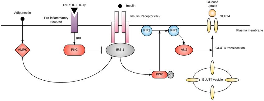
Int. J. Mol. Sci. 2018, 19, 3342                                                                     2 of 21

of glucose regulation during normal pregnancy, classification of GDM, forms of GDM, risk factors for
GDM, and consequences of GDM is first required.

1.1. Glucose Regulation during Healthy Pregnancy
     During healthy pregnancy, the mother’s body undergoes a series of physiological changes in order
to support the demands of the growing fetus. These include adaptations to the cardiovascular, renal,
hematologic, respiratory, and metabolic systems. One important metabolic adaptation is in insulin
sensitivity. Over the course of gestation, insulin sensitivity shifts depending on the requirements of
pregnancy. During early gestation, insulin sensitivity increases, promoting the uptake of glucose into
adipose stores in preparation for the energy demands of later pregnancy [5]. However, as pregnancy
progresses, a surge of local and placental hormones, including estrogen, progesterone, leptin, cortisol,
placental lactogen, and placental growth hormone together promote a state of insulin resistance [6].
As a result, blood glucose is slightly elevated, and this glucose is readily transported across the placenta
to fuel the growth of the fetus. This mild state of insulin resistance also promotes endogenous glucose
production and the breakdown of fat stores, resulting in a further increase in blood glucose and free
fatty acid (FFA) concentrations [7]. Evidence in animals suggests that, in order to maintain glucose
homeostasis, pregnant women compensate for these changes through hypertrophy and hyperplasia of
pancreatic β-cells, as well as increased glucose-stimulated insulin secretion (GSIS) [8]. The importance
of placental hormones in this process is exemplified by the fact that maternal insulin sensitivity returns
to pre-pregnancy levels within a few days of delivery [9]. For reasons that will be explored in this
review, the normal metabolic adaptations to pregnancy do not adequately occur in all pregnancies,
resulting in GDM.

1.2. Classification and Prevalence of Gestational Diabetes
      The American Diabetes Association (ADA) formally classifies GDM as “diabetes first diagnosed
in the second or third trimester of pregnancy that is not clearly either preexisting type 1 or type 2
diabetes” [1]. However, the exact threshold for a diagnosis of GDM depends on the criteria used,
and so far, there has been a lack of consensus amongst health professionals. It is now advised by
the ADA, the World Health Organization (WHO), the International Federation of Gynaecology and
Obstetrics, and the Endocrine Society, that the International Association of Diabetes and Pregnancy
Study Group (IADPSG) criteria be used in the diagnosis of GDM [10]. The IADPSG criteria was
developed based on the results of the Hyperglycemia and Adverse Pregnancy Outcomes (HAPO)
Study—a large multinational and multicenter study of 23,000 pregnant women [11]. One major finding
of the HAPO Study was a continuous risk of adverse maternal and fetal outcomes with increasing
maternal glycaemia—even below the diagnostic threshold for GDM—suggesting the that criteria for
intervention needed to be adjusted. The IADPSG therefore recommends that all women undergo a
fasting plasma glucose (FPG) test at their first prenatal visit (where a reading ≥92 mg/dL is indicative
of GDM), and that women with FPG
Int. J. Mol. Sci. 2018, 19, 3342 3 of 21
the highest prevalence of GDM at 24.2%, while the lowest prevalence was seen in Africa at 10.5%.
Almost 90% of cases of hyperglycemia in pregnancy occurred in low- and middle-income countries,
where access to maternal healthcare is limited. Even within-countries, GDM prevalence varies
depending on race/ethnicity and socioeconomic status. Aboriginal Australians, Middle Easterners,
and Pacific Islanders are the most at-risk groups for GDM [16]. Within the United States,
Native Americans, Hispanics, Asians, and African-American women are at a higher risk of GDM than
Caucasian women [17]. There is also some evidence that GDM prevalence varies by season, with more
diagnoses of GDM in summer than winter [18].
Table 1. Various criteria for gestational diabetes mellitus (GDM) diagnosis using oral glucose tolerance
test (OGTT).
Glucose Glucose Threshold (mmol/L)
Criteria Pregnancies Timing of OGTT Steps
Load (g)
Fasting 1h 2h 3h
O’Sullivan, 1964 All 24–28 weeks 2 100 5.0 9.2 8.1 6.9
WHO, 1999 All 24–28 weeks 1 75 7.0 — 7.8 —
American Diabetes 14–18 weeks for high
High and
Association (ADA), risk, 28–32 weeks for 2 100 5.3 10.0 8.6 7.8
medium risk
2004 medium risk
National Institute
for Health and Care
High risk As early as possible 1 75 5.6 — 7.8 —
Excellence (NICE),
2015
IADPSG, 2010
WHO, 2013 All 24–28 weeks 1 75 5.1 10.0 8.5 —
ADA, 2016
1.3. Forms of Gestational Diabetes
Outside of pregnancy, three distinct forms of diabetes mellitus are described: autoimmune
diabetes (type 1), diabetes occurring on a background of insulin resistance (type 2), and diabetes
as a result of other causes, including genetic mutation, diseases of the exocrine pancreas
(e.g., pancreatitis), and drug- or chemical-induced diabetes (such as after organ transplantation
or in the treatment of human immunodeficiency virus infection and acquired immune deficiency
syndrome (HIV/AIDS)) [1,19]. While there is evidence that GDM can occur in all three settings [20,21],
the vast majority (~80%) of GDM cases present as β-cell dysfunction on a background of chronic
insulin resistance, to which the normal insulin resistance of pregnancy is partially additive [22]. Thus,
affected women tend to have an even greater degree of insulin resistance than healthy pregnant women,
and therefore have further reductions in glucose utilization and increased glucose production and
FFA concentrations [23]. It is thought that β-cells deteriorate due to excessive insulin production in
response to excess energy consumption and insulin resistance, exhausting the cells over time. The fact
that this pathology closely resembles that of T2DM has spurred much debate about whether the two
diseases should be considered to be etiologically indistinct [24,25]. As this form of GDM is by far the
most common, it will be the focus of this review.
1.4. Risk Factors for Gestational Diabetes
Epidemiological studies of risk factors for GDM are limited and are typically afflicted by
confounding factors [26,27]. In addition, inconsistencies in diagnostic criteria for GDM and
measurements of risk factors make it difficult to compare findings across studies. Despite these
concerns, several risk factors for GDM emerge consistently. These include overweight/obesity [28],
excessive gestational weight gain [29], westernized diet [30], ethnicity [31], genetic polymorphisms [32],
advanced maternal age [33], intrauterine environment (low or high birthweight [34]), family andInt. J. Mol. Sci. 2018, 19, 3342 4 of 21
personal history of GDM [35], and other diseases of insulin resistance, such as polycystic ovarian
syndrome (PCOS) [26].
Each of these risk factors are either directly or indirectly associated with impaired β-cell
function and/or insulin sensitivity. For example, overweight and obesity are intrinsically linked
with prolonged, excessive calorie intake, which overwhelms β-cell insulin production and insulin
signaling pathways. Even independently of body mass index (BMI) and overall caloric intake, diet and
nutrition are associated with GDM. Diets that are high in saturated fats, refined sugars, and red and
processed meats are consistently associated with an increased risk of GDM [36,37], while diets high
in fiber, micronutrients, and polyunsaturated fats are consistently associated with a reduced risk of
GDM [38–40]. Saturated fats directly interfere with insulin signaling [41], and they can also induce
inflammation and endothelial dysfunction—both pathogenic factors in GDM [42]. On the other hand,
n-3 polyunsaturated fatty acids, including those derived from fish and seafood, have anti-inflammatory
properties [38]. The relationship between processed meat and GDM remains strong, even after
adjustment for fatty acids, cholesterol, heme iron, and protein content [43]. It has been suggested
that by-products related to the processing of meat could be responsible—such as nitrates (a common
preservative in processed meats), or advanced glycation end products (AGEs), which have both been
implicated in β-cell toxicity [44,45]. Interestingly, even independently of meat consumption, high
protein diets are associated with GDM [46–48]. One theory for this is the role of amino acids as
substrates for hepatic glucose production [49], and in hepatic lipotoxicity [50]. The inverse association
between dietary fiber and GDM may be the result of reduced appetite or slowed glucose absorption,
reducing demand on β-cells and insulin signaling mediators [39].
Low and high birthweight are likely risk factors for GDM because of their association with insulin
resistance. Low birthweight is often the result of undernutrition in the womb, either as a result
of maternal undernutrition or placental insufficiency. It is believed that the fetus compensates for
undernutrition in the womb by epigenetically altering the expression of genes that are involved
in fat storage, energy utilisation, and appetite regulation. Further, animal studies suggest that
undernutrition in utero is associated with reduced β-cell number [51]. These alterations persist
after birth—a phenomenon referred to as “developmental programming” [52]. While potentially
beneficial in times of famine, a mismatch between nutritional status in the womb and nutritional
status once born can contribute to the development of obesity and metabolic disease [53,54]. On the
opposite end of the spectrum, overnutrition in the womb—such as can occur in GDM—can result in
fetal overgrowth. These individuals are more likely to have experienced hyperglycemia and β-cell
fatigue even before birth, predisposing them to hyperglycemia during times of later metabolic stress,
such as during pregnancy [55].
1.5. Consequences of Gestational Diabetes
The importance of aiming to understand and effectively treat or prevent GDM is illustrated by
the wide-ranging consequences of GDM for both the mother and the fetus.
Mother—GDM increases the risk of a number of short-term and long-term maternal health issues.
In addition to the stress of normal pregnancy, GDM is associated with antenatal depression [56].
There is also an increased risk of additional pregnancy complications, including preterm birth and
preeclampsia, and, in many cases, surgical delivery of the baby is required [57]. Approximately 60% of
women with a past history of GDM develop T2DM later in life [58]. Each additional pregnancy also
confers a threefold increase in the risk of T2DM in women with a history of GDM. Further, women with
a previous case of GDM have a yearly risk of conversion to T2DM of ~2 to 3% [58]. Emerging evidence
also suggests that the vasculature of women with a prior case of GDM is permanently altered,
predisposing them to cardiovascular disease (CVD). A recent study reported a 63% increased risk of
CVD amongst women with a history of GDM, which was partly, but not fully, explained by BMI [59].
This is of major concern, as CVD is the number one cause of death in the world [60].Int. J. Mol. Sci. 2018, 19, 3342 5 of 21
Child—GDM also poses short- and long-term consequences for the infant. The aforementioned
increase in placental transport of glucose, amino acids, and fatty acids stimulate the fetus’s endogenous
production of insulin and insulin-like growth factor 1 (IGF-1). Together, these can cause fetal
overgrowth, often resulting in macrosomia at birth [61]. As previously mentioned, excess fetal insulin
production can stress the developing pancreatic β-cells, contributing to β-cell dysfunction and insulin
resistance, even prenatally [62]. Macrosomia is also a risk factor for shoulder dystocia—a form of
obstructed labor. Thus, babies of GDM pregnancies are usually delivered by caesarean section [63,64].
Once delivered, these babies are at increased risk of hypoglycemia, which is likely due to formed
dependence on maternal hyperglycemia (fetal hyperinsulinemia), which can contribute to brain injury
if not properly managed [65]. There is also evidence that GDM increases the risk of stillbirth [66].
In the long term, babies that are born of GDM pregnancies are at increased risk of obesity, T2DM, CVD,
and associated metabolic diseases. Children born to mothers with GDM have almost double the risk
of developing childhood obesity when compared with nondiabetic mothers, even after adjusting for
confounders such as maternal BMI [67,68], and impaired glucose tolerance can be detected as young
as five years old [69]. Females are therefore more likely to experience GDM in their own pregnancies,
contributing to a vicious intergenerational cycle of GDM [70].
2. Pathophysiology of Gestational Diabetes
The remainder of this review will discuss molecular processes underlying the pathophysiology of
GDM. GDM is usually the result of β-cell dysfunction on a background of chronic insulin resistance
during pregnancy and thus both β-cell impairment and tissue insulin resistance represent critical
components of the pathophysiology of GDM. In most cases, these impairments exist prior to pregnancy
and can be progressive—representing an increased risk of T2DM post-pregnancy [71]. A number
of additional organs and systems contribute to, or are affected by, GDM. These include the brain,
adipose tissue, liver, muscle, and placenta.
2.1. β-Cell Dysfunction
The primary function of β-cells is to store and secrete insulin in response to glucose load.
When β-cells lose the ability to adequately sense blood glucose concentration, or to release sufficient
insulin in response, this is classified as β-cell dysfunction. β-cell dysfunction is thought to be the result
of prolonged, excessive insulin production in response to chronic fuel excess [72]. However, the exact
mechanisms underlying β-cell dysfunction can be varied and complex [73,74]. Defects can occur
at any stage of the process: pro-insulin synthesis, post-translational modifications, granule storage,
sensing of blood glucose concentrations, or the complex machinery underlying exocytosis of granules.
Indeed, the majority of susceptibility genes that are associated with GDM are related to β-cell
function, including potassium voltage-gated channel KQT-like 1 (Kcnq1) and glucokinase (Gck).
Minor deficiencies in the β-cell machinery may only be exposed in times of metabolic stress, such as
pregnancy [75].
β-cell dysfunction is exacerbated by insulin resistance. Reduced insulin-stimulated glucose uptake
further contributes to hyperglycemia, overburdening the β-cells, which have to produce additional
insulin in response. The direct contribution of glucose to β-cell failure is described as glucotoxicity [76].
Thus, once β-cell dysfunction begins, a vicious cycle of hyperglycemia, insulin resistance, and further
β-cell dysfunction is set in motion.
Animal studies suggest that β-cell number is also an important determinant of glucose
homeostasis. For example, Zucker fatty (ZF) rats that were subjected to 60% pancreatectomy mostly
recover β-cell mass by one week post-surgery, but still develop hyperglycemia. In these cases,
the short-term but dramatic reduction in β-cell mass overburdens the remaining β-cells, resulting in
severely reduced glucose-stimulated insulin secretion and the depletion of internal insulin granule
stores [77]. Sprague Dawley rats, which are usually very resistant to the development of diabetes,
experience substantial loss of β-cell mass (50% reduction) by 15-weeks old when growth-restrictedInt. J. Mol. Sci. 2018, 19, 3342 6 of 21
in utero via bilateral uterine artery ligation [78]. This loss of β-cell mass has been linked to
epigenetic downregulation of pancreatic homeobox transcription factor (Pdx1), which is essential
for normal β-cell differentiation in the embryo [79]. Prolactin is also essential for adequate β-cell
proliferation, as demonstrated in mouse knockouts of the prolactin receptor (PrlR−/− ) [80]. In addition,
glucotoxicity is also thought to result in β-cell apoptosis over time [76]. Pancreatic samples from
T2DM patients can show a reduction of β-cell mass by 40–60% [81], but less than 24% loss after five
years of disease has also been reported [82]. Reduced β-cell hyperplasia may also play a role in GDM,
based on animal studies and limited post-mortem human studies [83]. Therefore, reduced β-cell mass,
reduced β-cell number, β-cell dysfunction, or a mix of all three contribute to GDM, depending on
the individual.
2.2. Chronic Insulin Resistance
Insulin resistance occurs when cells no longer adequately respond to insulin. At the molecular
level, insulin resistance is usually a failure of insulin signaling, resulting in inadequate plasma
membrane translocation of glucose transporter 4 (GLUT4)—the primary transporter that is responsible
for bringing glucose into the cell to use as energy (Figure 1). The rate of insulin-stimulated glucose
uptake is reduced by 54% in GDM when compared with normal pregnancy [84]. While insulin receptor
abundance is usually unaffected, reduced tyrosine or increased serine/threonine phosphorylation
of the insulin receptor dampens insulin signaling [85]. In addition, altered expression and/or
phosphorylation of downstream regulators of insulin signaling, including insulin receptor substrate
(IRS)-1, phosphatidylinositol 3-kinase (PI3K), and GLUT4, has been described in GDM [84]. Many of
these
Int. molecular
J. Mol. Sci. changes
2018, 19, x FOR PEER REVIEW persist beyond pregnancy [86]. 8 of 24
Figure 1Error! No text of specified style in document.. Simplified diagram of insulin signaling. Binding of insulin to the insulin receptor (IR) activates IRS-1. Adiponectin
Figure 1. Simplified diagram of insulin signaling. Binding of insulin to the insulin receptor (IR)
promotes IRS-1 activation through AMP-activated protein kinase (AMPK), while pro-inflammatory cytokines activate protein kinase C (PKC) via IκB kinase (IKK), which
activates IRS-1. Adiponectin promotes IRS-1 activation through AMP-activated protein kinase
inhibits IRS-1. IRS-1 activates phosphatidylinositol-3-kinase (PI3K), which phosphorylates phosphatidylinositol-4, 5-bisphosphate (PIP2) to phosphatidylinositol-3, 4, 5-
phosphate (PIP3). PIP3 activates Akt2, which promotes GLUT4 translocation and glucose uptake into the cell.
(AMPK), while pro-inflammatory cytokines activate protein kinase C (PKC) via IκB kinase (IKK),
which inhibits IRS-1. IRS-1 activates phosphatidylinositol-3-kinase (PI3K), which phosphorylates
phosphatidylinositol-4, 5-bisphosphate (PIP2) to phosphatidylinositol-3, 4, 5-phosphate (PIP3).
PIP3 activates Akt2, which promotes GLUT4 translocation and glucose uptake into the cell.
Several of the previously discussed risk factors for GDM are thought to exert their effects
by interfering with insulin signaling. For example, saturated fatty acids increase intracellular
concentrations of diacylglycerol within myocytes, activating protein kinase C (PKC) and inhibiting
tyrosine kinase, IRS-1 and PI3K [41]. Pro-inflammatory cytokines and adiponectin also modify this
process, as discussed below.
A diagram of the relationship between β-cell dysfunction, insulin resistance, and GDM is provided
in Figure 2.Int. J. Mol. Sci. 2018, 19, 3342 7 of 21
Int. J. Mol. Sci. 2018, 19, x FOR PEER REVIEW 9 of 24
Figure
Figure 2.2.β-cell, blood
β-cell, glucose,
blood and insulin
glucose, sensitivity
and insulin duringduring
sensitivity normalnormal
pregnancy and GDM.
pregnancy andDuring
GDM.
normal pregnancy,
During normal β-cellsβ-cells
pregnancy, undergo hyperplasia
undergo and and
hyperplasia hypertrophy
hypertrophy in order to to
in order meet
meetthe
themetabolic
metabolic
demands
demands of of pregnancy.
pregnancy. Blood
Blood glucose
glucose rises
rises as
as insulin
insulin sensitivity
sensitivity falls.
falls. Following
Followingpregnancy,
pregnancy, β-cells,
β-cells,
blood
blood glucose,
glucose, and
and insulin
insulin sensitivity
sensitivity return
return to normal. During
During gestational
gestational diabetes,
diabetes, β-cells
β-cells fail
fail to
to
compensate
compensatefor forthe
thedemands
demands ofof
pregnancy,
pregnancy,and, when
and, whencombined
combined withwith
reduced insulin
reduced sensitivity,
insulin this
sensitivity,
results in hyperglycemia.
this results in hyperglycemia.Following pregnancy,
Following β-cells,
pregnancy, bloodblood
β-cells, glucose, and insulin
glucose, sensitivity
and insulin may
sensitivity
return to normal
may return or may
to normal orremain impaired
may remain on a pathway
impaired towardtoward
on a pathway GDM inGDM future
inpregnancy or T2DM.
future pregnancy or
T2DM. Pancreas
Pancreas image obtained
image obtained fromNoun
from The The Noun Project
Project underunder
the the terms
terms andand conditionsofofthe
conditions theCreative
Creative
Commons Attribution
Commons Attribution (CC
(CCBY)BY)license
license(http://creativecommons.org/licenses/by/4.0/),
(http://creativecommons.org/licenses/by/4.0/), by byartist
artist Arif
Arif
Fajar Vulianto.
Fajar Vulianto.Int. J. Mol. Sci. 2018, 19, 3342 8 of 21
2.3. Neurohormonal Networks
Neurohormonal dysfunction has been implicated in the pathogenesis of diseases of insulin
resistance, such as that present in GDM. This network regulates appetite, active energy expenditure,
and basal metabolic rate, and it is made up of a complex network of central (e.g., cortical centers
that control cognitive, visual, and “reward” cues) and peripheral (e.g., satiety and hunger hormones)
signals [87,88]. These contribute to GDM by influencing adiposity and glucose utilization. This network
is highly regulated by the circadian clock, which may explain why pathological sleep disorders or those
individuals undertaking shift work are correlated with GDM rates [89,90]. Neural networks controlling
body weight are most likely set in early life, as demonstrated in animal studies. For example, rats that
are both under- and over-fed in early life experience epigenetic alteration of the regulatory set-point of
hypothalamic neurons [91,92]. This adds to the previously mentioned suggestion that predisposition
to GDM may be set in the womb.
Some of the most important regulators of neurohormonal metabolic control are adipokines—cell
signaling proteins that are secreted primarily by adipose tissue. These include leptin and adiponectin:
2.3.1. Leptin
Leptin is a satiety hormone secreted primarily by adipocytes in response to adequate fuel stores.
It primarily acts on neurons within the arcuate nucleus of the hypothalamus to decrease appetite and
increase energy expenditure. Specifically, leptin inhibits appetite-stimulators neuropeptide Y (NPY)
and agouti-related peptide (AgRP), and it activates the anorexigenic polypeptide pro-opiomelanocortin
(POMC) [93]. When leptin was first discovered, it was lauded as a potential treatment for obesity [94].
However, it was soon revealed that the majority of obese individuals do not respond to leptin,
and instead demonstrate leptin resistance. While leptin treatment is effective in obesity that is caused
by leptin and leptin receptor genetic polymorphisms, these are rare (Int. J. Mol. Sci. 2018, 19, 3342 9 of 21
stimulates insulin secretion, by upregulating insulin gene expression and exocytosis of insulin granules
from β-cells [105].
Adiponectin is also expressed at low concentration from the syncytiotrophoblast of the placenta
where it is regulated by cytokines, such as tumor necrosis factor alpha (TNF-α), interleukin (IL)-6,
interferon gamma (IFN-γ), and leptin [106]. The role of placental adiponectin in normal and
GDM pregnancy is unclear [107]. However, emerging evidence suggests adiponectin impairs
insulin signaling and amino acid transport across the placenta, limiting fetal growth. Therefore,
adiponectin gene methylation in the placenta is associated with maternal glucose intolerance and fetal
macrosomia [108].
2.4. Adipose Tissue
Originally believed to exist only as a passive depot of energy, the discovery of leptin in 1994
established adipose tissue as an essential endocrine organ. Adipose tissue both ensures that energy is
partitioned safely and it actively secretes circulatory factors, including adipokines (the aforementioned
leptin and adiponectin) and cytokines (such as TNF-α, IL-6, and IL-1β), which have wide-ranging
metabolic effects.
2.4.1. Energy Storage
The storage capability of adipose tissue is essential for metabolic health. This is exemplified
through two extremes: rare disorders in which white adipose tissue is absent lead to severe metabolic
syndrome, whereas some obese individuals (with excessive white adipose tissue) do not develop
metabolic syndrome at all [109]. Therefore, the ability to partition excess calories into adipose tissue
rather than ectopically in the liver, muscle, or pancreas, appears to serve as a protective measure.
Non-diabetic obese individuals exhibit adequate adipose tissue expansion in response to fuel surfeit,
and therefore maintain healthy blood glucose concentrations, sufficient β-cell compensation, and avoid
chronic insulin resistance [110,111]. In this way, key organs avoid glucose and fatty acid-induced
tissue damage. As previously mentioned, early pregnancy is marked by an increase in adipose tissue
mass, while later pregnancy promotes the mobilization of fats from adipose tissue in order to fuel
fetal growth. Both of these processes are thought to be limited in GDM [112]. GDM is associated
with reduced adipocyte differentiation and increased adipocyte size (hypertrophy), accompanied
by downregulated gene expression of insulin signaling regulators, fatty acid transporters, and key
adipogenic transcription factors, such as PPARγ [113]. The combination of insulin resistance and
reduced adipocyte differentiation hinders the tissue’s ability to safely dispose of excess energy,
contributing to gluco- and lipo-toxicity in other peripheral organs. Indeed, both T2DM and GDM are
associated with lipid deposition in muscle and liver [114,115].
2.4.2. Adipose Tissue Inflammation
Obesity, T2DM and GDM are associated with an increased number of resident adipose tissue
macrophages (ATM) that secrete pro-inflammatory cytokines, including TNF-α, IL-6, and IL-1β.
The importance of a low-grade inflammatory state in the pathogenesis of insulin resistance has recently
become apparent. Pro-inflammatory cytokines have been discovered to both impair insulin signaling
and inhibit insulin release from β-cells. These factors induce insulin resistance either by diminishing
insulin receptor (IR) tyrosine kinase activity, increasing serine phosphorylation of IRS-1, or through the
STAT3-SOCS3 pathway, which degrades IRS-1 [85,116]. Circulating concentrations of pro-inflammatory
cytokines are increased in GDM [107,117]. Plasma TNF-α, in particular, is strongly correlated with
insulin resistance [118]. Similarly, placental gene expression of TNF-α, IL-1β and their receptors has
been reported to be increased in GDM [118,119]. However, the relationship between pregnancy and
inflammation is complex. For example, Lappas et al. (2010) reported that GDM placentae secrete fewer
pro-inflammatory cytokines (3 of 16 studied: IL-1β, TNF-α and M1P1B) than healthy placentae (13 outInt. J. Mol. Sci. 2018, 19, 3342 10 of 21
of 16 studied) [120]. This suggests that, while chronic low-grade inflammation appears to be important
in the pathogenesis of GDM, the relationship may not be straightforward.
2.5. Liver
GDM is associated with upregulated hepatic glucose production (gluconeogenesis).
Gluconeogenesis is increased in the fasted state, and not adequately suppressed in the fed state [84].
This is not believed to be entirely the result of inaccurate glucose sensing due to insulin resistance,
as the majority of glucose uptake by the liver (~70%) is not insulin dependent. Common factors
between the insulin signaling pathway and the pathways controlling gluconeogenesis, such as PI3K,
might contribute to these effects [121]. Increased protein intake and muscle breakdown may also
stimulate the process by providing excess gluconeogenesis substrate [122]. Despite this, the liver does
not seem to be a primary pathogenic driver of T2DM or GDM [123].
2.6. Skeletal and Cardiac Muscle
Traditionally, skeletal muscle insulin resistance was believed to play a causal role in T2DM.
However, skeletal muscle insulin resistance now appears to be a consequence of hyperglycemia—a
protective measure to prevent metabolic stress and steatosis [124]. Even following a short period of
overfeeding, cardiac and skeletal muscle develop insulin resistance in order to divert the excess energy
into adipose tissue [125]. This is an important distinction when considering potential treatments for
GDM: attempts to directly reverse skeletal muscle insulin resistance, without reducing plasma glucose
concentrations, could be detrimental [123].
Separate to insulin sensitivity, T2DM and GDM are associated with a reduced number and function
of mitochondria within skeletal muscle cells [126]. This could be the result of genetics, early-life
programming, or chronic inactivity. Therefore, decreased number and function of mitochondria is
likely an additional contributor to reduced glucose utilization in GDM.
2.7. Gut Microbiome
There is emerging evidence that microbial organisms within the gut—the “gut microbiome”—
might contribute to metabolic diseases, including GDM. The gut microbiome can be influenced by
early-life events, such as preterm delivery and breastfeeding, and by events in later life, such as
diet composition and antibiotic use. The gut microbiome has been consistently reported to differ
between metabolically healthy and obese individuals, including during pregnancy [127]. Furthermore,
a study of stool bacteria in women with a past case of GDM reported a lower proportion of the
phylum Firmicutes and higher proportion of the family Prevotellaceae as compared with normoglycemic
pregnancy [128]. Similar associations have been observed in obesity [129], T2DM [130], fatty liver
disease [131], and elevated total plasma cholesterol [132]. Firmicutes metabolize dietary plant
polysaccharides. This may explain some of the dietary risk factors for GDM that are discussed
earlier. Both red meat and animal protein decrease levels of Firmicutes, while high dietary fiber increase
them [133]. However, the findings by Fugmann et al. (2015) remained after adjustment for dietary
habits [128]. Therefore, Firmicutes appear to be relevant to pathogenesis of GDM independent of diet,
although the mechanisms underlying this are unknown. Prevotellaceae are mucin-degrading bacteria
that may contribute to increased gut permeability. Gut permeability is regulated by tight junction
proteins, such as zonulin (ZO-1). Increased “free” plasma/serum ZO-1 is associated with type 1
diabetes (T1DM), T2DM [134], and GDM [135]. Increased gut permeability is thought to facilitate the
movement of inflammatory mediators from the gut into the circulation, promoting systemic insulin
resistance [134,136].
2.8. Oxidative Stress
Oxidative stress describes an imbalance between pro-oxidants and antioxidants in cells.
Oxidative stress can lead to cellular damage by interfering with the state of proteins, lipids andInt. J. Mol. Sci. 2018, 19, 3342 11 of 21
DNA, and has been implicated in the pathogenesis of many diseases, including GDM [137].
Reactive oxygen species (ROS) are described as free radical and nonradical derivatives of oxygen,
and include superoxide anion (O2 − ), hydroxyl radical (•OH) and hydrogen peroxide (H2 O2 ) [138].
A hyperglycemic environment is associated with oxidative stress, and GDM women have been reported
to overproduce free radicals and have impaired free-radical scavenging mechanisms [139]. ROS inhibit
insulin-stimulated glucose uptake by interfering with both IRS-1 and GLUT4 [140]. ROS also slow
glycogen synthesis in the liver and muscle. Pro-inflammatory cytokines, such as TNF-α, may also
contribute to oxidative stress by increasing the expression and the activation of ROS precursors,
like NADPH oxidase 4 (NOX4) [141].
Interestingly, iron supplementation in women already replete in iron is associated with GDM [142].
Several studies suggest that this relationship is the result of increased oxidative stress. Iron is a
transitional metal and it can catalyze the reaction from O2− and H2 O2 to the extremely reactive
•OH within mitochondria [143]. On the contrary, selenium and zinc are transitional metals that are
necessary for the activity of some antioxidant enzymes, which may explain their inverse association
with GDM [144].
Homocysteine—a non-protein α-amino acid that is formed by the demethylation of
methionine—is also thought to contribute to GDM via oxidative stress. Exposure of β-cells to even
small amounts of homocysteine results in dysfunction and impaired insulin secretion [145]. A recent
meta-analysis examined the relationship between serum homocysteine concentration and GDM in
ten eligible studies. The authors reported significantly higher homocysteine concentrations among
women with GDM as compared with those without GDM [146]. B vitamins, including folic acid, B2,
B6, and B12 are essential for homocysteine homeostasis, and this may be one reason why deficiencies
and imbalances of these micronutrients are associated with GDM [147].
2.9. Placental Transport
The placenta contributes to insulin resistance during pregnancy via its secretion of hormones
and cytokines. As the barrier between the maternal and fetal environments, the placenta itself is also
exposed to hyperglycemia and its consequences during GDM. This can impact transport of glucose,
amino acids, and lipids across the placenta:
Glucose—Glucose is the primary energy source for the fetus and the placenta, and therefore must
be readily available at all times. For this reason, insulin is not required for the placental transport
of glucose. Instead, glucose transport occurs via GLUT1, by carrier-mediated sodium-independent
diffusion [148]. However, the placenta still expresses the insulin receptor, and insulin signaling can
influence placental metabolism of glucose [149]. The receptiveness of the placenta to glucose uptake
means that it is particularly sensitive to maternal hyperglycemia, and this directly contributes to
increased fetal growth and macrosomia.
Protein—Amino acid transport across the placenta is also an important determinant of fetal growth.
GDM is associated with increased System A and L activity [150]. These can also be modulated by
pro-inflammatory cytokines, such as TNF-α and IL-6 [151]. Altered amino acid transport may also be
one mechanism by which excess protein intake contributes to GDM.
Lipids—Finally, while GDM has traditionally been described as a disease of hyperglycemia,
the rise in obesity-associated GDM has prompted a greater focus on the role of hyperlipidemia in
GDM. The majority of placental gene expression alterations in GDM occur in lipid pathways (67%),
as compared with glucose pathways (9%) [152]. Preferential activation of placental lipid genes is
also associated with GDM compared with T1DM [152]. These data correlate with the results of the
HAPO Study, which revealed independent effects of maternal obesity and glucose on excessive fetal
growth [153]. Therefore, it appears that GDM influences the placental transport of glucose, amino acids,
and fatty acids, and that all three must be considered when discussing the impact of GDM on placental
function and fetal growth.Int. J. Mol. Sci. 2018, 19, 3342 12 of 21
In addition to these alterations in placental transport, GDM has been associated with other
changes in the placenta. Some recent studies have reported that GDM is associated with placenta
global DNA hypermethylation [154]. Similarly, studies of the placental proteome have identified
differences in the expression of proteins between GDM and non-GDM placentas [155]. However,
more research is required before the role of placental epigenetic and proteomic modifications in GDM
is fully understood [156]. There has also been recent interest in small noncoding single-stranded
segments of RNA, called microRNAs (miRNAs), expressed in placental trophoblast cells. miRNAs
are involved in a number of cellular processes, including proliferation, differentiation, and apoptosis.
Emerging evidence suggests that exosomes containing miRNAs are shed from the placenta during
gestation and released into the maternal circulation, which can in turn influence the functioning of
other cells, potentially contributing to the pathogenesis of GDM [157,158]. Interestingly, exposure to
endocrine disrupting chemicals (EDCs), including bisphenol A (BPA—found in food packaging
materials and consumer products) has been associated with GDM, and it has been suggested
that this could be because EDCs induce exosome signaling from the placenta [159]. Interestingly,
EDCs including BPA have also been associated with alterations in methylation, perhaps linking the
Int. J.two
Mol. Sci.mechanisms [160]. A summary diagram of the pathophysiology of GDM is presented in Figure 143.of 24
2018, 19, x FOR PEER REVIEW
Figure 3. Organs involved in the pathophysiology of GDM (Images in this figure were obtained from The Noun Project under the terms and conditions of the Creative
Figure 3. Organs involved in the pathophysiology of GDM (Images in this figure were obtained from
Commons Attribution (CC BY) license (http://creativecommons.org/licenses/by/4.0/). Brain and Gut by Hunotika; Liver by Lavmik; Pancreas by Arif Fajar Vulianto;
The
Placenta Noun Project
by Charmeleon under
Design; Muscle thePetrishchev).
by Misha terms and conditions of the Creative Commons Attribution (CC BY)
license (http://creativecommons.org/licenses/by/4.0/). Brain and Gut by Hunotika; Liver by Lavmik;
Pancreas by Arif Fajar Vulianto; Placenta by Charmeleon Design; Muscle by Misha Petrishchev).
3. Opportunities and Considerations for Future Study
Uncovering the intricate molecular mechanisms underlying GDM is challenging, but necessary
for our greater understanding of the disease and how these could assist in the design of new treatments.
As β-cell dysfunction and insulin resistance are the hallmarks of GDM, the greatest emphasis should
be placed on further understanding the mechanisms underlying these processes. For example,
why do β-cells exhibit proper hyperplasia and hypertrophy in some pregnancies, but not others?
How could we modify these processes to bolster pancreatic function and prevent hyperglycemia
in at-risk individuals? As already mentioned, increasing insulin sensitivity could have unintended
consequences by promoting uptake of glucose into tissue where energy should not be stored, such as
the liver and skeletal muscle. Instead, investment into adipose-specific insulin sensitivity should
be examined. While improving adipose capacity (and in theory increasing adipose tissue mass)
might seem counterintuitive, in actuality, it should reduce hyperglycemia while ensuring that excessInt. J. Mol. Sci. 2018, 19, 3342 13 of 21
energy is stored safely. Of course, many of the mechanisms underlying GDM are not unique to GDM,
encompassing other common disorders of insulin sensitivity, such as T2DM, prediabetes, and PCOS.
Therefore, determination of pathways influencing development of these metabolic disorders may
also shed light on GDM, and potentially accelerate opportunities for prevention and/or treatment.
This is an important consideration, as the study of GDM (as a disorder of pregnancy) is limited for
ethical reasons. Finally, the ability to study large amounts of data through computer technology is
rapidly advancing the fields of genomics, epigenetics, proteomics, metagenomics (the microbiome),
and metabolomics (the study of the small-molecule intermediates and products of metabolism). It is
hopeful that the advancement of these large-scale techniques may assist in our understanding of the
pathogenesis of GDM in the future.
4. Conclusions
Pregnancy is a state of high metabolic activity, in which maintaining glucose homeostasis is
of upmost importance. When hyperglycemia is detected in the pregnant mother, this is referred to
as GDM, although controversy remains over diagnostic criteria. It is likely that genetic, epigenetic,
and environmental factors all contribute to the development of GDM, and that the mechanisms
involved are complex and advance over a substantial period of time. However, in the majority of cases,
pancreatic β-cells fail to compensate for a chronic fuel surfeit, leading to eventual insulin resistance,
hyperglycemia, and an increased supply of glucose to the growing fetus. There is also evidence
that adipose expandability, low-grade chronic inflammation, gluconeogenesis, oxidative stress,
and placental factors contribute to the pathology of GDM. Greater understanding of these processes and
their contribution to GDM is required in order to develop effective treatments and prevention strategies.
Author Contributions: J.F.P. primarily wrote the manuscript. J.L.S., C.M.R., P.N.B. and M.H.V. provided
supervisory and editorial assistance.
Funding: This research received no external funding.
Conflicts of Interest: The authors declare no conflict of interest.
References
1. American Diabetes Association. Classification and Diagnosis of Diabetes: Standards of Medical Care in
Diabetes—2018. Diabetes Care 2018, 41, S13–S27. [CrossRef] [PubMed]
2. International Diabetes Federation. IDF Diabetes Atlas, 8th ed.; IDF: Brussels, Belgium, 2017.
3. Feig, D.S.; Moses, R.G. Metformin Therapy during Pregnancy Good for the goose and good for the gosling
too? Diabetes Care 2011, 34, 2329–2330. [CrossRef] [PubMed]
4. Camelo Castillo, W.; Boggess, K.; Stürmer, T.; Brookhart, M.A.; Benjamin, D.K.; Jonsson Funk, M.
Association of Adverse Pregnancy Outcomes with Glyburide vs Insulin in Women with Gestational Diabetes.
JAMA Pediatr. 2015, 169, 452–458. [CrossRef] [PubMed]
5. Di Cianni, G.; Miccoli, R.; Volpe, L.; Lencioni, C.; Del Prato, S. Intermediate metabolism in normal pregnancy
and in gestational diabetes. Diabetes Metab. Res. Rev. 2003, 19, 259–270. [CrossRef] [PubMed]
6. Catalano, P.M.; Tyzbir, E.D.; Roman, N.M.; Amini, S.B.; Sims, E.A. Longitudinal changes in insulin release
and insulin resistance in nonobese pregnant women. Am. J. Obstet. Gynecol. 1991, 165, 1667–1672. [CrossRef]
7. Phelps, R.L.; Metzger, B.E.; Freinkel, N. Carbohydrate metabolism in pregnancy: XVII. Diurnal profiles of
plasma glucose, insulin, free fatty acids, triglycerides, cholesterol, and individual amino acids in late normal
pregnancy. Am. J. Obstet. Gynecol. 1981, 140, 730–736. [CrossRef]
8. Parsons, J.A.; Brelje, T.C.; Sorenson, R.L. Adaptation of islets of Langerhans to pregnancy: Increased islet cell
proliferation and insulin secretion correlates with the onset of placental lactogen secretion. Endocrinology
1992, 130, 1459–1466. [CrossRef] [PubMed]
9. Ryan, E.A.; O’Sullivan, M.J.; Skyler, J.S. Insulin Action During Pregnancy: Studies with the Euglycemic
Clamp Technique. Diabetes 1985, 34, 380–389. [CrossRef] [PubMed]
10. Chiefari, E.; Arcidiacono, B.; Foti, D.; Brunetti, A. Gestational diabetes mellitus: An updated overview.
J. Endocrinol. Investig. 2017, 40, 899–909. [CrossRef] [PubMed]Int. J. Mol. Sci. 2018, 19, 3342 14 of 21
11. HAPO Study Cooperative Research Group; Metzger, B.E.; Lowe, L.P.; Dyer, A.R.; Trimble, E.R.;
Chaovarindr, U.; Coustan, D.R.; Hadden, D.R.; McCance, D.R.; Hod, M.; et al. Hyperglycemia and adverse
pregnancy outcomes. N. Engl. J. Med. 2008, 358, 1991–2002. [CrossRef] [PubMed]
12. Egan, A.M.; Vellinga, A.; Harreiter, J.; Simmons, D.; Desoye, G.; Corcoy, R.; Adelantado, J.M.; Devlieger, R.;
Assche, A.V.; Galjaard, S.; et al. Epidemiology of gestational diabetes mellitus according to IADPSG/WHO
2013 criteria among obese pregnant women in Europe. Diabetologia 2017, 1–9. [CrossRef] [PubMed]
13. Williams, C.B.; Iqbal, S.; Zawacki, C.M.; Yu, D.; Brown, M.B.; Herman, W.H. Effect of selective screening for
gestational diabetes. Diabetes Care 1999, 22, 418–421. [CrossRef] [PubMed]
14. Griffin, M.E.; Coffey, M.; Johnson, H.; Scanlon, P.; Foley, M.; Stronge, J.; O’Meara, N.M.; Firth, R.G.
Universal vs. risk factor-based screening for gestational diabetes mellitus: Detection rates, gestation at
diagnosis and outcome. Diabet. Med. J. Br. Diabet. Assoc. 2000, 17, 26–32. [CrossRef]
15. Capula, C.; Chiefari, E.; Vero, A.; Arcidiacono, B.; Iiritano, S.; Puccio, L.; Pullano, V.; Foti, D.P.; Brunetti, A.;
Vero, R. Gestational Diabetes Mellitus: Screening and Outcomes in Southern Italian Pregnant Women.
Available online: https://www.hindawi.com/journals/isrn/2013/387495/ (accessed on 9 October 2018).
16. Zhu, Y.; Zhang, C. Prevalence of Gestational Diabetes and Risk of Progression to Type 2 Diabetes: A Global
Perspective. Curr. Diabetes Rep. 2016, 16, 7. [CrossRef] [PubMed]
17. Yuen, L.; Wong, V.W. Gestational diabetes mellitus: Challenges for different ethnic groups. World J. Diabetes
2015, 6, 1024–1032. [CrossRef] [PubMed]
18. Moses, R.G.; Wong, V.C.K.; Lambert, K.; Morris, G.J.; Gil, F.S. Seasonal Changes in the Prevalence of
Gestational Diabetes Mellitus. Diabetes Care 2016, 39, 1218–1221. [CrossRef] [PubMed]
19. Haneda, M.; Noda, M.; Origasa, H.; Noto, H.; Yabe, D.; Fujita, Y.; Goto, A.; Kondo, T.; Araki, E.
Japanese Clinical Practice Guideline for Diabetes 2016. J. Diabetes Investig. 2018, 9, 657–697. [CrossRef]
[PubMed]
20. Chiu, K.C.; Go, R.C.; Aoki, M.; Riggs, A.C.; Tanizawa, Y.; Acton, R.T.; Bell, D.S.; Goldenberg, R.L.;
Roseman, J.M.; Permutt, M.A. Glucokinase gene in gestational diabetes mellitus: Population association
study and molecular scanning. Diabetologia 1994, 37, 104–110. [CrossRef] [PubMed]
21. Damm, P.; Kühl, C.; Buschard, K.; Jakobsen, B.K.; Svejgaard, A.; Sodoyez-Goffaux, F.; Shattock, M.;
Bottazzo, G.F.; Mølsted-Pedersen, L. Prevalence and predictive value of islet cell antibodies and insulin
autoantibodies in women with gestational diabetes. Diabet. Med. J. Br. Diabet. Assoc. 1994, 11, 558–563.
[CrossRef]
22. Buchanan, T.A.; Xiang, A.H. Gestational diabetes mellitus. J. Clin. Investig. 2005, 115, 485–491. [CrossRef]
[PubMed]
23. Catalano, P.M.; Huston, L.; Amini, S.B.; Kalhan, S.C. Longitudinal changes in glucose metabolism during
pregnancy in obese women with normal glucose tolerance and gestational diabetes mellitus. Am. J.
Obstet. Gynecol. 1999, 180, 903–916. [CrossRef]
24. Pendergrass, M.; Fazioni, E.; DeFronzo, R.A. Non-insulin-dependent diabetes mellitus and gestational
diabetes mellitus: Same disease, another name? Diabetes Rev. 1995, 3, 566–583.
25. Zajdenverg, L.; Negrato, C.A. Gestational diabetes mellitus and type 2 diabetes: Same disease in a different
moment of life? Maybe not. Arch. Endocrinol. MeTable 2017, 61, 208–210. [CrossRef] [PubMed]
26. Ben-Haroush, A.; Yogev, Y.; Hod, M. Epidemiology of gestational diabetes mellitus and its association with
Type 2 diabetes. Diabet. Med. 2004, 21, 103–113. [CrossRef] [PubMed]
27. Metzger, B.E.; Buchanan, T.A.; Coustan, D.R.; de Leiva, A.; Dunger, D.B.; Hadden, D.R.; Hod, M.;
Kitzmiller, J.L.; Kjos, S.L.; Oats, J.N.; et al. Summary and Recommendations of the Fifth International
Workshop-Conference on Gestational Diabetes Mellitus. Diabetes Care 2007, 30, S251–S260. [CrossRef]
[PubMed]
28. Okosun, I.S.; Chandra, K.M.D.; Boev, A.; Boltri, J.M.; Choi, S.T.; Parish, D.C.; Dever, G.E.A. Abdominal
adiposity in U.S. adults: Prevalence and trends, 1960-2000. Prev. Med. 2004, 39, 197–206. [CrossRef] [PubMed]
29. Durnwald, C. Gestational diabetes: Linking epidemiology, excessive gestational weight gain, adverse
pregnancy outcomes, and future metabolic syndrome. Semin. Perinatol. 2015, 39, 254–258. [CrossRef]
[PubMed]
30. Zhang, C.; Tobias, D.K.; Chavarro, J.E.; Bao, W.; Wang, D.; Ley, S.H.; Hu, F.B. Adherence to healthy lifestyle
and risk of gestational diabetes mellitus: prospective cohort study. BMJ 2014, 349, g5450. [CrossRef]
[PubMed]Int. J. Mol. Sci. 2018, 19, 3342 15 of 21
31. Jenum, A.K.; Mørkrid, K.; Sletner, L.; Vange, S.; Torper, J.L.; Nakstad, B.; Voldner, N.; Rognerud-Jensen, O.H.;
Berntsen, S.; Mosdøl, A.; et al. Impact of ethnicity on gestational diabetes identified with the WHO and the
modified International Association of Diabetes and Pregnancy Study Groups criteria: A population-based
cohort study. Eur. J. Endocrinol. 2012, 166, 317–324. [CrossRef] [PubMed]
32. Anghebem-Oliveira, M.I.; Martins, B.R.; Alberton, D.; de Ramos, E.A.S.; Picheth, G.; de Rego, F.G.M. Type 2
diabetes-associated genetic variants of FTO, LEPR, PPARg, and TCF7L2 in gestational diabetes in a Brazilian
population. Arch. Endocrinol. MeTable 2017, 61, 238–248. [CrossRef] [PubMed]
33. Lao, T.T.; Ho, L.-F.; Chan, B.C.P.; Leung, W.-C. Maternal Age and Prevalence of Gestational Diabetes Mellitus.
Diabetes Care 2006, 29, 948–949. [CrossRef] [PubMed]
34. Pettitt, D.J.; Jovanovic, L. Low Birth Weight as a Risk Factor for Gestational Diabetes, Diabetes, and Impaired
Glucose Tolerance During Pregnancy. Diabetes Care 2007, 30, S147–S149. [CrossRef] [PubMed]
35. Levy, A.; Wiznitzer, A.; Holcberg, G.; Mazor, M.; Sheiner, E. Family history of diabetes mellitus as an
independent risk factor for macrosomia and cesarean delivery. J. Matern. Fetal Neonatal Med. 2010, 23,
148–152. [CrossRef] [PubMed]
36. Bowers, K.; Tobias, D.K.; Yeung, E.; Hu, F.B.; Zhang, C. A prospective study of prepregnancy dietary fat
intake and risk of gestational diabetes. Am. J. Clin. Nutr. 2012, 95, 446–453. [CrossRef] [PubMed]
37. Zhang, C.; Schulze, M.B.; Solomon, C.G.; Hu, F.B. A prospective study of dietary patterns, meat intake and
the risk of gestational diabetes mellitus. Diabetologia 2006, 49, 2604–2613. [CrossRef] [PubMed]
38. Taschereau-Charron, A.; Da Silva, M.S.; Bilodeau, J.-F.; Morisset, A.-S.; Julien, P.; Rudkowska, I. Alterations
of fatty acid profiles in gestational diabetes and influence of the diet. Maturitas 2017, 99, 98–104. [CrossRef]
[PubMed]
39. Zhang, C.; Liu, S.; Solomon, C.G.; Hu, F.B. Dietary Fiber Intake, Dietary Glycemic Load, and the Risk for
Gestational Diabetes Mellitus. Diabetes Care 2006, 29, 2223–2230. [CrossRef] [PubMed]
40. Bao, W.; Bowers, K.; Tobias, D.K.; Olsen, S.F.; Chavarro, J.; Vaag, A.; Kiely, M.; Zhang, C. Prepregnancy
low-carbohydrate dietary pattern and risk of gestational diabetes mellitus: A prospective cohort study. Am. J.
Clin. Nutr. 2014, 99, 1378–1384. [CrossRef] [PubMed]
41. Sivan, E.; Boden, G. Free fatty acids, insulin resistance, and pregnancy. Curr. Diabetes Rep. 2003, 3, 319–322.
[CrossRef]
42. Fung, T.T.; McCullough, M.L.; Newby, P.K.; Manson, J.E.; Meigs, J.B.; Rifai, N.; Willett, W.C.; Hu, F.B.
Diet-quality scores and plasma concentrations of markers of inflammation and endothelial dysfunction.
Am. J. Clin. Nutr. 2005, 82, 163–173. [CrossRef] [PubMed]
43. Zhang, C. Risk Factors for Gestational Diabetes: From an Epidemiological Standpoint. In Gestational Diabetes
during and after Pregnancy; Springer: London, UK, 2010; pp. 71–81. ISBN 978-1-84882-119-4.
44. Dahlquist, G. The aetiology of type 1 diabetes: An epidemiological perspective. Acta Paediatr. Oslo Nor. 1992
Suppl. 1998, 425, 5–10. [CrossRef]
45. Lijinsky, W. N-Nitroso compounds in the diet. Mutat. Res. 1999, 443, 129–138. [CrossRef]
46. Bao, W.; Bowers, K.; Tobias, D.K.; Hu, F.B.; Zhang, C. Prepregnancy Dietary Protein Intake, Major Dietary
Protein Sources, and the Risk of Gestational Diabetes Mellitus. Diabetes Care 2013, 36, 2001–2008. [CrossRef]
[PubMed]
47. Maslova, E.; Hansen, S.; Grunnet, L.G.; Strøm, M.; Bjerregaard, A.A.; Hjort, L.; Kampmann, F.B.;
Madsen, C.M.; Thuesen, A.B.; Bech, B.H.; et al. Maternal protein intake in pregnancy and offspring metabolic
health at age 9–16 y: Results from a Danish cohort of gestational diabetes mellitus pregnancies and controls.
Am. J. Clin. Nutr. 2017, ajcn128637. [CrossRef] [PubMed]
48. Pang, W.W.; Colega, M.; Cai, S.; Chan, Y.H.; Padmapriya, N.; Chen, L.-W.; Soh, S.-E.; Han, W.M.; Tan, K.H.;
Lee, Y.S.; et al. Higher Maternal Dietary Protein Intake Is Associated with a Higher Risk of Gestational
Diabetes Mellitus in a Multiethnic Asian Cohort. J. Nutr. 2017, 147, 653–660. [CrossRef] [PubMed]
49. Tremblay, F.; Lavigne, C.; Jacques, H.; Marette, A. Role of dietary proteins and amino acids in the pathogenesis
of insulin resistance. Annu. Rev. Nutr. 2007, 27, 293–310. [CrossRef] [PubMed]
50. Zhang, F.; Zhao, S.; Yan, W.; Xia, Y.; Chen, X.; Wang, W.; Zhang, J.; Gao, C.; Peng, C.; Yan, F.; et al. Branched
Chain Amino Acids Cause Liver Injury in Obese/Diabetic Mice by Promoting Adipocyte Lipolysis and
Inhibiting Hepatic Autophagy. EBioMedicine 2016, 13, 157–167. [CrossRef] [PubMed]
51. Garofano, A.; Czernichow, P.; Bréant, B. In utero undernutrition impairs rat beta-cell development.
Diabetologia 1997, 40, 1231–1234. [CrossRef] [PubMed]You can also read