SURVEY AND SUMMARY Cyclic di-AMP, a second messenger of primary importance: tertiary structures and binding mechanisms
←
→
Page content transcription
If your browser does not render page correctly, please read the page content below
Published online 25 February 2020 Nucleic Acids Research, 2020, Vol. 48, No. 6 2807–2829
doi: 10.1093/nar/gkaa112
SURVEY AND SUMMARY
Cyclic di-AMP, a second messenger of primary
importance: tertiary structures and binding
mechanisms
1,*,†
Jin He , Wen Yin1,† , Michael Y. Galperin 2,*
and Shan-Ho Chou 1,3,*
Downloaded from https://academic.oup.com/nar/article/48/6/2807/5755886 by guest on 07 December 2020
1
State Key Laboratory of Agricultural Microbiology, College of Life Science and Technology, Huazhong Agricultural
University, Wuhan, Hubei 430070, P. R. China, 2 National Center for Biotechnology Information, National Library of
Medicine, National Institutes of Health, Bethesda, MD 20894, USA and 3 Institute of Biochemistry and Agricultural
Biotechnology Center, National Chung Hsing University, Taichung 40227, Taiwan, Republic of China
Received December 16, 2019; Revised February 09, 2020; Editorial Decision February 10, 2020; Accepted February 21, 2020
ABSTRACT INTRODUCTION
Cyclic diadenylate (c-di-AMP) is a widespread sec- Cyclic bis(3 →5 ) dimeric adenosine monophosphate
ond messenger in bacteria and archaea that is in- (cyclic di-AMP or c-di-AMP) is a dinucleotide second
volved in the maintenance of osmotic pressure, messenger that is widespread in bacteria and archaea. It
response to DNA damage, and control of central was initially synthesized in 1985 as a potential inhibitor of
RNA polymerase (1); five years later, it was tested for its
metabolism, biofilm formation, acid stress resis-
(in)ability to replace the closely related cyclic diguanylate
tance, and other functions. The primary importance
(c-di-GMP) as an activator of the bacterial cellulose syn-
of c-di AMP stems from its essentiality for many bac- thase (2). The discovery of c-di-AMP in a biological system
teria under standard growth conditions and the abil- happened much later, in 2008, when it was serendipitously
ity of several eukaryotic proteins to sense its pres- found in the crystal structure of the DNA integrity scan-
ence in the cell cytoplasm and trigger an immune ning protein DisA from Thermotoga maritima, whose close
response by the host cells. We review here the ter- homolog in Bacillus subtilis is a sporulation checkpoint
tiary structures of the domains that regulate c-di- protein that senses DNA double-strand breaks (3,4). The
AMP synthesis and signaling, and the mechanisms of N-terminal domain of DisA, DisA N, has been identified
c-di-AMP binding, including the principal conforma- as a diadenylate cyclase (DAC), responsible for producing
tions of c-di-AMP, observed in various crystal struc- c-di-AMP from two molecules of ATP; this activity is
suppressed when DisA encounters branched DNA struc-
tures. We discuss how these c-di-AMP molecules are
tures of stalled replication forks (3,4). The DisA N domain
bound to the protein and riboswitch receptors and [Pfam database entry PF02457 and COG database entries
what kinds of interactions account for the specific COG1623 and COG1624 (5,6)] is often referred to as the
high-affinity binding of the c-di-AMP ligand. We de- DAC domain (3,7). Although it is generally not a good
scribe seven kinds of non-covalent– interactions idea to name a protein domain after a specific enzymatic
between c-di-AMP and its receptor proteins, includ- activity (which may be lacking or altered), we are using the
ing –, C–H–, cation–, polar–, hydrophobic–, same name here, as all DisA N domains characterized so
anion– and the lone pair– interactions. We also far exhibited the DAC activity.
compare the mechanisms of c-di-AMP and c-di-GMP Like its better-studied sibling c-di-GMP, c-di-AMP con-
binding by the respective receptors that allow these sists of two nucleotide moieties bound by 3 →5 phosphodi-
two cyclic dinucleotides to control very different bi- ester bridges that form a 12-atom central ribose-phosphate
ring. Both molecules can be seen in a wide range of confor-
ological functions.
mations, from two nucleobases located side-by-side (Figure
* To
whom correspondence should be addressed. Tel: +86 27 87280670; Fax: +86 27 87280670; Email: hejin@mail.hzau.edu.cn
Correspondence may also be addressed to Michael Y. Galperin. Tel: +1 301 435 5910; Email: galperin@ncbi.nlm.nih.gov
Correspondence may also be addressed to Shan-Ho Chou. Fax: +886 4 2285 3487; Email: shchou@nchu.edu.tw
†
The authors wish it to be known that, in their opinion, the first two authors should be regarded as joint First Authors.
Published by Oxford University Press on behalf of Nucleic Acids Research 2020.
This work is written by (a) US Government employee(s) and is in the public domain in the US.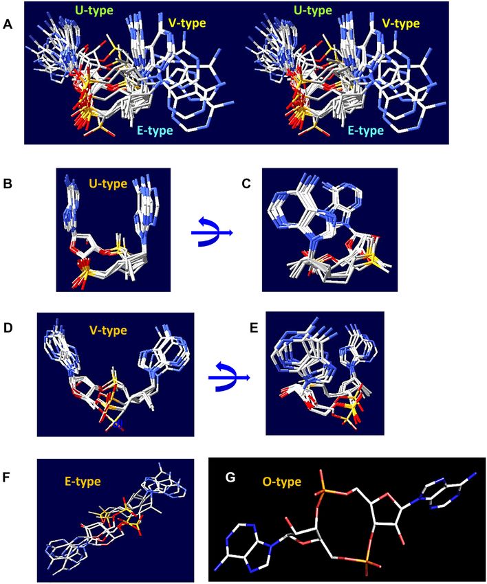
2808 Nucleic Acids Research, 2020, Vol. 48, No. 6
bination, and PgpH, which has an HD-type phosphohy-
drolase domain; see Table 1 for a comparison and refer-
ences (9–17) for reviews. A survey of the phylogenetic dis-
tribution of the respective domains identified the likely c-di-
AMP turnover machinery in most bacterial phyla and in the
archaeal phylum Euryarchaeota; see Supplementary Table
S1 or the respective entries in the Pfam and COG databases
(5,6). Remarkably, the major groups of bacteria that do not
produce c-di-AMP are alpha-, beta- and gamma subdivi-
sions of Proteobacteria (Supplementary Table S1), the same
ones that encode multiple c-di-GMP-related enzymes and
where the c-di-GMP signaling has been studied in most de-
tail (11,12). The mechanisms of c-di-AMP synthesis and hy-
Downloaded from https://academic.oup.com/nar/article/48/6/2807/5755886 by guest on 07 December 2020
drolysis and its physiological effects have been investigated
and reviewed in detail (13–20). This review is centered on
the structural aspects of c-di-AMP signaling and the spe-
cific interactions between c-di-AMP and its protein and ri-
boswitch receptors, something that has not been compre-
hensively addressed until now.
SECOND MESSENGER: SIGNAL TRANSDUCTION
BY CYCLIC DI-AMP
As a second messenger, c-di-AMP serves as a signaling
molecule that is being synthesized (or not synthesized) in re-
sponse to certain extra- and intracellular signals and trans-
mits this information by binding to certain cellular recep-
tors, RNA (riboswitches) or protein molecules. As it is, un-
fortunately, still the case with many other signaling sys-
Figure 1. Structures of c-di-GMP and c-di-AMP molecules. (A, B) U-type
conformations of c-di-GMP from the PDB entry 2RDE (in complex with
tems (21), the signals controlling c-di-AMP turnover re-
the V. cholerae protein PlzD) (A), and c-di-AMP from the PDB entry 4YP1 main poorly understood.
(in complex with the cation-proton antiporter CpaA from S. aureus) (B);
the two bases are oriented in an almost parallel fashion. (C, D) Extended Regulation of diadenylate cyclases
conformations of c-di-GMP from its complex with the EAL domain of
Klebsiella pneumoniae photoreceptor BlrP1, PDB: 3GFX (C), and c-di- All DACs characterized to date share the same DisA N do-
AMP from its complex with the RECON protein, PDB: 5UXF (D); the main (DAC, Pfam: PF02457), which consists of seven -
two bases are wide apart. (E, F) Widespread conformations of c-di-GMP,
a dimer from its complex with the cellulose synthase from Rhodobacter
strands forming a central -sheet that is flanked on both
sphaeroides, PDB: 4P02 (E), and c-di-AMP, a V-type monomer from its sides by five ␣-helices (3,22). However, they differ in the as-
complex with the YdaO riboswitch, PDB: 4QLN, with two adenine bases sociated domains (Supplementary Figure S1A), which ap-
pointed in the same direction but inclined by ∼45◦ from vertical (F). The pear to play regulatory roles (13,17). In this section, we
carbon atoms are in green (in panel E, also in light blue), N atoms are in briefly review what is known - and unknown - about these
blue, O in red and P in orange.
regulatory domains.
The most widespread version of DACs, exemplified by
the CdaA proteins of B. subtilis and Listeria monocytogenes,
1A, B) to fully stretched conformations where the bases are consists of a DAC domain (22) that is anchored in the mem-
far apart (Figure 1C, D). However, while c-di-GMP is typ- brane by three N-terminal transmembrane (TM) helices,
ically found in a dimer form with four guanine bases form- which carry several conserved residues (Supplementary Fig-
ing a parallel stack (Figure 1E), c-di-AMP is almost always ure S1B). In these two organisms, and apparently many oth-
seen in a monomer form with its adenine bases either paral- ers, the DAC activity of CdaA is controlled by the interac-
lel (Figure 1B) or arranged at an angle with each other (Fig- tion of its TM segments with the N-terminal TM fragment
ure 1F). The c-di-GMP conformations and binding mech- of the membrane-anchored extracytoplasmic protein CdaR
anisms have been analyzed in detail (8) and those of c-di- (formerly YbbR, PF07946 and COG4856), which is usu-
AMP are discussed below. ally encoded in the same conserved cdaA-cdaR operon – or
The two second messengers are also similar in that they in a longer cdaA-cdaR-glmM operon (23,24). The interac-
are both synthesized by dedicated (but distinct) nucleotidyl tion with CdaR dramatically increased the activity of CdaA
cyclase domains (DAC for c-di-AMP and GGDEF for c- from B. subtilis (23) but inhibited the enzyme from L. mono-
di-GMP) and both can be hydrolyzed by two different en- cytogenes (24). In some organisms, CdaA and CdaR are
zymes. For c-di-GMP, those hydrolases are the EAL and fused on a single polypeptide chain, forming a single pro-
HD-GYP domains, whereas c-di-AMP hydrolysis can be tein consisting of the DAC domain that is followed by one,
catalyzed by two distinct classes of phosphodiesterases, two, three, or four YbbR domains. High-resolution crys-
GdpP/DhhP, which have the DHH/DHHA1 domain com- tal structures of the ∼100-aa YbbR domain [Protein Data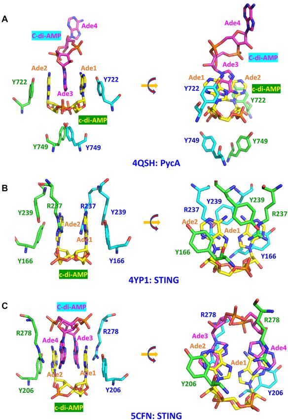
Nucleic Acids Research, 2020, Vol. 48, No. 6 2809
Table 1. Comparison of c-di-AMP- and c-di-GMP-mediated signaling
c-di-GMP c-di-AMP
Discovery year 1987 2008
Chemical properties
Structural formula C20 H24 N10 O14 P2 C20 H24 N10 O12 P2
Molecular weight 690.4 Da 658.4 Da
PDB structure code C2E (95 structures) 2BA (33 structures)
Oligomeric structure Monomer, dimer (tetramer) Monomer (dimer)
Intracellular levelsa 0.1–10 M 0.5–18.8 M
Turnover enzymes: gene and domain namesb , structures
Synthetase(cyclase) : DgcA, AdrA DisA, CdaA, CdaS, CdaM, CdaZ
GGDEF domain (PF00990, PDB: 1W25b ) DisA N (DAC) domain (PF02457, PDB: 3C1Y)
Hydrolase class I YhjH, YkuI, BlrP1 GdpP/PdeA, DhhP, DHH and DHHA1
EAL domain (PF00563, PDB: 2R6O) domains (PF01388/PF02272, PDB: 5XSN)
Hydrolase class II RpfG, PmGH, TM0186 PgpH
HD-GYP domain (COG2206, PDB: 4R8Z) HDc domain (PF01966, PDB: 4S1B)
Receptor types
Downloaded from https://academic.oup.com/nar/article/48/6/2807/5755886 by guest on 07 December 2020
Riboswitches Class I (PDB: 3IRW), Class II (PDB:3Q3Z) YdaO (PDB: 4QLN)
Bacterial proteinsa
Widespread c-di-NMP-binding domains PilZ domain (PF07238, PDB: 1YWU) RCK C domain (PF02080, PDB: 4XTT)
MshEN domain (PDB: 5HTL) CBS domain pair (PDB: 5KS7)
Histidine kinases CckA, HATPase domain (PDB: 5IDM) KdpD, USP-like domain (PDB: 2R8R)
Transcriptional regulators BldD (PDB: 5KHD); FleQ (PDB: 5EXX) BusR (RCK C domain); DarR (COG4977)
Eukaryotic proteins STING (PDB: 4EMT) STING (PDB: 6CFF)
DDX41 (PDB: 5GVR) RECON (PDB: 5UXF)
Phylogenetic distribution All bacterial phyla, Dictyostelium Most bacterial phyla, Euryarchaeota
Regulated processes Transcription, protein and polysaccharide secretion, DNA repair, sporulation, K+ transport, osmotic
motility, cell development, biofilm formation homeostasis, central metabolism, biofilm formation
a
Cyclic di-AMP levels in wild-type cells have been reported to range from 0.49–0.96 M in M. pneumoniae (33) and 1.7–5.1 M in B. subtilis (4) to 18.8 M in Synechococcus
elongatus (72).
b
Only some examples are presented, see text and Table 2 for more details and references. The domain names are from the Pfam (5) and COG (6) databases, protein and RNA
structures are from the PDB.
Bank (PDB) entries 3LYW and 5HQH] show that it consists tures such as Holliday junctions and stalled replication
of 7 -strands, sometimes with an additional helix in the forks, which inhibits c-di-AMP synthesis (3,4). The DisA-
middle, arranged in an elongated slightly bent barrel (Sup- encoding gene is often located in the conserved radA-disA
plementary Figure S1C); solution NMR structures (PDB: operon, and the RadA-type DNA repair ATPases from
2L3U, 2L5N) show much shorter -strands (25), suggest- B. subtilis, Bacillus thuringiensis, Mycobacterium smegma-
ing possible conformational flexibility of the YbbR domain. tis, Mycobacterium tuberculosis and Streptomyces coelicolor
The conserved cdaA-cdaR operon is found in the members have been shown to interact with the respective DisA pro-
of the Firmicutes and in certain Gram-negative bacteria teins, forming potential tripartite complexes with branched
from Acidobacteria, Chloroflexi, Deinococcus-Thermus, DNA that suppressed their DAC activity (29,30). Thus,
Spirochetes, Synergistes, and some other phyla (compare DisA interactions with branched DNA and/or RadA con-
COG1624 and COG4856, Supplementary Table S1). It re- trol the cellular levels of c-di-AMP, which therefore re-
mains unknown whether the YbbR domain binds pepti- flect the integrity of the chromosomal DNA and provide
doglycan or any other ligand and if so, how this signal is a sporulation checkpoint for B. subtilis. In addition to the
transmitted to its N-terminal TM segment. The product of Firmicutes, DisA-type DACs are found in the members of
the third gene of the cdaA-cdaR-glmM operon, phosphoglu- Actinobacteria, Thermotogae, and Dictyoglomi.
cosamine mutase GlmM, has been shown to interact with Certain spore-forming members of the Firmicutes, in-
CdaA and inhibit its DAC activity in B. subtilis, Lactococ- cluding B. subtilis, encode yet another form of DAC, CdaS,
cus lactis and Staphylococcus aureus, apparently by masking that combines the enzymatic DAC domain with an N-
the active site of the cyclase and preventing the formation terminal YojJ domain (Pfam: PF10372), which consists of
of the catalytically active dimers (26–28). While GlmM is an two long ␣-helices (32 and 28 aa, Supplementary Figure
essential enzyme of the peptidoglycan biosynthesis and is S1D) and is responsible for the formation of CdaS hex-
found in almost every bacterium, CdaA has a more limited amers (31,32). Studies of this enzyme in B. subtilis and B.
distribution (Supplementary Table S1). Accordingly, CdaA thuringiensis brought conflicting results. In B. subtilis, CdaS
from S.aureus has been shown to interact with its cognate was only expressed in the forespore compartment and dele-
GlmM but not with GlmMs from E. coli or Pseudomonas tion of the YojJ domain dramatically stimulated the DAC
aeruginosa, which do not encode CdaA or any other DACs activity of CdaS, prompting labeling it an ‘autoinhibitory
(28). A crystal structure of the CdaA-GlmM complex from domain’ (31). In B. thuringiensis, CdaS was expressed in
B. subtilis should help to understand the mechanisms of stationary-phase cells, stimulating sporulation and paras-
such selectivity. poral crystal formation, while the YojJ domain was required
The DNA integrity scanning protein DisA, the first for the DAC activity (32). B. subtilis spores were suggested
experimentally characterized form of DAC, is the only to contain some ligand that could interact with the YojJ do-
one where the signal controlling c-di-AMP synthesis has main to allow the formation of the catalytically active CdaS
been identified. DisA combines the DAC domain with a dimers and thereby stimulate c-di-AMP production in the
long linker domain and a C-terminal DNA-binding helix- germinating spore (31) but the nature of that ligand remains
hairpin-helix domain that binds to branched DNA struc- unknown.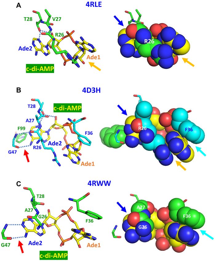
2810 Nucleic Acids Research, 2020, Vol. 48, No. 6
An experimentally characterized DAC from Mycoplasma Historically, the first characterized PDE was the GdpP
pneumoniae (33), referred to as CdaM, has a single N- (formerly YybT) enzyme from B. subtilis, a multidomain
terminal TM segment (an uncleavable signal peptide) that protein that, in addition to the C-terminal DHH and
anchors the enzyme in the membrane. Although it is tempt- DHHA1 enzymatic domains, contains two N-terminal
ing to suggest that this TM segment allows CdaM activity TM segments, followed by a ligand-binding PAS do-
to be regulated by the status of the membrane (e.g. its tur- main and a degenerate GGDEF (xGGDEF) domain (41).
gor), there is no data to support or disprove this idea. The construct consisting solely of the DHH/DHHA1 do-
Many members of the archaeal phylum Euryarchaeota main combination (which is similar to the bifunctional
encode CdaZ/DacZ-type DACs that combine the DAC do- oligoribonuclease/pAp phosphatase NrnA of B. subtilis,
main with a specific version of the PK C (Pfam: PF02887) PDB: 5IPP) was catalytically active, hydrolyzing c-di-AMP
domain (Supplementary Figure S1E), which serves as the with the Km of 1.3 M and Kcat of 0.55 s−1 (41). The PAS
ligand-binding domain for allosteric regulators of pyruvate domain was shown to play a regulatory role: it binds b-
kinase activity, such as pyruvate and glucose 6-phosphate type heme, which in turn can bind NO and CO; binding of
Downloaded from https://academic.oup.com/nar/article/48/6/2807/5755886 by guest on 07 December 2020
(34–36). This 110-aa ␣/ domain (Supplementary Figure NO, but not CO, stimulated the PDE activity of GdpP (46).
S1F) has also been seen binding such osmolytes as proline The role of the xGGDEF domain remains obscure; it was
and glycerol (34), which would be in line with the role of shown to possess some ATPase activity (41) but whether it
c-di-AMP in maintaining the osmotic balance in bacteria contributes to the regulation of the PDE activity remains
(see below). However, the residues that bind these osmolytes unknown. GdpP can also interact with the stringent re-
are not conserved in the CdaZ version of the PK C do- sponse factor (p)ppGpp, which serves as a competitive in-
main (Supplementary Figure S1E). While the DAC activity hibitor of its PDE activity (41). The phylogenetic distribu-
of CdaZ-type enzymes from Methanocaldococcus jannaschii tion of GdpP-type PDEs is limited to the Firmicutes and
and Haloferax volcanii has been experimentally demon- Tenericutes (Mollicutes), see Supplementary Table S1 or
strated (37,38), the ligands sensed by their N-terminal COG3887 in the COG database (6).
PK C domains remain to be identified. It is also not known The second type of c-di-AMP-specific PDE, PgpH, has
if ligand binding activates or inhibits the DAC activity of been initially characterized in B. subtilis and L. monocyto-
these enzymes. genes (26,42) and subsequently described in the cyanobac-
Certain members of the Planctomycetes, Verrucomicro- terium Synechocystis sp. PCC 6803 (47). This protein con-
biae, and Spirochetes phyla encode DACs that combine sists of three distinct domains: an extracellular 7TMR-
the DacZ architecture with yet another N-terminal reg- HDED domain preceded by a signal peptide, an inte-
ulatory domain (Supplementary Figure S1A), the 150- gral membrane domain with 7 TM helices (7TM-7TMR-
aa EIIA-like domain (Pfam: PF00359) of the phos- HD), and a cytoplasmic HD-type phosphodiesterase do-
phoenolpyruvate:sugar phosphotransferase system (PTS), main (14). The extracellular ligands sensed by the 7TMR-
which contains a conserved His residue (Supplementary HDED domain and presumably controlling the PDE ac-
Figure S1G,H) that can be phosphorylated in a PEP- tivity of PgpH still remain unknown. PgpH plays an im-
dependent reaction via the Enzyme I – HPr protein cascade portant role in cold shock response of L. monocytogenes: a
of the PTS (39). None of these DACs have been character- pgpH mutant was cold-sensitive, while cold-adapted strains
ized so far, and the regulation, if any, imposed by the EIIA- showed a 9- to 76-fold increase in the expression of the
like domains remains obscure. pgpH gene (48,49). This protein has a wide phylogenetic
Finally, as noted back in 2008 (3,7), some archaeal DACs distribution, being found, besides Firmicutes, in members
contain N-terminal PAS domains. These enzymes, found in of Bacteroidetes, Chlamydiae, Chloroflexi, Cyanobacteria,
the euryarchaeal order Methanomicrobiales, have the REC– Fusobacteria, Planctomycetes, Spirochaetes, and other bac-
PAS1–2 –DAC domain architecture, combining the ligand- terial phyla, as well as in the delta subdivision of Proteobac-
binding PAS domain with the phosphoacceptor receiver teria, see Supplementary Table S1 or COG1480 in the COG
(REC) domain (Supplementary Figure S1A). This puts the database (6).
DAC activity of these enzymes under the control of the two- The observation that the DHH/DHHA1 domain com-
component signal transduction systems, which are particu- bination of GdpP was capable of hydrolyzing c-di-AMP
larly complex in the organisms of this group (40). (41) suggested that the DhhP-type enzymes (COG0618),
There are also other forms of DACs with additional do- which consist solely of these two domains, should also func-
main architectures, such as DAC–TPR5 in Actinosynnema tion as c-di-AMP-specific PDEs. This suggestion proved to
mirum protein Amir 4183 (Supplementary Figure S1A), be partly correct, as this activity has been verified in sev-
which might be regulated by protein-protein interactions, eral members of the DhhP family from Borrelia burgdor-
but their partners remain to be identified. feri, M. smegmatis, M. tuberculosis, M. pneumoniae, S. au-
reus, Streptococcus pneumoniae and T. maritima (33,43,50–
54). However, other members of the same family have been
shown to also – or instead – catalyze the second step of c-di-
Regulation of c-di-AMP phosphodiesterases
AMP degradation, hydrolysis of pApA to AMP (33,51,53).
Two major classes of c-di-AMP-hydrolyzing phosphodi- Indeed, Supplementary Table S1 shows that many bac-
esterases (PDEs) have been described, those containing the teria encode two paralogous members of this family, of-
DHH/DHHA1 domain combination (GdpP and DhhP ten along with either GdpP (Mollicutes, Negativicutes) or
families) and those containing the HD phosphohydrolase PgpH (Bacteroidetes, Chloroflexi, Cyanobacteria, Plancto-
domain (26,41–45). mycetes, Spirochaetes, Thermotogae). However, in archaea,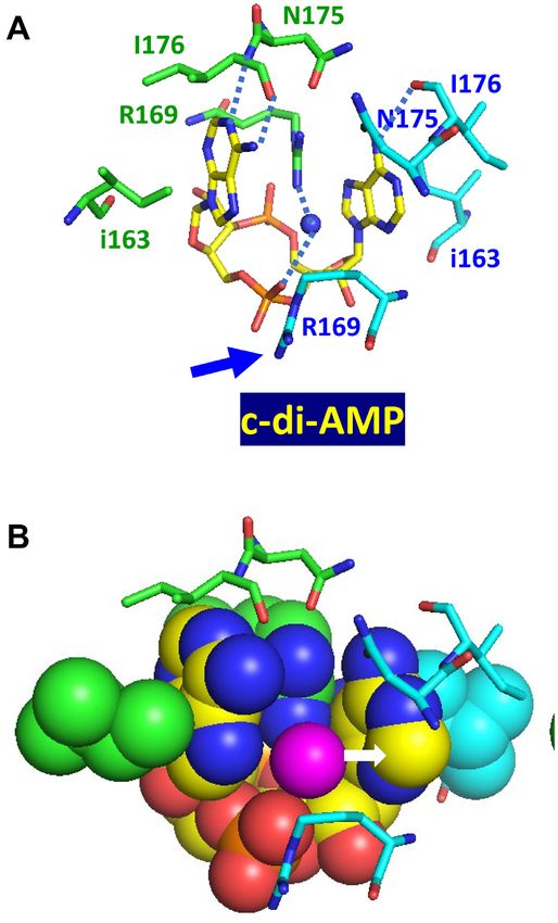
Nucleic Acids Research, 2020, Vol. 48, No. 6 2811

most members of Actinobacteria and in some members                  (Table 2). By contrast, the majority of B. subtilis CBS-
of Bacteroidetes, GdpP and PgpH are both missing and                containing proteins do not bind c-di-AMP (66). Accord-
the DhhP family enzymes are the only c-di-AMP hydro-                ingly, only a relatively small fraction of the widespread
lases (51,52). Further, Chlamydia trachomatis, Chlamydia            CBS domains (Supplementary Table S1) can be expected
avium and Chlamydia muridarum encode a CdaA-type DAC                to function as c-di-AMP receptors; those associated with
but do not encode any of the known c-di-AMP PDEs (Sup-              the voltage-gated chloride channel (PF00654) and mag-
plementary Table S1); the mechanisms of c-di-AMP hydrol-            nesium transporters of the CNNM (cyclin M, PF01595)
ysis in such organisms remain enigmatic.                            family look like plausible candidates.
                                                                 • K+ transport-regulating histidine kinases KdpD from
                                                                    several bacteria have been shown to bind c-di-AMP (Ta-
Signal transduction by c-di-AMP                                     ble 2) and the binding site has been localized to the USP-
The above sections show that the cellular levels of c-di-AMP        like (universal stress protein, Pfam: PF00582) domain of
can be regulated both by affecting its synthesis and hydrol-        this protein (59). In addition, binding of c-di-AMP by

                                                                                                                                   Downloaded from https://academic.oup.com/nar/article/48/6/2807/5755886 by guest on 07 December 2020
ysis. Studies of the bacteria with elevated or decreased c-di-      the K+ uptake protein KimA has been localized to its C-
AMP levels revealed its involvement in the following pro-           terminal cytoplasmic domain (62), which also belongs to
cesses:                                                             the USP superfamily (Supplementary Figure S2).
                                                                 • Two closely related c-di-AMP-regulated K+ uptake pro-
• Monitoring DNA damage (3,4)                                       teins from L. lactis, KupA and KupB (82), combine a 12-
• Controlling sporulation (4,31,32)                                 TM membrane transporter domain with a previously un-
• Controlling cell size and peptidoglycan homeostasis               characterized C-terminal cytoplasmic domain. This do-
  (53,55,56)                                                        main, which we refer to as the KupAC domain (Supple-
• Controlling osmotic pressure and envelope stress (16,55)          mentary Figure S3), can be predicted to serve as yet an-
  - K+ transport (33,57–62)                                         other receptor of c-di-AMP.
  - Osmolyte transport (63–65)                                   • Several more proteins, listed in Table 2, do not appear
  - Mg2+ transport (66)                                             to have dedicated c-di-AMP-binding domains. Instead,
• Controlling biofilm formation (67,68)                             c-di-AMP binding occurs at allosteric sites on the pro-
• Regulation of central metabolism, growth rate (50,69,70)          tein surface. One of such c-di-AMP binding proteins is
• Regulation of fatty acid biosynthesis (51,71)                     structurally related to the family of PII-like signal trans-
• Response to acid stress (43)                                      duction proteins that are involved in many pathways, of-
• Response to cold shock (48,49,71)                                 ten associated with nitrogen metabolism. It was originally
• Light-dark cycle in cyanobacteria (72)                            named PstA (PII-like signal transduction protein) (57),
• Regulation of competence (73).                                    which caused certain confusion, as that name had been
                                                                    previously used for the widespread membrane permease
   Finally, c-di-AMP contributes to virulence of several bac-       subunit of the bacterial ABC-type phosphate transporter
terial pathogens (50,53,74–79).                                     (83). In B. subtilis, this protein has been renamed from
   All these responses are mediated by c-di-AMP binding to          YaaQ to DarA (c-di-AMP receptor A) (84), which was
its RNA and protein receptors, which come from several dif-         not ideal either because the darA symbol is also used for
ferent protein families (Table 2). The principal c-di-AMP-          the genes for darobactin family peptide antibiotic and the
binding domains are as follows.                                     phage defense against restriction protein A. Since both
                                                                    DarA and PstA designations are used in the literature
• The RCK C (regulator of conductance of K+ C-terminal)             (69,84–87), we refer to this protein here as DarA/PstA.
  domain, referred to as the TrkA C (PF02080) domain             • Other c-di-AMP receptors without dedicated binding do-
  in Pfam (5). Four distinct classes of c-di-AMP recep-             mains but with experimentally characterized c-di-AMP
  tors containing this domain have been characterized; in           binding sites include pyruvate carboxylase and the eu-
  two cases, the structures of c-di-AMP–RCK C complexes             karyotic STING and RECON proteins.
  have been solved (Table 2). So far, most experimentally           Physiological responses to c-di-AMP have been described
  studied RCK C domains have been found to bind c-di-            in detail in several comprehensive reviews (13–20), which is
  AMP (66), although they are also widespread in alpha-,         why in subsequent sections, we discuss primarily the mech-
  beta- and gamma-proteobacteria that do not produce c-          anistic aspects of c-di-AMP-binding.
  di-AMP. Several widespread RCK C-containing domain
  architectures, including its associations with voltage-
                                                                 PRIMARY IMPORTANCE: ESSENTIALITY OF CYCLIC
  gated chloride channel (PF00654), sodium-sulfate sym-
                                                                 DI-AMP
  porter (PF00939), citrate transporter (PF03600) and
  aspartate-alanine exchanger (PF06826), remain to be ex-        Uniquely among known second messengers, c-di-AMP
  perimentally characterized.                                    plays important roles in representatives of all three domains
• Pairs of the CBS (cystathionine beta-synthase) domain          of life. Most bacteria and some archaea can produce c-di-
  (Pfam: PF00571), also referred to as the Bateman do-           AMP and it has been found to be essential for the growth of
  main (80,81), bind a variety of adenine derivatives and are    bacteria (23,26,33,56) and, recently, for an archaeon (38). In
  found in a wide variety of domain architectures. Two of        eukaryotic cells, c-di-AMP is not synthesized; its presence
  these domain combinations, along with the stand-alone          in cell cytoplasm is sensed by such proteins as STING, RE-
  CBS domain pairs, have been shown to bind c-di-AMP             CON, DDX41 and ERAdP and is perceived as a sign of an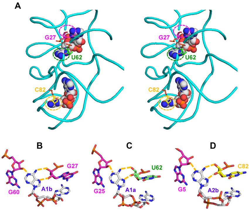
2812 Nucleic Acids Research, 2020, Vol. 48, No. 6
Table 2. Regulatory domains of c-di-AMP receptorsa
Receptor typeb Characterized examples
Structure, PDB
Organismc , accession numbers coded Estimated Kd , Me References
Riboswitch
ydaO (potE) Bacillus subtilis 4W90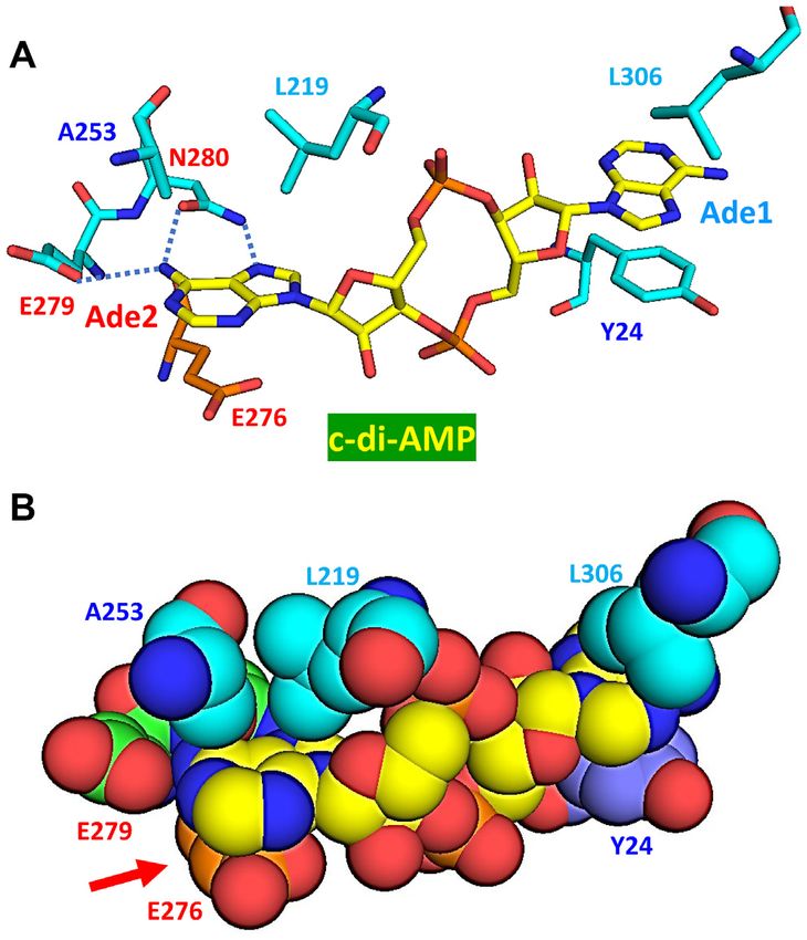
Nucleic Acids Research, 2020, Vol. 48, No. 6 2813
infection and a signal to trigger an immune response by the The essentiality of c-di-AMP is probably enhanced by its
host cells. other regulatory roles, such as the interaction of the c-di-
AMP riboswitch YdaO with its multiple targets (100) and
its involvement in controlling central metabolism (69,70,90)
and aerobic respiration (91) of bacterial cells.
Essentiality of c-di-AMP in bacteria
The very first attempts to prepare deletion mutants lacking c-di-AMP, an alarmone in eukaryotes
the DAC activity showed that c-di-AMP was essential for
such bacteria as B. subtilis (23,56), S. aureus (55), L. mono- Along with other cyclic nucleotide second messengers, c-
cytogenes (77), S. pneumoniae (53), Streptococcus agalactiae di-AMP serves as a signaling molecule that can be recog-
(88) and other species. The essentiality of c-di-AMP was nized by eukaryotic receptors and trigger an immune re-
also observed for the spirochete Borrelia burgdorferi (50), sponse in the host cells (101). The importance of this re-
the cell wall-less mollicute M. pneumoniae (33), and the ar- sponse is such that it uses several redundant mechanisms
Downloaded from https://academic.oup.com/nar/article/48/6/2807/5755886 by guest on 07 December 2020
chaeon H. volcanii (38), showing that this phenomenon is and relies on c-di-AMP binding by at least four distinct
not limited to the members of Firmicutes or just Gram- receptors. These include STING (stimulator of interferon
positive bacteria. In contrast, a disA deletion mutant of M. genes, formerly Tmem173), DDX41 (DEAD-box helicase
tuberculosis was viable; it showed no c-di-AMP production 41), RECON (reductase controlling NF-B, also known
and decreased virulence but no slowdown of growth (79). as aldo-keto reductase family 1 member C13, encoded by
A viable c-di-AMP-less mutant has also been constructed Akr1c13), ERAdP (CTD nuclear envelope phosphatase 1
in another actinobacterium, Streptomyces venezuelae, al- regulatory subunit 1 or C16orf69, encoded by Cnep1r1,
beit not in S. coelicolor (89). Likewise, in the cyanobac- formerly Tmem188), and possibly NLRP3 (C1orf7) in-
terium Synechococcus elongatus, a cdaA transposon mutant flammasome (102–106). STING and DDX41 trigger in-
with a dramatically decreased c-di-AMP level was viable but nate immunity response by activating the production of in-
showed a disrupted light-dark cycle (72), whereas a cdaA terferon, RECON and ERAdP help in the activation of
mutant of Synechocystis could not be obtained (47). On a pro-inflammatory antibacterial state, whereas NLRP3
the other hand, c-di-AMP proved to be non-essential for stimulates secretion of interleukin-1. In addition to c-di-
L. monocytogenes and S. aureus cells grown in chemically AMP, STING binds other cyclic dinucleotides, including c-
defined minimal media (90,91). In B. subtilis, a c-di-AMP- di-GMP and c-GMP-AMP (3 ,3 -cGAMP) synthesized by
lacking mutant could also grow on minimal media, but only bacterial pathogens, as well as the 2 ,3 -cGAMP that is syn-
at low (0.1 mM) levels of K+ ions (60). These observations thesized by the host cGMP-AMP synthetase (cGAS), acti-
indicated that the essentiality of c-di-AMP was dependent vated by the presence of foreign DNA (102,107). RECON
on the growth conditions, or, more generally, on its distinct binds c-di-AMP and 3 ,3 -cGAMP but not c-di-GMP or
roles as a second messenger. 2 ,3 -cGAMP (104). We discuss the detailed mechanisms of
Judging from the complementation studies, properties c-di-AMP binding by STING and RECON in the next sec-
of the DAC-missing suppressor mutants, and the func- tion.
tions of c-di-AMP-binding proteins, of all the c-di-AMP- The ability of the eukaryotic cells to sense c-di-AMP
mediated processes mentioned above, the most universal – raises several interesting questions. For example, is c-di-
and the most likely to make it essential for a variety of di- AMP production beneficial or detrimental for the host-
verse organisms––is maintaining the osmotic homeostasis associated bacteria? Group B streptococci actively degrade
through regulation of K+ uptake and, as a result, of the secreted c-di-AMP, ostensibly to decrease the inflammatory
cellular level of this cation (58–62,65,92,93). K+ plays sev- response by the host (108). In contrast, c-di-AMP secretion
eral important roles in bacterial, archaeal and eukaryotic by L. monocytogenes enhanced the cell-to-cell spread of this
cells, both as a major cation and as a component of the ri- organism between different hepatocytes, suggesting that it
bosome and a cofactor for some key cellular enzymes that could benefit the pathogen (109). In any case, intracytoplas-
cannot be replaced, e.g. by Na+ ions (94,95). It has been mic surveillance for foreign nucleic acids and individual nu-
proposed that the first live cells originated in a K+ -rich en- cleotides is an important phenomenon that deserves further
vironment (96) and only later, after the acquisition of so- study and promises a better insight into the immune mech-
phisticated ion pumps, ventured into the habitats with the anisms of the eukaryotic cell.
prevalence of Na+ over K+ (95). Some of the ancestral and
most widespread ATPases appear to be K+ -dependent (97). TERTIARY STRUCTURE: MECHANISMS OF CYCLIC
However, most environments contain far less K+ than Na+ . DI-AMP BINDING
Accordingly, most organisms possess a variety of K+ up-
Diversity of c-di-AMP conformations
take systems that allow them to maintain the K+ gradi-
ent with [K+ ]in >> [K+ ]out (98). Obviously, these systems As discussed above, c-di-AMP regulates a wide range of
are not foolproof and need to be regulated to prevent the functions in bacteria. This variety is partly due to the
unlimited accumulation of K+ ions, particularly when the diversity of conformations that c-di-AMP adopts when
cells grow in the media with relatively high K+ levels. In binding to diverse cellular receptors. A comparison of the
all cases where it has been studied, c-di-AMP was found to crystal structures of the c-di-AMP-protein and c-di-AMP-
do exactly that, by inhibiting K+ uptake via both high- and riboswitch complexes available in the PDB reveals a vari-
low-affinity transporters and by stimulating K+ exit via the ety of conformations of this interesting molecule (Figures 1
K+ /H+ antiporter CpaA (99). and 2). We could identify at least four principal conforma-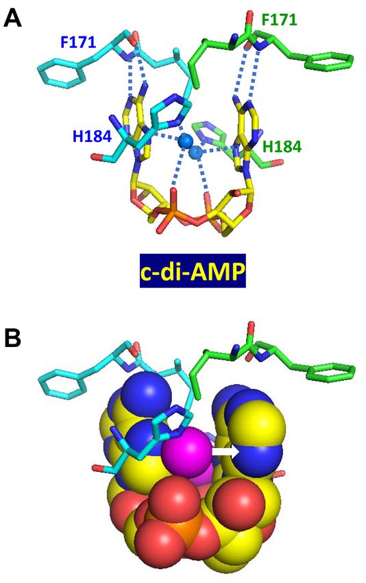
2814 Nucleic Acids Research, 2020, Vol. 48, No. 6

                                                                                                                                                          Downloaded from https://academic.oup.com/nar/article/48/6/2807/5755886 by guest on 07 December 2020
Figure 2. Diversity of c-di-AMP conformations. (A) Stereo view of three main conformations of c-di-AMP: the U-, V- and E-types. (B, C) Superposition of
U-type c-di-AMP molecules from PDB entries 4QSH1 , 4XTT, 4YP1, 5CFN, and 5F29 displayed from two different angles. (D,E) Superposition of V-type
c-di-AMP molecules from PDB entries 4RLE, 4RWW, 4WK1, 4D3H and 4S1B displayed in two different angles. (F) E-type c-di-AMP molecules from
PDB entries 5XSN, 5UXF and 4QSH2. (G) The O-type c-di-AMP from PDB entry 5KS7.

tion types, two of which have been discussed before (99,110),                 transport proteins KtrA (PDB: 4XTT) and CpaA (PDB:
while the other two have not.                                                 5F29), which both bind c-di-AMP via their C-terminal
                                                                              RCK C (PF02080) domains (99,111). This conformation of
U-type. The most widespread is the ‘U-type’ conforma-                         c-di-AMP was also seen in the structure of the pyruvate
tion of c-di-AMP, in which the two adenine bases are ori-                     carboxylase PycA from L. monocytogenes (PDB: 4QSH),
ented in an almost parallel fashion (Figures 1B and 2B).                      which binds c-di-AMP at the interface between the enzyme
This conformation was first seen by Witte and colleagues in                   monomers (69). In one of the crystal forms of PycA, how-
the structure of the T. maritima protein DisA (PDB: 3C1Y)                     ever, there is also another c-di-AMP molecule that bridges
that led to the initial discovery of c-di-AMP in a biologi-                   two c-di-AMPs from two different PycA monomers; each of
cal system (3). After that, a very similar U-type conforma-                   its adenine bases fits into the cavity between the adenines of
tion has been observed in the structures of S. aureus K+                      the U-shaped c-di-AMP (Supplementary Figure S4). The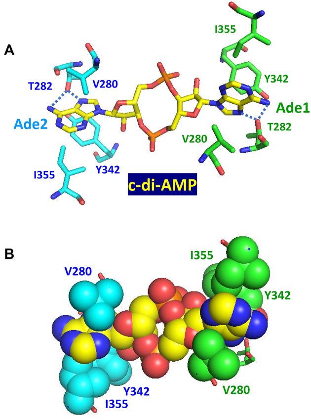
Nucleic Acids Research, 2020, Vol. 48, No. 6 2815
U-type conformation of c-di-AMP has also been seen in interactions that contribute to the tight binding of these
its complexes with the murine (PDB: 4YP1, Supplemen- cyclic dinucleotides.
tary Figure S4C) and porcine (PDF: 6IYF) molecules of
the STING protein (112). One more STING structure, the Hydrogen bonds. In our earlier review of c-di-GMP bind-
one from the sea anemone Nematostella vectensis (PDB: ing mechanisms, we noted the nearly universal presence
5CFN), contains two U-shaped c-di-AMP molecules (Sup- of Arg residues in all kinds of c-di-GMP-binding recep-
plementary Figure S4D), whose adenine bases are stacked tors (8). In most cases, these Arg residues formed hydro-
in a pairwise manner (113). A similar U-shaped conforma- gen bonds with O6 and N7 atoms of the guanine Hoog-
tion is also adopted by 3 ,3 -cGAMP in its complexes with steen edge, whereas Asp and Glu residues often formed
its synthase cGAS (PDB: 5VDT) and its riboswitch (PDB: hydrogen bonds with N1 and N(2) atoms of the Watson-
4YB1) (114,115). Crick edge of the guanine base (8); see reference (119) for
the nomenclature. In contrast, Arg residues are rarely in-
V-type. The structure of the c-di-AMP-specific PDE volved in c-di-AMP binding. Instead, there is a wide vari-
Downloaded from https://academic.oup.com/nar/article/48/6/2807/5755886 by guest on 07 December 2020
PgpH (42) from L. monocytogenes (PDB: 4S1B) revealed a ety of hydrogen bonds formed between N1, N3, N6 (amino
slightly different conformation, in which the adenine bases group), and N7 atoms of c-di-AMP adenines and assorted
point in the same direction but the respective glycosidic residues of the receptor proteins, either through their side-
bonds are inclined by approximately 45◦ , leaving more open chain groups (Asn, Thr or Glu) or backbone atoms (see be-
space between the adenines (Figures 1F and 2D). This con- low). Binding of c-di-AMP by its YdaO riboswitch is also
formation, referred to as ‘V-type’ (99), has been also ob- supported by multiple hydrogen bonds (Figure 3). In most
served in the c-di-AMP-riboswitch complexes from B. sub- cases, these hydrogen bonds are formed by adenine-specific
tilis (PDB: 4W90) and three thermophilic bacteria (PDB: nitrogen atoms and contribute to the specificity of the c-di-
4QLN, 4QK8 and 4QK9) (116–118). Essentially the same AMP binding.
V-type conformation is seen in the structures of the c-di-
AMP receptor DarA/PstA from S. aureus (PDB: 4D3H and π –π interaction. Base-base stacking has been found to be
4WK1), B. subtilis (PDB: 4RLE), and L. monocytogenes very important for stabilizing the DNA duplex structure.
(PDB: 4RWW) (84–87). The c-di-AMP molecule bound Accordingly, Phe and Tyr (and sometimes Trp) residues can
to the pyruvate carboxylase from L. lactis (PDB: 5VYZ) stack the adenine base of c-di-AMP with their side-chain
also displays a V-type conformation (Supplementary Fig- aromatic groups. Such an interaction has also been seen in
ure S4A), in contrast to the U-shaped one in L. monocy- the case of c-di-GMP binding to its receptors (8).
togenes (PDB: 4QSH) (Supplementary Figure S4B). One
could speculate that at higher concentrations of c-di-AMP, Cation–π interaction (mainly by the Arg guanidino cation).
the second c-di-AMP molecule binds to first one, insert- Cation– interaction has been found to provide a signif-
ing one of its bases between the two adenines of the first icant contribution to the stabilization of protein–cofactor
molecule and converting it from the V-shape into a U-shape and enzyme–substrate interactions (120–123). In c-di-
(Supplementary Figure S4C, D). GMP-binding proteins, Arg residues exhibited two distinct
types of interaction by their side-chain guanidino group: (i)
E-type. The third type of c-di-AMP conformation, seen binding the Hoogsteen side of the guanine base and (ii) pla-
in its complex with the murine RECON protein (PDB: nar stacking against the guanine base, causing significant
5UXF), is almost flat (Figures 1D and 2F) with the two ade- stabilization of the c-di-GMP/receptor complexes (8). Al-
nine bases looking in the opposite directions (104). We refer though the Arg guanidino group cannot bind adenine base
to this extended conformation of c-di-AMP as the ‘E-type’. from its Hoogsteen side (99), stacking with the adenine base
O-type. Finally, c-di-AMP bound to the c-di-AMP- is still possible, so the cation (guanidino group)– interac-
specific PDE GdpP from S. aureus (PDB: 5XSN) displays tion plays an important role in c-di-AMP–receptor interac-
another type of extended conformation with one adenine tions.
base almost orthogonal to the other one (45). We refer to
this conformation as ‘O-type’ (Figure 2G). The same con- Polar–π interaction (by the backbone peptide bond or from
formation is adopted by c-di-AMP in its complex with the Asn or Gln side-chain amide bond). Since the peptide bond
CBS domain dimer (PF00571) in the carnitine transporter is both planar and polar, it has been shown to stack with
subunit OpuCA from L. monocytogenes (PDB: 5KS7) (63) a guanine base of c-di-GMP bound to the allosteric in-
and by the second c-di-AMP molecule in the pyruvate car- hibitory site (I-site) of the GGDEF domain that suppresses
boxylase PycA structure from L. monocytogenes (the upper its diguanylate cyclase activity (124). In a similar fashion,
one in Supplementary Figure S4B) (69). c-di-AMP can interact with a protein receptor through the
Obviously, as has been previously observed for c-di-GMP wide upper region of its V-shape (the U-type conformation
(8), the distinct shapes of the c-di-AMP molecule allow is too narrow to accommodate a peptide segment, see be-
distinct modes of its interactions with diverse protein and low). Since Asn and Gln side-chain amide groups are also
RNA targets. planar, they can also stack with the adenine base.
C-H bond–π interaction. Although the C–H bond has
Non-covalent interactions involved in c-di-NMP binding
no charge or polarity, it can still interact with an aro-
Analysis of the c-di-AMP- and c-di-GMP-binding proteins matic group when it is situated perpendicularly to an aro-
and riboswitches identified the following principal types of matic ring (122,125–127). Such a special interaction mode
2816 Nucleic Acids Research, 2020, Vol. 48, No. 6
Downloaded from https://academic.oup.com/nar/article/48/6/2807/5755886 by guest on 07 December 2020
Figure 3. Binding of c-di-AMP to the YdaO riboswitch. (A) Stereo view of two c-di-AMP molecules (shown as spheres) bound to the YdaO riboswitch from
B. subtilis (PDB: 4W90), whose sugar-phosphate backbone is shown in cyan (116). C-di-AMP-binding ribose moieties of G27, U62, and C82 are shown
in stick representation and indicated by dashed circles. (B–D) Role of the ribose hydroxyl groups of the riboswitch in binding c-di-AMP (carbon atoms in
grey). Hoogsteen side hydrogen bonds between N6 and N7 atoms of adenine bases of c-di-AMP and the ribose 2 - and 3 -hydroxyls from riboswitch bases
G27 (B), U62 (C), and C82 (D) are coupled with adenine-N1 binding by 2 -hydroxyl groups of G60 (B), G25 (C) and G5(D). The abundance of hydrogen
bonds explains the much higher affinity of c-di-AMP binding by the riboswitch than by protein receptors.
is present in c-di-GMP–receptor complexes (8) and can also molecule is situated on top of an adenine base and coor-
be seen in the complex of c-di-AMP with the carnitine trans- dinates with three surrounding chemical moieties (see be-
porter OpuCA (64). low). A similar interaction can also be seen in the recently
reported complex (PDB: 6PFJ) of the S. venezuelae sigma
Hydrophobic–π interaction. The previous type of interac- factor WhiG with an anti-sigma RsiG and two c-di-GMP
tion depended on perpendicularity in forming the C–H– molecules (134,135).
bond. However, even when the C–H bond is not located
directly above the aromatic ring, it can still contribute to
the hydrophobic–aromatic group interaction. For example, c-di-AMP binding by the YdaO riboswitch
a triple Leu– interaction was shown to be crucial for c-di-
GMP binding to the MshEN domain, where replacement Like its sibling c-di-GMP, c-di-AMP has a specific ri-
of any of those Leu residues reduced the binding affinity boswitch that binds it with a very high affinity (Kd in
at least 10-fold (128). However, two nearby hydrophobic sub-nanomolar to nanomolar range), see Table 1 and ref-
groups should be enough for binding and long and bulky erence (100). While the structures of these riboswitches
hydrophobic side chains of Leu, Ile, Phe or Val can all par- are not related, changing just two bases in the c-di-GMP-
ticipate in such a ‘hydrophobic–’ interaction. binding riboswitch GEMM was enough to convert it into
one with a 4-fold preference for c-di-AMP over c-di-GMP
Anion–π interaction. While no anion– interaction has (136,137). Surprisingly, this apparently never happened in
been seen in the c-di-GMP receptor complexes, such an in- the course of the riboswitch evolution and the structure of
teraction is theoretically possible (122,129,130). Indeed, the the c-di-AMP-specific riboswitch YdaO is dramatically dif-
planar carboxylate group of Glu has been found to stack ferent from both class I and class II c-di-GMP-binding ri-
upon the adenine base of c-di-AMP in its complex with the boswitches. The riboswitch GEMM-I from Geobacter sul-
RECON protein (109), see below. furreducens has been shown to bind c-di-GMP and 3 ,3 -
cGAMP, but not c-di-AMP or 2 ,3 -cGAMP (138).
Lone pair–π interaction. In addition to a cation, neutral Structures of the c-di-AMP-riboswitch complexes from
C-H bond, and an anion that all interact with systems, B. subtilis (PDB: 4W90) and the thermophilic bacteria Cal-
a lone pair of electrons of the H2 O molecule can do that danaerobacter subterraneus (PDB: 4QLN), Thermoanaer-
as well (122,131–133). Such an interaction has been found obacter pseudethanolicus (PDB: 4QK8) and Thermovirga
in the crystal structures of c-di-AMP-bound RCK C do- lienii (PDB: 4QK9) revealed very similar binding modes
mains of CpaA and KtrA proteins (99,111). Here, a water that combined partial stacking of the adenine bases of c-Nucleic Acids Research, 2020, Vol. 48, No. 6 2817
di-AMP by the adenine bases of the riboswitch with mul-
tiple hydrogen bonds between the atoms of the ligand and
the riboswitch (116–118). As already noted, the planes of
these adenine bases are not parallel, they form a 10–30◦
angle (117). However, Hoogsteen edge atoms of c-di-AMP
adenines form hydrogen bonds with the ribose hydroxyls
of the sugar-phosphate backbone of the riboswitch, par-
ticularly those of U62, G27, and C82, as shown in Figure
3. In addition, the 2 -hydroxyls of c-di-AMP interact with
several bases of the riboswitch, whereas the central ribose-
phosphate ring of c-di-AMP (which is also present in c-di-
GMP) does not seem to form any bonds, ensuring bind-
ing specificity and the absence of competition by c-di-GMP.
Downloaded from https://academic.oup.com/nar/article/48/6/2807/5755886 by guest on 07 December 2020
Thus, several components of the c-di-AMP ligand partici-
pate in interactions with the riboswitch, which probably ac-
counts for the extremely tight binding (116–118).
Binding mechanisms in c-di-AMP–protein complexes
Crystal structures of several c-di-AMP–protein complexes
have been solved (Table 2), which allows taking a close look
at the c-di-AMP binding mechanisms.
c-di-AMP–RCK C domain complex. The c-di-AMP-
binding RCK C domain is a Rossmann-fold domain that
is found at the C-termini of several distinct potassium
transporters, including the K+ uptake protein KtrA,
K+ /H+ antiporter subunit KhtT, and Na+ /H+ and K+ /H+
antiporter CpaA; it is also found in the transcriptional
regulator BusR (Table 2). In its complex with a CpaA
dimer from S. aureus (PDB: 5F29), the U-shaped c-di- Figure 4. Binding of c-di-AMP to CpaA. Complex of U-shaped c-di-AMP
AMP is coordinated by lone-pair electrons from two water with two RCK C domains of S. aureus K+ /H+ antiporter CpaA (PDB:
5F29) (99) shows each water molecule (solid blue circles in (A) and magenta
molecules located between the adenine bases (Figure 4). spheres in (B)) coordinated by the N7 atom and phosphate oxygen atom of
Each of these water molecules is coordinated by the N7 c-di-AMP, as well as the N atom from the side chain of His184. The lone
atom of the adenine base and a phosphate oxygen atom pair electrons of oxygen atom form a water-mediated lone pair–adenine
from c-di-AMP, as well as the nitrogen atom from the interaction (indicated by a white arrow in B). The N1 and N6 atoms of c-
di-AMP adenines bind the backbone nitrogen and carbonyl oxygen atoms
side chain of His184, leaving a pair of electrons to form a of Phe171, respectively, of both RCK C domains.
lone pair–adenine interaction (indicated by a white arrow
in Figure 4B). Indeed, replacement of this His184 with
Ala resulted in a >300-fold weaker binding of c-di-AMP arrow in Figure 5B). In addition, the c-di-AMP molecule
(99). In addition, N1 and N6 atoms of each adenine base forms hydrogen bonds with the backbone nitrogen and car-
of c-di-AMP form hydrogen bonds with the backbone bonyl oxygen atoms, respectively, of the Asn175 residue.
nitrogen and carbonyl oxygen atoms, respectively, of the Thus, the same U-shaped molecule of c-di-AMP is bound
Phe171 residues of the CpaA dimer, while the oxygen atoms by the two RCK C domains by the same types of interac-
of the central ribose-phosphate ring form hydrogen bonds tions, albeit provided by totally different residues.
with the backbone nitrogen atom of Gly185. With the
exception of the last one, all these interactions are specific c-di-AMP–CBS domain complex. Interaction of c-di-
for the adenine base and account for the specificity of the AMP with the carnitine transporter subunit OpuCA,
c-di-AMP binding by the RCK C domain. demonstrated in B. subtilis, L. monocytogenes, S. aureus and
A similar lone pair–adenine interaction is seen in the c- S. agalactiae (Table 2), is important for controlling cel-
di-AMP complex with the RCK C domain of the S. au- lular turgor for normal bacterial growth and survival
reus KtrA receptor (PDB: 4XTT). Here, a water molecule is (63,64,66,88). The OpuCA subunit is an ATPase that binds
coordinated by the guanidino nitrogen atom of one of the c-di-AMP via the tandem of cystathionine beta-synthase
Arg169 residues and the phosphate oxygen atom, leaving domains of the ␣␣ structure (CBS pair), located at its
one lone pair of electrons to interact with the adenine base C-terminus. In its complex with an OpuCA dimer from
(Figure 5). However, in this case, the binding mode is asym- L. monocytogenes (PDB: 5KS7) (64), c-di-AMP adopts an
metric, and the side chain guanidino group of the Arg169 extended orthogonal (O-type) conformation, with the one
from the other RCK C monomer (marked by a blue arrow adenine base turned almost 90◦ relative to the other (Fig-
in Figure 5A) is turned sideways to prevent a steric clash ure 6). In this complex, both adenine bases of c-di-AMP
with the water oxygen atom. Thus, there is only one H2 O are involved in extensive C–H bond– interactions, sand-
lone pair–adenine interaction in KtrA (indicated by a white wiched between the C–H bonds of Val280 on one side and2818 Nucleic Acids Research, 2020, Vol. 48, No. 6
Downloaded from https://academic.oup.com/nar/article/48/6/2807/5755886 by guest on 07 December 2020
Figure 6. Binding of c-di-AMP to the CBS domains of the carnitine trans-
porter OpuCA. In this structure (PDB: 5KS7), c-di-AMP is in an O-type
conformation and both adenine bases are sandwiched between Val280 on
one side and Ile355 and Tyr342 on the other. In (A), blue dotted lines indi-
cate the hydrogen bonds between the side-chain hydroxyl group of Thr282
and the N6 and N7 atoms of each adenine base. The bottom part (B) shows
this binding site in a space-filling model.
Figure 5. Binding of c-di-AMP to KtrA. (A) Complex of U-shaped c-di-
AMP with two RCK C domains of S. aureus K+ transporter KtrA (PDB: ganisms: S. aureus (PDB: 4D3H and 4WK1), L. monocyto-
4XTT) (111) shows an H2 O lone pair–adenine interaction, in which a wa- genes (PDB: 4RWW), and B. subtilis (PDB: 4RLE) (84–87).
ter molecule is coordinated by the guanidino nitrogen atom of Arg169 and
the phosphate oxygen atom, with its lone pair electrons interacting with The top row in Figure 7 shows that when bound to B. subtilis
the adenine base, as shown in (B). Here, the binding mode is asymmet- DarA, c-di-AMP adopts a V-type conformation, which al-
ric and the side-chain guanidino group of Arg169 from the other RCK C lows a peptide fragment to pass through the upper region of
monomer (marked by a blue arrow) is turned sideways to prevent a steric the V-shape. This Arg26-Val27-Thr28 tripeptide is impor-
clash with the water oxygen atom.
tant for binding c-di-AMP. Arg26 plays a dual role in c-di-
AMP binding: its terminal guanidino group forms a cation–
of Ile355 on the other side, from each OpuCA monomer. interaction with the Ade1 base (indicated by an orange
The side-chain hydroxyl group of Thr282 forms two hydro- arrow), while the planar peptide bond between Arg26 and
gen bonds with the N6 and N7 atoms of each adenine base Val27 stacks with the Ade2 base to form a polar– inter-
and contributes to the specific recognition of c-di-AMP by action. These stacking interactions are clearly seen in the
OpuCA. In addition, oxygen atoms of the central ribose- space-filling model on the right (indicated by a blue arrow).
phosphate ring form hydrogen bonds with Arg358 residues In addition, the side-chain hydroxyl group of Thr28 forms
of both monomers, one of those cases where Arg residues two hydrogen bonds (indicated by blue dotted lines): with
are involved in c-di-AMP binding. the N3 atom of the Ade2 base and with the 2 -OH group of
the Ade1 adenosine ribose, thus contributing to the speci-
c-di-AMP–DarA/PstA complexes. The DarA/PstA pro- ficity of the interaction. This is different from what was seen
tein has a ferredoxin-like fold and is structurally related to above for OpuCA, where Thr282 also forms two hydrogen
the family of PII-like signal transduction proteins that are bonds, but with the N6 and N7 atoms of the adenine base.
involved in many pathways, often associated with nitrogen The middle row of Figure 7 shows c-di-AMP binding by
metabolism. There are already four separately solved struc- the DarA/PstA from S. aureus, which involves a similar
tures of c-di-AMP–DarA/PstA complexes from three or- tripeptide Arg26-Ala27-Thr28. The binding mechanism isNucleic Acids Research, 2020, Vol. 48, No. 6 2819
Downloaded from https://academic.oup.com/nar/article/48/6/2807/5755886 by guest on 07 December 2020
Figure 7. Binding of c-di-AMP to DarA/PstA proteins. Mechanisms of c-di-AMP binding to the DarA/PstA proteins from B. subtilis (PDB: 4RLE, top
row, A), S. aureus (PDB: 4D3H, middle row, B), and L. monocytogenes (PDB: 4RWW, bottom row, C) (84–86) are shown in the sticks view on the left and
as space-filling models on the right. The c-di-AMP molecules in V-type conformation are shown with carbon atoms in yellow. In (A), the guanidino group
of Arg26 stacks with Ade1, while the peptide bond linking it to Val27 stacks with Ade2 (indicated by an orange arrow and a blue arrow, respectively).
The dotted blue lines indicate hydrogen bonds between the -OH group of Thr28 and N3 atom of Ade2 and 2 -OH of its ribose. In (B), the guanidino
group and peptide bond of Arg26 also stack with the Ade1 and Ade2 and the -OH group of Thr28 also forms a hydrogen bond with N3 atom of Ade2.
The hydrogen bonds between the N and O atoms of the peptide bond of Gly47 and N1 and N6 atoms of Ade2 are indicated by a red arrow on the left
panel. Additional stacking of Ade1 by Phe36 is indicated by a cyan arrow on the right. In (C), the Arg residue of the tripeptide Arg26-Ala/Val27-Thr28 is
replaced by Gly, resulting in the loss of cation– stacking of the guanidino group with Ade1 base. However, this complex retains the polar- interaction
of the peptide bond with Ade2 (indicated by a blue arrow), the hydrogen bonds between backbone atoms of Gly47 and nitrogen atoms of Ade2 (indicated
by a red arrow), and the stacking of Ade1 by Phe36 (indicated by a cyan arrow).
essentially the same as in B. subtilis protein with additional each forming two hydrogen bonds with the backbone nitro-
stacking interactions of the phenyl groups of Phe36 on the gen atoms of Phe36 and Leu37 residues of the neighboring
Ade1 base and of Phe99 on the Ade2 base. Also, in addi- subunit. Here, c-di-AMP shows a more complex interaction
tion to the hydrogen bonds between the side-chain hydroxyl with the DarA/PstA protein than in the case of B. subtilis,
group of Thr28 and the N3 atom of Ade2 and the 2 -OH of which is consistent with an at least 10-fold higher binding
ribose (marked by blue dotted lines), the backbone nitro- affinity of S. aureus protein (see Table 2).
gen and carbonyl oxygen atoms of the nearby Gly47 residue The bottom row in Figure 7 shows c-di-AMP binding
form two hydrogen bonds with the N1 and N6 atoms of to the DarA/PstA from L. monocytogenes [PDB: 4RWW,
Ade2 (marked by blue dotted lines and a red arrow). Like (139)]. The c-di-AMP in this complex is also in the V-type
the previous one, this structure forms a homo-trimer, with conformation but the tripeptide motif is now Gly26-Ala27-
O1P and O2P oxygen atoms of the c-di-AMP central ring Thr28, losing the contribution of the side chain of Arg26You can also read