Neuropeptides Modulate Local Astrocytes to Regulate Adult Hippocampal Neural Stem Cells - Song Lab
←
→
Page content transcription
If your browser does not render page correctly, please read the page content below
Article

Neuropeptides Modulate Local Astrocytes to
Regulate Adult Hippocampal Neural Stem Cells
Graphical Abstract                                               Authors
                                                                 Brent Asrican, Josh Wooten,
                                                                 Ya-Dong Li, ..., Jessica Hu, Peng Jin,
                                                                 Juan Song

                                                                 Correspondence
                                                                 juansong@email.unc.edu

                                                                 In Brief
                                                                 Asrican et al. demonstrate that
                                                                 stimulating endogenous CCK
                                                                 neuropeptide release in adult dentate
                                                                 gyrus supports neurogenic proliferation
                                                                 of rNSCs through a dominant astrocyte-
                                                                 mediated glutamatergic signaling
                                                                 cascade. In contrast, reducing dentate
                                                                 CCK induced reactive astrocytes,
                                                                 decreased neurogenic proliferation of
                                                                 rNSCs, and upregulated genes involved
                                                                 in neuroinflammation.

Highlights
d   Stimulating dentate CCK release promotes rNSC
    depolarization and proliferation

d   CCK modulates local astrocytes to regulate rNSCs via
    glutamatergic gliotransmission

d   CCK interneuron stimulation recruits GABAergic inputs onto
    excitatory neurons

d   Reduced CCK induces reactive astrocytes and impairs
    neurogenic potential of rNSCs

            Asrican et al., 2020, Neuron 108, 349–366
            October 28, 2020 ª 2020 Elsevier Inc.
            https://doi.org/10.1016/j.neuron.2020.07.039                                         ll
ll

Article
Neuropeptides Modulate Local Astrocytes
to Regulate Adult Hippocampal Neural Stem Cells
Brent Asrican,1 ,2 ,7 Josh Wooten,1 ,3,7 Ya-Dong Li,1 ,2 ,8 Luis Quintanilla,1 ,5,8 Feiran Zhang,4,8 Connor Wander,1 ,6
Hechen Bao,1 ,2 Chia-Yu Yeh,1 ,2 Yan-Jia Luo,1 ,2 Reid Olsen,1 Szu-Aun Lim,1 Jessica Hu,1 Peng Jin,4 and Juan Song1 ,2 ,9 ,*
1Department   of Pharmacology, University of North Carolina at Chapel Hill, Chapel Hill, NC 27599, USA
2Neuroscience   Center, University of North Carolina at Chapel Hill, Chapel Hill, NC 27599, USA
3Genetics and Molecular Biology Curriculum, University of North Carolina at Chapel Hill, Chapel Hill, NC 27599, USA
4Department of Human Genetics, Emory University, Atlanta, GA 30322, USA
5Neuroscience Curriculum, University of North Carolina at Chapel Hill, Chapel Hill, NC 27599, USA
6Pharmacology Curriculum, University of North Carolina at Chapel Hill, Chapel Hill, NC 27599, USA
7These authors contributed equally
8These authors contributed equally
9Lead Contact

*Correspondence: juansong@email.unc.edu
https://doi.org/10.1016/j.neuron.2020.07.039

SUMMARY

Neural stem cells (NSCs) in the dentate gyrus (DG) reside in a specialized local niche that supports their
neurogenic proliferation to produce adult-born neurons throughout life. How local niche cells interact at
the circuit level to ensure continuous neurogenesis from NSCs remains unknown. Here we report the role
of endogenous neuropeptide cholecystokinin (CCK), released from dentate CCK interneurons, in regulating
neurogenic niche cells and NSCs. Specifically, stimulating CCK release supports neurogenic proliferation of
NSCs through a dominant astrocyte-mediated glutamatergic signaling cascade. In contrast, reducing den-
tate CCK induces reactive astrocytes, which correlates with decreased neurogenic proliferation of NSCs
and upregulation of genes involved in immune processes. Our findings provide novel circuit-based informa-
tion on how CCK acts on local astrocytes to regulate the key behavior of adult NSCs.

INTRODUCTION                                                         projections act on local niche cells to indirectly regulate rNSCs.
                                                                     Whether and how local niche cells interact within the DG to regu-
Radial glia-like neural stem cells (rNSCs) reside in a specialized   late rNSCs remains undefined.
niche within the adult dentate gyrus (DG) that consists of highly       To address this, we focused on DG cholecystokinin (CCK) in-
diverse cells, including excitatory granule cells (GCs) and mossy    terneurons, which co-release GABA and the neuropeptide CCK.
cells (MCs), distinct inhibitory interneurons, and astrocytes (Bao   For nearly 40 years, it has been known that CCK is highly abun-
and Song, 2018; Song et al., 2016). Diverse signaling from these     dant in adult hippocampus (Beinfeld et al., 1981). Within the
niche cells act on rNSCs to regulate hippocampal neurogenesis.       hippocampus, the DG is highly enriched with CCK2 receptors
Our previous studies have identified local parvalbumin (PV) inter-   (CCK2Rs) (Köhler and Chan-Palay, 1988; Kritzer et al., 1988).
neurons as a critical niche component regulating rNSCs through       However, the role of CCK signaling in regulating rNSCs and hip-
tonic GABAergic signaling (Song et al., 2012). Furthermore, our      pocampal neurogenesis remains unknown, largely due to a com-
recent studies showed that long-distance projection neurons          bination of technical and conceptual challenges, including (1)
send axonal collaterals to rNSCs or DG interneurons to directly      lack of selective tools to target dentate CCK cells; (2) complica-
or indirectly regulate rNSCs, respectively (Bao et al., 2017; Yeh    tions of multiple releasing components from CCK interneurons
et al., 2018). For instance, commissural projections from hilar      (GABA, CCK, and possibly glutamate) (Somogyi et al., 2004);
MCs send glutamatergic axons to rNSCs and dentate PV inter-          and (3) broad actions of CCK on the neurogenic niche cell types
neurons to dynamically regulate direct and indirect pathways         expressing CCK2Rs (Deng and Lei, 2006; Deng et al., 2010;
influencing rNSCs (Yeh et al., 2018). In addition, projections       Mu €ller et al., 1997).
from medial septal GABA neurons innervate dentate PV inter-             In this study, we have addressed each of these challenges and
neurons to regulate rNSCs (Bao et al., 2017). These studies re-      reported that dentate CCK interneurons, upon activation, utilize
vealed two fundamental modes of rNSC regulation: a direct            endogenous CCK to regulate multiple cell types in the neuro-
mode in which local or long-distance circuits directly act on        genic niche, including MCs, GCs, PV interneurons, and
and regulate rNSCs and an indirect mode in which long-distance       astrocytes. Though all of these niche cells can potentially

                                                                      Neuron 108, 349–366, October 28, 2020 ª 2020 Elsevier Inc. 349
ll
                                                                                                                             Article

regulate rNSCs, we found that CCK release induces depolariza-           tion of CCK interneurons depolarized rNSCs to !23.1 ± 7.2 mV
tion of rNSCs and promotes neurogenic proliferation of rNSCs            (Figures 1J and 1L–1N), which was prevented by CCK2R antag-
through a dominant astrocyte-mediated glutamatergic signaling           onist YM022 (Figures 1K and 1L–1N). To confirm that YM022 did
cascade. In contrast, reducing dentate CCK induces reactive as-         not affect dentate CCK interneurons during chemogenetic acti-
trocytes, which correlates with reduced neurogenic proliferation        vation, we performed Ca2+ imaging to record spontaneous
of rNSCs and upregulation of genes involved in immune                   Ca2+ events as an activity readout for dentate CCK cells (Fig-
processes, thus highlighting an anti-inflammatory role of CCK           ure S1B). During the time window in which we observed
in adult DG.                                                            CCK2R-dependent depolarization of rNSCs, CNO increased
                                                                        the mean frequency (not area) of Ca2+ events in DG hM3Dq+
RESULTS                                                                 CCK cells and was not affected by YM022 (Figures S1C and
                                                                        S1D). This confirmed that CNO-induced activation of DG CCK
Activation of Dentate CCK Interneurons Induces Robust                   cells was not affected by CCK2R antagonism. Together, these
Depolarization of rNSCs through CCK2R Signaling                         results suggest that activation of CCK interneurons induces
Historically, selective expression of transgenes in CCK cells has       rNSC depolarization via neuropeptide signaling instead of
been difficult due to off-target labeling of other cell types,          GABA or glutamate transmission.
including DG GCs and CA3/CA1pyramidal cells (Taniguchi
et al., 2011). Previously, intersectional genetics in triple trans-     CCK-Interneuron-Activity-Induced rNSC Depolarization
genic mice has been employed (Whissell et al., 2019). However,          Is Mediated by a CCK2R-Dependent
this approach labels all CCK neurons in the brain, prohibiting re-      Glutamatergic Relay
gion-specific manipulation. In our approach, we tested several          We sought to address whether CCK-activity-induced depolari-
serotypes of Cre-dependent AAVs in CCK-Cre mice to selec-               zation of rNSCs represents a direct or indirect effect on rNSCs
tively target dentate CCK interneurons. We found that properly          through other niche components. First, we recorded the Vm of
titered AAV2 (300 nL at 1.08 3 1011 viral genomes/mL) labeled           Nestin-GFP+ rNSCs upon bath application of non-sulfated
hilar CCK interneurons without off-target effects (Figure 1A).          CCK8 peptide, the most abundant form of CCK in the brain
Moreover, labeling of dentate CCK interneurons by mCherry re-           and selective agonist for CCK2Rs (Huang et al., 1989). Bath
mained restricted to the hilus throughout the dorsal-ventral axis       application of CCK8 in the presence of TTX, ionotropic GluR
(Figure S1A). Next, we validated the specificity and efficiency of      (iGluR) and group I metabotropic GluR (mGluR) antagonists
targeting dentate CCK interneurons with an established pro-             (APV+NBQX+AIDA), and GABAAR antagonist (bicuculline) did
CCK antibody (Beinfeld, 1985). We found that AAV2 labeled               not alter the Vm of rNSCs (Figures 2A, 2D, and 2E). In contrast,
~70% of pro-CCK+ cells, and nearly 100% of labeled cells                CCK8 application without these antagonists induced prominent
were pro-CCK+ (Figures 1B and 1C). Importantly, almost all cells        rNSC depolarization (Figures 2B, 2D, and 2E), which was absent
were GABA+, indicating that CCK interneurons are GABAergic              in YM022 (Figures 2C–2E). These data suggest the involvement
(Figures 1D and 1E). Activity-dependent neuropeptide release            of intermediates that relay CCK signaling onto rNSCs.
requires a ‘‘priming’’ step in which release of Ca2+ from intracel-        To dissect potential intermediates, we recorded rNSCs upon
lular stores prepares large dense core vesicle fusion with the          CNO application in the presence of various combinations of GA-
cellular membrane (Ludwig et al., 2002). Therefore, we adopted          BAR or GluR antagonists. In the presence of bicuculline, rNSCs
a chemogenetic approach using CNO-activated designer recep-             exhibited a robust depolarization that was not significantly
tors exclusively activated by designer drugs (Gq-DREADDs) to            different from ACSF (Figures 2F, 2I, and 2J), suggesting that
induce Ca2+ mobilization from intracellular stores (Armbruster          interneuron-mediated GABAergic components do not contribute
et al., 2007). We evaluated chemogenetic activation of CCK in-          to the rNSC depolarization. Next, we added NBQX, APV, and
terneurons by slice electrophysiology. Firing rates of CCK cells        AIDA to block both iGluRs and group I mGluRs. As a result, the
labeled with hM3Dq were robustly increased upon bath applica-           rNSC depolarization was completely abolished upon adding
tion of CNO in acute brain slices. (Figures 1F–1H). Interestingly,      CNO (Figures 2G, 2I, and 2J), suggesting that glutamatergic
dentate CCK interneurons exhibited distinct responses: some             components are necessary for CCK interneuron-mediated
were quiet at baseline, and CNO application depolarized them            rNSC depolarization. Furthermore, we separately examined the
and increased their firing rate (Figure 1F); while others were rela-    contribution of iGluRs or group I mGluRs in mediating CCK inter-
tively active at the baseline, and CNO application increased their      neuron-induced rNSC depolarization. We recorded rNSCs in the
firing rate without significant baseline shift (Figure 1G). These re-   presence of NBQX + APV or AIDA to isolate the contribution from
sults highlight the heterogeneity of dentate CCK interneurons           iGluRs or group I mGluRs, respectively. As a result, we observed
and confirmed the efficacy of chemogenetic activation of den-           significant reductions in the resultant depolarization magnitude
tate CCK interneurons.                                                  in rNSCs (Figures 2H and 2I). In addition, the percent of respond-
    To examine the net functional effect of endogenous CCK              ing rNSCs (Figure 2J) upon chemogenetic activation of dentate
release on rNSCs at the circuit level, we generated double-trans-       CCK interneurons was greatly reduced as compared to ACSF
genic CCK-Cre::Nestin-GFP mice and used Gq DREADDs                      or bicuculline. Delay times to depolarization in rNSCs after
(Figure 1I) to chemogenetically activate CCK interneurons and           CNO application varied, with an average of 13.4 ± 3.7 min and
record the membrane potential (Vm) of GFP+ rNSCs in acute               were not significantly different among various treatment condi-
brain slices. At baseline, rNSCs maintain an average Vm of              tions (Figure 2K). Furthermore, we directly confirmed the
!78.8 ± 3.2 mV (Figure 1L). Interestingly, chemogenetic activa-         presence of functional iGluRs and group I mGluRs in rNSCs.

350   Neuron 108, 349–366, October 28, 2020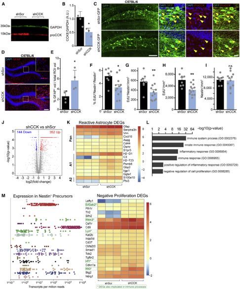
ll
Article

Figure 1. Local CCK Interneurons Depolarize rNSCs through CCK2R-Mediated Signaling
(A) AAV2, AAV5, or AAV8 expression of DIO-hM3Dq-mCherry in CCK-Cre mice using equivalent titers. Scale bar 200 mm.
(B) Colocalization of mCherry with ProCCK in hilar CCK interneurons after AAV2 injection in CCK-Cre mice. Scale bars 100 mm, 50 mm.
(C) Specificity (left) and efficiency (right) of DREADD expression in hilar CCK interneurons. Bars indicate mean ± SD for 4 animals.
(D) Colocalization of GABA in hM3D+ CCK interneurons. Arrows highlight colocalized cells. Scale bar 25 mm.
(E) Quantification of GABA colocalization. Bar indicates mean ± SD for 3 animals.
(F and G) Whole-cell recordings in acute slice of an initially silent (F) or initially active (G) hM3Dq+ CCK interneuron upon application of 10 mM CNO. Arrows indicate
start of drug application.
(H) Quantification of fold increase in spiking at peak chemogenetic activation. Bars indicate means ± SEM for 4 animals.
(I) hM3Dq-mCherry expression in hilar CCK interneurons near GFP+ rNSCs. Scale bar 100 mm.
(J and K) Sample whole-cell recordings of GFP+ rNSCs showing (J) large depolarization due to chemogenetic activation of CCK interneurons or (K) maintained
hyperpolarization in 2 mM YM022.
(L) Quantification of rNSC Vm before and during CNO application in ACSF or YM022. Bars indicate mean Vm ± SEM. p = 0.02(ACSF), 0.09(YM022) by paired t test
for (9,7) cells.
(M) Magnitude of depolarization due to CCK activation. p = 0.02 by Wilcoxon-Mann-Whitney test for (9,7) cells from (6,6) animals. Bars indicate mean ± SEM for
responding rNSCs (>10 mV; circles). Triangles = non-responding rNSCs.
(N) Percent of cells that depolarized by at least 10 mV. See also Figure S1.

                                                                                                               Neuron 108, 349–366, October 28, 2020 351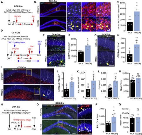
ll
                                                                                                                                                  Article

Figure 2. CCK-Induced rNSC Depolarization Is Mediated by a Glutamatergic Relay
(A–C) Sample changes in rNSCs Vm to 300 nM CCK8 peptide in the presence of (A) 100 mM APV, 10 mM NBQX, 100 mM AIDA, 50 mM bicuculline, and 1 mM TTX; (B)
in ACSF; or (C) in 1 mM YM022.
(D) Comparison of peptide-induced depolarization of rNSCs. p = (0.003,0.003) for (7,7,7) cells in (6,4,5) animals by Wilcoxon-Mann-Whitney test. Bars indicate
mean ± SEM for rNSCs that depolarized >10 mV from baseline (circles). ANBAT = APV, NBQX, bicuculline, AIDA, TTX.
(E) Percent of rNSCs in (D) that responded >10 mV to CCK8 application.
(F–H) Sample recordings of rNSC Vm to chemogenetic activation of CCK interneurons during (F) GABAAR blockade; (G) combined blockade of GABAARs, iGluRs
and mGluRs; and (H) when only GABAARs and iGluRs were blocked or when only GABAAR and mGluRs were blocked. Curve fits to data in red, yellow diamonds
indicate delay time to Vm deflection.
(I) Comparison of depolarization magnitude during CNO application under indicated conditions. One-way ANOVA showed an effect by condition F(4,41) = 10.18,
p = 8.1*10!6, and a post hoc Dunnett analysis gave individual p values versus ACSF of (0.95,0.018,0.002,0.0008) for (9,11,9,10,7) cells from (6,10,8,7,5) animals.
Bars indicate mean ± SEM for rNSCs that depolarized >10 mV (circles).
(J) Percent of rNSCs in (I) that depolarized >10 mV.
(K) Times from CNO application to initiation of depolarizing event. Bars indicate mean ± SEM. One-way ANOVA showed no effect of condition; F(3,23) = 1.66,
p = 0.2 for (6,10,5,5) responding cells from (5,9,4,4) animals. See also Figure S2.

352   Neuron 108, 349–366, October 28, 2020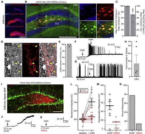
ll
Article

                                  (legend on next page)

          Neuron 108, 349–366, October 28, 2020 353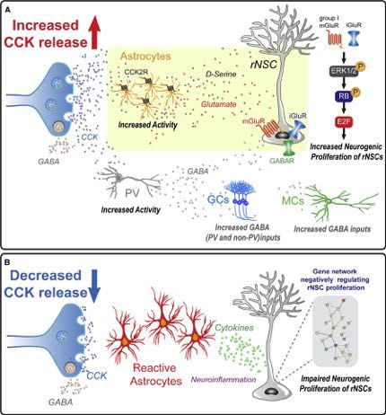
ll
                                                                                                                                                  Article

Specifically, bath application of AMPA, NMDA, or DHPG in the                      3K). These data suggest that CCK interneuron activation recruits
presence of TTX depolarized rNSCs (Figure S2). Together,                          CCK2R-dependent inhibitory inputs to both MCs and GCs.
these data suggest that CCK interneuron activity-induced                             Next, we sought to examine the inhibitory inputs to both MCs
rNSC depolarization requires activation of both iGluRs and group                  and GCs upon CCK interneuron activation. First, we repeated
I mGluRs.                                                                         the previous Ca2+ experiments in MCs and GCs in the presence
                                                                                  of bicuculline to block the GABAAR-mediated component. We
Activation of Dentate CCK Interneurons Increases                                  found the CCK-induced inhibitory effects onto MCs were
GABAergic Inputs onto Dentate Glutamatergic Neurons                               completely abolished in the presence of bicuculline (Figures 3C
We sought to identify sources of CCK-induced glutamatergic                        and 3E), similar to that observed in YM022, suggesting that
signaling onto rNSCs. Our recent electron microscopy and immu-                    all GABAergic inputs on MCs induced by CCK interneuron acti-
nohistology established that bushy processes of rNSCs wrap                        vation are CCK2R dependent. In contrast, blocking the
around glutamatergic synapses formed between MCs and GCs                          GABAAR-mediated component induced a significant increase
(Yeh et al., 2018). Therefore, we performed Ca2+ imaging in                       in the frequency (not area) of Ca2+ events in GCs (Figures 3H
MCs and GCs to examine the functional impact of CCK inter-                        and 3J), as well as a further reduction of the percent of GCs
neuron activity on these cells (Figure 3A). Interestingly, we                     with reduced Ca2+ frequency on top of that observed in YM022
observed a significant decrease in both frequency and area of                     (Figure 3I). This suggests that the GABAergic inputs on GCs
Ca2+ events in MCs upon CNO-induced chemogenetic activation                       induced by CCK interneuron activation contain both CCK2R-
of CCK interneurons, which was abolished in the presence of                       dependent and -independent components. It is interesting to
CCK2R blocker YM022 (Figures 3B, 3C, and 3E). These results                       note that CCK interneuron activation recruited GABAergic
suggest that CCK interneuron activation leads to decreased MC                     inputs to both MCs and GCs, but only MCs exhibited decreased
activity through a CCK2R-mediated mechanism. In contrast,                         Ca2+ activity, suggesting that MCs and GCs use differential
there was no significant change in frequency or area of Ca2+                      mechanisms to maintain their activity.
events in GCs upon CNO in ACSF or YM022 (Figures 3G, 3H,                             As GCs and MCs are well known to be inhibited by local PV
and 3J). We further quantified the percent of MCs and GCs that                    interneurons (Acsády et al., 2000; Espinoza et al., 2018), we
increased or decreased Ca2+ activity based on individual brain sli-               sought to determine whether dentate PV interneurons respond
ces under various pharmacological conditions. Slice-wise ana-                     to CCK activity by recording PV interneuron excitability upon
lyses showed that 78% of MCs exhibit decreased frequency                          CCK interneuron activation in CCK-Cre::PV-FLP mice (Fig-
and 67% of MCs exhibit decreased area of Ca2+ events upon                         ure 3L). We found significantly increased spiking rates in dentate
CNO activation of CCK interneurons (Figures 3D and 3F), and                       PV interneurons upon CNO application as measured by cell-
58% of GCs exhibit decreased Ca2+ frequency (not area) upon                       attached recordings. Importantly, this increase was abolished
the same manipulation (Figures 3I and 3K). Importantly, the                       by YM022 (Figures 3M and 3N), suggesting that dentate PV inter-
percent of MCs and GCs with decreased Ca2+ signals was signif-                    neurons represent CCK2R-dependent inhibitory inputs onto
icantly reduced in the presence of YM022 (Figures 3D, 3F, 3I, and                 both MCs and GCs. To confirm this, we recorded spontaneous

Figure 3. CCK Interneurons Increase GABAergic Inputs onto Dentate Glutamatergic Neurons in a CCK2R Manner
(A) Expression of hM3Dq-mCherry in CCK cells and GCaMP6 in MCs and GCs. Yellow arrows indicate GCaMP6+ GCs, white arrows indicate GCaMP6+ MCs.
Scale bars 200 mm, 50 mm.
(B) Ca2+ transients in MCs before and during chemogenetic activation of CCK interneurons in ACSF, 1 mM YM022, or 50 mM bicuculline. Gap indicates a 10 min
pause of image acquisition for CNO application.
(C) Frequency of MC Ca2+ events due to chemogenetic activation of CCK cells in ACSF, YM022, or bicuculline. Lines and markers indicate paired measurements
in individual MCs. Bars indicate mean ± SEM. Ca2+ frequency. p = (1*10!5,0.27,0.30) by paired t test for (56,52,87) MCs in (5,3,5) animals.
(D) Slice-wise analysis of the percent increasing (green), decreasing (purple), or unchanging (gray) MC Ca2+ frequencies per slice due to CNO application. Stacked
bars indicate means ± SEM. p = 0.003(ACSF:YM022), 0.60(YM022:bic), 0.0001(ACSF:bic) increasing and 0.02(ACSF:YM022), 0.86(YM022:bic), 0.004(ACSF:bic)
decreasing for (10,7,13) slices in (5,3,5) animals by unpaired t test.
(E) Area under MC Ca2+ traces during baseline and chemogenetic activation of CCK cells as in (C). p = (0.032,0.039,0.70).
(F) Slice-wise analysis of MC Ca2+ area as in (D). p = 0.047(ACSF:YM022), 0.81(YM022:bic), 0.047(ACSF:bic).
(G) Sample Ca2+ transients in GCs as in (B).
(H) Frequency data of GC Ca2+ as in (C) for (130,68,58) GCs in (5,3,3) animals. p = (0.71,0.39,0.003).
(I) Slice-wise analysis of GC frequencies (H) as in (D). p = 0.52(ACSF:YM022), 0.018(YM022:bic), 0.0013(ACSF:bic) increasing and 0.032(ACSF:YM022),
0.31(YM022:bic), 0.003(ACSF:bic) decreasing for (9,6,6) slices in (5,3,3) animals.
(J) Area data of GC Ca2+ (H) as described in (E). p = (0.52,0.52,0.07).
(K) Slice-wise analysis of GC Ca2+ area (J) as described in (F). p = 0.96(ACSF:YM022), 0.39(YM022:bic), 0.43(ACSF:bic).
(L) YFP expression in PV interneurons and mCherry expression in hM3Dq hilar CCK cells. Arrows indicate YFP+ PV+ cells. Scale bars 200 mm, 100 mm.
(M) Cell-attached recordings of PV spiking in ACSF or 1 mM YM022. The gap in the traces indicates a pause of recording during CNO addition.
(N) Quantification of mean ± SEM fold-changes in spike rates for (7,7) cells from (5,5) animals. p = 0.003 by Wilcoxon-Mann-Whitney test.
(O) Hilar CCK-cell targeted expression of hM3Dq-mCherry and patch pipette for recordings of GCs. Scale bar 50 mm.
(P) Sample inward IPSCs in GCs in ACSF, CNO, CNO + 200 nM u-agotoxin TK, or CNO + 50 mM bicuculine, using high-chloride internal solution.
(Q) Quantification of mean ± SEM IPSC frequencies for (12,7) GCs at baseline, during CNO, and then in CNO + ago. p = (0.007,0.048) by paired t test in (10,6)
animals.
(R) Mean ± SEM fold change in GC IPSC frequency for cells in (Q). p = 3*10!5 by unpaired t test. See also Figure S3.

354 Neuron 108, 349–366, October 28, 2020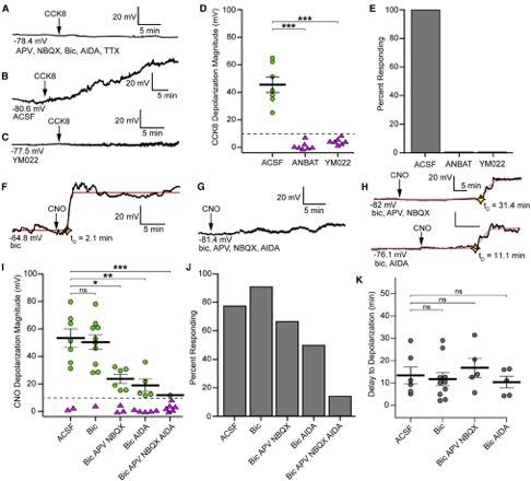
ll
Article

Figure 4. Dentate Astrocytes Respond to CCK Activity and Are Capable of Inducing rNSC Depolarization
(A) Expression of hM3Dq-mCherry in CCK cells and GCaMP6-LCK in astrocytes. Arrows highlight hM3Dq+ processes adjacent to GFAP+ astrocyte. Scale bars
100 mm, 20 mm.
(B) Sample astrocyte Ca2+ transients evoked by CCK interneuron activation. The gap in traces indicates a 10 min pause of image acquisition for CNO addition.
(C) Astrocyte Ca2+ frequencies due to activation of CCK cells in ACSF, 1 mM YM022, or 50 mM bicuculline + 1 mM CPG. Lines and markers indicate paired
measurements of individual ROIs. Bars indicate mean ± SEM. Ca2+ frequency for (282,278,219) ROIs in (3,3,3) animals. p = (1.3*10!12,0.49,1.8*10!9) by paired
t test.
(D) Area under Ca2+ traces in (C). p = (0.083,0.47,0.50) by paired t test. Bars indicate mean ± SEM.
                                                                                                                            (legend continued on next page)

                                                                                                        Neuron 108, 349–366, October 28, 2020 355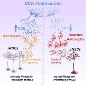
ll
                                                                                                                                                  Article

inhibitory postsynaptic currents (sIPSCs) in GCs upon CCK inter-                  GABA components do not play a major role in CCK-dependent
neuron activation in u-agotoxin TK, a selective blocker for P/Q                   astrocyte activation. Furthermore, bath application of CCK8
type Ca2+ channels unique to PV cells (Hefft and Jonas, 2005;                     peptide in the presence of TTX increased frequency and area
Wilson et al., 2001) (Figures 3O and 3P). As a result, we observed                of Ca2+ events in astrocytes (Figures 4E and 4F), suggesting
a 3-fold increase in sIPSCs upon CNO-induced activation of                        that CCK directly acts on astrocytes without other neuronal
CCK cells (Figures 3Q and 3R), which was reversed to 36% of                       intermediates.
the initial CNO-level frequency in u-agotoxin TK. All inward cur-                    We next sought to determine whether direct activation of
rents were confirmed GABAergic by addition of bicuculline (Fig-                   astrocytes could depolarize rNSCs. We infected DG astro-
ure 3P). Furthermore, by considering the fractional proportion of                 cytes with Gq-DREADDs (Figure 4G) and found that CNO
sIPSC frequencies that were u-agotoxin TK sensitive and insen-                    application significantly increased the frequency (not area) of
sitive upon CNO and at baseline (Figure S3A), we estimated the                    intracellular astrocyte Ca2+ (Figures 4H–4J). We then recorded
PV contribution to the CCK-dependent increase in GC sIPSCs to                     the Vm of rNSCs from Nestin-GFP mice upon astrocyte activa-
be ~77% (Figure S3B, also see STAR Methods). Together, these                      tion (Figure 4K). We found that chemogenetic activation of
data confirmed that dentate CCK interneuron activation re-                        dentate astrocytes was sufficient to induce depolarization of
cruited both CCK2R-dependent PV inputs and other CCK2R-in-                        rNSCs (Figures 4L–4O). Furthermore, this depolarization was
dependent GABAergic inputs onto GCs.                                              significantly reduced by iGluR and group I mGluR antagonists
                                                                                  and resulted in fewer responding rNSCs. Together, these re-
Dentate Astrocytes Respond to CCK Activity and                                    sults indicate that dentate astrocytes are responsive to CCK
Astrocyte Activation Is Sufficient to Induce rNSC                                 interneuron activation in a CCK2R-dependent manner, and
Depolarization                                                                    this activation is sufficient to cause rNSC depolarization pre-
Since the activities of local glutamatergic neurons are either in-                sumably via release of gliotransmitters (such as glutamate
hibited or unchanged during CCK interneuron activation, we                        and D-serine) that act on iGluRs and group I mGluRs
sought to identify an alternative source of glutamate-mediated                    on rNSCs.
rNSC depolarization. Ca2+ elevation in astrocytes has been
widely shown to promote release of gliotransmitters, including                    CCK-Activity-Induced rNSC Depolarization Requires
glutamate and D-serine (Fiacco and McCarthy, 2018; Hamilton                       Dentate Astrocyte Intermediates
and Attwell, 2010; Savtchouk and Volterra, 2018), which could                     To determine if dentate astrocytes serve as an intermediate in
act on GluRs to depolarize rNSCs. However, it is yet unclear if                   CCK-induced depolarization of rNSCs, we interfered with astro-
DG astrocytes respond to CCK and whether astrocytes act as                        cyte signaling by application of a glial-specific Krebs cycle inhib-
mediators of the CCK induced changes in rNSCs. To address                         itor Fluorocitric acid (FC) (Pabst et al., 2016; Swanson and Gra-
this, we performed Ca2+ imaging in astrocytes during chemoge-                     ham, 1994). Strikingly, chemogenetic activation of dentate CCK
netic activation of dentate CCK cells. Confocal imaging                           cells failed to induce rNSC depolarization when astrocyte
confirmed that DG CCK processes reside adjacent to astro-                         signaling was disrupted by FC (Figures 5A and 5B). Importantly,
cytes (Figure 4A), thus providing morphological support for                       rNSCs retained their ability to depolarize in response to gluta-
the interaction between these cells. Functionally, we observed                    mate (Figure S4A), suggesting that FC does not disrupt the ma-
a significant increase in the frequency (not area) of astrocytic                  chinery for glutamatergic receptor signaling.
Ca2+ events upon chemogenetic activation of CCK cells (Fig-                          As chemogenetic activation of dentate CCK cells increased
ures 4B–4D), which was abolished in YM022 (Figures 4C and                         the Ca2+ transients in astrocytes, we attempted to quench astro-
4D). Since GABA signaling can activate astrocytes (Mederos                        cytic Ca2+ signaling through infusion of the Ca2+ chelator BAPTA
and Perea, 2019) and our study found that CCK interneurons re-                    in astrocytes using a patch pipette (Pabst et al., 2016). Because
cruit GABAergic inputs, we recorded astrocyte Ca2+ in the pres-                   astrocytes are coupled by gap junctions, BAPTA infusion of a
ence of GABAA and GABAB receptor antagonists. As a result,                        single astrocyte has the ability to buffer Ca2+ in large astrocyte
we still observed significant CNO-induced increases in the fre-                   networks (Araque et al., 1998; Pabst et al., 2016). We first
quency of Ca2+ events (Figures 4C and 4D), suggesting that                        confirmed that infusing BAPTA into a single astrocyte was

(E) Astrocyte Ca2+ events frequencies to 300 nM CCK8 peptide while in 1 mM TTX for 142 ROIs from 3 animals. p = 0.014 by paired t test. Bars indicate
mean ± SEM.
(F) Area under Ca2+ traces in (E). p = 0.03 by paired t test. Bars indicate mean ± SEM.
(G) Astrocytic targeting of hM3Dq for chemogenetic stimulation. Arrows highlight colocalized expression in GFAP+ cells. Scale bars 100 mm, 20 mm.
(H) Sample astrocyte Ca2+ transients in hM3D+ astrocytes due to CNO application. The gap in traces indicates a 10 min pause of image acquisition for CNO addition.
(I) Frequencies of astrocyte Ca2+ due to chemogenetic activation of astrocytes for 166 regions from 3 animals. p = 0.0006 by paired t test. Bars indicate
mean ± SEM.
(J) Area under Ca2+ traces in (I). p = 0.31 by paired t test. Bars indicate mean ± SEM.
(K) Expression of hM3Dq in astrocytes in Nestin-GFP mouse. Scale bar 100 mm.
(L) Recording of a GFP+ mCherry! rNSC during chemogenetic stimulation of astrocytes.
(M) Vm of rNSCs before and during CNO application. Bars indicate mean ± SEM. p = 0.006 by paired t test for 7 cells.
(N) Magnitude of CNO-induced depolarization in either ACSF or 100 mM APV + 10 mM NBQX + 100 mM AIDA. p = 0.038, by unpaired t test for all (7,7) cells from (4,3)
animals. Bars indicate mean ± SEM for rNSCs responding >10 mV (circles). Triangles = non-responders.
(O) Percent of cells that depolarized >10 mV.

356 Neuron 108, 349–366, October 28, 2020
ll
Article
Figure 5. CCK-Induced rNSC Depolarization Requires Astrocyte Intermediates
(A) Reduced rNSC depolarization when slices are preincubated in 100 mM FC.
(B) Vm of rNSCs due to chemogenetic activation of CCK cells in FC treated slices. Bars indicate mean Vm ± SEM. p = 0.02 by paired t test for 7 cells from 4
animals. Darker blue lines, rNSCs responding >10 mV.
(C) Expression of mCherry and GCaMP6-LCK in GFAP+ astrocytes for pipette targeting and BAPTA filling. Scale bars 100 mm, 20 mm.
(D) Ca2+ traces in hilar astrocytes at baseline and then after BAPTA infusion into a single mCherry+ astrocyte.
(E) Frequencies of nearby astrocyte Ca2+ events due to intracellular BAPTA infusion. Lines and markers indicate paired measurements in individual ROIs. Bars
indicate mean ± SEM frequencies for 107 astrocytic ROIs in 3 animals. p = 2.3*10!13 by paired t test.
(legend continued on next page)
Neuron 108, 349–366, October 28, 2020 357ll
Article
sufficient to interfere with the DG astrocyte network as reflected or p130PH conditions, significantly different from controls (Fig-
by significant reductions in both frequency and area of astrocyte ures 5Q and 5R). Together, these data confirmed that dentate
Ca2+ events (Figures 5C–5F). We then recorded the Vm of rNSCs astrocytes are the major glutamatergic intermediaries for CCK-
upon chemogenetic activation of CCK cells after astrocytic Ca2+ activity-induced rNSC depolarization.
depletion (Figures 5G and S4B) and found that CCK-activity-
induced rNSC depolarizations were largely abolished (Figures CCK Interneuron Stimulation Promotes Neurogenic
5H and 5I). Importantly, rNSCs remained responsive to bath Proliferation of rNSCs via MAPK/ERK Signaling and
application of glutamate after astrocyte BAPTA infusion Increases Neurogenesis
(Figure S4C). Vm has been implicated as a functional determinant of prolifera-
CCK-evoked Ca2+ increases in astrocytes are independent tion and differentiation in non-neuronal cells, such as embryonic
of extracellular Ca2+ concentrations (Mu €ller et al., 1997) and stem cells (Levin, 2014). Quiescent cells remain hyperpolarized,
likely involve PLC induced release of Ca2+ from intracellular whereas proliferation events are often accompanied by mem-
stores via IP3-mediated intracellular cascades (Dufresne brane depolarization (Aprea and Calegari, 2012). Transduction
et al., 2006; Lee et al., 2011). We therefore disrupted dentate of extracellular niche signals activates cell surface receptors
astrocyte function by buffering mobile cytosolic IP3 through that in turn trigger intracellular signaling cascades that control
expression of the pleckstrin homology domain of PLC-like-pro- activation and quiescence in adult rNSCs. It has been shown
tein p130 (p130PH) (Xie et al., 2010). We injected AAVs con- that both NMDARs and group I mGluRs activate MAPK/ERK1/
taining p130PH construct along with GCaMP6-LCK and 2 pathways via upstream effectors in vitro (Wang et al., 2007).
confirmed co-expression in GFAP+ astrocytes (Figure 5J). We We sought to determine whether pERK signaling facilitates
first validated the efficacy of p130PH in disrupting Ca2+ activity rNSC activation upon CCK interneuron stimulation in vivo. We
of DG astrocytes by bilaterally injecting mice with p130PH + first examined expression of immediate early gene c-Fos in den-
GCaMP6 on one side and mRFP + GCaMP6 on the other tate CCK interneurons 1.5 h after CNO intraperitoneal injection
side. As a result, we found a significant reduction in the area (Figure 6A). The density of c-Fos+ mCherry+ CCK interneurons
of Ca2+ events in dentate astrocytes (not frequency) at the in the hM3Dq group was significantly higher compared to con-
p130PH side as compared to the control side at baseline (Fig- trols (Figures 6B and 6C), thus confirming in vivo activation of
ures 5K and 5L). Furthermore, CCK8 induced a significant in- CCK interneurons.
crease in the frequency (not area) of astrocytic Ca2+ events Next, we examined expression of pERK in rNSCs 80 min af-
on the control side, with no significant change in either fre- ter CNO intraperitoneal injection (Figures 6D, 6E, and S5A) and
quency or area of astrocytic Ca2+ events on the p130PH side observed a significant increase in the density of pERK+ rNSCs
(Figures 5M and 5N). Using the ability of p130PH to suppress in hM3Dq mice compared to control mice (Figure 6F). As a
the activity of astrocytes, we then determined whether dentate mitogen-activated protein kinase (MAPK), ERK regulates cell
astrocytes served as the relay that couples CCK activity to proliferation through promotion of G1/S cell cycle progression
rNSC depolarization. Whole-cell electrophysiological record- (Chambard et al., 2007; Zhang and Liu, 2002). Specifically,
ings from GFP+ rNSCs displayed a significant reduction in activation of ERK signaling drives hyper-phosphorylation of
the depolarization magnitude of Vm with fewer responding retinoblastoma protein (RB), releasing RB’s inhibition of E2F
rNSCs upon chemogenetic activation of CCK neurons when transcription factors, which in turn activates genes required
astrocyte IP3 mobilization was disrupted (Figures 5O and 5P). for the G1/S transition (Duronio and Xiong, 2013). To address
Quantification of the depolarization magnitude and percent of whether hyper-phosphorylated RB (pRB) mediates CCK inter-
responding rNSCs was consistently reduced in FC, BAPTA, neuron activity-dependent rNSC activation, we examined pRB
(F) Area under Ca2+ traces in (E). p = 1.9*10!5 by paired t test. Bars indicate mean ± SEM.
(G) CCK-Cre::Nestin-GFP animal with (top) BAPTA filling pipette on a mCherry+ astrocyte in the hilus and (bottom) recording configuration from GFP+ rNSC. Scale
bar 50 mm.
(H) Vm recording from a GFP+ rNSCs during CCK chemogenetic activation after high BAPTA.
(I) Mean ± SEM Vm of rNSCs due to CCK activation after astrocytic BAPTA. p = 0.16 by paired t test for 7 cells in 6 animals. Darker purple lines, rNSCs re-
sponding >10 mV.
(J) mRFP-p130PH and GCaMP6-LCK expression in DG astrocytes. Arrows indicate colocalization in GFAP+ astrocytes. Scale bars 100 mm, 20 mm.
(K) Basal astrocyte Ca2+ frequencies in control or p130PH injected hemispheres. Bars indicate mean ± SEM frequency. p = 0.11 for (275,236) astrocyte ROIs in
3 animals by unpaired t test.
(L) Area under Ca2+ traces in (K). p = 0.009. Bars indicate mean ± SEM.
(M) Frequencies of DG astrocyte Ca2+ events due to 300 nM CCK8 peptide in p130PH or control hemisphere. Baseline data are identical to (K). p = (0.005,0.11) for
(275,236) astrocyte ROIs in 3 animals by paired t test. Bars indicate mean ± SEM.
(N) Area under Ca2+ traces in (M). p = (0.3,0.16). Bars indicate mean ± SEM.
(O) Vm from a GFP+ rNSC from p130PH-injected DG upon chemogenetic activation of CCK cells.
(P) Vm of rNSCs due to CCK activation in p130PH-injected DGs. p = 0.13 by paired t test for 10 cells in 5 animals. Bars indicate mean Vm ± SEM. Darker red lines,
rNSCs responding >10 mV.
(Q) Magnitude of depolarization due to chemogenetic CCK activation in indicted conditions. One-way ANOVA showed a significant effect of condition
F(3,29) = 9.01, p = 0.002, and Dunnett post hoc analysis showed each are significantly reduced from ACSF. p = (0.009,0.009,0.004) for (9,7,7,10) cells from
(6,4,6,5) animals. Bars indicate mean ± SEM for rNSCs responding >10 mV (circles). Triangles, non-responding rNSCs.
(R) Percent of cells that depolarized >10 mV. See also Figure S4.
358 Neuron 108, 349–366, October 28, 2020ll
Article
Figure 6. CCK Interneuron Stimulation Promotes Proliferation of rNSCs via MAPK/ERK Signaling and Increases Neurogenesis
(A) Experimental paradigm for in vivo stimulation of CCK interneurons and c-Fos analysis.
(B) DG c-Fos expression after CCK chemogenetic activation. Arrows indicate hilar mCherry+ c-Fos+ cells. Scale bars 100 mm, 20 mm.
(C) Density of c-Fos labeled hilar mCherry+ cells. p = 0.034 for (3,4) animals by Student’s t test. Bars indicate means ± SD.
(D) Experimental paradigm for quantification of phospho-ERK, phospho-RB, or EdU uptake in response to chemogenetic stimulation of CCK interneurons.
(E) Phospho-ERK+ rNSCs in the DG 80 min after IP CNO. Scale bar 10 mm. Arrows highlight pERK+ Nestin+ adult rNSCs.
(F) Density of pERK+ rNSCs for (4,5) animals. Bars indicate means ± SD. p = 0.013 by Student’s t test.
(G) Phospho-RB+ rNSCs in the DG 5 h after IP CNO. Scale bar 10 mm. Arrow highlights a pRB+ Nestin+ rNSC.
(H) Density of pRB+ rNSCs for (5,4) animals. Bars indicate means ± SD. p = 0.008 by Student’s t test.
(I) EdU uptake in rNSCs after 4 days of CNO drinking water. Scale bars 100 mm, 20 mm. Arrow highlights an EdU+ Nestin+ adult rNSC.
(J–M) Proportion (J) and density (K) of proliferating adult neural stem cells in the DG, density of all proliferating cells (L), and total rNSC pool (M). Bars indicate
means ± SD. p = (0.03,0.05,0.80,0.005) for (6,5) animals by Student’s t test.
(N) Experimental paradigm for quantification of neurogenic proliferation in response to in vivo chemogenetic activation of CCK interneurons.
(O) DCX+ adult-born immature neurons after 3 weeks of CNO drinking water. Scale bars 100 mm, 20 mm. Arrows indicate DCX+ EdU+ cells.
(P–Q) Density (P) of DCX+ EdU+ immature neurons and rNSC pool (Q) after in vivo chemogenic stimulation of CCK interneurons. Bars indicate means ± SD.
p = (0.047,0.29) for (4,6) animals by Student’s t test. See also Figure S5.
Neuron 108, 349–366, October 28, 2020 359ll
Article
Figure 7. Reduced Dentate CCK Induces Reactive Astrocytes and Decreases rNSCs Proliferation Potentially via Neuroinflammatory-Related
Gene Profiles
(A and B) Western blot and densitometry of hippocampal lysates 1 month after injection of shCCK or shScr AAVs. p = 0.02 from (3,3) animals by Student’s t test.
Bars indicate means ± SD.
(legend continued on next page)
360 Neuron 108, 349–366, October 28, 2020ll
Article
expression in rNSCs 5 h after CNO injection (Figures 6D, 6G, wondered whether reduced CCK contributes to an adverse
and S5B). As a result, we observed a significant increase in state of astrocytes. To address this, we examined GFAP
the density of pRB+ rNSCs (Figure 6H), suggesting that CCK expression, an established marker for astrocyte reactivity (Lid-
interneuron activity promotes G1/S cell cycle progression in delow and Barres, 2017), in the dentate astrocytes by quanti-
rNSCs. Then, we examined the incorporation of EdU in rNSCs fying the volume of GFAP immunofluorescence in mice in-
and newborn progeny (Figure 6D). We assessed EdU incorpo- jected with shCCK or scrambled control (shScr). Strikingly,
ration by counting EdU+ Nestin+ rNSCs with radial processes GFAP expression in shCCK injected mice was significantly
(Figure 6I) after 4 days of CNO drinking water. Stereological increased as compared to shScr controls (Figures 7D and
analysis showed that chemogenetic activation of local CCK in- 7E), suggesting that reduced CCK induces reactive astro-
terneurons led to a significant increase in the percent and den- cytes. To examine the functional effects of reduced CCK on
sity of Nestin+ EdU+ rNSCs (Figures 6J and 6K). As activated rNSCs, we labeled dividing cells through 4 EdU injections
rNSCs give rise to proliferating progeny, we examined the 1 month after AAV injection and examined EdU incorporation
density of EdU+ cells and observed a significant increase of in Nestin+ rNSCs and EdU+ proliferating progeny. Stereologi-
EdU incorporation upon chemogenetic activation of CCK in- cal quantification showed a significant decrease in the percent
terneurons (Figure 6L) without altering the Nestin+ rNSC pool and density of EdU+ Nestin+ rNSCs (Figures 7F and 7G) and
(Figure 6M). These results suggest that dentate CCK inter- total EdU+ proliferating progeny (Figure 7H) without altering
neuron activation promotes neurogenic proliferation of rNSCs the Nestin+ rNSC pool (Figure 7I).
and increases production of proliferating progeny. We also To explore mechanisms linking reactive astrocytes and
examined the integrity of activated astrocytes and found that deceased rNSC proliferation upon CCK knockdown, we per-
chemogenetic activation of CCK interneurons did not induce formed RNA-seq of DGs microdissected from shCCK or shScr
changes in astrocyte morphology or GFAP expression in mice (Tables S1 and S2). Our RNA-seq analysis identified 496
hM3Dq-injected animals (Figures S5C and S5D). differentially expressed genes (DEGs), including 144 downre-
To determine the long-term effects of dentate CCK inter- gulated and 352 upregulated transcripts (Figure 7J; Table
neuron activity in regulating neurogenesis and the rNSC pool, S3). We compared the RNA-seq data with a recently published
we labeled a starting proliferating population with 5 EdU injec- transcriptional profile of reactive astrocytes induced by neuro-
tions followed by 3-week chronic stimulation of dentate CCK inflammation or ischemia (Clarke et al., 2018) and found that
interneurons through CNO drinking water (Figure 6N). Stereo- 17 reactive-astrocyte associated genes were differentially ex-
logical analysis revealed a significant increased density of pressed in the DGs of shCCK mice (Figure 7K; Table S4).
DCX+ EdU+ immature neurons without exhaustion of the These DEGs included standard reactive astrocyte genes
rNSC pool (Figures 6O–6Q). Together, these results suggest (PAN: 8/17), such as GFAP and Vimentin; A1-reactive genes
that CCK interneuron stimulation promotes stable neurogenic associated with neuroinflammation (A1: 7/17), such as C3
proliferation via MAPK/ERK signaling and activation of G1/S and Cxcl10; and A2-reactive genes associated with ischemia,
cell cycle regulators, resulting in increased production of prolif- such as S100a10 and Ptx3 (A2: 2/17). These data suggest that
erating progeny and immature neurons without depleting the reducing CCK mainly induces A1 reactive astrocytes associ-
rNSC pool. ated with neuroinflammation. Additionally, gene ontology and
biological pathway analysis revealed many DEGs enriched in
Reduced Dentate CCK Induces Reactive Astrocytes, biological pathways involved in immune processes and innate
Decreased Neurogenic Proliferation of rNSCs, and inflammatory responses (Figure 7L; Table S5), supporting the
Upregulation of Genes Involved in Neuroinflammation involvement of innate immune cells, including astrocytes.
To complement the gain-of-function approach for increased Furthermore, of 21 DEGs relating to negative regulation of
CCK, we took a loss-of-function approach to reduce dentate cell proliferation (Table S6), 19 were also expressed in neural
CCK by AAV delivery of shRNA against CCK (shCCK) (Arey precursor cells based on a published single-cell RNA-seq pro-
et al., 2014). We confirmed efficiency of shCCK by western file of Nestin+ cells in adult DG (Shin et al., 2015) (Figure 7M).
blot (Figures 7A and 7B) and immunostaining (Figure 7C) These data support our observed correlation between
1 month after viral injection. Based on our finding that reduced CCK and decreased proliferation of rNSCs (Figures
increased CCK induces astrocyte gliotransmission, we 7F–7I). Interestingly, several of these highly expressed DEGs
(C) Reduced CCK immunofluorescence in the hilus of shCCK treated animals. Arrows indicate targeted GFP+ hilar cells. Scale bars 100 mm, 20 mm.
(D) GFAP reactivity after knockdown of DG CCK. Scale bar 100 mm.
(E) Quantification of %GFAP immunofluorescence per volume for (4,4) animals. p = 0.03 by unpaired t test. Bars indicate means ± SD.
(F–I) Proportion (F) and density (G) of proliferating rNSCs, density (H) of total proliferating cells, and total (I) rNSC pool after shCCK. Bars indicate means ± SD.
p = (0.011,0.009,0.002,0.81) for (8,12) animals by Student’s t test.
(J) Volcano plot of fold changes in gene expression due to shCCK, highlighting upregulated and downregulated DEGs.
(K) Heatmap of 17 DEGs related to reactive astrocyte states from 3 animals in each treatment group. Pan-reactive, type A1, and type A2 reactive astrocyte genes
are indicated. Color indicates direction and relative magnitude of FPKMs.
(L) Gene Ontology terms identified from analysis of all 496 DEGs identifying immune processes and negative regulation of proliferation.
(M) Transcript expression in rNSCs (based on Shin et al., 2015) and heatmap of 21 DEGs related to negative control of proliferation. Color indicates direction and
relative magnitude of FPKMs. Green highlighted genes are additionally implicated in immune processes. See also Figure S6.
Neuron 108, 349–366, October 28, 2020 361ll
Article
Figure 8. Model of Dentate CCK Interneuron Regulation of rNSCs through Differential Astrocyte States
(A) Dentate CCK terminals, via CCK2Rs, induce glutamate release from astrocytes, which activate iGluRs and mGluRs on rNSCs and induce intracellular signaling
cascades including phosphorylation of ERK1/2, translocation to the nucleus, hyperphosphorylation of RB, and unbinding of transcription factor E2F. Unbound
E2F can drive transcription of genes that promote cell cycle progression. PV interneurons, meanwhile, are excited by CCK, and contribute to GABAergic inhibition
of local glutamatergic circuitry.
(B) Conversely, reduced CCK levels reflect pathogenic states and result in immune activation, neuroinflammation, and reactive astrocyte states, leading to
inactivation of rNSCs, likely through cytokine signaling and gene expression changes that negatively regulate proliferation.
in rNSCs, including Eif2ak2, Ifitm3, Lyn, Irf1, and Ifit3, were DISCUSSION
jointly implicated in both negative proliferation pathways and
immune system processes (Figures 7M and S6). Together, We demonstrated that stimulating CCK release supports neuro-
these results suggest that reduced CCK induces astrocyte genic proliferation of rNSCs through the trophic effects of CCK
reactivity and increases innate immune responses, which in on dentate astrocytes to promote gliotransmission onto rNSCs
turn leads to decreased proliferation of rNSCs and newborn (Figure 8A). In contrast, reducing dentate CCK induced reactive
progeny. astrocyte states, which correlated with decreased neurogenic
362 Neuron 108, 349–366, October 28, 2020ll
Article
proliferation of rNSCs and upregulation of genes involved in in- Signaling Mechanisms Underlying CCK-Activity-
flammatory responses (Figure 8B). Dependent Regulation of Local Astrocytes
Our study identifies dentate CCK interneurons as a critical niche
NSCs Are Capable of Incorporating Dominant Niche component that utilizes an endogenous neuropeptide to regulate
Signals to Determine Their Actions rNSCs via local astrocytes. Specifically, local astrocyte states are
Many diverse cell types and signaling pathways within the regulated by endogenous levels of dentate CCK: high dentate
specialized local niche of the DG can simultaneously act on CCK promotes neurogenic proliferation of rNSCs and production
rNSCs and influence their behavior. This raises a key question: of newborn progeny through an astrocyte-mediated glutamater-
how do rNSCs integrate such diverse signals to ultimately gic signaling cascade; while low dentate CCK reduces neuro-
remain quiescent or become activated? We found that activa- genic proliferation of rNSCs and production of newborn progeny
tion of dentate CCK interneurons increased the activity of both potentially through neuroinflammatory signaling cascades
dentate astrocytes and PV interneurons through CCK2R- associated with reactive astrocytes. The signaling mechanisms
mediated mechanisms. Our previous studies implicated inhib- underlying CCK modulation of dentate astrocytes remain to be
itory transmission with rNSC quiescence, since bath applica- determined. We show that dentate astrocytes respond to CCK
tion of a GABAAR agonist hyperpolarized rNSCs (Yeh et al., signals with increased Ca2+ events via CCK2Rs, suggesting a
2018) and optogenetic activation of dentate PV interneurons direct mechanism mediated by astrocyte CCK2Rs. It remains to
reduced activation of rNSCs through GABAAR signaling be determined whether reduced CCK leads to aberrant Ca2+ sig-
(Song et al., 2012). In contrast, our current study found that nals in reactive astrocytes, which could contribute to proinflam-
CCK interneurons promote rNSC depolarization through an matory cytokine release. Accumulating evidence has suggested
astrocytic intermediary and increased rNSC proliferation via that reactive astrocytes exhibit aberrant Ca2+ signals, and the
GluR signaling. Simultaneous activation of dentate astrocytes patterns of aberrant Ca2+ depend upon distinct pathological
and PV interneurons should therefore exert opposing effects conditions (Shigetomi et al., 2019; Vincent et al., 2010). Future
on the Vm and proliferation status of rNSCs. Despite such studies will be required to characterize the Ca2+ signals of reactive
opposing niche signals, the dominant effects on rNSCs ap- astrocytes associated with reduced dentate CCK.
peared to be rNSC depolarization and increased neurogenic
proliferation of rNSCs through GluR-mediated ERK/MAPK Reduced CCK, Reactive Astrocytes, and NSC Regulation
signaling cascades. However, in the case of reduced CCK in One striking finding from our study is that reduced dentate CCK
the DG, we found decreased rNSC proliferation, suggesting appears to exert a detrimental effect on local astrocytes by
that glutamatergic niche signaling was no longer dominant in switching them to an A1 reactive state associated with neuroin-
this context. Indeed, besides increased gliotransmission of flammation, suggesting that CCK normally acts as an anti-in-
glutamate (Madeira et al., 2015; Pirttimaki et al., 2013), reac- flammatory molecule in adult DG. The anti-inflammatory role of
tive astrocytes release cytokines, which appear to be the CCK has been implicated in the peripheral system (Meng et al.,
dominant niche signals that contribute to reduced proliferation 2002; Miyamoto et al., 2012; Oehlers et al., 2017), but not in
of rNSCs (Wu et al., 2012). the central nervous system nor in the context of adult rNSC regu-
lation. Since CCK knockdown in the DG induced reactive
Potential Role of Other Niche Cells Recruited by Dentate astrocytes and neuroinflammation, this raised a question on
CCK Interneuron Stimulation the potential mechanism linking reactive astrocytes and
Besides activating dentate PV interneurons and astrocytes, deceased rNSC proliferation. Interestingly, our RNA-seq anal-
stimulating CCK interneurons led to increased GABAergic inhibi- ysis found that the majority of DEGs involved in both negative
tion of both MCs and GCs. Interestingly, the GABAergic inputs to proliferation and immune processes were associated with inter-
MCs were CCK2R dependent, while GABAergic inputs to GCs feron-g (IFNg) signaling, including Eif2ak2, Ifitm3, Irf1, and Ifit3
were both CCK2R dependent and independent. Importantly, (based on GeneCards), posing the possibility that upregulated
blocking the GABA component upon CCK interneuron stimula- IFNg signaling upon reduced CCK may contribute to decreased
tion abolished the inhibition of MCs and induced significant rNSC proliferation. Supporting this, recent studies showed
increases in GC activity, suggesting that CCK interneuron stim- increased proliferation of adult precursor cells in the adult olfac-
ulation exerts an inhibitory tone on dentate excitatory neurons in tory bulbs and DGs of IFNg knockout mice (Monteiro et al., 2016;
order to prevent hyperexcitability of the DG. The inhibitory tone in Pereira et al., 2015). The classical sources of IFNg are T cells and
the neurogenic niche is critical for maintaining the NSC pool by natural killer cells in the peripheral system (Monteiro et al., 2017);
preventing DG hyperexcitability (Bao et al., 2017; Sierra et al., however, our RNA-seq data provided strong support for the
2015). In addition, activation of dentate PV interneurons pro- involvement of resident immune cells, including astrocytes and
moted survival of neuroblasts and increased adult-born GCs microglia. Based on our findings that astrocytes are extremely
(Song et al., 2013). Consistent with this, we found that chronic responsive to dentate CCK, we speculate that reactive astro-
activation of local CCK interneurons promoted survival of cytes may be the source of IFNg upon reduced CCK. Future ex-
newborn neurons and preserved the rNSC pool. This suggests periments with specific manipulation of IFNg in astrocytes upon
that the combination of astrocyte-mediated glutamatergic reduced CCK could identify the role of IFNg in regulating rNSC
signaling and inhibitory tone in the DG induced by CCK inter- behavior.
neuron activation collectively supports sustainable hippocampal Recent human studies have provided tremendous support for
neurogenesis. reduced CCK in Alzheimer’s disease (AD) patients. For instance,
Neuron 108, 349–366, October 28, 2020 363ll
Article
a recent microarray analysis of human hippocampal tissues Univ.) for AAV knockdown vector, Dr. Garret Stuber (Univ. Washington) for
showed that physical exercise significantly increased hippocam- GCaMP virus, Dr. Margery Beinfeld (Tufts Univ.) for pro-CCK antibody, and
Dr. Shinghua Ding’s lab for AAV2/5-GFAP-p130PH (University of Missouri-
pal CCK transcription, while AD significantly reduced it
Columbia). Confocal microscopy was performed at the UNC Neuroscience
(Berchtold et al., 2019). Another study showed that higher levels Microscopy Core funded by P30 NS045892 and U54 HD079124. This work
of CCK in cerebrospinal fluid is negatively correlated with AD was supported by grants awarded to J.S. from American Heart Association,
susceptibility (Plagman et al., 2019). Moreover, emerging evi- Whitehall Foundation, Alzheimer’s Association and NIH (MH111773,
dence has revealed impaired rNSC behavior and hippocampal MH122692, AG058160, and NS104530); and NARSAD Young Investigator
neurogenesis in AD mouse models (Fu et al., 2019; Mu and Award (26017) to B.A.
Gage, 2011) and human patients (Crews et al., 2010; Gomez-
Nicola et al., 2014; Jin et al., 2004; Li et al., 2008; Lovell et al., AUTHOR CONTRIBUTIONS
2006; Moreno-Jiménez et al., 2019; Perry et al., 2012). Further- J.S. conceived the project. J.S., B.A., and J.W. designed experiments and
more, human AD patients display aberrant astrocytes with rami- wrote the manuscript. B.A. performed surgeries, electrophysiology, and cal-
fied processes and altered GFAP isoforms (Kamphuis et al., cium imaging; J.W. performed surgeries, immunohistology, confocal imaging,
2012, 2014), and AD mice exhibit significant astrogliosis (Oakley and in vivo analysis; F.Z. and P.J. performed RNA-seq analysis. Y.-D.L., L.Q.,
et al., 2006). Therefore, AD pathological hallmarks, including pla- C.W., C-Y.Y., H.B., Y.-J.L., R.O., S-A.L., and J.H. assisted experiments and
analysis.
ques and tangles, may induce reduced endogenous CCK and
reactive astrocytes as a potential mechanism for impaired
rNSCs and hippocampal neurogenesis. Given the critical role DECLARATION OF INTERESTS
of adult-born neurons in regulating cognitive functions, The authors declare no competing interests.
increasing CCK release to promote neurogenic potential of
NSCs and reduce inflammatory responses may represent a Received: November 18, 2019
novel strategy to treat neuropathological conditions associated Revised: June 12, 2020
with aberrant hippocampal neurogenesis and neuroinflamma- Accepted: July 29, 2020
Published: September 1, 2020
tion, such as aging, AD, and epilepsy.
REFERENCES
STAR+METHODS
Acsády, L., Katona, I., Martı́nez-Guijarro, F.J., Buzsáki, G., and Freund, T.F.
Detailed methods are provided in the online version of this paper (2000). Unusual target selectivity of perisomatic inhibitory cells in the hilar re-
and include the following: gion of the rat hippocampus. J. Neurosci. 20, 6907–6919.
Aprea, J., and Calegari, F. (2012). Bioelectric state and cell cycle control of
d KEY RESOURCES TABLE Mammalian neural stem cells. Stem Cells Int. 2012, 816049.
d RESOURCE AVAILABILITY Araque, A., Sanzgiri, R.P., Parpura, V., and Haydon, P.G. (1998). Calcium
B Lead Contact elevation in astrocytes causes an NMDA receptor-dependent increase in the
B Materials Availability frequency of miniature synaptic currents in cultured hippocampal neurons.
B Data and Code Availability J. Neurosci. 18, 6822–6829.
d EXPERIMENTAL MODEL AND SUBJECT DETAILS Arey, R.N., Enwright, J.F., 3rd, Spencer, S.M., Falcon, E., Ozburn, A.R., Ghose,
B Animals S., Tamminga, C., and McClung, C.A. (2014). An important role for cholecys-
d METHOD DETAILS tokinin, a CLOCK target gene, in the development and treatment of manic-
like behaviors. Mol. Psychiatry 19, 342–350.
B Stereotaxic Injections
Armbruster, B.N., Li, X., Pausch, M.H., Herlitze, S., and Roth, B.L. (2007).
B Electrophysiological recordings
2+ Evolving the lock to fit the key to create a family of G protein-coupled receptors
B Ca Imaging
potently activated by an inert ligand. Proc. Natl. Acad. Sci. USA 104,
B Tissue process and Immunohistochemistry 5163–5168.
d QUANTIFICATION AND STATISTICAL ANALYSIS Bao, H., and Song, J. (2018). Treating Brain Disorders by Targeting Adult
B Electrophysiological quantification Neural Stem Cells. Trends Mol. Med. 24, 991–1006.
B Calcium analysis
Bao, H., Asrican, B., Li, W., Gu, B., Wen, Z., Lim, S.A., Haniff, I., Ramakrishnan,
B Stereological quantification C., Deisseroth, K., Philpot, B., et al. (2017). Long-Range GABAergic Inputs
B Microdissection of the dentate gyrus Regulate Neural Stem Cell Quiescence and Control Adult Hippocampal
B RNA-Seq library prep and analysis Neurogenesis. Cell Stem Cell 21, 604–617 e605.
B Statistics Beinfeld, M.C. (1985). Cholecystokinin (CCK) gene-related peptides: distribu-
tion and characterization of immunoreactive pro-CCK and an amino-terminal
SUPPLEMENTAL INFORMATION pro-CCK fragment in rat brain. Brain Res. 344, 351–355.
Beinfeld, M.C., Meyer, D.K., Eskay, R.L., Jensen, R.T., and Brownstein, M.J.
Supplemental Information can be found online at https://doi.org/10.1016/j. (1981). The distribution of cholecystokinin immunoreactivity in the central ner-
neuron.2020.07.039. vous system of the rat as determined by radioimmunoassay. Brain Res.
212, 51–57.
ACKNOWLEDGMENTS Berchtold, N.C., Prieto, G.A., Phelan, M., Gillen, D.L., Baldi, P., Bennett, D.A.,
Buchman, A.S., and Cotman, C.W. (2019). Hippocampal gene expression pat-
We thank members of Song lab for comments and discussions. We also thank terns linked to late-life physical activity oppose age and AD-related transcrip-
Dr. Bryan Roth lab (UNC) for chemogenetic approach, Dr. Ralph DiLeone (Yale tional decline. Neurobiol. Aging 78, 142–154.
364 Neuron 108, 349–366, October 28, 2020You can also read