Protein Engineering of Candida - antarctica Lipase A Anders G. Sandström
←
→
Page content transcription
If your browser does not render page correctly, please read the page content below
Protein Engineering of Candida
antarctica Lipase A
Enhancing Enzyme Properties by Evolutionary and Semi-Rational
Methods

Anders G. Sandström
© Anders G. Sandström, Stockholm 2010

Cover picture: The Hand That Shapes, Anders Sandström, 2010.
Photo by Richard Lihammar.

ISBN 978-91-7447-202-8

Printed in Sweden by US-AB, Stockholm 2010
Distributor: Department of Organic Chemistry, Stockholm University

                                             ii
”Strength is not an absolute value. To be strong is
                 to evolve. Mutability is strength.”
     – Trevor Goodchild, Æon Flux: End Sinister

                                    Till min familj

                            iii
iv
Abstract

Enzymes are gaining increasing importance as catalysts for selective
transformations in organic synthetic chemistry. The engineering and design
of enzymes is a developing, growing research field that is employed in
biocatalysis. In the present thesis, combinatorial protein engineering
methods are applied for the development of Candida antarctica lipase A
(CALA) variants with broader substrate scope and increased enantio-
selectivity. Initially, the structure of CALA was deduced by manual
modeling and later the structure was established by X-ray crystallography.
The elucidation of the structure of CALA revealed several biocatalytically
interesting features. With the knowledge derived from the enzyme structure,
enzyme variants were produced via iterative saturation mutagenesis (ISM), a
powerful protein engineering approach. Several of these variants were highly
active and enantioselective towards bulky esters. Furthermore, an
extensively combinatorial protein engineering approach was developed and
investigated. A CALA variant with a spacious substrate binding pocket that
can accommodate an unusually bulky substrate, an ester derivate of the non-
steroidal anti-inflammatory drug (S)-ibuprofen, was obtained with this
approach.

                                     v
vi
List of Publications

This thesis is based on the following papers, referred to in the text by their
Roman numerals I-V.

I          Prediction of the Candida antarctica Lipase A Protein
           Structure by Comparative Modeling and Site-Directed
           Mutagenesis
           Kasrayan, A.; Bocola, M.; Sandström, A. G.; Lavén, G.;
           Bäckvall, J.-E. ChemBioChem 2007, 8, 1409–1415.

II         X-ray Structure of Candida antarctica Lipase A Shows a
           Novel Lid Structure and a Likely Mode of Interfacial
           Activation
           Ericsson, D. J.; Kasrayan, A.; Johansson, P.; Bergfors, T.;
           Sandström, A. G.; Bäckvall, J.-E.; Mowbray, S. L. J. Mol. Biol.
           2008, 376, 109–119.

III        Directed Evolution of Candida antarctica Lipase A Using an
           Episomaly Replicating Yeast Plasmid
           Sandström, A. G.; Engström, K.; Nyhlén, J.; Kasrayan, A.;
           Bäckvall, J.-E. Protein Eng. Des. Sel. 2009, 22, 413–420.

IV         Directed Evolution of an Enantioselective Lipase with Broad
           Substrate Scope for Hydrolysis of α-Substituted Esters
           Engström, K.; Nyhlén, J.; Sandström, A. G.; Bäckvall, J.-E. J.
           Am. Chem. Soc. 2010, 132, 7038–7042.

V          Highly Combinatorial Reshaping of the Candida antarctica
           Lipase A Substrate Pocket Using an Extremely Condensed
           Library
           Sandström, A. G.; Wikmark, Y.; Engström, K.; Nyhlén, J.;
           Bäckvall, J.-E. Manuscript.

           Reprints were made with the kind permission of the publishers

                                      vii
Related papers by the author, but not submitted as part of this
      thesis:

VI    Influence of δ-Functional Groups on the Enantiorecognition
      of Secondary Alcohols by Candida antarctica Lipase B.
      Nyhlén, J.; Martín-Matute, B.; Sandström, A. G.; Bocola, M.;
      Bäckvall, J.-E. ChemBioChem 2008, 9, 1968–1974.

VII   Highly Enantioselective Resolution of β-Amino Esters by
      Candida antarctica Lipase A Immobilized in Mesocellular
      Foam: Application to Dynamic Kinetic Resolution.
      Shakeri, M.; Engström, K.; Sandström, A. G.; Bäckvall, J.-E.
      ChemCatChem 2010, 5, 534–538.

                              viii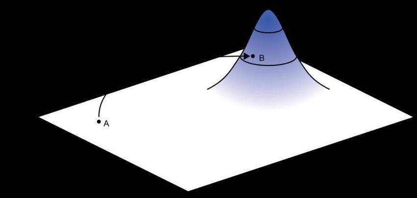
Contribution to Publications

I     Performed molecular biology experimental, expressed enzyme,
      performed activity assay, active-site titration, protein purification
      and sequence data analysis. Wrote parts of the paper.

II    Designed and performed molecular biology experimental,
      expressed enzyme and performed protein purification.

III   Designed and performed molecular biology experimental, protein
      purification, screening (in part) and sequence data analysis.
      Determined kinetic constants. Wrote the paper.

IV    The methods from paper III was extended to aromatic substrates.
      Practically, I performed minor sequence data analysis.

V     Conceived, designed and performed molecular biology
      experimental, screening (in part), activity assay, protein
      purification and sequence data analysis. Wrote the paper.

                                 ix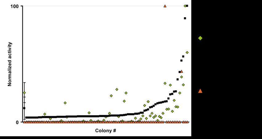
x
Table of Contents

Abstract ......................................................................................................... v

List of Publications .................................................................................... vii

Contribution to Publications ..................................................................... ix

Abbreviations............................................................................................. xiv

Amino Acid Abbreviations ........................................................................ xv

1. Introduction ........................................................................................... 17
    1.1 Introduction to Enzymes ................................................................................. 17
    1.2 Enzymes as Catalysts in Organic Chemistry ............................................... 17
    1.3 Enzymatic Kinetic Resolution ......................................................................... 18
    1.4 Lipases and Serine Hydrolases ...................................................................... 19
    1.5 Candida antarctica Lipase A ........................................................................... 21
    1.6 Protein Engineering .......................................................................................... 22
        1.6.1 Natural and Directed Evolution ............................................................. 22
        1.6.2 Random Protein Engineering ................................................................. 23
        1.6.3 Site-Specific Protein Engineering.......................................................... 24
        1.6.4 Semi-Rational Protein Engineering ....................................................... 25
    1.7 Objectives .......................................................................................................... 27

2. Determination of the Candida antarctica Lipase A Protein
Structure (Paper I and II) ....................................................................... 28
    2.1 Introduction ....................................................................................................... 28
    2.2 Recombinant Production of CALA .................................................................. 29
    2.3 Manual Structure Modelling ............................................................................ 30
        2.3.1 Alanin-Scanning ....................................................................................... 30
        2.3.2 Active Site Titration................................................................................. 31
    2.4 X-ray Structure ................................................................................................. 33
    2.5 Conclusions........................................................................................................ 35

3. Directed Evolution of Candida antarctica Lipase A for Enhanced
Enantioselectivity (Paper III and IV) .................................................... 36
    3.1 Introduction ....................................................................................................... 36
    3.2 Preparation of the Episomally Replicating Yeast Expression Vector
    pBGP1-CALA ............................................................................................................. 37

                                                              xi
3.3 Directed Evolution of CALA for Increased Enantioselective Towards 4-
Nitrophenyl 2-Methylheptanoate .......................................................................... 38
3.3.1 4-Nitrophenyl 2-Methylheptanoate as Model Substrate ................... 38
3.3.2 Selection of Mutable Sites...................................................................... 38
3.3.3 Production of Libraries ............................................................................ 40
3.3.4 Library Screening .................................................................................... 41
3.3.5 Kinetic Investigation and Model Analysis of Enantioselective
Variants................................................................................................................ 42
3.4 Directed Evolution of CALA towards 4-Nitrophenyl 2-Phenylpropanoate
.................................................................................................................................... 43
3.4.1 4-Nitrophenyl 2-Phenylpropanoate as Model Substrate ................... 43
3.4.2 Library Screening .................................................................................... 44
3.4.3 Substrate Scope ...................................................................................... 45
3.4.3 Kinetic Resolution of 2-Phenylpropanoates with Different Alcohol
Moieties ................................................................................................................ 46
3.4.4 Models of Enantioselective Enzyme Variants...................................... 47
3.5 Mechanistic Investigations via Site-Directed Mutagenesis ....................... 48
3.5 Conclusions........................................................................................................ 49
4. Combinatorial Reshaping of the Substrate Pocket (Paper V) ..... 51
4.1 Introduction ....................................................................................................... 51
4.2 Experimental Outline ....................................................................................... 52
4.3 Results and Discussion .................................................................................... 53
4.3.1 Combinatorial Library Design ................................................................ 53
4.3.2 Mutagenesis and Homologous Recombination ................................... 56
4.3.3 Functional Diversity of the Library ....................................................... 56
4.3.4 Library Screening towards Ibuprofen Ester ........................................ 57
4.3.5 Back Mutations......................................................................................... 58
4.3.6 Enzyme Models ........................................................................................ 59
4.3.7 Protein Fitness Landscapes .................................................................... 60
4.3.8 Combinatorial Substrate Pocket Sculpting .......................................... 61
4.3.9 Other Considerations .............................................................................. 62
4.4 Conclusions........................................................................................................ 63
5. Concluding Remarks ............................................................................ 64
Acknowledgments ..................................................................................... 65
References .................................................................................................. 67
xiixiii
Abbreviations

CALA            Candida antarctica
                                 Candida
                                   lipase Aantarctica lipase A
CALB            Candida antarctica
                                 Candida
                                   lipase Bantarctica lipase B
CAST            Combinatorial active-site
                                 combinatorial
                                          saturation
                                                 active-site
                                                     test saturation test
Cfu             Colony forming units
                                colony forming units
DKR             Dynamic kinetic resolution
                                 dynamic kinetic resolution
DNA             Deoxyribonucleic deoxyribonucleic
                                 acid             acid
ee              Enantiomeric excess
                                enantiomeric excess
epPCR           Error-prone PCR
ESRF            European synchrotron radiation facility
HIC             Hydrophobic interaction
                                 hydrophobic
                                        chromatography
                                             interaction chromatography
IPTG            Isopropyl β-D-1-thiogalactopyranoside
ISM             Iterative saturationisopropyl β-D-1-thiogalactopyranoside
                                      mutagenesis
KR              Kinetic resolution
LED             Lipase engineeringkinetic
                                    database
                                          resolution
mRNA            Messenger RNA
MSA             Multiple sequence alignment
NSAID           Non-steroidal anti-inflammatory
                                  non-steroidal anti-inflammatory
                                                 drug             drug
PCR             Polymerase chain polymerase
                                 reaction   chain reaction
RCSB PDB        Research Collaboratory for Structural Bio-
                informatics Protein Data Bank
RNA             Ribonucleic acid ribonucleic acid
SDM             Site-directed mutagenesis
                                  site-directed mutagenesis

                              xiv
Amino Acid Abbreviations
Abbreviation
Amino acid name
Three-letter Single-letter
Ala A Alanine
Arg R Arginine
Asn N Aspargine
Asp D Aspartic acid (Aspartate)
Cys C Cystein
Gln Q Glutamine
Glu E Glutamic acid (Glutamate)
Gly G Glycine
His H Histidine
Ile I Isoleucine
Leu L Leucine
Lys K Lysine
Met M Methionine
Phe F Phenylalanine
Pro P Proline
Ser S Serine
Thr T Threonine
Trp W Tryptophan
Tyr Y Tyrosine
Val V Valine
xvxvi
1. Introduction

1.1 Introduction to Enzymes
In 1897 Eduard Buchner discovered that yeast extracts can ferment sugars to
alcohols and that the process was promoted by substances found in the
extract. Wilhelm Kühne had already introduced the term enzyme in 1878, to
describe such „non-living‟ catalysts.1 Enzymes are biocatalysts, and as such
in principle work like other catalysts – it decreases the activation energy via
transition state stabilization, leading to an increased rate of the reaction. It is
essential to understand that catalysts (such as enzymes) never alter a
chemical equilibrium.
   Compared to other catalysts, many enzymes show a remarkable
specificity. This specificity is popularly believed to be due to an „induced fit‟
of the enzyme to the shape of the substrate.2 The induced fit mainly
influences the initial binding, and not the catalytic process itself. Yet, at the
same time many enzymes also show a high degree of promiscuity, i.e. they
can catalyze reactions and accept substrates that are not natural substrates for
the enzyme.3 The immense catalytic ability has been mainly explained as a
result of the preorganization found in the active site of enzymes.4-5 The
transition state is stabilized by the electrostatic environment, which is the
main contributor of the lowering of the activation barrier compared to the
corresponding reaction in water. Other disputed hypotheses have been put
forward over the years, which claim that strain, protein dynamics, low
barrier hydrogen bonds or quantum tunneling is the main contributor of
catalytic activity.6 Another popular theory has been the ground state
destabilization idea, i.e. shielding the transition state from solvation effects.7
However, as mentioned above, it is now largely accepted that enzymes work
by transition state stabilization.5,8 Some enzymes, such as carbonic
anhydrase, have reached so called catalytic perfection, where the chemical
reaction occurs so fast that it is only limited by the diffusion of the reactants
entering and leaving the active site.9

1.2 Enzymes as Catalysts in Organic Chemistry
Enzymes have been used by mankind since early history. One of the oldest
applications has been the fermentation of carbohydrates to alcoholic

                                                                                17
beverages. There is evidence that even Mesopotamian people in 6000 BC fermented sweet fruits to produce wine.10 The understanding of what occurred in the fermentation process was of course limited. We now understand that it is Saccharomyces cerevisiae, bakers‟ yeast, which carries out an anaerobic oxidation of carbohydrates to form ethanol. Saccharomyces cerevisiae was used by the Bayer corporation already in the 1930‟s to form a precursor to ephedrine, L-phenylacetylcarbinol via whole-cell biotransformation of benzaldehyde.11 Ever since then, the use of enzymes for biotransformations have slowly but steadily gained momentum. Enzymes are used in industry either isolated or in living whole-cell systems. Many energy-efficient processes have been developed using enzymes, as many enzymes have their temperature optimum at room temperature.12 Enzymes are large polypeptides that are easy to produce with modern recombinant gene technology. The use of enzymes will most likely increase in an energy- and resource-conscious world. A vision of the future is the concept of „microbial cell factories‟, the idea of utilizing genetically engineered microbes, with entire biosynthetic pathways (catalyzed by several enzymes) incorporated.13-14 1.3 Enzymatic Kinetic Resolution Many molecules can exist as non-superimposable mirror images of each other. Such molecules are considered to be chiral. These „mirror‟ images of a chiral molecule are called enantiomers. This fundamental discovery was made in the 19th century by Louis Pasteur who separated the enantiomeric crystals of sodium ammonium tartrate; the crystal shapes were mirror- images of each other.15 Enantiomers have the same physical properties provided that they are in an achiral environment. Biological organisms contain a large quantity of enantiopure molecules, and therefore constitute chiral environments. Amino acids and sugars occur predominantly in one enantiomeric form in nature. Enzymes and cellular receptors are made up of only L-amino acids, thus they are enantiomerically pure. Nature is pervaded by this homochirality, and it is essential for the existence of terrestrial life.16 Many modern drugs are chiral, and the two enantiomers of the compound can often interact with the organism in completely different ways. Methods for the preparation of enantiopure compounds are thus highly relevant for the production of pharmaceuticals. One of the methods available is kinetic resolution (KR). KR relies on the rate difference between two enantiomers in the transformation from substrate to product (Scheme 1). KR can be achieved by the use of a chiral catalyst, for example an enzyme.17 18
Scheme 1. (S)-selective enzymatic kinetic resolution
Enzymes have the advantage of having a defined topology in the active
site where the catalytic reaction occurs. Compared to chiral ligands, the
active site of an enzyme most often has considerably larger defined space
where the chiral recognition occurs. Thus, in many cases, extremely high
enantioselectivity can be obtained.18 The enantioselectivity of the reaction is
defined by the E-value,19 which has been introduced to specify the
selectivity, as
k
fast
E
k
slow
1.4 Lipases and Serine Hydrolases
Lipases (EC 3.1.1.3) are currently the most used class of enzymes in
chemoenzymatic reactions and kinetic resolutions.20 The reaction catalyzed
by lipases in nature is the hydrolysis of water-insoluble esters such as lipids.
Lipases have a tendency to increase their activity in presence of high lipid
concentration; this is assumed to be caused by a change in enzymatic
conformation when in close contact with a non-polar surface, such as lipid
droplets. This phenomenon is called the interfacial activation. It has been
suggested that a hydrophobic „lid‟ is responsible for this effect; the lid
covers the active site, and swings open and immerses itself in the
hydrophobic media when the lid comes in contact with the lipid phase.21
Lipases have been used for hydrolysis of esters, and for the reverse
reaction, the synthesis of esters in organic solvents. Some lipases can also be
used for acylating amines for the formation of amide bonds.22 Enzymes were
thought to be unstable in organic solvents; however, Klibanov et al.
discovered that that many enzymes are actually stabilized by dry unpolar
solvents.23 The proposed reason is that in these dry solvents the native
conformation is kept and the enzymes do not unfold.
Many lipases display enantioselectivity, a highly useful property. Lipases
have been applied to perform kinetic resolution of many different substrates.
Our research group have used KR to great extent, and also in combination
with transition metal-catalysed racemisation of the chiral substrates, which
has been coined dynamic kinetic resolution (DKR) (for comprehensive
reviews, see refs.17,24-27). This method has been applied to produce several
interesting compounds in high yields and enantiopurity.28-30
19In comparison with the oxidoreductases, which are relying on either expensive cofactors (such as NADPH/NADP+) with regeneration systems or whole cell-systems, many lipases can be used in vitro without any special additives.31 All serine hydrolases (which includes lipases, esterases and serine proteases) work via a similar molecular mechanism.32 Three amino acid residues, the so called „catalytic triad‟, are key players. An acid residue (aspartate or glutamate) coordinates to a histidine, which in turn works as a charge relay residue.33 The histidine withdraws a proton from the nucleophilic serine. The now activated serine works as the nucleophile attacking the ester carbonyl, and the formed oxyanion of the tetrahedral intermediate is stabilized by the so called the oxyanion hole (Figure 1).34 Figure 1. The serine hydrolase reaction mechanism for the hydrolysis of an ester. The Candida antarctica lipase A (CALA) catalytic machinery is displayed.35 In the free enzyme, the nucleophilic Ser184* is coordinating to His366, which in turn is hydrogen bonding to Asp334. In step 1, the ester enters the active site, and is attacked by the activated nucleophilic serine. The oxyanion is stabilized by Asp95 and the nitrogen backbone of Gly185. In step 2, the alcohol leaves, and in step 3, water attacks the carbonyl of the acylated serine, and a new tetrahedral intermediate is formed. Finally, in step 4, the acid is released, serine is reconstituted, and the catalytic cycle is completed. * The author would like to point out in the sake of clarity, that in papers I–V, the numbering of amino acid residues is numbered +10 in comparison to the original Novozyme cloning publication.38 The +10 numbering of residues is also used in this thesis. 20
1.5 Candida antarctica Lipase A
Several Japanese research expeditions were sent out in the 1960s‟ to sample
Antarctic soils to examine the microbiological flora. Soil and water samples
were assayed in the McMurdo dry valleys. The yeast Candida antarctica
was found in a sample from lake sediment at 9 m depth, from a hypersaline
lake, Lake Vanda.36 Lake Vanda is perennially covered by ice.37
    Researchers at Novo Nordisk A/S (now Novozymes) isolated two lipases
from Candida antarctica. The two lipases, called Candida antarctica lipase
A and B (CALA and CALB, respectively) were both found to be highly
thermostable, and were cloned into Aspergillus oryzae.38
    Homology analysis of the CALA and CALB peptide amino acid
sequences and DNA sequences reveals close relationship to the
basidiomycetous fungi Pseudozyma aphidis39, Kurtzmanomyces sp. I-1140
and Ustilago maydis.41 P. aphidis have highly homologous genes to the two
lipases from C. antarctica. U. maydis, also called corn smut, is a well
studied pathogen found on maize. Pseudozyma aphidis was curiously first
isolated from the faeces of aphids. The Pseudozyma aphidis strain DSMZ
7072542 was used in our laboratory for the in-house isolation of CALA and
CALB. The isolated CALA gene contains a single silent mutation, and the
CALB gene gives rise to two surface located amino acid substitutions that
differ from the original Novozyme publication.38,43
    CALB has been used for vast numbers of biotransformations and kinetic
resolutions of many substrates, and is probably the single most used enzyme
for kinetic resolutions. CALA has not found such broad application yet, but
it has some interesting properties that are currently exploited.
    CALA is a monomeric 431 amino acid residues single peptide lipase,
weighing 45 kDa, with a pH optimum at 7.44-45 CALA is, as previously
mentioned, highly thermostable, and is claimed to be one of the most
thermostable lipases known.46 Novozymes has produced CALA in
Aspergillus oryzae and is marketing the lipase as Novozyme 735. Regarding
the preference of esters, CALA prefers medium to long chain lengths of the
alcohol and acid moieties.46 CALA is known to exhibit a weak interfacial
activation.45 CALA has also the interesting property that it has an sn-2
preference towards triglycerides.47 In triglycerides, sn-2 is a designation of
the center carbon of the glycerol moiety. The sn-2 preference can be used for
selective substitutions on triglycerides, which could be useful for the
preparation of fat replacement products and in theory, covalently tethered
drug hidden in a triglyceride-like compound.
    CALA has found use for the preparation of highly enantiopure β-amino
acids/esters, which holds large promise as building blocks for important drug
candidates, such as specific protease inhibitors.48-50 CALA has also shown
the unusual trait of being able to hydrolyse esters with tertiary alcohol
moieties.51-52 Tertiary alcohols are used as a protective group in synthetic

                                                                           21
organic chemistry, and the specific removal of such groups can be of great interest. Enantiopure tertiary alcohols are also interesting, and the enrichment via kinetic resolution could prove very useful. In literature, mutational studies of CALA are quite sparse, but some information can be found in Novozyme patents. One CALA variant has been reported, with the modifications F145W† and F149W, which is claimed to have a fourfold increase in the activity towards glycerol tributyrate.53-54 1.6 Protein Engineering Enzymes are proteins, and as such biopolymers, produced by the cells to facilitate various molecular processes such as metabolism and replication of DNA. As with all proteins, their formation is based on the „central dogma‟; transcription of DNA to produce mRNA, transport of the mRNA to the ribosome, where the mRNA is translated and the protein is synthesized. The ribosome is a large RNA-protein complex which synthesizes polypeptides, using mRNA as a template, and amino acids as building blocks.55 The polypeptide is processed, and folded into a defined structure, and the protein is formed.1 The fact that the genetic information is coupled to the protein phenotype facilitates the adaption of protein properties via the modification of genetic information. Protein engineering is the deliberate modification of these properties, by the use of molecular biology techniques. This field is currently expanding rapidly, and several techniques have been established, or are in the process of being established.56 1.6.1 Natural and Directed Evolution The British naturalist Charles Darwin developed his theory of evolution in the mid-19th century.57 The theory of evolution can be roughly summarized as follows: Diversification: Copy X (parents) into several Y (offspring). Introduce slight variations in the Ys. Throw away all X. Selection (natural or non-natural): Only Y that has traits that grant „survivability‟ are kept, the other Ys are discarded. Reproduction: Remaining Y (offspring) becomes X (parents). Go to first step and repeat. This simple iterative process has created all the variation in natural biological life, observed so far. „Survivability‟ is an abstract concept; in † A note on amino acid residue substitutions; F145W, or Phe145Trp, means that phenylalanine residue no. 145 has been replaced with tryptophan in that particular enzyme variant. 22
biological science the term fitness is used, where it indicates an organisms‟
capacity to replicate its genetic material.58 In non-natural selection, such as
in directed evolution, it can be any arbitrary property that the researcher
selects for.
   The refinement and development by breeding and selection of
domesticated livestock, dogs and cultivated grass are based on evolution.
The information carrier in living organisms, the inheritable genetic code, is
DNA. Variation in the genetic code can be introduced by several processes,
such as mutations by exchanges of bases in DNA, or sexual recombination.59
   Directed evolution is a method used in protein engineering, where the
power of non-natural selection is utilized to improve desired properties of
proteins. The iterative process, the essence of directed evolution, facilitates
these stepwise improvements.60-63 Molecular biology techniques and
recombinant DNA technologies have steadily improved over the last
decades. Many of these methods have found usage in directed evolution
procedures, where they are used for introducing protein diversity.56,64-66

1.6.2 Random Protein Engineering
One of the first techniques used for directed evolution was the error-prone
PCR (epPCR) technique. It is based on the non-perfect replication of DNA
in the polymerase chain reaction (PCR).67 Misincorporation of nucleotides
occurs over the entire replicated sequence. By altering the concentration of
magnesium and manganese ions it is possible to modify the amount of
erroneously incorporated nucleotides in the replicated DNA.65 It gives rise
to a pool of mutated sequences, a so called „library‟ of mutants. A word on
definition: a mutant gene gives rise to a protein variant.
   Classical error-prone PCR for directed evolution requires neither crystal
structure of the protein in question, nor any special knowledge of the
mechanism of the enzyme, or of the active site. The majority of amino acids
found in an enzyme are generally quite far from the active site. Thus, there is
slim chance of hitting a residue involved in substrate binding and that is
influencing activity, and this approach may therefore require screening of
very large libraries.68-69
   Another technique that usually does not require prior knowledge of the
structure is the gene (DNA) shuffling techniques.70 A multitude of DNA
shuffling techniques have been developed, such as ITCHY, SCRATCHY
and SCOPE, etc.62,71-72 They all have in common that they are based on
recombining more-or-less homologous sequences, for example homologous
enzymes derived from different species.73

                                                                            23
1.6.3 Site-Specific Protein Engineering Site-directed mutagenesis (SDM) is currently one of the most used mutagenesis methods in protein engineering.74 It is based on the use of primers, short oligonucleotides used in the polymerase chain reaction (PCR) step, which are not completely complementary to the sequence being amplified. The non-complementary nucleotides are introduced in the amplified sequence. The template sequence is preferably a plasmid, a circular extra-chromosomal body of DNA (Figure 2). Figure 2. A brief overview of the site-directed mutagenesis. The starting point is a template; a double-stranded plasmid, which contains the gene of interest. In step 1, the plasmid is denatured by heat, and primers anneal to the complementary strand. The PCR is carried out in step 2. The mutagenic primer is incorporated in the amplified DNA-fragments. In step 3, complementary, mutagenic plasmid strands anneal to each other and form double stranded nicked open-circular plasmids. The PCR has ended, and reaction mixture is worked up and transformed into the bacterium Escherichia coli that repair the nicked plasmid. The non-complementary nucleotides usually codes for a substituted amino acid. As a triplet codon in a nucleotide sequence specifies a single amino acid, often up to three nucleotides are substituted. This method can be used for the rational modification of enzyme active sites. It can for example also be used for „knocking out‟ enzyme functionality or removing protein- protein interactions by substituting catalytically important residues or charged surface residues, with the „inactive‟ residue alanin, which is known as „alanin-scanning‟.75 SDM can also be used for the saturation of a single site, using „degenerate‟ primers, which are randomized in their nucleotide composition at specific sites. This can give rise to small libraries of protein variants, where a specific amino acid is substituted by a random residue. 24
1.6.4 Semi-Rational Protein Engineering
   The size and utility of the protein libraries generated are important
parameters when deciding what protein engineering strategy that should be
pursued.76 Factors such as cost and labor time for screening are reasons to
keep the library size as small as possible.77 Protein engineering methods that
focus on the active site are known to have a higher chance of influencing
catalytic properties.69 These methods generally create small libraries, as only
a few amino acid residues are targeted. One development of SDM was
conceived in the group of Manfred T. Reetz. The technique is called
combinatorial active-site saturation test (CAST), which is based on the
simultaneous randomization of a few amino acid sites, in close sequence
proximity, using one single primer pair.78 Two or three amino acid residues
are generally subjected simultaneously to mutagenesis. The reason for
choosing more than one amino acid to mutate is the potential synergistic
conformational and electrostatic effects that may appear. Amino acid residue
pairs surrounding the active site are usually the target for the saturation.
These active site-focused libraries have been used with good results for the
improvement of activity and enantioselectivity.79 Iterative rounds of
mutagenesis of the active site often give rise to highly synergistic effects. 80
CASTing has been used in an iteratively manner (coined iterative saturation
mutagenesis, ISM) by the Reetz group to change diverse properties such as
thermostability and enantioselectivity.81 CASTing (and site-directed
mutagenesis) requires knowledge of the substrate binding, and preferably the
mechanism, of the enzyme and associated amino acids. This knowledge is
often derived from the X-ray structure of the enzyme in question.
   The ability to determine the composition of nucleotides at certain
positions when designing primers gives rise to different sets of potentially
encoded amino acids (Figure 3).

    LibF_for 5’ CACGGCGGCACGCCCNNKAGCNNKAAGGACACCTTT 3’
    LibF_rev 5’ AAAGGTGTCCTTMNNGCTMNNGGGCGTGCCGCCGTG 3’

Figure 3. An asymmetric, degenerate primer pair, used for CASTing. Here NNK
degeneracy is used, which code for all 20 possible natural amino acid residues. N
uses all the four nucleotides, K use thymine and guanine, and M (complementary to
K) use adenine and cytosine.

   Clouthier et al. created libraries with NDT degeneracy with successful
results.82 In the primer synthesis, the following codes are used; N uses all the
four nucleotides, D use adenine, guanine, and thymine, and T only thymine.
NDT degeneracy gives rise to a reduced set of amino acids; only 12 amino
acids are coded for. This enables smaller library sizes, at an expense of
missing potential positive hits. There is an issue to what level of amino acid
sets can be reduced, as this can be difficult to discern a priori.64,77,83

                                                                              25
Mutational suggestions may not only be rationally deducted from the three-dimensional structure information, but computational and bioinformatical based-methods are also used to a high degree. For example, the degree of amino acid residue conservation derived from a multiple sequence alignment (MSA) can be used for the elucidation of a residues‟ mutability.84-85 Combinatorial libraries with small sets of amino acid residues have been used for the generation of consensus libraries with84 or without86-87 phylogenetic bias, for the development of thermostable enzymes. The structure-based multiple-sequence alignment 3DM database has been used for suggesting mutational sites and „allowed‟ residues.88 Also, computationally designed combinatorial libraries have generated broad functional diversity for fluorescent proteins.89 In these described methods each mutational site is randomized with a small set of amino acid residues. Indeed, information from statistical and computational methods assists modern protein engineering in an increasing extent.90-91 26
1.7 Objectives
The main scientific aim of this thesis has been the exploration of CALA‟s
structural mutability and biocatalytic potential for kinetic resolution. This
thesis covers the entire process from the structural determination of CALA,
to the protein engineering using structure-based directed evolution methods,
to the acquirement of several enantioselective enzyme variants. The main
substrate focus has been on chiral α-methyl carboxylic acid substrates. This
class of compounds contain several interesting NSAIDs (non-steroidal anti-
inflammatory drugs) such as ibuprofen and naproxen (Figure 4).92

Figure 4. Ibuprofen and Naproxen, two pharmaceuticals that are bulky chiral α-
methyl carboxylic acids.

   CALA was chosen as it had the right prerequisites for developing
enantioselectivity towards these substrates. It was reported to be highly
thermostable, and able to accept large substrates. The directed evolution
method ISM was assumed to be an efficient process to obtain high
enantioselectivity, but required a structure of the enzyme in question.
Therefore, a crucial objective was the determination of the structure of
CALA.
    Also, one objective was the assessment and development of different
protein engineering methods. The development of an efficient method that
would radically alter the substrate binding pocket of an enzyme was
imperative as obstacles were encountered during the development of more
active and enantioselective CALA variants toward esters containing the
bulky ibuprofen moiety.

                                                                                 27
2. Determination of the Candida antarctica Lipase A Protein Structure (Paper I and II) 2.1 Introduction For the development of an enzyme with increased enantioselectivity, directed evolution is an excellent approach. It was decided early on to use CALA for the development of a highly enantioselective lipase towards large substrates, as it was considered to have the prerequisites necessary for the project. The CASTing technique had been proven advantageous for the development of a highly enantioselective Pseudomonas aeruginosa lipase.78 As previously mentioned, the CASTing technique requires an X-ray structure or a homology model for the selection of amino acid residues that may influence the property screened for. As an X-ray structure of CALA did not yet exist, a homology model was considered as an acceptable alternative for the project. A comparison of the amino acid sequence revealed that there were no available enzyme X-ray structures sufficiently related to CALA. The crystallization and determination of an X-ray structure of a novel enzyme was also seen as quite difficult. The closest related available structures were Pseudomonas putida esterase (14% sequence identity) and Pseudomonas fluorescens esterase (14%).93 CALA could however easily be identified as belonging to the large α/β hydrolase fold family. The possibility of creating a manually modeled structure was considered, based on the generic α/β hydrolase fold (Figure 5). It was assumed that it would be possible to produce the model if the active site residues could be determined. A hypothesis we had was that the catalytic residues could be identified by knocking out functionality via SDM. That information was assumed to give enough knowledge for the creation of a manually constructed 3D-model of CALA. 28
Figure 5. Secondary structure diagram of an idealised α/β-hydrolase. The catalytic
                                                                                 94
residues are indicated with black dots. Adapted from Nardini and Dijkstra, 1999.

2.2 Recombinant Production of CALA
First efforts to overproduce CALA was done in Escherichia coli, using
different pET-plasmids (Novagen) in the host strain Origami2(DE3)
(Novagen). The Origami2 strains are stated to be able to express proteins
with folding difficulties.95 The pET-plasmids all contain the LacZ-promoter
which are induced by isopropyl β-D-1-thiogalactopyranoside (IPTG).96
Unfortunately, the enzyme yield was unsatisfactory and we were also faced
with problem with insoluble enzyme aggregates in the cellular pellet, so
called inclusion bodies.
   Several parameters were evaluated for the expression of CALA using the
cold-induced pCOLD (TaKaRa) vector.97 Expression was more reliable at 15
°C, which also appeared to be true for pET-vectors. The requirement to use
low expression temperature indicates that bacteria are stressed by the
eukaryotic enzyme expression. Several co-transformed chaperone-producing
plasmids were also tested, but of them none gave any satisfactory results.98
   As a high-purity enzyme is required for the activity assay, the E. coli
expression system had to be discarded and the methylotrophic yeast Pichia
pastoris was instead used for expression.99 The secretory expression
facilitates protein purification. P. pastoris has the ability to secrete proteins
of interest, when the α-mating signal peptide from Saccharomyces cerevisiae
is attached to the N-terminus.100 Using the pPICZ-vector (Invitrogen), which
integrates into the AOX1-locus, 101 properly folded CALA enzyme could be
expressed in satisfactory yield (~200 mg L-1). The AOX1-promoter is
induced by addition of methanol in small concentrations, which triggers
strong expression. The secretory expression of CALA was confirmed by
SDS-PAGE of supernatant and cell lysate.

                                                                                29
2.3 Manual Structure Modelling 2.3.1 Alanin-Scanning An „alanin-scanning‟ (as described in chapter 1.6.3) was carried out by SDM, where several CALA variants were produced with key residues replaced by alanin. The enzyme variants produced were purified using hydrophobic interaction chromatography (HIC). The enzyme variants were assayed by an easy colorimetric reaction, by the hydrolysis of 4-nitrophenyl hexadecanoate (1) (Figure 6). The release of 4-nitrophenol (pKa 7.08)102, was monitored spectrophotometrically at 410 nm, in basic buffers. Figure 6. 4-nitrophenyl hexadecanoate. Several putative key amino acid residues were assayed, based on the ordered configuration found in other serine hydrolases.94 For example, it is known that the nucleophilic serine always comes before the acidic contributor in residue sequence order (Figure 5). If enzyme activity was extinguished, the amino acid residue replaced by alanine was potentially one of the catalytic residues. After screening 16 amino acid residues it was found that the enzyme variants Ser184Ala, Ser210Ala, His366Ala, His330Ala and Asp334Ala displayed profound reduced activity (Table 1). His330 could be ruled out as a participant as it preceded Asp334 in the sequence order, which does not comply with the canonical α/β-hydrolase fold (Figure 5). The Ser210Ala variant showed also reduced activity, and we could not completely rule out this residue as an active participant. The Ser184 and Ser210 both appeared in a lipase consensus motif associated with the nucleophilic serine; GXSXG.103 The associated sequences were GYSGG for Ser184 and GASHG for Ser210.93 30
Table 1. Hydrolytic enzymatic activity of selected variants.
Entry Enzyme variant U mg-1[a]
1 Wild type 138 7.0
2 S184A 0.2 1.1
3 H366A 6.9 1.7
4 H330A 4.3 2.9
5 E298A 65 19
6 E314A 69 6.6
7 E308A 18.3 6.8
8 D334A 1.8 2.1
9 I301A 129 14
10 L367A 122 29
11 T118A 121 8.4
12 V120A 88 13
13 W129A 153 13
14 Y317A 39 12
15 Y183A 8.7 3.4
16 S210A 12 1.1
[a] One unit (U) of activity was defined as the amount of enzyme that released 1 µmol p-
nitrophenol per minute under our assay conditions.
2.3.2 Active Site Titration
To distinguish between Ser184 or Ser210 as the nucleophilic residue, an
active site titration was carried out.104 Compound 2 was prepared by
coupling the fluorophore 4-methylumbelliferone with a phosphonate
compound (Figure 7). Compound 2 irreversible binds to the nucleophilic
serine, and can be used to determine whether an enzyme actually contains a
functional active site or not. The nucleophilic serine attacks the phosphorus
and the fluorescent moiety is released, resulting in an enzyme that is
irreversibly inhibited. The released fluorophore can be quantified by
fluorometry, and the fluorescence should display a linear correlation with the
amount of free active sites. The wild type CALA and the variants Ser184Ala
and Ser210Ala were subjected to active site titration. The wild type and
Ser210Ala both displayed correlation between fluorescence and amount of
enzyme. Fluorescence did not increase with increasing Ser184Ala enzyme
concentration, thus revealing that Ser184 was indeed the active site
nucleophile.
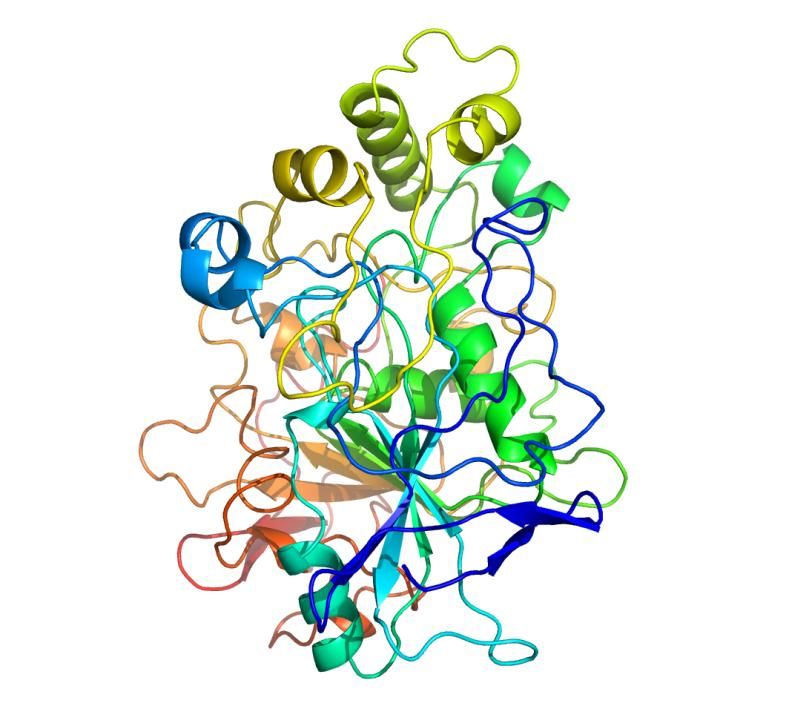
31Figure 7. The phosphonate compound used for active site titration. The residues Asp334, His366 and Ser184 were finally established to be the catalytic triad participants. The assembled data could now be used for the knowledge-based construction of the model. Candida rugosa hydrolase (PDB ID‟s 1CRL & 1CLE) was used as a reference model; even though it has low sequence identity to CALA, it has similar substrate specificity profile. The structure of the model was built up in the program SwissPdb Viewer105, and was allowed to settle in probable configuration based on molecular dynamics and energy minimization using the software package MOLOC (Figure 8).106 Figure 8. The manually modelled CALA structure, in a cartoon representation. CALA is coloured beginning with blue at the N-terminus, going through the rainbow to red at the C-terminus. 32
2.4 X-ray Structure
In parallel to our modeling efforts, a project was initiated with the intention
to obtain an X-ray structure of CALA. Overproduction was carried out as
previously described, using the previously designed pPICZ-CALA vector for
homologous recombination. Production of CALA was straightforward, and
purification was ensured to be of the highest quality, using standard HIC.
HIC is suitable for lipases, as lipases have hydrophobic patches on the
surface that has affinity for the hydrophobic resin.107
CALA crystallized under several conditions, and high-quality crystals
were obtained by hanging-drop vapour diffusion. Data was collected from
European Synchrotron Radiation Facility (ESRF), Grenoble. Unfortunately,
the data did not turn out to be very interpretable at first, as no homologous
structures existed that could help the molecular replacement. Using
selenomethionine, and anomalous scattering, was considered, as this would
give rise to better diffraction data.108 Unfortunately, selenomethionine
incorporation requires quite complex growth and expression conditions.109
Selenomethionine replaces methionine in the translated protein, but
unfortunately CALA had quite few methionine codons, only 2, which were
viewed as insufficient.
Crystals were instead soaked in a uranyl chloride (OU2Cl2) solution,
which gave strong anomalous scattering in the ESRF beamline. The
diffraction data were sufficient for starting to build a structure, but this was
slightly hampered by the lack of homologous structures. Finally a
corroborated structure could be produced, at 2.2 Å resolution (Figure 9).
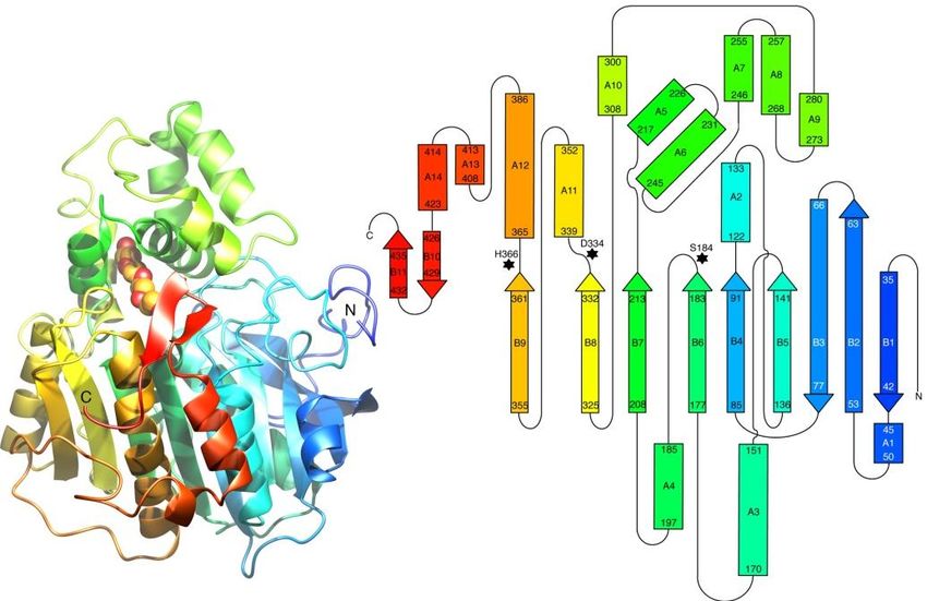
33Figure 9. The X-ray structure of CALA; a cartoon representation and a topology
diagram of the fold. CALA is coloured beginning with blue at the N-terminus, going
through the rainbow to red at the C-terminus. The C-terminal flap (red), which
blocks entrance to the active site, is consisting of residues Gly426 to Gly436. The
large „cap‟ (green) is located between residues 217–308.
The structure revealed a couple of interesting surprises, such as that
CALA turned out to have a flap covering the active site. The lid-like flap is
consisting of the C-terminal residues Gly426 to Gly436. This flexible flap is
most likely coupled to CALA‟s interfacial activation.45 A large „cap‟
(residues 217–308) is a unique feature; this structure thus represents the first
described in a new lipase subfamily. The acyl-binding site is a narrow,
~30 Å long tunnel that accepts long carboxylic acids. A putative
glycosylation site is found at Asn291, and there was a hint that glycosylation
had occurred based on the electron density maps.
The structure confirmed the results from the previous activity assays
regarding the amino acid residues of the catalytic triad. The catalytic triad
was identified as Ser184, His366 and Asp334, and the conformation around
the residues was more or less identical to that of the manual model. The
surroundings of the active site showed some difference compared to the
manual model. One of the most surprising details of the oxyanion hole is that
Asp95 seems to be a crucial component. This was unexpected, since the
acidic group is an unusual residue for stabilizing a negatively charged
reaction intermediate. The importance of Asp95 is supported by the fact that
it is highly conserved, being very rarely replaced by asparagine, or even less
34frequently, glutamine, in some distant relatives. A computational prediction
of pKa-values for ionisable protein residues, using PROPKA 2.0, suggested
that Asp95 has a high pKa of 7.9.110 This increases the plausibility that this
acidic residue can stabilize the oxyanion. The future will resolve whether
this claim is correct. An extensive review has described the different families
of oxyanion holes, and it is difficult to fit CALA into these defined
families.111
The uniqueness of the CALA-homologous sequences has resulted in the
designation of six homologous families and one new superfamily (called the
“Candida antarctica lipase A like” superfamily) in the Lipase Engineering
Database (LED).112 The X-ray structure is deposited at the RCSB Protein
Data Bank under the PDB ID: 2VEO.35
The manually constructed model and the X-ray structure are different in
some aspects. The manually constructed model was bound to have some
minor flaws, and one of these flaws originated from a misinterpretation of
the results from the study of CALA‟s interfacial activation.45 These data
were interpreted as there was no authentic interfacial activation in CALA
and the model was therefore not equipped with an active-site flap. The
modeled protein structure did also display an atypical Ramachandran plot.113
2.5 Conclusions
Protein structure determination is crucial for modern protein engineering, as
site-specific directed evolution techniques are becoming more powerful and
practical. The first part of this chapter demonstrates a novel knowledge-
based structure prediction approach. The latter part presents the resolved
structure of CALA; the first structure from an unexplored α/β-hydrolase
subfamily. The CALA fold will facilitate the generation of homology models
of potentially catalytically interesting enzymes.
Based on the X-ray structure of CALA it is concluded that the CALA
indeed have a C-terminal active-site flap, covering the active site. Molecular
modeling indicates that this flap is quite flexible, and that it is probably
responsible for the slight interfacial activation that has been observed. 45 The
manual model proved correct in the assumptions regarding the active site
residues.
353. Directed Evolution of Candida antarctica Lipase A for Enhanced Enantioselectivity (Paper III and IV) 3.1 Introduction The acquirement of the 3D-structure of CALA was a crucial key objective for the planned structure-based directed evolution projects. One aim was to achieve high enantioselectivity towards several chiral carboxylic acids. From the start, the targeting of two interesting substrate families was intended. The first target was the chiral allenic acids, and the second was the arylpropanoic acids. The arylpropanoic acids are highly interesting as they form basis for the „profen‟-group of pharmaceuticals. Early on, when the first draft of the manually constructed CALA model was completed, the first attempt at CASTing was performed. The allenic model substrate 3 was used as a target for increased enantioselectivity (Figure 10).114 Ester 3 was of interest to us as appealing reactions of allenic compounds has been developed in our group.115-117 Allenes are also interesting substrates as chiral allenes has intriguing axial chirality. Compound 3 had previously been used as substrate in the development of an enantioselective Pseudomonas aeruginosa lipase, also using CASTing.78 Figure 10. 4-nitrophenyl 4-cyclohexyl-2-methylbuta-2,3-dienoate. The pET22b+-vector in the E. coli Origami2(DE3) expression strain was used in the first saturation mutagenesis libraries. Mutations were carried out at the following four residue pairs, Thr97/Leu99, Gly185/His188, Thr263/Leu264 and Val311/Gln312. None of these libraries produced anything of significance. After the X-ray structure had been obtained it was realized that these sites are too far away from the active site to have any influence on enantioselectivity or substrate specificity. One library, Gly185/His188, is located just next to the active site, and as such, could 36
conceivably influence activity. However, this area is conserved due to
structural importance, for example backbone nitrogen of Gly185 is
responsible for oxyanion stabilization and therefore catalytic activity can be
severely reduced by even small perturbations.
   After experimenting with temperature and IPTG concentration, and
bacterial host strains, some improvement could be achieved (as described in
chapter 2.2). The realization how detrimental the bacterial lysation procedure
was for activity, and the insufficient enzyme yields, forced us to look at
other options for enzyme expression.
   A switch to a more efficient yeast expression system resulted in better
yields of protein and with higher purity. However, hydrolytic activity
towards the allenic substrate 3 was unsatisfactory. Even though the
expression levels were higher, the reaction was very slow, and appeared to
level off. Only marginal conversion of 3 occurred after several days, even
weeks of incubation. Strong product inhibition cannot be ruled out, and the
studies on 3 were abandoned for the time being.

3.2 Preparation of the Episomally Replicating Yeast
Expression Vector pBGP1-CALA
The novel P. pastoris episomally replicating pBGP1 vector118 was examined
to see whether it could be used for the expression of CALA libraries. Protein
expression from this vector is driven by the strong constitutively active
glyceraldehyde 3-phosphate dehydrogenase promoter.119 The CALA gene
was cloned into pBGP1, amplified, and transformed into P. pastoris (Figure
11).
                          AmpR
                                                    Alpha factor

                                                         CALA
                                        pBGP CalA
                                         5873 bp

                                                        pGAP fragment
                     PARS1

                                 ZeoR

Figure 11. The pBGP1-CALA vector, used for the expression of CALA, and a
template for CASTing.

                                                                           37
After examining expression levels of P. pastoris transformed with pBGP1-CALA, it was concluded that the plasmid was highly useful for the expression of CALA. In the supernatant, CALA reached a concentration of approximately 100 mg L-1. 3.3 Directed Evolution of CALA for Increased Enantioselective Towards 4-Nitrophenyl 2- Methylheptanoate 3.3.1 4-Nitrophenyl 2-Methylheptanoate as Model Substrate Interest was shifted towards ester 4, which could be a model substrate for other interesting chiral α-methyl carboxylic acids (Figure 12). It was assumed that this substrate could work as an analogue to more bulky substrates, such as the „profen‟-like substrates. The hydrolytic activity of CALA towards 4 was tested to validate that the activity was sufficient for screening purposes. The activity was more than acceptable for the amount of enzyme that could be produced in 96 deep-well plates using P. pastoris pBGP1-CALA. The E value was also determined to be 5.1 towards the (S)- enantiomer. Figure 12. 4-nitrophenyl 2-methylheptanoate. 3.3.2 Selection of Mutable Sites Selection of amino acids were based on a model with (S)-4 bound in the active site in tetrahedral intermediate form (Scheme 2). This intermediate is found in the first step in the catalytic cycle. In this model the C-terminal flap had to be bent open to accommodate the 4-nitrophenyl moiety. 38
You can also read