Recent developments in mitochondrial medicine (Part 1) - 4open
←
→
Page content transcription
If your browser does not render page correctly, please read the page content below
4open 2021, 4, 2
Ó V. Weissig & M. Edeas, Published by EDP Sciences, 2021
https://doi.org/10.1051/fopen/2021002

                                                                                                                         Available online at:
                                                                                                                        www.4open-sciences.org

    REVIEW ARTICLE

Recent developments in mitochondrial medicine (Part 1)
Volkmar Weissig1,* and Marvin Edeas2,3
1
  Midwestern University, College of Pharmacy, Department of Pharmaceutical Sciences, 19555 N. 59th Avenue,
  Glendale, AZ 85308, USA
2
  Université de Paris, INSERM U1016, Institut Cochin, CNRS UMR8104, Faculté de médecine Cochin-Port Royal,
  75014 Paris, France
3
  Laboratory of Excellence GR-Ex, 75015 Paris, France

            Received 15 February 2021, Accepted 15 April 2021

            Abstract – Research into elucidating structure and function of mitochondria has been quite steady between
            the time of discovery during the end of the 19th century until towards the late 1980’s. During the 1990s there
            was talk about a “comeback” of this organelle reflecting a widely revitalized interest into mitochondrial research
            which was based on two major discoveries made during that time. The first was the etiological association
            between human diseases and mitochondrial DNA mutations, while the second revealed the crucial function
            of mitochondria during apoptosis. The March 5th, 1999 issue of Science even featured a textbook image of a
            mitochondrion on its front cover and was entirely dedicated to this organelle. Whilst the term “comeback”
            might have been appropriate to describe the general excitement surrounding the new mitochondrial discoveries
            made during the 1990s, a term for describing the progress made in mitochondrial research during the last two
            decades is difficult to find. Between 2000 and 2020 the number of publications on mitochondria has skyrock-
            eted. It is now widely accepted that there hardly exists any human disease for which either the etiology or
            pathogenesis does not seem to be associated with mitochondrial malfunction. In this review we will discuss
            and follow several lines of mitochondrial research from their early beginnings up to the present. We hope to
            be able to convince the reader of what we expressed about a decade ago, that the future of medicine will come
            through mitochondria.

            Keywords: Biochemistry, Cells, DQAsome, Lipoplexe, Heteroplasmy, miRNA, Mitochondrial medicine,
            Mitochondrial diseases, Mitochondrial DNA, Mitochondrial dysfunction, Mitochondrial research,
            Mitochondrion, Mitochondrium, mitomiRs, mtDNA, Organelle, Physiology, PNA, Research, Science

Introduction                                                                    Mitochondria contain their own DNA and as shown by
                                                                            Doug Wallace and his group in 1980 [3] are maternally
    All human cells, except erythrocytes, harbor mitochon-                  inherited. A recently claimed biparental inheritance of
dria in the form of a tubular network constantly undergoing                 mitochondria in humans [4] however has triggered some
fission and fusion. Known as the “powerhouse of the cell ”,                  contentious discussions [5, 6]. Based on correlating mito-
they are at the juncture of energy-producing pathways, as                   chondrial DNA (mtDNA) variations between populations
the “cell’s arsenal ” they play a decisive role in programmed               from around the world Wallace was able to reconstruct
cell death (apoptosis). In addition, mitochondria are                       origin and migration patterns of women in ancient times,
involved in a multitude of anabolic, catabolic, and signaling               which gave rise to the speculation about a “Mitochondrial
pathways.                                                                   Eve” the most recent female from whom all humans might
    According to the endosymbiotic theory mitochondria                      have descended.
are descended from once free-living bacteria which were                         Mitochondrial DNA encodes for 13 polypeptides essen-
engulfed by prokaryotic cells. This today generally accepted                tial for energy production; all other mitochondrial proteins
evolutionary theory was proposed for the first time in 1966                  encoded by nuclear DNA are imported from the cytoplasm.
by Lynn (Sagan) Margulis who had, as she remembers [1],                     The amount of mitochondrial mass per cell organized as a
to submit her corresponding manuscript to about 15 differ-                  sub-micro- to nano-structured tubular network extending
ent journals before it was accepted by the Journal of                       three-dimensionally around the nucleus and throughout
Theoretical Biology [2].                                                    the cytoplasm depends on the energy demands of the cell.
                                                                            Organs with the highest energy consumption are the brain,
*Corresponding author: vweiss@midwestern.edu                                skeletal muscle, the kidney cortex, liver, heart and the
This is an Open Access article distributed under the terms of the Creative Commons Attribution License (https://creativecommons.org/licenses/by/4.0),
              which permits unrestricted use, distribution, and reproduction in any medium, provided the original work is properly cited.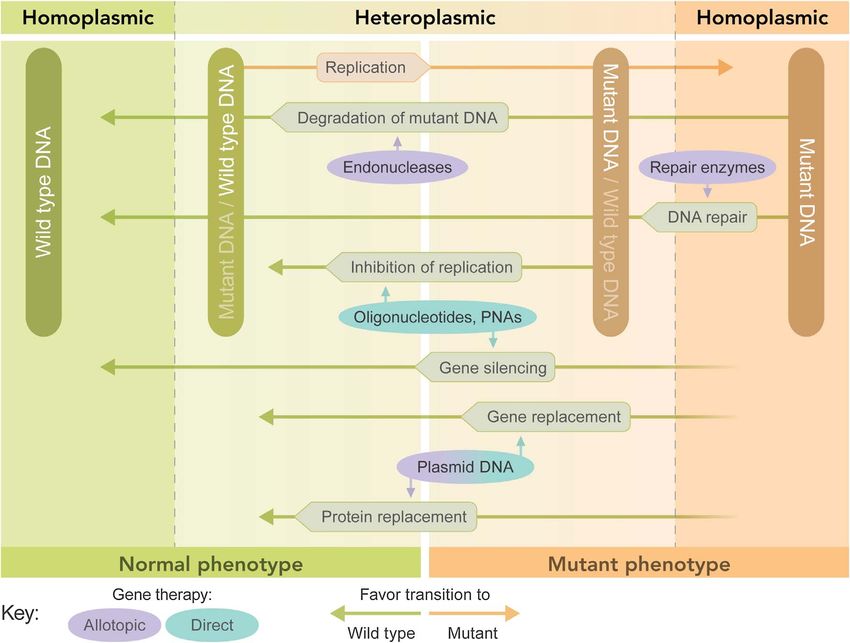
2                                       V. Weissig and M. Edeas: 4open 2021, 4, 2

visual system. Though mitochondrial malfunction as the           be visible under the light microscope. Light microscopy
cause of human disease was described for the first time in        however does not allow for a more detailed structural
1962 [7], the genetic basis for mitochondrial diseases           analysis of mitochondria.
remained elusive until 1988 [8]. Subsequent research over            Their major structural features, which are an outer
the last three decades has revealed that there is hardly         membrane and a highly convoluted inner membrane, were
any human disease the etiology or pathogenesis of which          made visible for the first time during the early 1950’s when
is not associated with mitochondrial malfunction, a realiza-     George Emil Palade (1912–2008, Nobel Prize Physiology
tion leading us [Marvin Edeas and Volkmar Weissig (ME &          1974) investigated the organelle under the electron micro-
VW)] to conclude that “the future of medicine will come          scope [16, 17]. Based on Palade’s work a model for the inner
through mitochondria” [9].                                       structure of mitochondria was developed which dominated
    In this review, written for “non-mitochondriologists”, we    the literature for up to 50 years (Fig. 1A). According to this
outline about 170 years of mitochondrial research whilst         model, mitochondria have two aqueous sub compartments,
emphasizing the major groundbreaking discoveries and             the intermembrane space (IMS) between the outer and the
developments made since the beginning of the new century.        inner membrane and the so-called mitochondrial matrix.
Because we (ME & VW) have been hosting in Berlin                 Here it should be noted that although the term “aqueous
(Germany) the Annual World Conference on Targeting               sub compartments” is still widely used, it is misleading.
Mitochondria since 2010 (the 12th edition will take place        Due to the presence of between 1100 and 1400 [18] distinct
in October 2021), we are fortunate to know on a personal         proteins, the mitochondrial matrix “constitutes a biochemi-
level many of the major investigators who are pushing mito-      cal reaction environment with a highly complex structure”
chondrial research forward. In this review, we highlight         [19]. The viscosity of the mitochondrial matrix solvent has
their contributions to the field of mitochondrial medicine.       been determined to be between 2.69 and 3.32 cP, which
                                                                 can, under a variety of physiological and pathological
                                                                 conditions increase up to 30-fold [19]. For comparison, the
From “thread-like granules” to “cristae                          viscosity of water is about 1 cP.
junctions”                                                           In Palade’s model no barrier exists between the space
                                                                 inside the cristae and the IMS. This model became obsolete
    Studies using light microscopy on cells during the           towards the end of the 1990’s when Perkins and Frey
second half of the 19th century uncovered the existence of       investigated mitochondrial morphology by electron micro-
small subcellular granules [10] which in shape and size were     scope tomography [20, 21] and discovered junctions at the
similar to bacteria as observed by Richard Altmann (1852–        neck of cristae (Fig. 1B). Due to their narrow diameter of
1900) who named them “bioblasts” in 1890 [10, 11]. Based on      about 12–40 nm, these junctions were proposed to impede
the Greek, eight years later, Carl Benda (1857–1932) coined      the flow of proteins and metabolites towards and away from
the term “mitochondria” in 1898 [12], with “mitos” meaning       cristae thereby creating an aqueous compartment distinct
thread and “chondrion” meaning grain. Noteworthy,                from the IMS and also separated from the mitochondrial
although obviously already recognized at the end of the          matrix.
19th century, the thread-like morphology of mitochondria             Studies conducted during the last decade, [22] have
only became fully appreciated at the beginning of the 21st       revealed major protein complexes which control the
century when it was revealed that the maintenance of the         formation of cristae and their junctions, among them optic
tubular (thread-like) network characteristic of mitochon-        atrophy 1 protein (OPA1) and the mitochondrial contact
dria is critical for human health [13–15]. The appearance        site and cristae organization system (MICOS) (Fig. 1B).
of mitochondria as individual objects usually indicates a            Controlling cristae junctions is crucial for mitochondrial
severely damaged mitochondrial network as present in             function. In the mid-1960s, Charles Hackenbrock reported
unhealthy, dying cells or as an artifact of sample processing    that ultrastructural changes of mitochondrial cristae were
for microscopic images. Unfortunately, the depiction of          associated with metabolic changes [23–25]. In 2002, Stanley
mitochondria in text books as single bean-like shaped            Korsmeyer and co-workers showed that cristae remodeling
bodies seems to be still very common.                            is an essential step during a highly complex pathway lead-
    By 1930, most cytologists had recognized mitochondria        ing to apoptosis (programmed cell death) [26]. Finally, a
as well-defined and ubiquitous cellular structures, but there     large body of work during the last 20 years (the reader is
was no agreement as to their function. In 1890, Richard          referred to an excellent recently published review [22])
Altmann proposed them to be the basic units of cellular          revealed that malfunctioning cristae formation or cristae
activity, other proposals made suggested them to be the          remodeling is associated with several human diseases. Just
center for genetic information, protein synthesis, respiration   to name a few, mutations in the OPA1 gene have been
and lipid synthesis [10].                                        linked to optic atrophy associated with deafness and
    The average single mitochondrion, disconnected from          dementia, as well as encephalomyopathy, and cardiomy-
the tubular mitochondrial network by fixation and staining        opathy (reviewed in [22]). Further studies about mitochon-
procedures of tissues samples, appears as a bean-shaped          drial cristae formation will have “important implications for
organelle with approximately the size of Escherichia coli,       understanding human disease linked to various forms of
up to 2 lm long and 1 lm wide, i.e. it is large enough to        mitochondrial dysfunction” [22].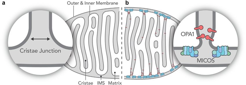
V. Weissig and M. Edeas: 4open 2021, 4, 2                                                  3

Figure 1. (A) The “old” and (B) the “new” model of the inner structure of mitochondria. Instead of the large openings connecting the
intercristae space to the intermembrane space present in the old model (A), narrow tubular openings (crista junctions) connect these
spaces in the new model (B) thereby creating a third aqueous intramitochondrial compartment. Panel (B) also shows the presence of
two out of several protein complexes which control the formation and remodeling of the cristae junctions. This schematic illustration is
based on figures in [22].

From “Atmungsferment” to “Respirasomes”                               between 1939 and 1941 Fritz Albert Lipmann (1899–1986,
                                                                      Nobel Prize in Physiology or Medicine in 1953) identified
     Exactly how substrates are being oxidized in aerobic             ATP as the main bearer of chemical energy in the cell,
organism was a hot topic discussion in the early 1900s.               coining also the term “energy-rich phosphate bonds” [38–40].
For example, Heinrich Otto Wieland (1877–1957, Nobel                      Also during the 1940s, based on progress made in devel-
laureate in Chemistry in 1927) proposed that oxidation in             opment of cell fractionation techniques, the activities of
living tissues involves the removal of the hydrogen atom              enzymes for fatty acid oxidation and the TCA cycle were
(dehydrogenation) instead of the addition of oxygen [10].             found to be associated with mitochondrial fractions [10]
Otto Warburg (1883–1970, Nobel Prize in Physiology or                 and in 1953 Edward Charles Slater (1917–2016) proposed
Medicine in 1931) advocated in 1914 for an iron compound,             a scheme for the mechanism of oxidative phosphorylation
which he termed “Atmungsferment” (“Breathing Enzyme”)                 involving chemical intermediates [41, 42]. These develop-
as being the chief catalyst for activating and transferring           ments in mitochondrial research did not go unnoticed; the
oxygen in biological systems [27]. Subsequently, Warburg              legendary term “Powerhouse of the cell” was coined in an
succeeded in identifying cytochrome oxidase as the enzyme             article for Scientific American by Philip Siekevitz (1918–
that reacts with oxygen. In addition to finding the final               2009) in 1957 [43]. In 1961 it was suggested by Robert Joseph
cytochrome (cytochrome a3), Warburg also characterized                Paton Williams (1926–2015) that protons instead of Slater’s
nicotinamide adenine dinucleotide (NAD) as a hydrogen                 energy-rich intermediates served to deliver energy to ATP
carrier in biological oxidation reactions. Interestingly,             synthase [44]. In the same year Peter Dennis Mitchell
cytochromes were already described back in 1884 by Mac                (1920–1992) postulated his chemiosmotic hypothesis [45–47]
Munn as respiratory pigments that function in the transfer            for which he was awarded the Nobel Prize in Chemistry in
of oxygen [28]. Charles Alexander Mac Munn’s (1852–1911)              1978. By and in 1962 the major players of the respiratory
work however wasn’t fully recognized and accepted until               chain, i.e. enzymes with coenzymes, were purified, reconsti-
40 years later when David Keilin (1887–1963) started in               tuted and termed Complexes I–IV by Youssef Hatefi et al.
1925 publishing a series of investigations about cytochrome           [48–51] and by 1976 the fluid model of the inner mitochon-
systems of pigments in aerobic cells [29].                            drial membrane (IMM) organization was established [52, 53].
     The next milestone towards revealing the secrets of                  Since then it has become a widely believed dogma that
biological oxidation and respiration was the description of           all enzyme complexes, CI-CIV including the ATP synthase
the citric acid cycle or tricarboxyllic acid (TCA) or Krebs           (Complex V) are independent diffusible proteins associated
cycle by Sir Hans Adolf Krebs (1900–1981, Nobel Prize in              with the IMM and connected by small electron carriers shut-
Physiology or Medicine in 1953) in 1937 [30–34], who in turn          tling between them. However, this paradigm of how the
built upon the earlier work by Albert Szent-Gyoergyi (1893–           yeast and mammalian system of oxidative phosphorylation
1986, Nobel Prize in Physiology or Medicine in 1937), Carl            is organized fundamentally changed around 2000. Using a
Martius (1906–1993) and Georg Franz Knoop (1875–1964)                 mild one-step protocol for the isolation of membrane protein
[7]. Hans Karl Heinrich Adolf Lohman (1898–1978) discov-              complexes, namely blue-native polyacrylamide gel elec-
ered ATP in 1929 [35] but its connection to and vital role            trophoresis (BN-PAGE), Schragger et al were able to isolate
in cell respiration wasn’t established until 1937 [36]. Two           supramolecular structures of the mitochondrial oxidative
years later Volodymyr Oleksandrovych Belitser (1906–                  phosphorylation electron transfer chain (OXPHOS-ETC).
1988) and Elena Tsibakova introduced the term “oxidative              They found that in mammalian mitochondria, almost all
phosphorylation” [37] and about during the same time,                 complex I is assembled into supercomplexes comprising
4 V. Weissig and M. Edeas: 4open 2021, 4, 2
Figure 2. (A) “Classical” schematic representation of the mitochondrial oxidative phosphorylation (OXPHOS) system showing its
individual components separated from each other, (M) matrix, (IMS) intermembrane space, (IM) inner membrane. (B) Schematic
model of the organization of individual components of the respiratory chain (complexes I, III and IV) into a respiratory string. Both
figure panels were created based on corresponding figures in [55].
complexes I and III and up to four copies of complex IV, towards the end of the 1950s malfunctioning mitochondria
which led the authors to propose a model for a network of as a cause of human diseases. In 1962, he published a paper
respiratory chain complexes they named “respirasome” entitled “A case of severe hypermetabolism of nonthyroid
[54]. During the next 10 years (2000–2010) additional origin with a defect in the maintenance of mitochondrial
studies demonstrated that that complexes I and III in mam- respiratory control: a correlated clinical, biochemical, and
mals and I, III, and IV in plants actually behave as single morphological study” [7]. The ailment described in his
units kinetically, which led to the assumption of substrate paper, caused by an extensive uncoupling of mitochondrial
channeling. Furthermore, recently it has been found that respiration in skeletal muscle tissue, soon became known as
respirasomes seem to be organized into respiratory strings, “Luft disease”. Rolf Luft is nowadays widely acknowledged
as schematically shown in Figure 2 (reviewed in [55]. as the “Father of Mitochondrial Medicine” and the Rolf
Finally, in 2016 the first detailed organization of a Luft Foundation invites nominations for the “Rolf Luft
respirasome was published by Yang’s group from Tsinghua Award in Diabetes and Endocrinology Research” each year.
University. The authors presented “a 5.4 Å cryo-electron Luft’s paper from 1962 spurred intense investigations into
microscopy structure of the major 1.7 MDa SCI1III2IV1 the possible role of mitochondria in the etiology of human
respirasome purified from porcine heart” [56]. Disruption disease. About 25 years later Hans Rudolf Scholte, in a
of the formation of such supercomplexes or strings of respi- comprehensive review of the biochemical basis of mitochon-
rasomes ultimately can cause OXPHOS malfunction result- drial disease, was able to classify over 120 diseases. Luft
ing in pathologic changes [57]. Studying the detailed himself wrote in 1994 “From Scholte’s and subsequent
interactions within this huge molecular machinery will reviews several basic principles in mitochondrial pathology
eventually provide important information for drug design emerged. First, some mitochondrial diseases affect only
and screening [58]. one tissue, most often skeletal muscle and brain but also
liver, heart, kidneys or endocrine glands. Other organs
may be involved secondarily. The disease may originate as
The recognition of mitochondrial dysfunctions a specific defect in mitochondrial function, but a variety of
as a cause of human diseases genetic and environmental factors may contribute to the
phenotype” [59].
Rolf Luft (1914–2007), a neurologist and endocrinolo- However, the genetic basis for mitochondrial diseases
gist at the Karolinska Institute in Sweden, discovered remained elusive until 1988 (see below).V. Weissig and M. Edeas: 4open 2021, 4, 2 5
The “Decade of the Mitochondrion” mitochondria-targeted gene therapy, Peter Seibel wrote in
1995
On March 5th in 1999 the editors of Science chose a “Successes in classical gene therapies have been
textbook image of a mitochondrion as the front cover, achieved by placing a corrected copy of a defective
which was unprecedented for this journal. It was a way nuclear gene in cells. A similar gene replacement
for the Science editors to “officially” acknowledge the approach for a mutant mitochondrial genome is
1990s as the “decade of the mitochondrion”. Two key dis- invariably linked to the use of a yet unavailable
coveries were made during this decade: the first one actually mitochondrial transfection vector” [65].
continues where Luft and Scholte (see above) left off. Two
papers, one in Science and the other one in Nature (both Seibel’s group launched the hunt for the “Holy Grail” by
came out in 1988) revealed that mutations and deletions demonstrating that DNA coupled covalently to a short
in mitochondrial DNA were associated with human dis- “mitochondrial leader peptide” can enter mitochondria
eases. Ian James Holt and co-workers described the link of via the protein import pathway, which at that time
deletion mutations of mitochondrial DNA (mtDNA) and seemed to open a novel way for gene-, antisense-RNA-
mitochondrial myopathies [60], and Douglas Wallace and or antisense-DNA-delivery for mitochondrial molecular
his team reported the association of Leber’s Hereditary therapies.” [65]
Optic Neuropathy (LHON) with mtDNA point mutations A mitochondria-targeted transfection vector, in 1995
[8]. It should be noted that mitochondrial DNA was according to Seibel et al. still unavailable [65], presented
discovered back in 1963 by Nass & Nass, who described itself during the same time when one of us (VW) discovered
“intramitochondrial fibers with DNA characteristics” [61] by chance that dequalinium chloride, a mitochondri-
and was sequenced in 1981 [62]. Consequently, during the otropic cationic bolaamphiphile, was able to self-assemble
1990’s most of Luft’s and Scholte’s “classical” mitochondrial into cationic liposome-like vesicles, named “DQAsomes”
disorders were found to be linked to mtDNA mutations. (DeQAlinium-based liposomes) [66, 67]. Liposomes are
The second key discovery involves the crucial role of man-made vesicles between 50 and 1000 nm in diameter in
mitochondria during programmed cell death (apoptosis), which one or more phospholipid bilayer membranes
which however we will not further discuss in this review. encapsulate an aqueous inner space. Following the synthesis
The interested reader is referred to a review from 2007 by of cationic phospholipid derivatives in 1987 by Phillip
Susan Elmore [63]. To highlight the excitement this Felgner’s group [68], liposomes could be rendered cationic
key discovery has triggered, we would like to cite what which in turn enabled them to form complexes with nega-
Brown, Nicholls ad Cooper, three key players in this field, tively charged DNA. The striking similarity of DQAsomes
wrote in 1999 in their preface to “Mitochondria and Cell with cationic liposomes widely used during the 1990s for
Death” [64]: nuclear-targeted gene therapy seemed to qualify DQAsomes
“Who would have believed it?! Mitochondria, the as the mitochondrial transfection vector of choice [69, 70].
powerhouses of cellular life, are also motors of cell DQAsomes have been shown to meet all pre-requisites
death. Few would have accepted this even 5 years for a mitochondrial transfection vector, they bind and
ago”. condense pDNA forming “DQAplexes”, which are taken up
by cells followed by their early endosomal release (just like
it is known from cationic liposome/DNA complexes aka
Manipulation of the mitochondrial genome “Lipoplexes”) and upon contact with mitochondrial
membranes DQAplexes become destabilized releasing a
Finding an effective and reliable way to manipulate the
portion of the DNA from its cationic carrier [71–74].
mitochondrial genome has been the “Holy Grail” of mito-
Finally, it was demonstrated that oligonucleotides as
chondrial medicine ever since defective mitochondrial
well as plasmid DNA conjugated to a mitochondrial leader
DNA has been linked to human diseases. Due to the
sequence co-localize with mitochondria when delivered into
multi-copy nature of mtDNA, several principal strategies
mammalian cells via DQAsomes [75]. Yet the barrier lying
can be followed, which are correcting mtDNA mutations /
between co-localization with mitochondrial membranes and
deletions, selectively degrading mutated mtDNA circles,
functional DNA expression inside mitochondria proved to
selectively inhibiting the replication of mutated mtDNA
be unsurmountable at that time. However, several years
and replenishing mitochondria with wild-type copies of
later two groups have reported DQAsome-based transgene
mtDNA (Fig. 3). All of these approaches have been and
expression in mammalian mitochondria. In 2011, Lyrawati
are still being explored since the 1990s and though all of
et al. described the expression of Green Fluorescence
them were and still are promising, the “hunt for the Holy
Protein (GFP) GFP in the mitochondrial compartment
Grail” is ongoing.
using DQAsome-mediated delivery of an artificial mini-
Mitochondrial gene therapy mitochondrial genome [76] and Bae reported in 2017, an
enhanced mitochondrial transfection efficiency in HeLa cells
Worldwide, during the 1990’s over 1500 nuclear-tar- and dermal fibroblasts when using a modified DQAsome
geted gene therapy trials have been conducted exploring a formulation called DQA80 [77]. Though both reports seem
variety of nuclear-targeted viral and non-viral transfection to be a promising start, despite their limitations as
vectors. Relating to a potentially similar approach towards discussed most recently [78], any routine mitochondrial6 V. Weissig and M. Edeas: 4open 2021, 4, 2
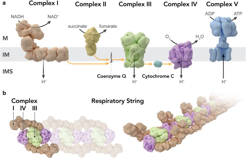
Figure 3. Graphical representation of the effect of various mitochondrial gene therapeutic approaches on mitochondrial
heteroplasmy. A cell can go from homoplasmic wild type (dark Green) to homoplasmic mutant (dark ochre) and pass stages of
heteroplasmy where either wild type (light green) or mutant (light ochre) mtDNA is dominant. The threshold determines the
transition from normal phenotype to mutant phenotype. Ovals represent therapeutic intervention and rectangles represent natural
processes. Arrows that do not contact the DNA boxes indicate that the process does not change the ratio or content of DNA.
Figure recreated based on a corresponding figure in [145].
transgene expression in the laboratory and above all any 1990’s the mechanism of mitochondrial protein import have
mitochondrial gene therapy-based treatment of primary been studied extensively [82–84]. In contrast to mitochon-
mitochondrial DNA diseases in the clinic have to be judged drial proteins imported from the cytoplasm, no imported
as being elusive. An alternative mitochondria-targeted tRNAs have been found inside mammalian mitochondria
nanocarrier, called Mito-Porter, has been designed by [85] though evidence had been presented for the transport
Hideyoshi Harashima’s group. The Mito-Porter is a of nuclear encoded tRNA into mitochondria in a variety
liposome-based carrier that delivers its macromolecular of non-mammalian organism [86]. The mechanisms by
cargo to the mitochondrial interior via membrane fusion which negatively charged RNAs can cross both hydropho-
mediated by an octaarginine peptide (R8) on the liposome bic mitochondrial membranes began to be unraveled in
surface [79, 80]. More recently, in 2020, Harashima’s group the late 1990’s by Ivan Tarassov’s group [87, 88]. During
encapsulated wild-type mitochondrial pre-tRNA(Phe) into the last 20 years, a substantial amount of data has been
MITO-Porters and then transfected cells with mitochondria accumulated demonstrating that in all eukaryotes non-
carrying a mutated version of tRNA (Phe). As result, the coding RNAs are being transcribed from the nuclear
mutation rate of tRNA(Phe) was decreased, and this genome followed by their transport into mitochondria,
therapeutic effect was sustained even on the 8th day after where they play a role in mitochondrial gene expression
transfection [81]. [89]. Today it has been well established that “the mitochon-
drial RNome represents an intricate mixture of the intrinsic
RNA-based approaches towards the manipulation transcriptome and the extrinsic RNA importome” [89] and
of the mitochondrial genome efforts are aimed at exploring mitochondrially imported
RNAs as a tool to further study mitochondrial DNA and
The majority of proteins functioning in mitochondria pathologies associated with its malfunction. For example,
are imported from the cytoplasm and by the end of the exploiting the pathway of RNA import into mitochondriaV. Weissig and M. Edeas: 4open 2021, 4, 2 7
a recent study investigated the possibility of establishing a Limited success of allotopically expressing mitochondrial
CRISPR-Cas9 system targeting mtDNA. Ivan Tarassov’s genes was described for the first time in yeast in 1988 [96,
group tested in vitro and in vivo mitochondrially targeted 97], endorsed for biomedical use by Aubrey de Grey in
Cas9 versions and a set of mitochondria-specific guide 2000 [98] and successfully applied to human cells for the
RNAs regarding their effect on mtDNA copy numbers. first time in 2002 as described in two papers. Manfredi
The authors describe this system as “more complicated et al. demonstrated the rescue of a deficiency in ATP
for use than previously found for nuclear DNA” and chose synthesis by transfer of the mitochondrial ATP6 gene to
as title for their paper “Can Mitochondrial DNA be the nucleus [99]. Guy et al. allotopically expressed the
CRISPRized: Pro and Contra” [90]. mitochondrial ND4 subunit gene of complex I, which was
also the first point mutation in the mitochondrial genome
Shifting heteroplasmy linked to Leber’s Hereditary Optic Neuropathy [100]. The
latter approach advanced in 2018 to clinical Phase I, II
Since mutations or deletions normally do not affect all and III trials (reviewed in [101]). The authors of these
mtDNAs, wild-type and mutated mtDNA can coexist in a studies found an improvement in vision when they injected
cell, a phenomenon called heteroplasmy. Depending on the viral vector carrying a modified ND4 gene into only one
the cell type, in particular on the cell’s energy demand, a eye while using the other eye as a control for disease pro-
minimal number of mtDNA circles have to be damaged gression [102, 103].
to cause clinical symptoms, which is called the threshold
effect. Shifting the level of heteroplasmy by selectively 5.5. Mitochondrial transplantation
inhibiting the replication of the mutant mtDNA and
A review paper published in August 2020 asked the
thereby allowing propagation of only the wild-type mole-
question “if (can). . . transplant(ing) fully functional mito-
cule was proposed for the first time in 1997 by Robert
chondria directly into defective cells (to treat mitochondrial
Lightowlers group in Newcastle, UK [91]. To confirm the
disorders). Could this be too good to be true?” [104]. Two
validity of such a novel strategy they synthesized peptide
years earlier, a paper carried the headline “Mitochondrial
nucleic acids (PNAs) complementary to human mtDNA
transplantation in humans: “magical” cure or cause for con-
templates containing certain mutations known to cause
cern?” [105]. Both of these papers mirror the skepticism
human pathologies. Using an in vitro replication run-off
with which many reports claiming the effective transplanta-
assay under physiological conditions, the authors confirmed
that their antigenomic PNAs specifically inhibited replica- tion of mitochondria into cells, tissues and patients have
tion of mutant but not wild-type mtDNA templates [91]. been met. The paper from August 2020 states
Following Lightowlers’ pioneering work, designing ways to “. . . all these data (about effective mitochondrial
manipulate heteroplasmy in cells became a hot area of transplantation) challenge our understanding of how
research for the next 20 years, “but until recently, a viable organelles behave in cells, and we should consequently
method has proven elusive” [92]. Most recent efforts towards demand high levels of rigour to support these claims”
the selective degradation of mutated mtDNA involve zinc- [104].
finger nucleases (ZFNs) [93] and transcription activator-like
effector nuclease (TALENS) [94, 95], both of which are It comes to mind that Laplace once said “the weight of
enzymes able to selectively target single nucleotide muta- evidence for an extraordinary claim must be proportional
tions. Both enzymes have genetically been modified to to its strangeness”, which much later was rephrased by
express at their N termini a mitochondrial leader peptide Carl Sagan (1934–1996), into “extraordinary claims
to ensure their transport into the mitochondrial matrix require extraordinary evidence” [106].
and both enzymes have recently been successfully tested It is not our aim here to investigate exactly how extraor-
in mice carrying a heteroplasmic pathogenic variant in dinary all those claims are, we just want to briefly outline
mt-tRNAAla [94, 95]. Both enzymes are introduced into cells the history of mitochondrial transplantation. First of all,
via nuclear-targeted transfection using viral vectors, and to anyone who believes in Lynn Petra Margulis’ (1938–
the remaining problem is the crossing of the BBB by the 2011) (1967 Lynn Sagan) endosymbiotic theory of the
vector, which is of importance considering that neurodegen- origin of eukaryotic cells [2] it should not come as a total
erative diseases have been linked to mtDNA mutations / surprise that mammalian cells might have the ability to
deletions within the CNS [92]. internalize exogenous mitochondria considering that their
progenitor (prokaryotic) cells must have had that ability.
Bypassing manipulating of the mitochondrial genome However, experimental evidence was lacking until the
via allotopic expression beginning of the 1980s, when two papers provided data in
support of a cells ability to internalize exogenous mitochon-
In contrast to the above described direct mitochondrial dria [107, 108]. Both of these papers apparently fell into
gene therapy this approach involves the nuclear-cytosolic oblivion, because a 2006 PNAS paper [109] was asking
expression of a protein originally encoded by mtDNA now the question whether cells with nonfunctional mitochondria
mutated and ensuring its transport into the mitochondria could be repaired by transfer of functional mitochondria
matrix by utilizing mitochondrial leader peptides recog- without referencing them. The authors answered their
nized by the mitochondrial protein import machinery. own question by demonstrating data which led them to8 V. Weissig and M. Edeas: 4open 2021, 4, 2
conclude that mitochondrial transfer between cells can res- 600 human microRNAs have been identified [137]. The
cue aerobic respiration [109] though the exact mechanism of existence of microRNA in mitochondria, termed “mitomiR”
mitochondrial transfer was left open. This reported mito- was established between 2009 and 2012 [138–143]. This
chondrial transfer between cells however was immediately new field of mitochondrial microRNAs is currently flourish-
doubted in the same year by Csordas who wrote that “these ing. Our 11th World Congress on Targeting Mitochondria
results do not establish whether it was only mtDNA or whole held virtually on October 29–30, 2020 dedicated an entire
functional mitochondria that were transferred” [110]. session to the role of non-coding RNAs in the nuclear-
During the same period one of us (VW) conducted his mitochondrial cross talk and their potential application for
own investigations in collaboration with Keshav Singh’s RNA medicine (https://www.targeting-mitochondria.com).
laboratory and we were able to demonstrate that xenogeneic The major focus on current mitomiR research lies on eluci-
transfer of murine mitochondria into human cells lacking dating the mechanistic details of the transport of micro-
functional mitochondria can, for a limited time, functionally RNAs from the nucleus into mitochondria, on exploring
restore respiration in cells lacking mtDNA [111]. Two years the possibility of mitochondrial genome generated micro-
later, in 2009, James McCully and his team at the Harvard RNAs and on trying to understand how mitomiRs regulate
Institute of Medicine tested the hypothesis whether trans- mitochondrial gene expression, mitochondrial biogenesis
plantation of mitochondria isolated from remote tissue and mitochondrial functions [131].
unaffected by ischemia into the ischemic zone before reper-
fusion could enhance cellular viability during reperfusion.
They succeeded in showing that injection of respiration- Conclusion
competent mitochondria could significantly decrease infarct
size in a surgical ischemia/reperfusion model [112]. Subse- In 2001 one of us (VW) wrote in the preface for a Theme
quently, McCully’s group published a series of papers vali- Issue about “Drug and DNA delivery to mitochondria” that
dating their approach [113–123] and several other groups, “the field of mitochondria is currently one of the fastest
using different clinical models, followed [124–127]. In conclu- growing discipline in biomedicine” [144]. This statement
sion we believe that it is certainly justified to keep demand- from 20 years ago was made perhaps a bit prematurely,
ing “high levels of rigor to support all data” [104] but we are considering the remarkable progress the field of mitochon-
not so sure whether implying the involvement of “magic” for drial medicine has made during the last two decades. In
mitochondrial transplantation [105] is appropriate. Part II of our review we will continue to outline and high-
light most recent developments in mitochondrial research.
Gene replacement – The “3 Parent Baby”
Tremendous success has been achieved in recent years in Abbreviations
trying to avoid pathogenic mtDNA transmission between
generations via so-called mitochondrial replacement therapy BBB Blood Brain Barrier
(MRT), which involves several different reproductive tech- BN-PAGE Blue-Native Polyacrylamide Gel
nologies designed to replace the mitochondria in eggs from Electrophoresis
affected women. This revolutionary approach to essentially C. elegans Caenorhabditis elegans
stop the maternally transmission of defect mtDNA to Cas9 Caspase 9
their newborns was pioneered at Newcastle University CNS Central Nervous System
[128–130]. The children born by utilizing this approach will CRISPR Clustered Regularly Interspaced Short
have nuclear DNA from both parents and healthy mtDNA Palindromic Repeats
from another woman as a third person, hence the term GFP Green Fluorescence Protein
“3 Parent Baby”. Currently, an ongoing clinical trial in the IMM Inner Mitochondrial Membrane
UK is designed to assess the outcome of such mitochondrial IMS Intermembrane Space
donation from a healthy woman to a woman carrying MICOS Mitochondrial contact site and Cristae
mtDNA mutations on the first 75 children born [92]. Organization System
mtDNA Mitochondrial DNA
Mitochondrial MicroRNAs (mitomiRs) NAD Nicotinamide Adenine Dinucleotide
OPA 1 Optic Atrophy (gene, protein) 1
MicroRNAs (miRNAs) are noncoding RNA molecules OXPHOS-ETC Oxidative Phosphorylation Electron
containing between 18 and 25 nucleotides which act gener- Transfer Chain
ally as post-transcriptional regulators of gene expression via TCA Tricarboxylic Acid
pairing with complementary mRNA thereby affecting tRNA transfer Ribonucleic Acid
multiple cellular processes like cell growth, proliferation, dif-
ferentiation, embryonic development and apoptosis [131].
The first miRNA (not mitochondrial!) was discovered in Conflict of interest
the early 1990s in C. elegans [132] and miRNAs were
recognized as a new specific group of biological regulators Volkmar Weissig is Senior Board member at Life
by the early 2000’s [133–136]. As of 2015, approximately Sciences-Medicine of 4open, published by EDP Sciences.V. Weissig and M. Edeas: 4open 2021, 4, 2 9
Both authors did not take any action to influence the stan- 11. Altmann R (1890), Die Elementarorganismen und ihre
dard submission or peer-review processes and reports no Beziehungen zu den Zellen, 1. Auflage, Von Veit & Comp
conflict of interest. The authors alone are responsible for Verlag, Leipzig. Deutsches Textarchiv. https://www.
deutschestextarchiv.de/altmann_elementarorganismen_
the content and writing of this manuscript. This manu- 1890/9. Access: April 9, 2021.
script contains original material that has been previously 12. Benda C (1898), Ueber die Spermatogenese der Vertebraten
published and is appropriately cited. The authors have no und höherer Evertebraten, II. Theil: Die Histiogenese der
conflicts of interest to declare. Spermien. Archiv Anatomie und Physiologie 73, 393–398.
13. Chan DC (2006), Mitochondrial fusion and fission in
mammals. Annu Rev Cell Dev Biol 22, 79–99. https://doi.
Acknowledgments org/10.1146/annurev.cellbio.22.010305.104638.
14. Chen H, Chan DC (2005), Emerging functions of mam-
The authors would like to thank all members of the malian mitochondrial fusion and fission. Hum Mol Genet 14,
World Mitochondrial Society (Paris, France) for their 2, R283–R289. https://doi.org/10.1093/hmg/ddi270.
strong support of and for their active participation in 15. Griffin EE, Graumann J, Chan DC (2005), The WD40
our annual World Conference of Targeting Mitochondria protein Caf4p is a component of the mitochondrial fission
machinery and recruits Dnm1p to mitochondria. J Cell Biol
series. The authors would also like to thank Thomas 170, 237–248. https://doi.org/10.1083/jcb.200503148.
Splettstoesser (https://www.scistyle.com) for the creation 16. Palade GE (1952), The fine structure of mitochondria.
of Figures 1–3. Anat Rec 114, 427–451. https://doi.org/10.1002/ar.
1091140304.
17. Palade GE (1953), An electron microscope study of the
References mitochondrial structure. J Histochem Cytochem 1, 188–211.
https://doi.org/10.1177/1.4.188.
1. Margulis L (1996), Gaia is a tough bitch, Chapter 7, page 18. Calvo SE, Mootha VK (2010), The mitochondrial pro-
129–151, in: J. Brockman (Ed.), Third Culture: Beyond the teome and human disease. Annu Rev Genomics Hum Genet.
Scientific Revolution, Simon and Schuster, Touchstone, 11, 25–44. https://doi.org/10.1146/annurev-genom-082509-
Rockefeller Center, New York. ISBN: 0-684-80359-3. 141720.
2. Sagan L (1967), On the origin of mitosing cells. J Theor Biol 19. Koopman W (2019), Viscosity and macromolecular
14, 3, 225–274. https://doi.org/10.1016/0022-5193(67) crowding affects size-dependent protein diffusion and con-
90079-3. formation in the mitochondrial matrix, University of
3. Giles RE, Blanc H, Cann HM, Wallace DC (1980), Maternal Cambridge, MRC Mitochondrial Biology Unit Seminars.
inheritance of human mitochondrial DNA. Proc Natl Acad Sci http://talks.cam.ac.uk/talk/index/120409 (accessed March
USA 77, 11, 6715–6719. https://doi.org/10.1073/pnas.77. 15, 2021).
11.6715. 20. Perkins GA, Renken CW, Song JY, Frey TG, Young SJ,
4. Luo S, Valencia CA, Zhang J, Lee NC, Slone J, Gui B, Lamont S, Martone ME, Lindsey S, Ellisman MH (1997),
Wang X, Li Z, Dell S, Brown J, Chen SM, Chien YH, Hwu Electron tomography of large, multicomponent biological
WL, Fan PC, Wong LJ, Atwal PS, Huang T (2018), structures. J Struct Biol 120, 219–227. https://doi.org/
Biparental inheritance of mitochondrial DNA in humans. 10.1006/jsbi.1997.3920.
Proc Natl Acad Sci USA 115, 51, 13039–13044. https://doi. 21. Perkins GA, Song JY, Tarsa L, Deerinck TJ, Ellisman MH,
org/10.1073/pnas.1810946115. Frey TG (1998), Electron tomography of mitochondria from
5. Rius R, Cowley MJ, Riley L, Puttick C, Thorburn DR, brown adipocytes reveals crista junctions. J Bioenerg
Christodoulou J (2019), Biparental inheritance of mito- Biomembr 30, 5, 431–442. https://doi.org/10.1023/a:
chondrial DNA in humans is not a common phenomenon. 1020586012561.
Genet Med 12, 2823–2826. https://doi.org/10.1038/s41436- 22. Kondadi AK, Anand R, Reichert AS (2019), Functional
019-0568-0. interplay between cristae biogenesis, mitochondrial dynam-
6. Lutz-Bonengel S, Parson W (2019), No further evidence for ics and mitochondrial DNA integrity. Int J Mol Sci 20, 17,
paternal leakage of mitochondrial DNA in humans yet. Proc 4311. https://doi.org/10.3390/ijms20174311.
Natl Acad Sci USA 116, 1821–1822. https://doi.org/ 23. Hackenbrock CR (1966), Ultrastructural bases for
10.1073/pnas.1820533116. metabolically linked mechanical activity in mitochondria.
7. Luft R, Ikkos D, Palmieri G, Ernster L, Afzelius B (1962), A I. Reversible ultrastructural changes with change in meta-
case of severe hypermetabolism of nonthyroid origin with a bolic steady state in isolated liver mitochondria. J Cell Biol
defect in the maintenance of mitochondrial respiratory 30, 2, 269–297. https://doi.org/10.1083/jcb.30.2.269.
control: a correlated clinical, biochemical, and morpholog- 24. Hackenbrock CR (1968), Chemical and physical fixation of
ical study. J Clin Invest 41, 1776–1804. https://doi.org/ isolated mitochondria in low-energy and high-energy states.
10.1172/JCI104637. Proc Natl Acad Sci USA 61, 2, 598–605. https://doi.org/
8. Wallace DC, Singh G, Lott MT, Hodge JA, Schurr TG, 10.1073/pnas.61.2.598.
Lezza AM, Elsas LJ 2nd, Nikoskelainen EK (1988), Mito- 25. Hackenbrock CR (1968), Ultrastructural bases for metabol-
chondrial DNA mutation associated with Leber’s hereditary ically linked mechanical activity in mitochondria. II. Elec-
optic neuropathy. Science 242, 1427–1430. https://doi.org/ tron transport-linked ultrastructural transformations in
10.1126/science.3201231. mitochondria. J Cell Biol 37, 2, 345–369. https://doi.org/
9. Edeas M, Weissig V (2013), Targeting mitochondria: 10.1083/jcb.37.2.345.
strategies, innovations and challenges: The future of 26. Scorrano L, Ashiya M, Buttle K, Weiler S, Oakes SA,
medicine will come through mitochondria. Mitochondrion Mannella CA, Korsmeyer SJ (2002), A distinct pathway
13, 389–390. https://doi.org/10.1016/j.mito.2013.03.009. remodels mitochondrial cristae and mobilizes cytochrome c
10. Tzagoloff A (1982), Mitochondria, Plenum Press, New during apoptosis. Dev Cell 2, 1, 55–67. https://doi.org/
York. ISBN 0-306-40799-X. 10.1016/s1534-5807(01)00116-2.10 V. Weissig and M. Edeas: 4open 2021, 4, 2
27. Otto AM (2016), Warburg effect(s)-a biographical sketch of 50. Hatefi Y, Haavik AG, Fowler LR, Griffiths DE (1962),
Otto Warburg and his impacts on tumor metabolism. Cancer Studies on electron transfer system. Reconstitution of elec-
Metab 4, 5. https://doi.org/10.1186/s40170-016-0145-9. tron transfer system. J Biol Chem 237, 2661–2669PMID:
28. Mac Munn CA (1886), Researches on Myohaematin and the 13905326.
Histohaematins. Philos Trans R Soc Lond 177, 267–298. 51. Hatefi Y, Haavik AG, Griffiths DE (1962), Studies on
29. Keilin D (1925), On cytochrome, a respiratory pigment, electron transfer system. Preparation and properties of
common to animals, yeast, and higher plants. Proc. R. Soc. mitochondrial Dpnh-Coenzyme Q reductase. J Biol Chem
Lond. 98, 312–339. 237, 1676–1680. PMID: 13905327.
30. Krebs HA, Johnson WA (1937), Metabolism of ketonic 52. Hochli M, Hackenbrock CR (1976), Fluidity in mitochon-
acids in animal tissues. Biochem J 31, 4, 645–660. drial-membranes - thermotropic lateral translational motion
https://doi.org/10.1042/bj0310645. of intramembrane particles. Proc Natl Acad Sci USA 73, 5,
31. Krebs HA (1937), Dismutation of pyruvic acid in Gonococcus 1636–1640. https://doi.org/10.1073/pnas.73.5.1636.
and Staphylococcus. Biochem J 31, 4, 661–671. https://doi. 53. Hackenbrock CR, Höchli M, Chau RM (1976), Calorimetric
org/10.1042/bj0310661. and freeze fracture analysis of lipid phase-transitions and
32. Krebs HA (1937), The intermediate metabolism of carbo- lateral translational motion of intramembrane particles in
hydrates. Lancet 2, 736–738. mitochondrial membranes. Biochim Biophys Acta 455, 2,
33. Krebs HA (1937), The role of fumarate in the respiration of 466–484. https://doi.org/10.1016/0005-2736(76)90318-7.
Bacterium coli commune. Biochem J 31, 11, 2095–2124. 54. Schagger H, Pfeiffer K (2000), Supercomplexes in the
https://doi.org/10.1042/bj0312095. respiratory chains of yeast and mammalian mitochondria.
34. Krebs HA, Johnson WA (1937), Acetopyruvic acid EMBO Jl 19, 1777–1783. https://doi.org/10.1093/emboj/
(ay-diketovaleric acid) as an intermediate metabolite in 19.8.1777.
animal tissues. Biochem J 31, 5, 772–779. https://doi.org/ 55. Dudkina NV, Kouril R, Peters K, Braun HP, Boekema EJ
10.1042/bj0310772. (2010), Structure and function of mitochondrial supercom-
35. Roskoski R (2018), Adenosine Triphosphate (ATP). plexes. Biochim Biophys Acta 1797, 664–670. https://doi.
AccessScience, McGraw-Hill. https://doi.org/10.1036/1097- org/10.1016/j.bbabio.2009.12.013.
8542.010700. 56. Gu J, Wu M, Guo R, Yan K, Lei J, Gao N, Yang M (2016),
36. Kalckar H (1937), The significance of phosphorylation in The architecture of the mammalian respirasome. Nature
kidney tissue. Skand Arch Physiol 77, 46–47. 537, 639–643. https://doi.org/10.1038/nature19359.
37. Belitser VA, Tsibakova ET (1939), The mechanism of 57. Lenaz G, Genova ML (2010), Structure and organization of
phosphorylation associated with respiration. Biokhimiia 4, mitochondrial respiratory complexes: a new understanding
516–535. of an old subject. Antioxid Redox Signal 12, 961–1008.
38. Lipmann F (1939), Role of phosphate in pyruvic acid https://doi.org/10.1089/ars.2009.2704.
dehydrogenation. Nature 144, 381–382. 58. Guo R, Gu J, Wu M, Yang M (2016), Amazing structure of
39. Lipmann F (1940), A phosphorylated oxidation product of respirasome: unveiling the secrets of cell respiration. Protein
pyruvic acid. J Biol Chem 134, 463–464. Cell 7, 854–865. https://doi.org/10.1007/s13238-016-0329-7.
40. Lipmann F (1941), Metabolic generation and utilization of 59. Luft R (1994), The development of mitochondrial medicine.
phosphate bond energy. Adv Enzymol Rel S Bi 1, 99–162. Proc Natl Acad Sci USA 91, 19, 8731–8738. https://doi.
41. Slater EC, Holton FA (1953), Oxidative phoshorylation org/10.1073/pnas.91.19.8731.
coupled with the oxidation of alpha-ketoglutarate by heart- 60. Holt IJ, Harding AE, Morgan-Hughes JA (1988), Deletions
muscle sarcosomes. 1. Kinetics of the oxidative phosphory- of muscle mitochondrial DNA in patients with mitochon-
lation reaction and adenine nucleotide specificity. Biochem drial myopathies. Nature 331, 717–719. https://doi.org/
Jl 55, 3, 530–544. https://doi.org/10.1042/bj0550530. 10.1038/331717a0.
42. Slater EC (1953), Mechanism of phosphorylation in the 61. Nass MM, Nass S (1963), Intramitochondrial fibers with
respiratory chain. Nature 172, 4387, 975–978. https://doi. DNA characteristics. I. Fixation and electron staining
org/10.1038/172975a0. reactions. J Cell Biol 19, 593–611. https://doi.org/10.1083/
43. Siekevitz P (1957), Powerhouse of the cell. Scientific jcb.19.3.593.
American 197, 131–144. 62. Anderson S, Bankier AT, Barrell BG, de Bruijn MH,
44. Williams RJ (1961), Possible Functions of chains of Coulson AR, Drouin J, Eperon IC, Nierlich DP, Roe BA,
catalysts. J Theor Biol 11–17, 1961. https://doi.org/ Sanger F, Schreier PH, Smith AJ, Staden R, Young IG
10.1016/0022-5193(61)90023-6. (1981), Sequence and organization of the human mitochon-
45. Mitchell P (1961), Coupling of phosphorylation to electron drial genome. Nature 290, 457–465. https://doi.org/
and hydrogen transfer by a chemi-osmotic type of mechanism. 10.1038/290457a0.
Nature 191, 144–148. https://doi.org/10.1038/191144a0. 63. Elmore S (2007), Apoptosis: a review of programmed cell
46. Mitchell P (1961), Conduction of protons through membranes death. Toxicol Pathol 35, 4, 495–516. https://doi.org/
of mitochondria and bacteria by uncouplers of oxidative 10.1080/01926230701320337.
phosphorylation. Biochem J 81, 1, P24. ISSN: 0264–6021. 64. Brown GC, Nicholls DG, Cooper CE (Eds.) (1999),
47. Mitchell P (1961), Chemiosmotic coupling in oxidative and Mitochondria and cell death, Princeton University Press.
photosynthetic phosphorylation. Biochem J 79, 3, P23. ISBN: 0-691-05026-0.
ISSN:0264–6021. 65. Seibel P, Trappe J, Villani G, Klopstock T, Papa S,
48. Hatefi Y, Haavik AG, Griffiths DE (1962), Studies on Reichmann H (1995), Transfection of mitochondria: strat-
electron transfer system. 41. Reduced coenzyme Q (Qh2)- egy towards a gene therapy of mitochondrial DNA diseases.
Cytochrome C reductase. J Biol Chem 237, 1681–1685. Nucleic Acids Res 23, 10–17. https://doi.org/10.1093/nar/
PMID: 13905328. 23.1.10.
49. Fowler LR, Richardson SH, Hatefi Y (1962), A rapid 66. Weissig V, Lasch J, Erdos G, Meyer HW, Rowe TC, Hughes
method for preparation of highly purified cytochrome J (1998), DQAsomes: a novel potential drug and gene
oxidase. Bioch Biophys Acta 64, 170–173. https://doi.org/ delivery system made from Dequalinium. Pharm Res 15,
10.1016/0006-3002(62)90770-9. 334–337. https://doi.org/10.1023/a:1011991307631.V. Weissig and M. Edeas: 4open 2021, 4, 2 11
67. Weissig V (2011), From serendipity to mitochondria- Ther Nucleic Acids 20, 687–698. https://doi.org/10.1016/j.
targeted nanocarriers. Pharm Res 28, 11, 2657–2668. omtn.2020.04.004.
https://doi.org/10.1007/s11095-011-0556-9. 82. Schatz G (1998), Protein transport. The doors to organelles.
68. Felgner PL, Gadek TR, Holm M, Roman R, Chan HW, Nature 395, 6701, 439–440. https://doi.org/10.1038/26620.
Wenz M, Northrop JP, Ringold GM, Danielsen M (1987), 83. Koehler CM, Jarosch E, Tokatlidis K, Schmid K, Schweyen
Lipofection: a highly efficient, lipid-mediated DNA-trans- RJ, Schatz G (1998), Import of mitochondrial carriers
fection procedure. Proc Natl Acad Sci USA 84, 21, 7413– mediated by essential proteins of the intermembrane space.
7417. https://doi.org/10.1073/pnas.84.21.7413. Science 279, 5349, 369–373. https://doi.org/10.1126/science.
69. Weissig V, Torchilin VP (2001), Cationic bolasomes with 279.5349.369.
delocalized charge centers as mitochondria-specific DNA 84. Kaldi K, Neupert W (1998), Protein translocation into
delivery systems. Adv Drug Deliv Rev 49, 1–2, 127–149. mitochondria. BioFactors 8, 3–4, 221–224. https://doi.org/
https://doi.org/10.1016/s0169-409x(01)00131-4. 10.1002/biof.5520080308.
70. Weissig V, Torchilin VP (2001), Towards mitochondrial 85. Entelis NS, Kolesnikova OA, Martin RP, Tarassov IA
gene therapy: DQAsomes as a strategy. J Drug Targeting 9, (2001), RNA delivery into mitochondria. Adv Drug Deliv
1, 1–13. https://doi.org/10.3109/10611860108995628. Rev 49, 1–2, 199–215. https://doi.org/10.1016/s0169-409x
71. Lasch J, Meye A, Taubert H, Koelsch R, Mansa-ard J, (01)00135-1.
Weissig V (1999), Dequalinium vesicles form stable com- 86. Schneider A (1994), Import of RNA into mitochondria.
plexes with plasmid DNA which are protected from DNase Trends Cell Biol 4, 8, 282–286. https://doi.org/10.1016/
attack. Biol Chem 380, 6, 647–652. https://doi.org/ 0962-8924(94)90218-6.
10.1515/BC.1999.080. 87. Entelis NS, Krasheninnikov IA, Martin RP, Tarassov IA
72. Weissig V, D’Souza GG, Torchilin VP (2001), DQAsome/ (1996), Mitochondrial import of a yeast cytoplasmic tRNA
DNA complexes release DNA upon contact with isolated (Lys): possible roles of aminoacylation and modified nucle-
mouse liver mitochondria. J Control Release 75, 3, 401–408. osides in subcellular partitioning. FEBS Lett 384, 1, 38–42.
https://doi.org/10.1016/s0168-3659(01)00392-3. https://doi.org/10.1016/0014-5793(96)00259-1.
73. D’Souza GG, Rammohan R, Cheng SM, Torchilin VP, 88. Tarassov IA, Martin RP (1996), Mechanisms of tRNA
Weissig V (2003), DQAsome-mediated delivery of plasmid import into yeast mitochondria: an overview. Biochimie 78,
DNA toward mitochondria in living cells. J Control Release 6, 502–510. https://doi.org/10.1016/0300-9084(96)84756-0.
92, 1–2, 189–197. https://doi.org/10.1016/s0168-3659(03) 89. Jeandard D, Smirnova A, Tarassov IA, Barrey E, Smirnov A,
00297-9. Entelis N (2019), Import of non-coding rnas into human
74. Weissig V, Lizano C, Torchilin VP (2000), Selective DNA mitochondria: A critical review and emerging approaches.
release from DQAsome/DNA complexes at mitochondria- Cells 8, 3, 286. https://doi.org/10.3390/cells8030286.
like membranes. Drug Delivery 7, 1, 1–5. https://doi.org/ 90. Loutre R, Heckel AM, Smirnova A, Entelis N, Tarassov I
10.1080/107175400266722. (2018), Can mitochondrial DNA be CRISPRized: Pro and
75. D’Souza GG, Boddapati SV, Weissig V (2005), Mitochon- contra. IUBMB Life 70, 12, 1233–1239. https://doi.org/
drial leader sequence–plasmid DNA conjugates delivered 10.1002/iub.1919.
into mammalian cells by DQAsomes co-localize with mito- 91. Taylor RW, Chinnery PF, Turnbull DM, Lightowlers
chondria. Mitochondrion 5, 5, 352–358. https://doi.org/ RN (1997), Selective inhibition of mutant human mito-
10.1016/j.mito.2005.07.001. chondrial DNA replication in vitro by peptide nucleic acids.
76. Lyrawati D, Trounson A, Cram D (2011), Expression of Nat Genet 15, 2, 212–215. https://doi.org/10.1038/ng0297-
GFP in the mitochondrial compartment using DQAsome- 212.
mediated delivery of an artificial mini-mitochondrial gen- 92. Russell OM, Gorman GS, Lightowlers RN, Turnbull
ome. Pharm Res 28, 11, 2848–2862. https://doi.org/ DM (2020), Mitochondrial diseases: hope for the future.
10.1007/s11095-011-0544-0. Cell 181, 1, 168–188. https://doi.org/10.1016/j.cell.2020.02.
77. Bae Y, Jung MK, Song SJ, Green ES, Lee S, Park HS, Jeong 051.
SH, Han J, Mun JY, Ko KS, Choi JS (2017), Functional 93. Minczuk M, Papworth MA, Miller JC, Murphy MP,
nanosome for enhanced mitochondria-targeted gene deliv- Klug A (2008), Development of a single-chain, quasi-dimeric
ery and expression. Mitochondrion 37, 27–40. https://doi. zinc-finger nuclease for the selective degradation of mutated
org/10.1016/j.mito.2017.06.005. human mitochondrial DNA. Nucleic Acids Res 36, 12,
78. Weissig V, Lozoya M, Yu N, D’Souza GGM (2021), 3926–3938. https://doi.org/10.1093/nar/gkn313.
DQAsomes as the prototype of mitochondria-targeted 94. Bacman SR, Kauppila JHK, Pereira CV, Nissanka N,
pharmaceutical nanocarriers: An Update. Methods Mol Miranda M, Pinto M, Williams SL, Larsson NG, Stewart
Biol, in press. JB, Moraes CT (2018), Author correction: MitoTALEN
79. Yamada Y, Akita H, Kamiya H, Kogure K, Yamamoto T, reduces mutant mtDNA load and restores tRNA(Ala) levels
Shinohara Y, Yamashita K, Kobayashi H, Kikuchi H, in a mouse model of heteroplasmic mtDNA mutation. Nat
Harashima H (2008), MITO-Porter: A liposome-based carrier Med 24, 12, 1940. https://doi.org/10.1038/s41591-018-
system for delivery of macromolecules into mitochondria via 0234-0.
membrane fusion. Biochim Biophysi Acta 1778, 2, 423–432. 95. Bacman SR, Kauppila JHK, Pereira CV, Nissanka N,
https://doi.org/10.1016/j.bbamem.2007.11.002. Miranda M, Pinto M, Williams SL, Larsson NG, Stewart
80. Yamada Y, Furukawa R, Yasuzaki Y, Harashima H (2011), JB, Moraes CT (2018), MitoTALEN reduces mutant
Dual function MITO-Porter, a nano carrier integrating both mtDNA load and restores tRNA(Ala) levels in a mouse
efficient cytoplasmic delivery and mitochondrial macro- model of heteroplasmic mtDNA mutation. Nat Med 24, 11,
molecule delivery. Mol Ther 19, 8, 1449–1456. https://doi. 1696–1700. https://doi.org/10.1038/s41591-018-0166-8.
org/10.1038/mt.2011.99. 96. Law RH, Farrell LB, Nero D, Devenish RJ, Nagley P
81. Kawamura E, Maruyama M, Abe J, Sudo A, Takeda A, (1988), Studies on the import into mitochondria of yeast
Takada S, Yokota T, Kinugawa S, Harashima H, Yamada Y ATP Synthase subunit-8 and subunit-9 encoded by artificial
(2020), Validation of gene therapy for mutant mitochondria nuclear genes. FEBS Lett 236, 2, 501–505. https://doi.org/
by delivering mitochondrial RNA using a MITO-Porter. Mol 10.1016/0014-5793(88)80086-3.You can also read