Pro-inflammatory activation following demyelination is required for myelin clearance and oligodendrogenesis
←
→
Page content transcription
If your browser does not render page correctly, please read the page content below
ARTICLE
Pro-inflammatory activation following
demyelination is required for myelin clearance and
oligodendrogenesis
Maria Inês Cunha1,2,3*, Minhui Su1,2*, Ludovico Cantuti-Castelvetri1,2, Stephan A. Müller2, Martina Schifferer1,2, Minou Djannatian1,2,
Ioannis Alexopoulos1,2, Franziska van der Meer4, Anne Winkler4, Tjakko J. van Ham5, Bettina Schmid2, Stefan F. Lichtenthaler2,6,7,
Christine Stadelmann4, and Mikael Simons1,2,6,8
Downloaded from http://rupress.org/jem/article-pdf/217/5/e20191390/860143/jem_20191390.pdf by guest on 30 December 2020
Remyelination requires innate immune system function, but how exactly microglia and macrophages clear myelin debris after
injury and tailor a specific regenerative response is unclear. Here, we asked whether pro-inflammatory microglial/macrophage
activation is required for this process. We established a novel toxin-based spinal cord model of de- and remyelination in
zebrafish and showed that pro-inflammatory NF-κB–dependent activation in phagocytes occurs rapidly after myelin injury.
We found that the pro-inflammatory response depends on myeloid differentiation primary response 88 (MyD88). MyD88-
deficient mice and zebrafish were not only impaired in the degradation of myelin debris, but also in initiating the generation
of new oligodendrocytes for myelin repair. We identified reduced generation of TNF-α in lesions of MyD88-deficient animals,
a pro-inflammatory molecule that was able to induce the generation of new premyelinating oligodendrocytes. Our study shows
that pro-inflammatory phagocytic signaling is required for myelin debris degradation, for inflammation resolution, and for
initiating the generation of new oligodendrocytes.
Introduction
Remyelination is a regenerative process that occurs naturally in 2013; Dombrowski et al., 2017). While the innate immune system
demyelinated lesions proceeding through the processes of oli- plays an important role in tissue repair, uncontrolled phagocyte
godendrocyte progenitor cell (OPC) proliferation and differen- activation can also cause tissue damage (Lloyd and Miron, 2019).
tiation (Franklin and Ffrench-Constant, 2017). Remyelination A key open question is how the innate immune system instructs
can occur in diseases such as multiple sclerosis (MS) but often the regenerative process. In the case of myelin injury, the
fails during the progressive phase of the disease (Dendrou et al., question arises whether and how phagocytes clearing away the
2015; Reich et al., 2018). An unmet but urgent medical need is damaged myelin initiate a specific response leading to OPC
therefore the development of myelin repair–promoting thera- proliferation and differentiation. While the regenerative prop-
pies (Plemel et al., 2017). To achieve this goal, an understanding erties of microglia and macrophages have been recognized, it is
of the function of the cells of the innate immune system in unclear how the cells obtain information on the nature of the
myelin repair is critical. Damage to myelin triggers innate im- damaged target and how a tailored regenerative response is
mune cell activation, including microglia and monocyte-derived generated. A central theme in innate immune system biology is
macrophages, which respond by proliferating and migrating to that sensing occurs by damage or pattern-recognition receptors
the area of injury (Foote and Blakemore, 2005). The cellular such as the TLRs that recognize unique structures associated
response after injury occurs in distinct phases, starting with with infection or tissue damage (Takeda et al., 2003). For ex-
immune cell activation, followed by immune phenotype adap- ample, the difference in the outcome of phagocytosis of apo-
tation, and ending with resolution of the response (Miron et al., ptotic cells and pathogens is due to the interaction of TLRs with
.............................................................................................................................................................................
1
Institute of Neuronal Cell Biology, Technical University Munich, Munich, Germany; 2German Center for Neurodegenerative Diseases, Munich, Germany; 3Graduate
Program in Areas of Basic and Applied Biology, Abel Salazar Biomedical Sciences Institute, University of Porto, Porto, Portugal; 4Department of Neuropathology, University
of Göttingen Medical Center, Göttingen, Germany; 5Department of Clinical Genetics, Erasmus MC, University Medical Center Rotterdam, Rotterdam, Netherlands;
6Munich Cluster of Systems Neurology (SyNergy), Munich, Germany; 7Neuroproteomics, School of Medicine, Klinikum rechts der Isar, Technical University of Munich,
Munich, Germany; 8Max Planck Institute of Experimental Medicine, Göttingen, Germany.
*M.I. Cunha and M. Su contributed equally to this paper; Correspondence to Mikael Simons: msimons@gwdg.de.
© 2020 Cunha et al. This article is distributed under the terms of an Attribution–Noncommercial–Share Alike–No Mirror Sites license for the first six months after the
publication date (see http://www.rupress.org/terms/). After six months it is available under a Creative Commons License (Attribution–Noncommercial–Share Alike 4.0
International license, as described at https://creativecommons.org/licenses/by-nc-sa/4.0/).
Rockefeller University Press https://doi.org/10.1084/jem.20191390 1 of 23
J. Exp. Med. 2020 Vol. 217 No. 5 e20191390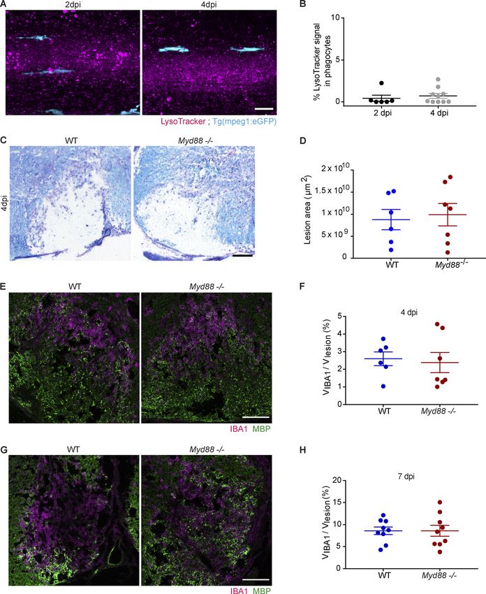
macromolecules of microbial origin, leading to activation of the together with Tg(mbp:eGFP-CAAX) to visualize axonal and myelin
transcription factors NF-κB and interleukin-1 receptor families, loss in the same lesions. We found that LPC injections resulted in a
which dictate the outcome of the innate immune response reduction of mCherry-CAAX fluorescence signal by only ∼3%,
(Hochreiter-Hufford and Ravichandran, 2013). while eGFP-CAAX signal dropped by ∼25% at 2 dpi. By serial
Here, we asked whether pro-inflammatory signaling is nec- scanning electron microscopy, we confirmed that only a small
essary to initiate the regenerative response that occurs upon fraction of axons was lost in the lesions (∼4%, n = 2). Furthermore,
demyelinating injury. TLRs constitute an extended family of using scanning electron microscopy at 2 dpi, we observed myelin
proteins; thus, we decided to delete myeloid differentiation fragments and phagocytes engulfing myelinated axons, providing
primary response 88 (MyD88), a universal adapter protein re- further evidence that LPC injection results in demyelinating le-
quired for the induction of inflammatory cytokines downstream sions. At 4 dpi, we observed demyelinated axons, but also axons
of all TLRs (except TLR3; Kawai and Akira, 2007). Given that with loose myelin wraps, possibly representing remyelination
many of the mechanisms that link inflammation to regeneration (Fig. 2, A–D).
are evolutionary conserved (Tsarouchas et al., 2018), we em- Phagocytes play an important role in myelin debris clearance
ployed a trans-species approach by combining work in zebrafish and in initiating the regenerative response in mice (Kotter et al.,
and mice in order to ask whether and how myelin phagocytosis 2006; Miron et al., 2013; Cantuti-Castelvetri et al., 2018). To
Downloaded from http://rupress.org/jem/article-pdf/217/5/e20191390/860143/jem_20191390.pdf by guest on 30 December 2020
induces the instructive inflammatory cell activation necessary further validate the LPC-induced model of de- and remyelina-
for the regenerative response. tion in zebrafish, we used loss of function mutants in the colony-
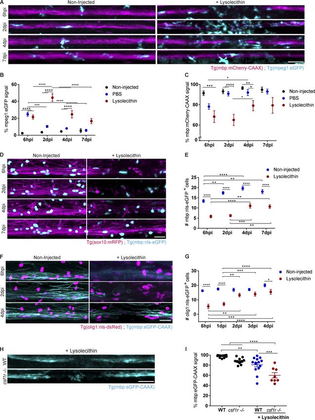
stimulating factor 1 receptor a and b (csf1ra and csf1rb), which
are almost devoid of microglia (Oosterhof et al., 2018). When
Results LPC-induced lesions were induced in csf1ra−/−;b−/− (csf1r−/−)
Establishment of a lysolecithin (LPC)-induced model of spinal mutant larvae and compared with WT controls, we observed
cord demyelination in zebrafish larvae that remyelination was impaired, providing evidence for a role
Focal demyelinating lesions can be induced in the white matter of microglia in remyelination in the larval zebrafish spinal cord
of mice and rats by local injection of LPC, a membrane- (Fig. 1, H and I). Thus, we have established an LPC-induced
dissolving chemical (Jeffery and Blakemore, 1995; Kotter et al., model of spinal cord demyelination in the larval zebrafish,
2006). Since zebrafish represent a highly versatile model system characterized by rapid demyelination, phagocyte influx, and loss
to study myelin biology, we wondered whether we could adapt of oligodendrocytes, which is followed by repopulation of the
the LPC-induced model of myelin injury to the spinal cord of lesion with oligodendrocytes and subsequent remyelination. The
zebrafish larvae. Using a stereotactic injection device, we in- reproducible pattern of de- and remyelination, together with the
jected a solution of 4 nl of LPC into the thoracic spinal cord of 6 d conserved role of microglia/macrophages in myelin repair, al-
postfertilization (dpf) double transgenic Tg(mbp:mCherry-CAAX) lowed us to use this system to analyze the function of phagocytes
and Tg(mpeg1:eGFP) larvae to visualize myelin sheaths and in myelin repair.
phagocytes, respectively. We used larvae at 6 dpf, as this rep-
resents an age when myelination in the spinal cord is largely Impaired phagocyte resolution in spinal cord lesions of myd88
completed and the population of resident phagocytes of the mutant zebrafish
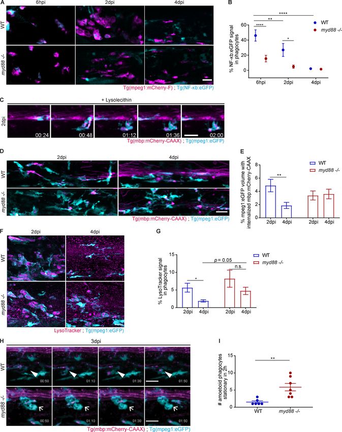
central nervous system (CNS) is already established. We ob- To determine whether pro-inflammatory signaling pathways
served a rapid recruitment of eGFP+ phagocytes into the lesion are induced in phagocytes after myelin injury, we used double
site, with a peak of phagocyte infiltration at 2 d postinjection transgenic Tg(NF-κb:eGFP) and Tg(mpeg1:mCherry-CAAX) zebra-
(dpi) and a progressive decline in the following 5 d (Fig. 1, A–C). fish. The Tg(NF-κb:eGFP) fish line was previously shown to serve
PBS injection resulted in influx of phagocytes shortly after the as a sensitive reporter to monitor NF-κB activity (Kanther et al.,
injection, but numbers had already returned to baseline values 2011). Upon lesion induction, we found that there was a strong
at 2 dpi. We assessed myelination integrity by determining increase in eGFP signal in mCherry+ phagocytes within 6 hpi,
mCherry-CAAX signal in LPC-injected zebrafish larvae (see indicative of NF-κB activation. This was followed by a pro-
Materials and methods) and observed a reduction at 6 h post- gressive decrease during the course of lesion evolution, pro-
injection (hpi) and 2 dpi, followed by an increase at 4 and 7 dpi. viding evidence that NF-κB activation in phagocytes is an early
The drop in the signal at 2 dpi was not a result of bleaching, as it event after myelin injury (Fig. 3, A and B). Myeloid differenti-
was only observed in LPC-injected and not in PBS-injected or ation factor 88 (Myd88) is a key adaptor for inflammatory sig-
noninjected fish (Fig. 1, A–C). In addition, the reduction in naling pathways downstream of TLRs, culminating in the
mCherry-CAAX signal was accompanied by a loss of myelinating activation of NF-κB signaling, resulting in the expression of
oligodendrocytes as assessed by counting the number of eGFP+ cytokines, chemokines, and type I IFNs (Wesche et al., 1997). We
cells in Tg(mbp:nls-eGFP) that express eGFP in the nucleus of used a myd88 mutant zebrafish line (myd88−/−; van der Vaart
mature oligodendrocytes (Fig. 1, D and E). Furthermore, using et al., 2013) to determine whether this key inflammatory sig-
Tg(olig1:nls-mApple) to assess the recruitment of OPCs, we found naling pathway is required for phagocyte activation in demye-
a decrease at 6 hpi and 1 dpi, which was followed by an increase linating lesions. When we injected LPC into the spinal cord of
back to normal numbers within the following days (Fig. 1, F and G). double transgenic Tg(NF-κb:eGFP) and Tg(mpeg1:mCherry-CAAX)
To characterize the extent of axonal damage after LPC in- myd88−/− zebrafish larvae, we found a pronounced reduction in
jections, we performed confocal imaging using Tg(cntn1b:mCherry) eGFP within mCherry+ phagocytes at 6 hpi and 2 dpi compared
Cunha et al. Journal of Experimental Medicine 2 of 23
Pro-inflammatory activation required for remyelination https://doi.org/10.1084/jem.20191390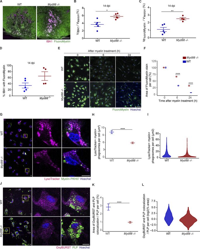
Downloaded from http://rupress.org/jem/article-pdf/217/5/e20191390/860143/jem_20191390.pdf by guest on 30 December 2020

Figure 1. The zebrafish larvae LPC model of de- and remyelination. (A–C) Confocal maximum intensity z-projections of a spinal cord lesion show the
myelinated spinal cord (magenta) and phagocytes (cyan) over time. Phagocyte infiltration was determined by the total amount of mpeg1:eGFP signal in the
lesion. Myelination was determined by the total amount of mbp:mCherry-CAAX signal in the dorsal spinal cord. n = 9–12, 15–16, 13–20, and 4–8 animals at
6 hpi, 2 dpi, 4 dpi, and 7 dpi, respectively. (D and E) Confocal maximum intensity z-projections of a spinal cord lesion show the myelinated spinal cord
(magenta) and the nuclei of mature oligodendrocytes (cyan) over time in noninjected larvae and larvae injected with LPC. The number of mature oligoden-
drocytes was determined by counting the mbp:nls-eGFP positive nuclei. n = 20–21, 16–21, 20–22, and 12–15 animals at 6 hpi, 2 dpi, 4 dpi, and 7 dpi, re-
spectively. (F and G) Confocal maximum intensity z-projections of the spinal cord lesion show the myelinated spinal cord (cyan) and the nuclei of

Cunha et al.                                                                                                         Journal of Experimental Medicine      3 of 23
Pro-inflammatory activation required for remyelination                                                            https://doi.org/10.1084/jem.20191390
oligodendrocyte precursor cells (magenta) over time in noninjected larvae and larvae injected with LPC. The number of OPCs was determined by counting the
olig1:nls-mApple–positive nuclei. n = 8–10, 12–16, 16, 17, and 16 or 17 animals at 6 hpi, 1 dpi, 2 dpi, 3 dpi, and 4 dpi, respectively. (H and I) Confocal maximum
intensity z-projections of the spinal cord lesion at 4 dpi show the myelinated spinal cord (cyan). Myelination was determined by the total amount of mbp:eGFP-
CAAX signal in the dorsal spinal cord. n = 8–14 animals. Lateral views of the lesion site are shown; anterior is left and dorsal is down. All data are mean ± SEM
(error bars). *, P < 0.05; **, P < 0.01; ***, P < 0.001; ****, P < 0.0001 by two-way ANOVA test in B, C, E, and G or one-way ANOVA in I, with Tukey’s multiple
comparisons test. Scale bars: 50 µm in A and H; 20 µm in D and F.
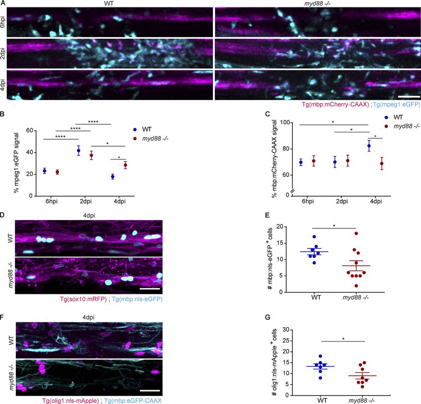
with control, showing that phagocyte activation is effectively between 7 and 21 dpi (Keough et al., 2015; Wang and Kotter,
suppressed (Fig. 3, A and B). 2018). Using Luxol Fast Blue staining, we found that lesion
Next, we determined the recruitment of phagocytes into le- sizes at 4 dpi were similar in WT and Myd88−/− mice (Fig. S1, C
sions of myd88−/− and control fish and found that it did not differ and D). Next, we used immunohistochemistry (IHC) to deter-
at 6 hpi and 2 dpi. However, when lesions were analyzed at mine the number of IBA1+ phagocytes. We found that the density
4 dpi, we observed a higher number of phagocytes in myd88−/− of IBA1+ cells did not differ at 4 and 7 dpi in lesions of WT and
larvae than in control, suggesting that inflammation resolution MyD88-deficient mice (Fig. S1, E–H). However, when lesions
Downloaded from http://rupress.org/jem/article-pdf/217/5/e20191390/860143/jem_20191390.pdf by guest on 30 December 2020
is impaired in mutants (Fig. 5, A and B). To determine the un- were analyzed at 14 dpi, we observed a higher density of IBA1+
derlying reason, we analyzed myelin debris clearance by phag- phagocytes in lesions of MyD88-deficient mice (Fig. 4, A and B).
ocytes. Time-lapse imaging of double transgenic Tg(mbp: In addition, there was an increase in the total number of IBA1+
mCherry-CAAX) and Tg(mpeg1:eGFP) zebrafish showed that phagocytes with accumulation of intracellular FluoroMyelin+
phagocytes were actively taking up and stripping off myelin myelin debris within mutant lesions (Fig. 4 C), although the
from axonal tracts in LPC-induced lesions at 2 dpi in control fish percentage of FluoroMyelin+/IBA1+ phagocytes was not statisti-
(Fig. 3 C and Video 1). Hence, we assessed the accumulation of cally significantly altered between WT and mutant mice (Fig. 4 D).
myelin debris, as determined by the incorporation of mCherry+ To determine the mechanism of impaired myelin clearance,
myelin particles within eGFP+ phagocytes and found that it was we prepared primary cultures of microglia from WT and
within a similar range in double transgenic Tg(mbp:mCherry- MyD88-deficient mice and performed myelin debris phagocy-
CAAX) and Tg(mpeg1:eGFP) myd88−/− and control fish at this time tosis assays. To ensure that most myelin debris were internal-
point, suggesting that phagocytosis was not visibly disturbed. ized by the cells, microglia cultures were treated with myelin for
We then sought to determine the rate of myelin debris 2 h, and the degradation was assayed in the following 6 and 24 h.
clearance by quantifying the amount of mCherry+ myelin within While the amount of internalized myelin debris did not differ
eGFP+ phagocytes in lesions at 2 and 4 dpi and found that control directly after the uptake, we found a slower decrease of Fluo-
fish efficiently cleared away myelin debris. However, when this roMyelin+ myelin debris in MyD88-deficient microglia at 6 and
analysis was performed in myd88−/−, accumulation of myelin 24 h after internalization (Fig. 4, E and F). After phagocytosis has
debris was found within phagocytes at 4 dpi (Fig. 3, D and E). occurred, phagosomes rapidly fuse with lysosomes, forming
Because lysosomes are the organelles responsible for degrada- phagolysosomes, which can be labeled by LysoTracker. Thus, the
tion, we analyzed whether lysosomal biogenesis was impaired in extent of phagosome-to-lysosome fusion can be assayed by de-
myd88−/− phagocytes. However, we found that the number of termining the colocalization of the internalized cargo with ly-
lysosomes in phagocytes that had declined in control lesions sosomes shortly after the uptake. We used PKH67 to label myelin
from 2 to 4 dpi, as determined by LysoTracker, remained high in debris and LysoTracker to mark lysosomes and observed fewer
myd88−/− fish at 4 dpi (Fig. 3, F and G). In noninjected larvae, myelin-PKH67+LysoTracker+ phagosomes in MyD88-deficient
eGFP+ phagocytes were almost completely devoid of Lyso- microglia (Fig. 4, G–I). A crucial reaction during and after the
Tracker+ lysosomes (Fig. S1, A and B). uptake of phagocytic cargo is the activation of the respiratory
To visualize how phagocytes were handling ingested myelin burst pathway, which leads to the release of ROS required to
debris, we used time-lapse imaging to follow the phagocytes in break down the phagocytic material and to promote phagosome
3 dpi lesions of myd88−/− and control fish. Although most cells maturation. We determined the extent of respiratory burst in-
were highly motile both in mutants and in controls, there were duction after myelin debris uptake in WT and MyD88-deficient
higher numbers of amoeboid and stationary phagocytes in microglia by quantifying ROS. We found a significant reduction
myd88−/− fish (Fig. 3, H and I; and Videos 2 and 3). Collectively, in ROS-derived signal colocalizing with myelin debris particles
these data provide evidence that phagocytes in myd88−/− ze- in MyD88-deficient microglia compared with WT microglia, as
brafish larvae are able to phagocytose myelin but fail to degrade determined by the OxyBURST assay (Fig. 4, J–L). In summary, these
myelin debris after the uptake has occurred. data provide evidence for impaired myelin debris degradation
within the phagolysosomal pathway of MyD88-deficient microglia.
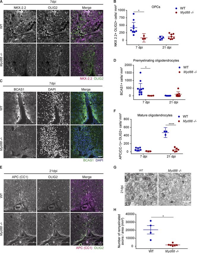
Myelin clearance is impaired in MyD88-deficient microglia
To understand the roles of pro-inflammatory signaling in a Oligodendrogenesis and remyelination are impaired after
mammalian model of demyelination, we performed LPC in- LPC-induced myelin injury in MyD88 mutant zebrafish and
jections into the spinal cord of WT and MyD88-deficient mice. In mice
lesioned animals, demyelination is normally complete within To determine the impact of impaired phagocyte resolution on
4 d, followed by a repair process that occurs in WT animals remyelination, we injected LPC into the spinal cord of double
Cunha et al. Journal of Experimental Medicine 4 of 23
Pro-inflammatory activation required for remyelination https://doi.org/10.1084/jem.20191390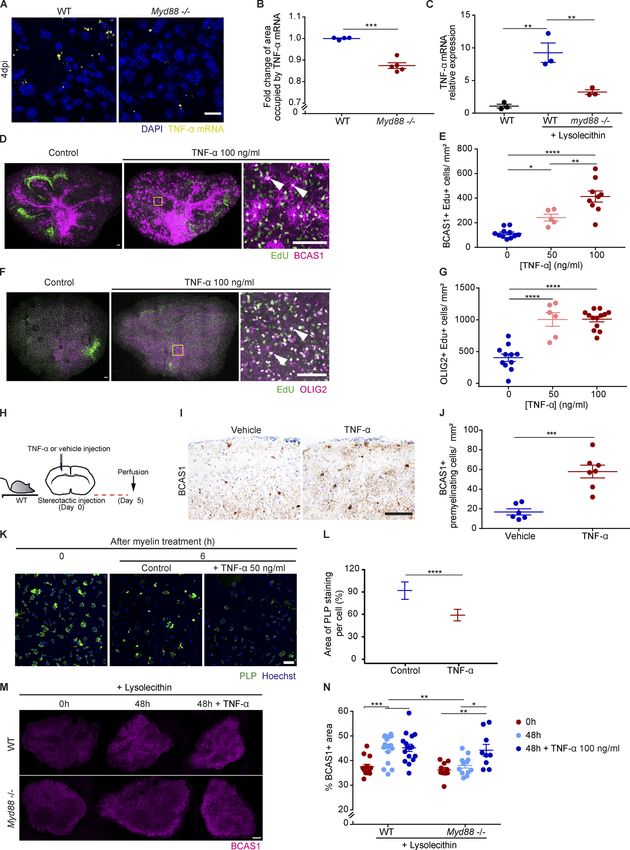
Downloaded from http://rupress.org/jem/article-pdf/217/5/e20191390/860143/jem_20191390.pdf by guest on 30 December 2020

Figure 2. Characterization of myelin and axonal damage in the zebrafish larvae LPC model. (A–C) Confocal maximum intensity z-projections of the
spinal cord show the myelinated spinal cord (cyan) and the axons (magenta) at 2 dpi in noninjected larvae and larvae injected with LPC. n = 16 or 17 animals.
(D) Electron micrographs of serial cross sections of the spinal cord lesion at 2 and 4 dpi in WT larvae injected with LPC. (D, a, b, and d–f) Close-up images
showing myelin fragments in a demyelinating lesion at 2 dpi. (D, b and c) Close-up images showing a phagocyte (blue) engulfing myelinated axons at 2 dpi. (D, c
and e) Close-up images showing lysosomes inside phagocytes (arrow in e) at 2 dpi. (D, b, d, f, and j) Close-up images showing demyelinated axons (asterisks in
d and j, at 2 and 4 dpi, respectively). (D, g, h, j, and k) Close-up images showing the presence of partially myelinated axons at 4 dpi (arrowheads in g and k). All
data are mean ± SEM (error bars). *, P < 0.05; ****, P < 0.0001 by one-way ANOVA, with Tukey’s multiple comparisons test in B or Kruskal-Wallis test with
Dunn’s multiple comparisons test in C. Scale bars: 50 µm in A; 5 µm in D.

Cunha et al.                                                                                                                  Journal of Experimental Medicine         5 of 23
Pro-inflammatory activation required for remyelination                                                                     https://doi.org/10.1084/jem.20191390
Downloaded from http://rupress.org/jem/article-pdf/217/5/e20191390/860143/jem_20191390.pdf by guest on 30 December 2020

Figure 3. Delayed clearance of ingested myelin debris by myd88−/− phagocytes in zebrafish larvae. (A and B) Confocal z-sections of a spinal cord lesion
show NF-κB activation (cyan) in recruited phagocytes (magenta) over time. NF-κB activation was determined by the colocalized signal of NF-κb:eGFP with
mpeg1:mCherry. n = 9 or 10, 7, and 10 or 11 animals at 6 hpi, 2 dpi, and 4 dpi, respectively. (C) Confocal maximum intensity z-projections from a 2 h time-lapse
video (Video 1) of the spinal cord lesion at 2 dpi showing the myelinated spinal cord and myelin debris (magenta) and phagocyte (cyan) movement in WT larvae.
(D and E) Confocal maximum intensity z-projections of the spinal cord lesion at 2 dpi and 4 dpi show the myelinated spinal cord and myelin debris (magenta) in

Cunha et al.                                                                                                               Journal of Experimental Medicine        6 of 23
Pro-inflammatory activation required for remyelination                                                                  https://doi.org/10.1084/jem.20191390
phagocytes (cyan). The amount of internalized myelin was determined by the colocalized signal of mbp:mCherry-CAAX within mpeg1:eGFP. n = 11–15 and 15–17
animals at 2 and 4 dpi, respectively. (F and G) Confocal maximum intensity z-projections of the spinal cord lesion at 4 dpi, showing lysosomes (magenta) in
phagocytes (cyan). The amount of lysosomes was determined by the colocalized signal of LysoTracker with mpeg1:eGFP. n = 8–10 and 14–25 animals at 2 and
4 dpi, respectively. (H and I) Confocal maximum intensity z-projections from 2 h time-lapse videos (Videos 2 and 3) of the spinal cord lesion at 3 dpi show the
myelinated spinal cord and myelin debris (magenta) and phagocyte (cyan) movement. Arrowheads show actively moving phagocytes, and short arrows show
amoeboid stationary phagocytes. n = 6 or 7 animals. Lateral views of the lesion site are shown; anterior is left and dorsal is down. All data are mean ± SEM
(error bars). *, P < 0.05; **, P < 0.01; ****, P < 0.0001; n.s. indicates no significance, by two-way ANOVA test with Tukey’s multiple comparisons test in B, D,
and E or Sidak’s multiple comparisons test in G and unpaired t test with Welch’s correction in J. Scale bars: 20 µm.

transgenic Tg(mpeg1:eGFP) and Tg(mbp:mCherry-CAAX) myd88                         evidence that remyelination, but not myelination, is impaired in
mutant and control larval zebrafish and determined the myeli-                    MyD88 mutants.
nated area over time. We found that the reduction in myelinated
area associated with demyelination at 6 hpi and 2 dpi was not                    TNF-α promotes oligodendrogenesis
different in myd88 mutant and control fish. Demyelination is                     MyD88-dependent inflammatory signaling pathways trigger the
followed by remyelination, which can be visualized by the                        generation of cytokines, chemokines, and growth factors. To

                                                                                                                                                                        Downloaded from http://rupress.org/jem/article-pdf/217/5/e20191390/860143/jem_20191390.pdf by guest on 30 December 2020
subsequent recovery of mCherry-CAAX signal. While signal                         examine whether myelin debris phagocytosis in microglia in-
increase was observed in control fish at 4 dpi, it did not recover               duces the secretion of proteins that regulate oligodendrogenesis,
in myd88 mutant fish (Fig. 5, A and C). We further analyzed                      we performed proteome analyses. WT and MyD88-deficient
myelinating oligodendrocytes at 4 dpi using double transgenic                    microglia cultured under serum-free media supplemented
Tg(sox10:mRFP) and Tg(mbp:nls-eGFP) fish that express eGFP in                    with 0.2% BSA were incubated with purified myelin debris for
the nucleus of oligodendrocytes and found that the number of                     4 h to induce phagocytosis, followed by a 16-h chase before
mature oligodendrocytes also did not recover in myd88 mutants                    harvesting the samples for mass spectrometry analysis. We
compared with control (Fig. 5, D and E). To determine whether                    compared differences in protein abundance in WT and MyD88-
this was a consequence of lack of early progenitor cells, we in-                 deficient microglia treated with myelin debris or not. Using log2
duced lesions in double transgenic Tg(mbp:eGFP-CAAX) and                         fold change of ±0.5 (>1.41-fold or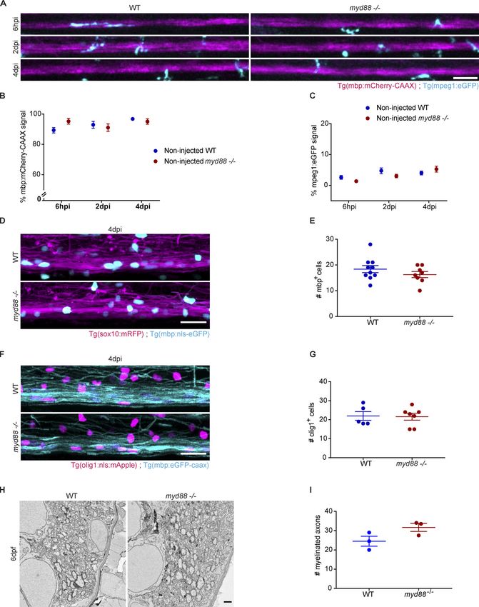
Downloaded from http://rupress.org/jem/article-pdf/217/5/e20191390/860143/jem_20191390.pdf by guest on 30 December 2020

Figure 4. Phagocyte retention in LPC-induced lesions of Myd88−/− mice and impaired phagosome maturation. (A and B) Proportion of the volume
occupied by IBA1+ cells in the lesions (devoid of FluoroMyelin) of WT and Myd88−/− mice at 14 dpi. n = 4 or 5 lesions. (C and D) Total accumulation of myelin
lipids in IBA1+ cells in mouse spinal cord lesions at 14 dpi, analyzed by the proportion of the volume (V) occupied by both FluoroMyelin (green) and IBA1
(magenta; colocalization, white), shown in A, in the lesions (C) and analyzed per total amount of IBA1 signal (D). n = 4 or 5 lesions. (E and F) Degradation of
myelin lipids in cultured microglia. The area of FluoroMyelin in each cell was normalized to the average area of FluoroMyelin per cell at the initial time point (0 h
after myelin treatment). n = 3 independent experiments. 6 h after myelin treatment Myd88−/− versus WT: P < 2.2 × 10⁻¹⁶; effect size (Cohen’s d) = 0.542;

Cunha et al.                                                                                                                   Journal of Experimental Medicine         8 of 23
Pro-inflammatory activation required for remyelination                                                                      https://doi.org/10.1084/jem.20191390
1,188 Myd88−/− cells and 1,016 WT cells. 24 h after myelin treatment: Myd88−/− versus WT: P < 2.2 × 10⁻¹⁶; effect size (Cohen’s d) = 0.657; 988 Myd88−/− cells
and 947 WT cells. (G–I) Phagosome maturation in myelin-loaded microglia. (G) Myelin+ phagosomes (myelin labeled with PKH67, green) and endolysosomes
(LysoTracker Red+, magenta) were detected by live-cell imaging. (H and I) Fusion of phagosomes containing myelin debris with endolysosomes, as analyzed by
the area of the overlap between myelin debris and LysoTracker in each cell, 1 h after treatment with myelin debris. n = 3 independent experiments. Myd88−/−
versus WT: P = 1.911 × 10⁻⁹; effect size (Cohen’s d) = −0.430; 573 Myd88−/− cells and 352 WT cells. (J–L) Oxidative activity in cultured microglia after
phagocytosing myelin debris. Representative overview and inset. The organelles labeled with OxyBURST BSA (magenta) identified ROS. Internalized myelin
debris was identified by PLP (green). n = 3 independent experiments. Myd88−/− versus WT: P = 4.388 × 10⁻⁶; effect size (Cohen’s d) = −0.345; 750 Myd88−/− cells
and 378 WT cells. Data are mean ± SEM (error bars) in B, C, and D and 95% CI in F, H, and K. *, P < 0.05; **, P < 0.01; ****, P < 0.0001 by unpaired t test with
Welch’s correction in B, C, F, H, and K. Scale bars: 100 µm in A; 50 µm in E; and 10 µm in G and J.

with immunohistochemistry against IBA1 at 4 dpi and found                        oligodendrogenesis after myelin injury. We used mice and ze-
that ∼80% of the TNF-α–positive cells colocalized with IBA1                      brafish lacking MyD88, the canonical adaptor for inflammatory
(Fig. S5). In addition, real-time PCR of zebrafish spinal cord                   signaling pathways downstream of TLRs, and found that lesioned
lesions showed that TNF-α mRNA expression was induced in                         animals are impaired not only in phagosome maturation and
lesions of control but to a much lesser degree in myd88 mutant                   myelin debris clearance but also in initiating the regenerative

                                                                                                                                                                        Downloaded from http://rupress.org/jem/article-pdf/217/5/e20191390/860143/jem_20191390.pdf by guest on 30 December 2020
zebrafish (Fig. 8 C). Thus, TNF-α represents a candidate cyto-                   response necessary for the generation of new oligodendrocytes.
kine induced in phagocytes after demyelinating injury that                       We identified TNF-α as one factor that is produced to a lesser
may regulate oligodendrogenesis.                                                 extent in lesions from MyD88-deficient mice and zebrafish and
    To determine the effects of TNF-α on the formation of new                    is able to promote efficient OPC generation. Our study under-
oligodendrocytes, organotypic cerebellar slice cultures (OCSCs)                  scores the role of microglia/macrophage activation in clearing
were treated with recombinant TNF-α and 5-ethynyl-29-deoxyur-                    myelin debris and initiating the secretion of pro-regenerative
idine (EdU) and immunostained with antibodies against OLIG2 and                  molecules for myelin repair. Our study concurs with earlier
BCAS1 to quantify the number of newly generated and premyeli-                    work, in which a role for innate immune cells and mediators in
nating and myelinating oligodendrocytes, respectively. We ob-                    stimulating remyelination was shown (Miron et al., 2013). It is
served a marked induction of EdU+BCAS1+ and of EdU+OLIG2+                        now well known that phagocytes are important for the rapid
oligodendrocytes (Fig. 8, D–G). Furthermore, we performed ster-                  clearance of myelin debris to remove repulsive signals and to
eotactic injections of TNF-α into the cortex of WT mice and per-                 pave the way for oligodendrocyte differentiation and remyeli-
formed IHC 5 d after the injections. As observed in the slice culture            nation (Kotter et al., 2006; Syed et al., 2011). Such regenerative
experiments, we detected a striking increase in the number of                    properties have been associated with anti-inflammatory, immune-
premyelinating BCAS1+ oligodendrocytes upon TNF-α injections                     regulatory M2 microglia/macrophages, which secrete growth
(Fig. 8, H–J). We also assessed the effect of TNF-α in promoting                 factors that drive OPC differentiation (Miron et al., 2013;
myelin debris clearance in microglia. When microglia cultures                    Lloyd et al., 2019). Thus, it is conceivable that the initial pro-
were treated with myelin for 2 h and the degradation of PLP was                  inflammatory response is required to activate OPC proliferation
assayed in the following 6 h, we found that TNF-α was able to                    and survival, while polarization of phagocytes into an anti-
enhance degradation (Fig. 8, K and L). Finally, we determined                    inflammatory phenotype that occurs later during lesion evolu-
whether TNF-α is able to rescue lesion recovery in the absence of                tion is important to generate the factors for OPC differentiation
MyD88. We used OCSCs, which we treated for 16 h with LPC, as an                  (Miron et al., 2013). Pro-inflammatory activation of microglia
established ex vivo model of demyelination (Birgbauer et al., 2004;              and macrophages has long been implicated in inducing harm to
Zhang et al., 2011). After LPC treatment, we exchanged the medium                the CNS by secreting damage-inducing molecules such as pro-
and continued the incubation with or without recombinant TNF-α                   inflammatory cytokines or ROS (Block et al., 2007; Perry and
for 48 h, followed by immunostaining against BCAS1 to determine                  Teeling, 2013). This Janus-faced nature of activated microglia is
the number of premyelinating and myelinating oligodendrocytes.                   particularly striking in active MS lesions, in which areas of ac-
We found that BCAS1 immunoreactivity increased in WT but not                     tive de- and remyelination coexist. TNF-α is one example of a
Myd88-deficient slices 48 h after LPC treatment (Fig. 8, M and N).               pro-inflammatory factor with such a dual role.
However, when TNF-α was added into the medium, we observed                           A major question will be to resolve how TNF-α regulates pro-
an increase in BCAS1 immunoreactivity in Myd88-deficient slices.                 survival and proliferative responses as opposed to programmed
Notably, TNF-α treatment of WT slices did not result in a further                cell death pathways. Studies in experimental autoimmune en-
increase in BCAS1 immunoreactivity (Fig. 8, M and N). Thus, our                  cephalomyelitis have shown that the blockage of TNF-α leads to
data provide evidence that MyD88-dependent signaling induces                     reduced pathology (Brambilla et al., 2011; Steeland et al., 2017).
the expression of TNF-α after a demyelinating injury, which is able              One possibility is that the aggravating effects of TNF-α are
to promote myelin debris degradation and the formation of new                    mediated by interaction with TNFR1, while the pro-regenerative
oligodendrocytes.                                                                effects are attributed to signaling through TNFR2. One study
                                                                                 suggested that the transmembrane form of TNF-α was respon-
                                                                                 sible for promoting remyelination by engagement of TNFR2,
Discussion                                                                       while soluble TNF-α inhibited myelin repair (Karamita et al.,
In this study, we provide evidence that pro-inflammatory acti-                   2017). Our data show that injection of soluble TNF-α into the
vation is required for myelin debris degradation and for                         mouse cortex or addition onto myelinating slice cultures was

Cunha et al.                                                                                                               Journal of Experimental Medicine        9 of 23
Pro-inflammatory activation required for remyelination                                                                  https://doi.org/10.1084/jem.20191390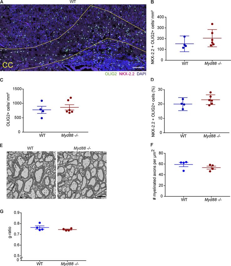
Downloaded from http://rupress.org/jem/article-pdf/217/5/e20191390/860143/jem_20191390.pdf by guest on 30 December 2020
Figure 5. Delayed phagocyte efflux in myd88−/− zebrafish larvae with impaired remyelination. (A–C) Confocal maximum intensity z-projections of the
spinal cord lesion over time show the myelinated spinal cord (magenta) and phagocytes (cyan) in WT and myd88−/− larvae injected with LPC. Phagocyte
infiltration was determined by the total amount of mpeg1:eGFP signal in the lesion. Myelination was determined by the total amount of mbp:mCherry-CAAX
signal in the dorsal spinal cord. n = 15–19, 11–17, and 13–17 animals at 6 hpi, 2 dpi, and 4 dpi, respectively. (D and E) Confocal maximum intensity z-projections
of the spinal cord lesion at 4 dpi show the myelinated spinal cord (magenta) and the nuclei of mature oligodendrocytes (cyan). The number of mature oli-
godendrocytes was determined by counting the number of mbp:nls-eGFP–positive nuclei. n = 7 or 10 animals. (F and G) Confocal maximum intensity
z-projections of the spinal cord lesion at 4 dpi show the myelinated spinal cord (cyan) and the nuclei of OPCs (magenta). The number of OPCs was determined
by counting the number of olig1:nls-mApple–positive nuclei. n = 7 or 8 animals. Lateral views of the lesion site are shown; anterior is left and dorsal is down. All
data are mean ± SEM (error bars). *, P < 0.05; ****, P < 0.0001 by two-way ANOVA test, with Tukey’s multiple comparisons test in B and C and unpaired t test
with Welch’s correction in E and G. Control noninjected larvae WT and myd88−/− animals are included in the statistical analysis in B and C and are represented
in Fig. S3, A–C. Scale bars: 50 µm in A; 20 µm in D and F.

sufficient to induce the generation of (pre)myelinating BCAS1+                        Recently, single-nucleus RNA sequencing from postmortem
oligodendrocytes. This pro-regenerative effect of TNF-α may                        human brain from patients with MS and from unaffected con-
explain why clinical trials in MS using recombinant TNFR or                        trols revealed a depletion of OPCs in MS (Jäkel et al., 2019).
antibodies against TNF-α were not only ineffective, but even                       Furthermore, studies using 14C carbon dating in human tissue
worsened the disease in some patients (van Oosten et al.,                          provided little evidence for OPC proliferation in fully remyeli-
1996).                                                                             nated shadow plaques. In contrast, OPC proliferation was clearly

Cunha et al.                                                                                                                  Journal of Experimental Medicine         10 of 23
Pro-inflammatory activation required for remyelination                                                                     https://doi.org/10.1084/jem.20191390
Downloaded from http://rupress.org/jem/article-pdf/217/5/e20191390/860143/jem_20191390.pdf by guest on 30 December 2020

Figure 6. Defective remyelination in the spinal cord of Myd88−/− mice. (A and B) OPCs in demyelinated lesions in mouse spinal cords at 7 dpi. The nuclei of
OPCs were identified by the expression of both NKX-2.2 (magenta) and OLIG2 (green; colocalization, white). n = 3–9 lesions. (C and D) Premyelinating oli-
godendrocytes expressing BCAS1 (green) in mouse spinal cord lesions at 7 dpi. n = 4–10 lesions. (E and F) Mature oligodendrocytes expressing both APC (CC1;
magenta, cytoplasm) and OLIG2 (green, nucleus) in mouse spinal cord lesions at 21 dpi. n = 3–7 lesions. (G and H) Remyelination in mouse spinal cord lesions at
21 dpi, quantified by the density of remyelinated axons in semi-thin sections. n = 4–6 lesions. Data are mean ± 95% CI (error bars) in B, D, and F or SEM in H.
*, P < 0.05; ****, P < 0.0001 by two-way ANOVA test, with Sidak’s multiple comparisons test in B, D, and F and unpaired t test with Welch’s correction in H.
Scale bars: 100 µm in A, C, and E; 10 µm in G.

Cunha et al.                                                                                                               Journal of Experimental Medicine       11 of 23
Pro-inflammatory activation required for remyelination                                                                  https://doi.org/10.1084/jem.20191390
Downloaded from http://rupress.org/jem/article-pdf/217/5/e20191390/860143/jem_20191390.pdf by guest on 30 December 2020
Figure 7. Proteome analysis of cultured microglia identified TNF-α as a candidate regulator of the response to myelin debris. (A) Volcano plot of
regulated proteins in vehicle-treated Myd88−/− versus vehicle-treated WT. (B) Volcano plot of regulated proteins in myelin-treated Myd88−/− versus myelin-
treated WT. The negative log10 transformed P value is plotted against the log2 transformed LFQ intensity ratios for each protein. Proteins with a P value 0.05 are indicated as blue circles. The hyperbolic curves indicate the threshold of a permutation-
based FDR correction for multiple hypotheses (FDR: P = 0.05, s0 = 0.1). (C) Heat map of the top 35 regulated proteins, which were consistently quantified in all
comparisons. The log2 fold changes of the proteins are indicated in a color scale from blue to red. (D) Heat map of significant upstream regulators predicted by
Ingenuity Pathway Analysis. The negative log10 P value was multiplied with the sign of the related z-score. Blue color indicates a decreased activation, whereas
red color indicates an increased activation with the corresponding upstream regulator.

observed in patients with aggressive disease (Yeung et al., 2019).                work analyzing the uptake of bacteria and apoptotic cells in
These data suggest that inflammation associated with more                         peripheral macrophages provided evidence that TLR-signaling
aggressive disease favors OPC proliferation. Given that pro-                      is able to control the mode of phagosome maturation. Engage-
inflammatory mediators can induce oligodendrogenesis, it will                     ment of bacteria with TLR receptors was found to alter the
be important to determine the effects of immune-modulatory                        kinetics of phagosome maturation by triggering an inducible
or immune-suppressive drugs currently in use to treat MS in                       mode of phagocytosis, resulting in more efficient clearance.
remyelination.                                                                    The inducible mode is distinguished from a constitutive mode
   Our study also provides evidence for a role of microglia ac-                   of phagosome maturation by its enhanced phagosome-to-lyso-
tivation in promoting myelin degradation. We found more ex-                       some fusion rate and by the induction of antimicrobial defense
tensive myelin debris accumulation within microglia in lesioned                   systems, such as nicotinamide adenine dinucleotide phosphate
MyD88-deficient animals than in control. How does MyD88-                          oxidase and nitric oxide synthase (Blander and Medzhitov,
dependent signaling regulate myelin debris clearance? Previous                    2004). Thus, differences in the cellular response that occurs

Cunha et al.                                                                                                                Journal of Experimental Medicine        12 of 23
Pro-inflammatory activation required for remyelination                                                                   https://doi.org/10.1084/jem.20191390
Downloaded from http://rupress.org/jem/article-pdf/217/5/e20191390/860143/jem_20191390.pdf by guest on 30 December 2020

Cunha et al.                                                Journal of Experimental Medicine    13 of 23
Pro-inflammatory activation required for remyelination   https://doi.org/10.1084/jem.20191390
Figure 8. TNF-α expression is induced after a demyelinating injury and increases the number of premyelinating oligodendrocytes. (A and B) Confocal
maximum intensity z-projections of RNAscope in situ hybridization targeting TNF-α in mouse spinal cord lesions at 4 dpi. TNF-α was determined by the total
amount of signal in the lesion. n = 4 or 5 lesions. (C) TNF-α mRNA in spinal cord lesions of zebrafish larvae was determined by quantitative RT-PCR of lesions at
6 hpi. n = 3 independent experiments. (D–G) After 5 or 6 DIV, OCSCs were treated with 50 or 100 ng/ml recombinant mouse TNF-α and EdU for 48 h. (D and E)
Newly formed myelinating oligodendrocytes were labeled by EdU (green) and expressed BCAS1 (magenta; examples of colocalization, white arrowheads) in the
cell body. n = 5–11 slices in two experiments. (F and G) Newly generated oligodendrocytes that expressed OLIG2 (magenta) and incorporated EdU (green;
examples of colocalization, white arrowheads). n = 6–13 slices in two experiments. (H) Schematic representation of cortical injection of TNF-α or vehicle in WT
mice. (I) IHC of BCAS1 on day 5 after stereotactic injection of vehicle or TNF-α in WT mice. (J) Quantification of BCAS1+ cells with a premyelinating morphology
in the subpial cortex day 5 after stereotactic injection of vehicle or TNF-α. (K and L) 2 h after myelin debris treatment, TNF-α (50 ng/ml) was added to cultured
microglia for 6 h. The amount of PLP per cell at 6 h was normalized to the initial amount of PLP per cell (0 h). Myelin treatment control versus TNF-α: P = 3.125 ×
10−6; effect size (Cohen’s d) = 0.224; 703 cells in the control group and 1,252 cells in the TNF-α treatment group were sampled randomly. n = 3 independent
experiments. (M and N) After 7 DIV, OCSCs were treated with 0.5 mg/ml LPC for 16 h and subsequently with TNF-α (100 ng/ml) for 48 h, and BCAS1
immunoreactivity was determined in the slices. n = 9–17 slices in four to six independent experiments. All data are mean ± SEM (error bars). *, P < 0.05; **,
P < 0.01; ***, P < 0.001; ****, P < 0.0001 by unpaired t test with Welch’s correction in B, J, and L; one-way ANOVA test with Tukey’s multiple comparisons
test in C, E, and G; and two-way ANOVA test with Tukey’s and Sidak’s multiple comparisons tests in N. Scale bars: 20 µm in A; 100 µm in D, F, and I; 60 µm in
K; and 500 µm in M.

                                                                                                                                                                           Downloaded from http://rupress.org/jem/article-pdf/217/5/e20191390/860143/jem_20191390.pdf by guest on 30 December 2020
upon uptake of apoptotic and bacterial cargo could be deter-                       medium. For the fish to be raised to adulthood, at 1 dpf, embryos
mined by the phagocytic route taken. It is interesting that                        were sorted in clutches of 30 per Petri dish and bleached, fol-
myelin debris appears to be processed in the system for host                       lowed by addition of pronase to facilitate their hatching. At 5
defense against infection. The reason why myelin debris em-                        dpf, larvae were transferred to 3.5-liter tanks in the facility and
ploys this route of phagocytosis could lie in its unique struc-                    kept in co-culture with rotifers. After 11 dpf, artemia and powder
ture. Myelin is a multi-layered, tightly packed membrane                           food were added to their diet. Adult fish were fed artemia and
enriched in lipids that are difficult to break down. Hence, it is                  dry food. For the larvae to be used in experiments, 1-phenyl
possible that microglia activation is required to induce anti-                     2-thiourea (Sigma; P7629) was added to the Petri dish at 1 dpf
microbial defense for its degradation.                                             in order to prevent pigmentation; at 5 dpf, larvae were trans-
    It is important to note that we used a global knockout of                      ferred to 600-ml beakers filled with 300 ml of E3 medium,
MyD88 in the mouse and zebrafish. As a consequence, we do not                      where they started to be fed with powder food once per day until
know whether MyD88-dependent signaling outside of the in-                          the end of the experiments. The following fish lines were used:
nate immune system contributed to the phenotype we describe.                       Tg(mpeg1:eGFP) (Ellett et al., 2011), Tg(mbp:eGFP-CAAX) (Almeida
MyD88 is highly enriched in microglia, although low levels                         et al., 2011), Tg(mbp:mcherry-CAAX) (Mensch et al., 2015), Tg(o-
present in oligodendrocytes cannot be excluded, where it can act                   lig1:nls-mApple) (Auer et al., 2018), Tg(mbp:nls-eGFP) (Karttunen
downstream of pathogen-derived TLR2 agonist–induced signal-                        et al., 2017), Tg(sox10:mRFP) (Kirby et al., 2006), Tg(mpeg1:
ing to block oligodendrocyte maturation (Sloane et al., 2010).                     mCherry-F) (Nguyen-Chi et al., 2014), Tg(NF-κb:eGFP) (Ogryzko
Our study argues that in the absence of such agonists, the                         et al., 2014), csf1raj4e1/j4e1; csf1rbre01/re01 (Oosterhof et al., 2018), and
pro-inflammatory–mediated induction of OPC proliferation                           myd88 hu3568/hu3568 (van der Vaart et al., 2013).
dominates. We provide evidence that generation of TNF-α is
triggered in a MyD88-dependent fashion after myelin injury,                        Mouse husbandry
but it is likely that other pro-inflammatory mediators also                        All animal experiments were performed according to German
contribute to the regenerative response. In the future, it will be                 animal welfare law and local regulations for animal ex-
important to extend this study to analyze whether inflammatory                     perimentation (Regierung von Oberbayern). The genotype
cytokine signaling–induced OPC stimulation can provide an                          of Myd88−/− mice (Adachi et al., 1998) was confirmed by gen-
approach for promoting remyelination. We envision that the                         otyping. The DNA fragments were amplified by PCR using the
novel model of de- and remyelination in the spinal cord of ze-                     forward primer 59-GTTGTGTGTGTCCGACCGT-39 and reverse
brafish larvae established in this work, together with rapid gene                  primer 59-GTCAGAAACAACCACCACCATGC-39. The PCR pro-
targeting, chemical screening, and visualization techniques, will                  gram using GoTaq G2 DNA polymerase (M7845; Promega) was
provide a powerful system to identify pathways and small                           94°C for 3 min; 35 cycles of 94°C for 30 s, 66°C for 1 min, and
molecules with the potential to enhance regeneration.                              72°C for 1 min; and 72°C for 2 min followed by cooling. The DNA
                                                                                   fragments amplified from Myd88−/− were 353 bp and 266 bp
                                                                                   from WT.
Material and methods
Zebrafish husbandry                                                                Zebrafish genotyping
Zebrafish lines were handled according to local animal welfare                     Fin clips from anesthetized 3 mo adult fish or whole larvae were
regulations and kept under standard conditions at the German                       lysed in a solution of Proteinase K (17 mg/ml) and Tris-EDTA
Center for Neurodegenerative Diseases fish facility in Munich,                     buffer for 4 h at 55°C, followed by Proteinase K inactivation at
Germany. Adult zebrafish were mated in individual breeding                         65°C for 5 min. Genomic DNA was then amplified using the
boxes overnight, and the eggs were collected the morning after                     specific primers (myd88 forward: 59-GAGGCGATTCCAGTAACA
in Petri dishes. Fertilized eggs were raised at 28.5°C in E3                       GC-39, myd88 reverse: 59-GAAGCGAACAAAGAAAAGCAA-39,

Cunha et al.                                                                                                                  Journal of Experimental Medicine        14 of 23
Pro-inflammatory activation required for remyelination                                                                     https://doi.org/10.1084/jem.20191390
csf1rb forward: 59-CTTGCTGACAAATCCAGCAG-39, and csf1rb thresholded NF-κB signal and phagocytes. The plug-in Surface-
reverse: 59-GAGCTAACCGGACAAACTGG-39), and the product Surface Coloc of Imaris was used to determine the value of co-
was digested with the appropriate restriction enzymes (MseI for localization, and the final result was expressed as a percentage of
myd88 and MspI for csf1rb) for 2 h. Undigested and digested PCR the colocalized surface per total amount of phagocyte surface.
products were loaded in a 2% gel for gel electrophoresis at 180 V The same method was applied to the quantification of lysosomes
for 50 min. Gels were scanned using a BioRad Gel Doc XR+ and myelin inside the phagocytes. The quantification of the
system. number of mature oligodendrocytes or OPCs was performed by
manually counting the mbp:nls-eGFP– or olig1:nls-mApple–
LPC-induced demyelination in the spinal cord of zebrafish positive nuclei, respectively, in the dorsal spinal cord in maxi-
larvae mum intensity projections. Images were thresholded using the
LPC has been applied previously to the adult optic nerve of adult IJ-IsoData method.
zebrafish (Münzel et al., 2014). Here, we used larvae at 6 dpf
anesthetized in E3 medium with tricaine (Pharmaq Limited; MS- Quantitative RT-PCR of zebrafish lesions
222) and placed individually in an agarose mold under a mi- For assessment of the gene expression in lesions, 100 lesions
croscope. Larvae were positioned laterally, and the medium was were collected per genotype and snap-frozen in dry ice. RNA
Downloaded from http://rupress.org/jem/article-pdf/217/5/e20191390/860143/jem_20191390.pdf by guest on 30 December 2020
removed as much as possible. A Nanoliter 2010 + Micro4 Con- was isolated using the RNeasy Mini Kit (Qiagen; 74134) following
troller injection setup was used to perform the injections. Glass the QIAshredder dissociation protocol. cDNA was obtained using
needles (World Precision Instruments; 504949) were pulled the SuperScript III First-Strand Synthesis system (Thermo
using a DMZ-Universal puller and fully filled with mineral oil. Fisher; 18080051). Quantitative RT-PCR was performed with
2000 nl of oil was infused, and 500 nl of air was withdrawn in PowerUP SYBR Green Master Mix (Thermo Fisher; A25780),
order to form an air bubble between the oil and the solution to using a Roche LightCycler 480 Instrument II Real-Time PCR
be injected; right after, 900 nl of a 40% solution of LPC (L-α- system. The following primers were used: tnfa forward: 59-CTC
lysophosphatidylcholine from egg yolk; Sigma; L4129) in PBS CATAAGACCCAGGGCAAT-39, tnfa reverse: 59-ATGGCAGCCTTG
was withdrawn. The setup was set to inject 4 nl at a rate of 23 GAAGTGAA-39, ef1a forward: 59-AGCAGCAGCTGAGGAGTGAT-
nl/s, and larvae were injected in the spinal cord at the level of 39, and ef1a reverse: 59-GTGGTGGACTTTCCGGAGT-39. Samples
the yolk sac. Immediately after injection, larvae were trans- were run in triplicates, and expression levels were normalized
ferred to a beaker with 300 ml of E3 medium, given powder to the housekeeping gene elf1a. The ΔΔCt method was used to
food, and put back in the incubator (28.5°C). Both concentration determine the relative gene expression. Experiments were
and volume to be injected were previously tested. performed as biological triplicates.
In vivo confocal imaging of zebrafish larvae LPC-induced demyelination in the spinal cord of mice
For live imaging, larvae were anesthetized with tricaine and Stereotactic injection of LPC in the spinal cord was performed in
mounted laterally in 0.8% low melting agarose in E3 medium, Myd88−/− mice that were 9–12 wk old and age-matched WT
using a glass-bottom dish filled with E3 medium and tricaine. C57BL/6J mice after at least 2 wk of acclimatization to the animal
Larvae were imaged with a Leica TCS SP8 confocal laser scan- unit. The reagents for injection were prepared under the cell
ning microscope with a climate chamber (28.5°C), using 20 × culture hood. 1% LPC was prepared by dissolving L-α-lyso-
0.75 numerical aperture (NA) air or 40 × 1.1 NA water immer- phosphatidylcholine from egg yolk in PBS, pH 7.4 (Gibco;
sion objectives and 488-nm and 552-nm lasers. For the quanti- 10010056). 3% Monastral blue was prepared by dissolving
fication of lysosomes, LysoTracker Red DND-99 dye (Thermo Copper (II) phthalocyanine-tetrasulfonic acid tetrasodium salt
Fisher; L7528) was used by adding 10 µM of the dye to larvae (Sigma; 274011) in deionized water, and the solution was passed
immersed in E3 medium in a 24-well plate. Larvae were incu- through an 0.45-µm filter and autoclaved. Prior to injection, 1 µl
bated for 1 h 30 min in the dark at 28.5°C, followed by four of 3% Monastral blue was mixed with 25 µl of 1% LPC. Glass
washing steps with fresh E3. After imaging, larvae were re- Capillaries for Nanoliter 2010, fire polished 2 (World Precision
moved out of the agarose, sacrificed, and genotyped in case Instruments; 504949 or 4878) were pulled using the P-1000
imaged larvae were a result of heterozygous incrosses. Next Generation Micropipette Puller (Sutter Instrument). The
program had the following parameters: Heat 530, Pull 0, Vel 60,
Image analysis of live imaging experiments in zebrafish larvae Time 250, Pressure 500, Ramp 520, Microinjection–BF100.50.10,
Automated quantification of myelination, phagocyte infiltration, TipNanoliter 2010 Injector with MICRO4 controller (World Preci- used for IHC, whereas the other was used for Luxol Fast Blue
sion Instruments) as previously described (Cantuti-Castelvetri stain. The slides were stored at −20°C or −80°C.
et al., 2018). After the spinal cord was exposed, the capillary was
positioned 0.55 mm lateral to the dorsal artery and lowered Semi-thin sections
1.15 mm into the tissue. At each injection site, 1 µl of 1% LPC The spinal cord was sectioned coronally using a vibratome (Leica
containing 0.12% Monastral blue was injected at a speed of 150 Biosystems) to prepare samples for semi-thin sections. 20%
nl/min. 1 min after LPC was delivered, the capillary was slowly gelatin was prepared by dissolving gelatin powder (Merck Mil-
retracted. After injection, the skin was sutured, and the wound lipore; 1040781000) in PBS by stirring and heating at 60°C.
was disinfected and sealed with the tissue adhesive Histoacryl Before vibratome sectioning, the aliquots of 20% gelatin were
(B. Braun). After the operation, the animals were injected i.p. thawed from −20°C and heated at 37°C with shaking. A segment
with 250 µl of 0.9% NaCl (normal saline solution) to compensate of spinal cord (shorter than 3 mm) was freshly dissected from
for the loss of blood. The analgesic buprenorphine was injected the mouse and embedded in 20% gelatin in an embedding mold
s.c. at a dose of 0.1 mg/kg. When 0.5 mg/kg body weight me- (Sigma; E4140-1EA) on ice. 200-µm coronal sections were cut at
detomidine, 5.0 mg/kg midazolam, and 0.05 mg/kg fentanyl was a speed of 0.4–0.5 mm/s. The coronal sections were fixed with a
used for anesthesia, 2.5 mg/kg atipamezole, 0.5 mg/kg flumaz- solution containing 4% PFA and 2.5% glutaraldehyde in Karlsson
Downloaded from http://rupress.org/jem/article-pdf/217/5/e20191390/860143/jem_20191390.pdf by guest on 30 December 2020
enil, and 1.2 mg/kg naloxone was injected i.p. for the animals to & Schultz buffer, under a coverslip in a 24-well plate at 4°C until
wake up. The animals were kept on a heating pad at 37°C until further processing. The spinal cord sections were embedded in
they were awake. They were supplied with wet powder food in a epon and cut at a thickness of 500 nm using the Leica Ultracut S
100-mm Petri dish in addition to regular food and water. The ultramicrotome. Semi-thin sections were stained with methyl-
wet powder food was refreshed daily until the animals’ hind ene blue-azure II to visualize the lipid-rich areas such as myelin.
limbs could function properly. The animals were injected s.c.
with buprenorphine 1 or 2 d after surgery. Preparation of myelin and fluorescent labeling
Myelin was isolated from the brains of 12 adult (at least 8-wk-
Preparation of tissue samples for histology old) WT C57BL/6 mice using the Beckman Optima XL-80 Ul-
The mice were fixed by transcardial perfusion to prepare sam- tracentrifuge with the SW 28 swinging-bucket rotor. Ultra-Clear
ples for histology. Adult animals were anesthetized by i.p. in- Thinwall 38.5-ml tubes (Beckman Coulter; 344058) were ster-
jection with 100–200 µl of 14% chloral hydrate (prepared in ilized with UV under the laminar hood. The Hepes-EDTA buffer
water; Sigma; C8383). The blood was washed out by transcardial was prepared as 10 mM Hepes and 5 mM EDTA, pH 7.4, filtered,
perfusion with filtered, ice-cold PBS, pH 7.4, for 5 min until clear and stored at 4°C. 0.32 M, and 0.85 M sucrose solutions were
liquid came out of the right atrium and the liver turned pale. The prepared freshly in cold 10 mM Hepes, pH 7.4 (Hepes buffer),
animals were then perfused with fresh, filtered, ice-cold 4% and filtered. Deionized water was also filtered and kept cold. The
paraformaldehyde (PFA) in PBS for 10–15 min until they stiff- mice were euthanized in a CO2 chamber followed by decapita-
ened. The brain was taken out of the skull and postfixed in 4% tion. Three or four brains were put in a Dounce homogenizer
PFA at 4°C overnight. The spine was taken out. The bones on the containing 4 ml of Hepes buffer. The brains were first homog-
ventral side were removed to expose the ventral spinal cord, and enized mechanically using the Dounce tissue grinder set. The
then the spine was fixed in 4% PFA at 4°C for 3–6 d. If only the homogenate was transferred to a conical tube and topped up to
spinal cord was needed for histology, the mice were anesthetized 5 ml with Hepes buffer. The homogenate was then sonicated for
in a CO2 chamber and euthanized by decapitation. The spine was 5 min using the Sonifier W-250 D (Branson). The brain ho-
dissected and fixed in the same way. mogenate was stored at −20°C.
After postfixation, the tissue was washed in PBS and pro- The isolation and purification of myelin were done at 4°C or
cessed for cryoprotection. The tissue not required for processing on ice. 5 ml of brain homogenate was overlaid on a stepwise
could be stored in PBS containing 0.1% sodium azide at 4°C. The 0.32/0.85 M sucrose gradient in an ultracentrifugation tube. The
spinal cord was dissected out of the bone. The brain and spinal first centrifugation was done at 23,800 rpm (54,000 ×g) for
cord were immersed in 30% sucrose (prepared in PBS on the day 35 min. The myelin fraction was collected from the interphase
of use) at 4°C for 3 d, until the tissue sank to the bottom of the using a P1000 pipette. Deionized water was added to the myelin
conical tube. The tissue was immersed in 1:1 mixture of 30% to remove small membrane fragments by osmotic shock. Myelin
sucrose in PBS and Tissue-Tek O.C.T. Compound (Sakura) at was pelleted by centrifugation at 23,800 rpm (54,000 ×g) for
room temperature with shaking at 400 rpm overnight. The 18 min. The supernatant was aspirated carefully, and the pellets
tissue was transferred to O.C.T. and incubated at room tem- were resuspended with water. Myelin was washed with water
perature for 4–5 h (spinal cord) or 1 d (brain). Afterward, the and centrifuged at 9,500 rpm (9,000 ×g) twice for 18–22 min.
tissue was embedded in O.C.T. on dry ice. The frozen tissue was The supernatant was removed; 1 ml of Hepes buffer was added to
stored at −20°C or −80°C. each pellet, and the pellets were kept at −20°C until the purifi-
Coronal sections of spinal cords and brains were cut at a cation steps. The crude products of myelin were purified by
thickness of 12 µm using a cryostat. The sections were mounted repeating the stepwise density gradient centrifugation, osmotic
on SuperFrost Plus Microscope Slides (Thermo Fisher; shock, and two washing steps. The Hepes buffer for cell culture
J1800AMNZ). The adjacent spinal cord sections were mounted was prepared from the 1 M Hepes (Gibco; 15630056), and the pH
on alternating slides so that in each pair of slides one slide was was measured by pH-indicator strips (Merck Millipore; 109543)
Cunha et al. Journal of Experimental Medicine 16 of 23
Pro-inflammatory activation required for remyelination https://doi.org/10.1084/jem.20191390You can also read