Altered White Matter Microstructural Organization in Post-Traumatic Stress Disorder across 3,049 Adults: Results from the PGC-ENIGMA PTSD ...
←
→
Page content transcription
If your browser does not render page correctly, please read the page content below
bioRxiv preprint first posted online Jun. 20, 2019; doi: http://dx.doi.org/10.1101/677153. The copyright holder for this preprint
       (which was not peer-reviewed) is the author/funder, who has granted bioRxiv a license to display the preprint in perpetuity.
                                    It is made available under a CC-BY-NC-ND 4.0 International license.

     Altered White Matter Microstructural Organization in Post-Traumatic Stress Disorder across 3,049
                         Adults: Results from the PGC-ENIGMA PTSD Consortium

 Emily L Dennis PhD1-4, Seth G Disner PhD5,6, Negar Fani PhD7, Lauren E Salminen PhD 2, Mark Logue PhD 8-11, Emily K
    Clarke MSc12,13, Courtney C Haswell PhD12,13, Christopher L Averill BSc14, Lee A Baugh PhD15-17, Jessica Bomyea
  PhD18,19, Steven E Bruce PhD20, Jiook Cha PhD21,22, Kyle Choi MPH23, Nicholas D Davenport PhD5,6, Maria Densmore
BSc24,25, Stefan du Plessis PhD 26, Gina L Forster PhD15,16,27, Jessie L Frijling PhD28, Atilla Gönenc PhD29,30, Staci Gruber
PhD29,30, Daniel W Grupe PhD31, Jeffrey P Guenette MD32, Jasmeet Hayes PhD33, David Hofmann MSc34, Jonathan Ipser
    PhD35, Tanja Jovanovic PhD7,36, Sinead Kelly PhD1, Mitzy Kennis PhD37,38, Philipp Kinzel BSc1,39, Saskia BJ Koch
 PhD28,40, Inga Koerte MD,PhD1,39, Sheri Koopowitz MSocSc35, Mayuresh Korgaonkar PhD41, John Krystal MD14, Lauren
AM Lebois PhD30,42, Gen Li PhD43,44, Vincent A Magnotta PhD45, Antje Manthey BSc46, Geoffrey J May MD47-50, Deleene S
 Menefee PhD51,52, Laura Nawijn PhD28,53, Steven M Nelson PhD47,48,50, Richard WJ Neufeld PhD24,54-56, Jack B Nitschke
  PhD57, Daniel O'Doherty BSc58, Matthew Peverill BA59, Kerry Ressler MD,PhD7,30,42, Annerine Roos PhD60, Margaret A
   Sheridan PhD61, Anika Sierk PhD46, Alan Simmons PhD18,19, Raluca M Simons PhD16,62, Jeffrey S Simons PhD17,62,
Jennifer Stevens PhD7, Benjamin Suarez-Jimenez PhD21,22, Danielle R Sullivan PhD8,9, Jean Théberge PhD24,25,63, Jana K
Tran PhD64, Leigh van den Heuvel MD26, Steven JA van der Werff PhD65,66, Sanne JH van Rooij PhD7, Mirjam van Zuiden
PhD28, Carmen Velez MSc3,67, Mieke Verfaellie PhD9,68, Robert RJM Vermeiren MD,PhD65, Benjamin SC Wade PhD3,67,69,
Tor Wager PhD70, Henrik Walter MD,PhD46, Sherry Winternitz MD30,71, Jonathan Wolff BSc42, Gerald York MD72,73, Ye Zhu
  PhD43,44, Xi Zhu PhD21,22, Chadi G Abdallah MD14, Richard Bryant PhD74, Judith K Daniels PhD75, Richard J Davidson
 PhD31,57,76, Kelene A Fercho PhD15-17, Carol Franz PhD19,77, Elbert Geuze PhD37,38, Evan M Gordon PhD47,48,50, Milissa L
  Kaufman MD,PhD30,71, William Kremen PhD18,19,77, Jim Lagopoulos PhD58, Ruth A Lanius PhD24,25,55, Michael J Lyons
  PhD78, Stephen R McCauley PhD79, Regina McGlinchey PhD30,80, Katie A McLaughlin PhD81, William Milberg PhD30,80,
    Yuval Neria PhD21,22, Miranda Olff MD,PhD28,82, Soraya Seedat PhD60, Martha Shenton PhD1,83, Scott R Sponheim
  PhD5,6, Dan J Stein PhD35, Murray B Stein MD19,84, Thomas Straube PhD34, David F Tate PhD3,67, Nic JA van der Wee
  MD,PhD65,66, Dick J Veltman MD, MD,PhD53, Li Wang PhD43,44, Elisabeth A Wilde PhD3,85,86, Paul M Thompson PhD2,
                        Peter Kochunov PhD87, Neda Jahanshad PhD2*, Rajendra A Morey MD12,13*

1        Psychiatry Neuroimaging Laboratory, Brigham & Women’s Hospital, Boston, MA.
2        Imaging Genetics Center, Stevens Neuroimaging & Informatics Institute, Keck School of Medicine of USC, Marina
         del Rey, CA.
3        Department of Neurology, University of Utah, Salt Lake City, UT.
4        Stanford Neurodevelopment, Affect, and Psychopathology Laboratory, Stanford, CA.
5        Minneapolis VA Health Care System, Minneapolis, MN.
6        Department of Psychiatry, University of Minnesota, Minneapolis, MN.
7        Department of Psychiatry and Behavioral Sciences, Emory University School of Medicine, Atlanta, GA.
8        National Center for PTSD, VA Boston Healthcare System, Boston, MA.
9        Department of Psychiatry, Boston University School of Medicine, Boston, MA.
10       Biomedical Genetics, Boston University School of Medicine, Boston MA.
11       Department of Biostatistics, Boston University School of Public Health, Boston MA.
12       Brain Imaging and Analysis Center, Duke University, Durham, NC.
13       VISN 6 MIRECC, Durham VA, Durham, NC.
14       Clinical Neuroscience Division, National Center for PTSD; Department of Psychiatry, Yale University School of
         Medicine, New Haven, CT.
15       Division of Basic Biomedical Sciences, Sanford School of Medicine, University of South Dakota, Vermillion, SD.
16       Center for Brain and Behavior Research, University of South Dakota, Vermillion, SD.
17       Sioux Falls VA Health Care System, Sioux Falls, SD.
18       Center of Excellence for Stress and Mental Health, VA San Diego Healthcare System, La Jolla, CA.
19       Department of Psychiatry, University of California, San Diego, La Jolla, CA.
20       Department of Psychological Sciences, Center for Trauma Recovery, St. Louis, MO.
21       Department of Psychiatry, Columbia University Medical Center, New York, USA.
22       New York State Psychiatric Institute, New York, NY.
23       Health Services Research Center, University of California, San Diego, CA.
24       Department of Psychiatry, Western University, London, Ontario, Canada.
25       Imaging Division, Lawson Health Research Institute, London, Ontario, Canada.
26       Department of Psychiatry, Stellenbosch University, Stellenbosch, South Africa.
27       Brain Health Research Centre, Department of Anatomy, University of Otago, Dunedin, 9054, New Zealand.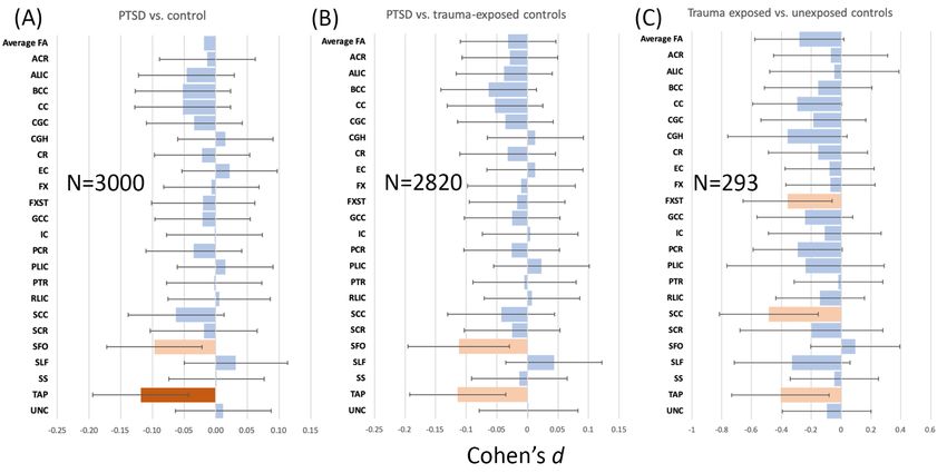
bioRxiv preprint first posted online Jun. 20, 2019; doi: http://dx.doi.org/10.1101/677153. The copyright holder for this preprint
      (which was not peer-reviewed) is the author/funder, who has granted bioRxiv a license to display the preprint in perpetuity.
                                   It is made available under a CC-BY-NC-ND 4.0 International license.

28      Department of Psychiatry, Amsterdam University Medical Centers, Location Academic Medical Center, University
        of Amsterdam, Amsterdam, The Netherlands.
29      Cognitive and Clinical Neuroimaging Core, McLean Hospital, Belmont, MA.
30      Department of Psychiatry, Harvard Medical School, Boston, MA.
31      Center for Healthy Minds, University of Wisconsin-Madison.
32      Division of Neuroradiology, Brigham and Women's Hospital, Boston, MA.
33      Department of Psychology, The Ohio State University, Columbus, OH, USA.
34      Institute of Medical Psychology and Systems Neuroscience, University of Münster.
35      Dept of Psychiatry, University of Cape Town, Cape Town, South Africa.
36      Department of Psychiatry and Behavioral Neuroscience, Wayne State University School of Medicine, Detroit, MI.
37      Brain Center Rudolf Magnus, Department of Psychiatry, UMCU, Utrecht, The Netherlands.
38      Brain Research and Innovation Centre, Ministry of Defence, Utrecht, The Netherlands.
39      Department of Child and Adolescent Psychiatry, Psychosomatic and Psychotherapy, Ludwig-Maximilians-
        Universität, Munich, Germany.
40      Donders Institute for Brain, Cognition and Behavior, Centre for Cognitive Neuroimaging, Radboud University
        Nijmegen, Nijmegen, The Netherlands.
41      Brain Dynamics Centre, Westmead Institute of Medical Research, University of Sydney, Westmead, NSW.
42      Division of Depression and Anxiety Disorders, McLean Hospital, Belmont, MA.
43      Laboratory for Traumatic Stress Studies, CAS Key Laboratory of Mental Health, Institute of Psychology, Chinese
        Academy of Sciences, Beijing, China.
44      Department of Psychology, University of Chinese Academy of Sciences, Beijing, China.
45      Departments of Radiology, Psychiatry, and Biomedical Engineering, University of Iowa, Iowa City, IA.
46      University Medical Centre Charite, Berlin, Germany.
47      VISN 17 Center of Excellence for Research on Returning War Veterans, Waco, TX.
48      Department of Psychology and Neuroscience, Baylor University, Waco, TX.
49      Department of Psychiatry and Behavioral Science, Texas A&M Health Science Center, Bryan, TX.
50      Center for Vital Longevity, School of Behavioral and Brain Sciences, University of Texas at Dallas, Dallas, TX.
51      Menninger Department of Psychiatry, Baylor College of Medicine, Houston, TX
52      South Central MIRECC, Houston, TX.
53      Department of Psychiatry, Amsterdam University Medical Centers, Location VU University Medical Center, VU
        University, Amsterdam, The Netherlands.
54      Department of Psychology, Western University, London, ON, Canada.
55      Department of Neuroscience, Western University, London, Ontario, Canada.
56      Department of Psychology, University of British Columbia, Okanagan, British Columbia, Canada.
57      Department of Psychiatry, University of Wisconsin-Madison.
58      University of the Sunshine Coast, Birtinya, QLD, Australia.
59      Department of Psychology, University of Washington, Seattle, WA.
60      SU/UCT MRC Unit on Risk and Resilience in Mental Disorders, Department of Psychiatry, Stellenbosch
        University, Stellenbosch, South Africa.
61      Department of Psychology and Brain Sciences, University of North Carolina at Chapel Hill.
62      Department of Psychology, University of South Dakota, Vermillion, SD.
63      Department of Medical Biophysics, Western University, London, ON, Canada.
64      Houston Fire Department, Houston, TX.
65      Psychiatry, LUMC, Leiden, The Netherlands.
66      Leiden Institute for Brain and Cognition, Leiden, The Netherlands.
67      Missouri Institute of Mental Health and University of Missouri, St Louis, MO.
68      Memory Disorders Research Center, VA Boston Healthcare System, Boston, MA.
69      Ahmanson-Lovelace Brain Mapping Center, Department of Neurology, University of California, Los Angeles, Los
        Angeles, CA.
70      Dartmouth College.
71      Division of Women's Mental Health, McLean Hospital, Belmont, MA.
72      Joint Trauma System, 3698 Chambers Pass, Joint Base San Antonio, Fort Sam Houston, TX.
73      Alaska Radiology Associates, Anchorage, AK.
74      School of Psychology, University of New South Wales, Sydney, NSW.
75      Department of Clinical Psychology, University of Groningen, Groningen, The Netherlands.
76      Department of Psychology, University of Wisconsin-Madison.
77      Center for Behavior Genetics of Aging, University of California, San Diego, La Jolla, CA.
78      Dept. of Psychological & Brain Sciences, Boston University, Boston, MA.
79      Departments of Physical Medicine and Rehabilitation, Neurology, and Pediatrics, Baylor College of Medicine,
        Houston, TX.
80      Geriatric Research Educational and Clinical Center and Translational Research Center for TBI and Stress
        Disorders, VA Boston Healthcare System.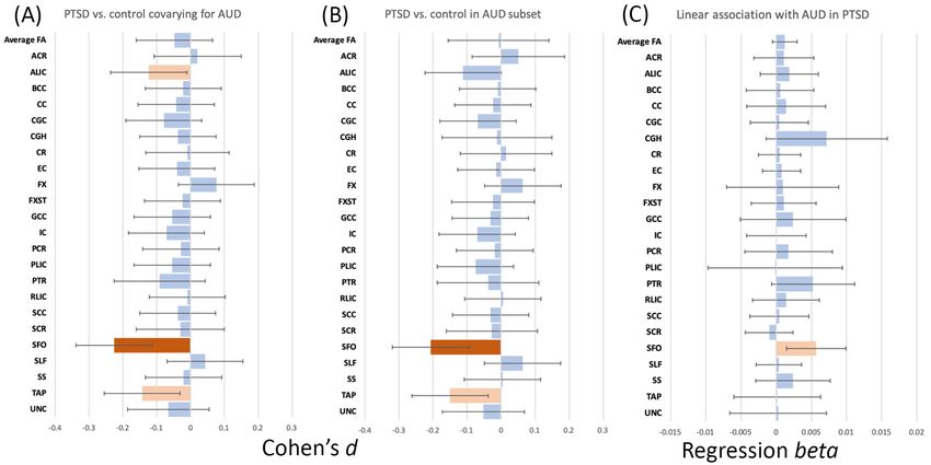
bioRxiv preprint first posted online Jun. 20, 2019; doi: http://dx.doi.org/10.1101/677153. The copyright holder for this preprint
      (which was not peer-reviewed) is the author/funder, who has granted bioRxiv a license to display the preprint in perpetuity.
                                   It is made available under a CC-BY-NC-ND 4.0 International license.

81      Department of Psychology, Harvard University.
82      Arq National Psychotrauma Center, Diemen, the Netherlands.
83      VA Boston Healthcare System, Brockton Division, Brockton, MA.
84      Department of Family Medicine and Public Health, University of California, San Diego, La Jolla, CA.
85      George E. Whalen Veterans Affairs Medical Center, Salt Lake City, UT.
86      H. Ben Taub Department of Physical Medicine and Rehabilitation, Baylor College of Medicine, Houston, TX.
87      Maryland Psychiatric Research Center, University of Maryland School of Medicine, Baltimore, MD.

Please address correspondence to:
Dr. Emily Dennis
Psychiatry Neuroimaging Laboratory, Brigham and Women’s Hospital
edennis@bwh.harvard.edu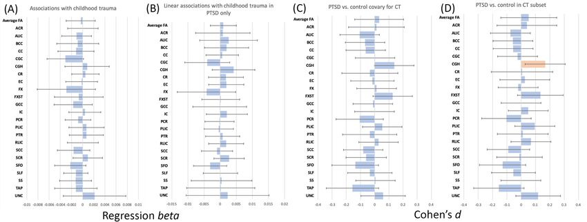
bioRxiv preprint first posted online Jun. 20, 2019; doi: http://dx.doi.org/10.1101/677153. The copyright holder for this preprint
     (which was not peer-reviewed) is the author/funder, who has granted bioRxiv a license to display the preprint in perpetuity.
                                  It is made available under a CC-BY-NC-ND 4.0 International license.

Abstract

       A growing number of studies have examined alterations in white matter organization in people

with posttraumatic stress disorder (PTSD) using diffusion MRI (dMRI), but the results have been

mixed, which may be partially due to relatively small sample sizes among studies. Altered structural

connectivity may be both a neurobiological vulnerability for, and a result of, PTSD. In an effort to find

reliable effects, we present a multi-cohort analysis of dMRI metrics across 3,049 individuals from 28

cohorts currently participating in the PGC-ENIGMA PTSD working group (a joint partnership between

the Psychiatric Genomics Consortium and the Enhancing NeuroImaging Genetics through Meta-

Analysis consortium). Comparing regional white matter metrics across the full brain in 1,446

individuals with PTSD and 1,603 controls (2152 males/897 females) between ages 18-83, 92% of

whom were trauma-exposed, we report associations between PTSD and disrupted white matter

organization measured by lower fractional anisotropy (FA) in the tapetum region of the corpus

callosum (Cohen’s d=-0.12, p=0.0021). The tapetum connects the left and right hippocampus,

structures for which structure and function have been consistently implicated in PTSD. Results

remained significant/similar after accounting for the effects of multiple potentially confounding

variables: childhood trauma exposure, comorbid depression, history of traumatic brain injury, current

alcohol abuse or dependence, and current use of psychotropic medications. Our results show that

PTSD may be associated with alterations in the broader hippocampal network.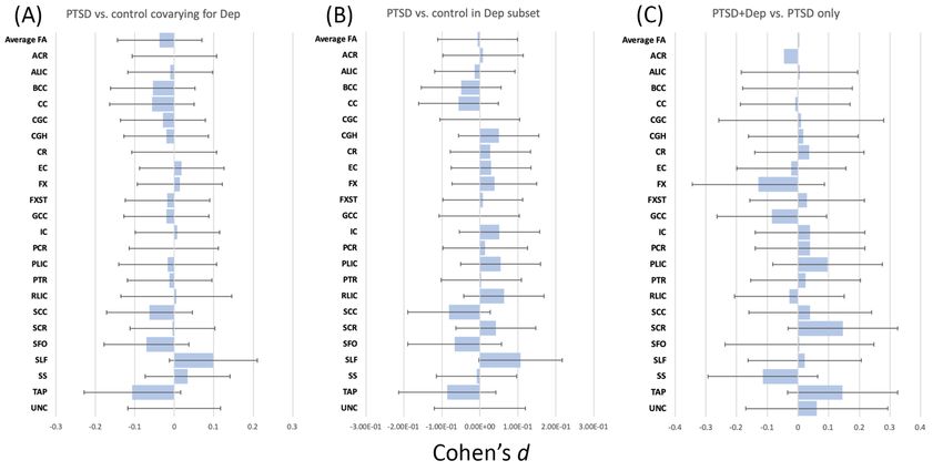
bioRxiv preprint first posted online Jun. 20, 2019; doi: http://dx.doi.org/10.1101/677153. The copyright holder for this preprint
      (which was not peer-reviewed) is the author/funder, who has granted bioRxiv a license to display the preprint in perpetuity.
                                   It is made available under a CC-BY-NC-ND 4.0 International license.

Introduction

        Posttraumatic stress disorder (PTSD) is a debilitating mental health condition with a lifetime

prevalence varying globally between 1-9% 1, with higher rates in women. Rates of PTSD are higher in

populations exposed to greater levels of trauma, such as combat veterans 2 and civilians in conflict

zones 3. In addition to trauma type, genetics, and other sociological, psychological, and biological

factors, individual differences in brain structure and function may explain vulnerability to developing

PTSD following exposure to trauma, may result from trauma, or may be exacerbated by PTSD 4.

Diffusion MRI (dMRI) is able to model white matter tracts and assess microstructural organization 5.

Fractional anisotropy (FA) is the most commonly used metric of microstructural organization,

reflecting the degree to which water is diffusing along the axon (axially) as compared with across it

(radially). Greater FA can reflect higher myelination, axonal diameter, or fiber density. Mean diffusivity

(MD) reflects the average magnitude of diffusion across all directions, axial diffusivity (AD) is diffusion

along the primary eigenvector (the dominant fiber direction), and radial diffusivity (RD) estimates

diffusion perpendicular to the primary eigenvector. Altered microstructural organization is associated

with several different psychiatric disorders and could constitute a risk factor and/or a consequence of

the disorders.

        There is a lack of mechanistic evidence on the effects of stress and trauma on white matter

structure. Exposure to trauma could lead to white matter damage, as excessive glucocorticoid levels

can be neurotoxic and can impact myelination 6,7. Studies of white matter microstructure in PTSD

have reported inconsistent results. The majority report that PTSD is associated with lower FA 8–24, but

some report higher FA 25–31, higher and lower FA in different regions 32, or null results 33–35.

Alterations in the cingulum bundle are frequently reported 9–13,16,18,21,23–29,31,32,36, with differences also

observed in the uncinate, corpus callosum, and corona radiata 14,16,18,19,24,26,29. Inconsistent findings

may be partially due to the use of hypothesis-driven rather than whole brain approaches, choice of

analytic pipeline, selection of diffusion metrics, gender-specific studies, homogeneity of single cohort

samples such as trauma-exposed vs. unexposed controls, and focus on military vs. civilian samples.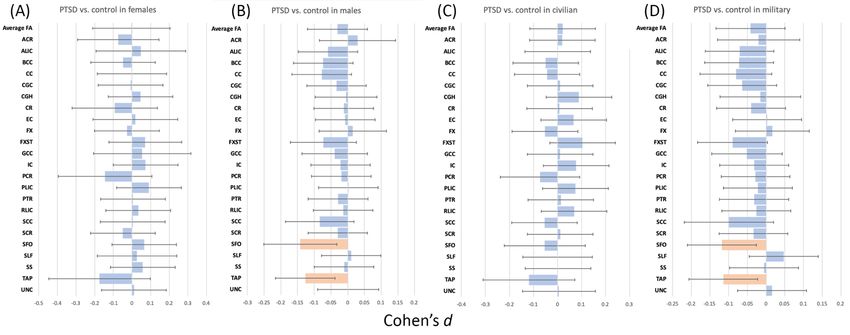
bioRxiv preprint first posted online Jun. 20, 2019; doi: http://dx.doi.org/10.1101/677153. The copyright holder for this preprint
     (which was not peer-reviewed) is the author/funder, who has granted bioRxiv a license to display the preprint in perpetuity.
                                  It is made available under a CC-BY-NC-ND 4.0 International license.

       The PGC-ENIGMA PTSD working group is an international collaborative effort of the

Psychiatric Genomics Consortium and the Enhancing NeuroImaging Genetics through Meta-Analysis

(ENIGMA) consortium that aims to increase statistical power through meta- and mega-analyses of

PTSD neuroimaging biomarkers. This collaborative approach has led to the largest PTSD

neuroimaging study to date, reporting smaller hippocampal volume in PTSD 37. Here, we applied this

approach to investigate the microstructural organization of white matter in PTSD. The ENIGMA DTI

workflow 38, which has successfully identified white matter compromise in schizophrenia 39, bipolar

disorder 40, major depression 41, and 22q11.2 deletion syndrome 42, among others, was used by 28

cohorts to process their DTI data locally. We hypothesized the largest effects of compromised

microstructure will be evident in the fronto-limbic tracts, such as the cingulum, uncinate, fornix, and

corpus callosum; these tracts are strongly implicated in behavioral deficits of PTSD such as emotion

regulation, working memory, and episodic memory 10,16,19,20,24,43.

Materials and Methods

Study Samples

       The PGC-ENIGMA PTSD DTI analysis included 28 cohorts from 7 countries totaling 1,603

healthy controls and 1,446 individuals with PTSD (either formally diagnosed or with CAPS-4>40, see

Supplementary Figure 1). The age range across cohorts was 18-83 years; all but two older Vietnam

era cohorts had an average age between 29-50. Of the 3,049 participants included in these analyses,

2,073 (68%) were from military cohorts, which resulted in a disproportionate number of males (70%).

The majority of cohorts included trauma-exposed controls (e.g., combat, community violence, intimate

partner violence, N=1,603), although some included trauma-unexposed controls (N=122), and one

included no control group. Table 1 contains demographic and clinical information for each cohort. All

participants provided written informed consent approved by local institutional review boards. Quality

control was completed by each site, with visual quality checking and outlier detection. Details on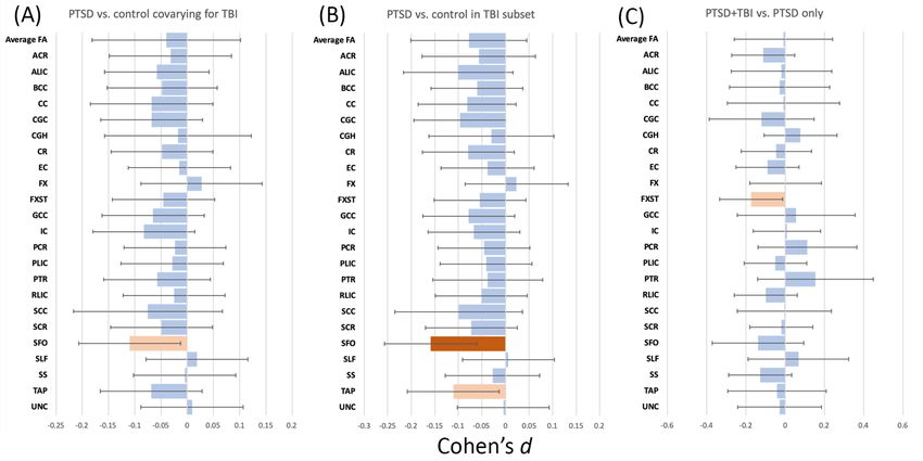
bioRxiv preprint first posted online Jun. 20, 2019; doi: http://dx.doi.org/10.1101/677153. The copyright holder for this preprint
      (which was not peer-reviewed) is the author/funder, who has granted bioRxiv a license to display the preprint in perpetuity.
                                   It is made available under a CC-BY-NC-ND 4.0 International license.

ENIGMA-DTI methods 39, inclusion/exclusion criteria, and clinical information may be found in

Supplementary Note 1, Supplementary Table 1, and Supplementary Note 2, respectively.

Image Acquisition and Processing

        The acquisition parameters for each cohort are provided in Supplementary Table 2.

Preprocessing, including eddy current correction, echo-planar imaging-induced distortion correction

and tensor fitting, was carried out at each site. Recommended protocols and quality control

procedures are available on the ENIGMA-DTI and NITRC (Neuroimaging Informatics Tools and

Resources Clearinghouse) webpages. Harmonization of preprocessing schemes was not enforced

across sites to accommodate site- and acquisition-specific pipelines. Once tensors were estimated,

they were mapped to the ENIGMA DTI template and projected onto the ENIGMA-DTI template and

were averaged within ROIs (http://enigma.ini.usc.edu/protocols/dti-protocols/). Further details and

ROI abbreviations can be seen in Supplementary Note 1.

Statistical Analysis

        For each cohort/study, a linear model was fit using the ppcor and matrixStats packages in R

3.1.3, with the ROI FA as the response variable and PTSD and covariates as predictors. For

cohorts/studies including more than one data collection site, site was included as a fixed dummy

variable in the site-level analysis. As in prior ENIGMA disease working group meta-analyses 39, a

random-effects inverse-variance weighted meta-analysis was conducted at a central coordinating site

(the University of Southern California Imaging Genetics Center) in R (metafor package, version 1.99–

118 http://www.metafor-project.org/) to combine individual cohort estimated effect sizes (see Figure

2). Cohen’s d for the main effect of group and unstandardized β coefficients (regression parameters)

for continuous predictors were computed with 95% confidence intervals. We used the Cohen’s d

calculation that accounts for covariates in the fixed effects model, using the following equation:

                                                                  + ',-, ∗ (%0123 + %567896: )
                                              !"ℎ$%& ' ) ~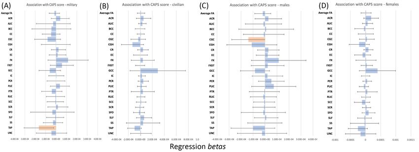
bioRxiv preprint first posted online Jun. 20, 2019; doi: http://dx.doi.org/10.1101/677153. The copyright holder for this preprint
         (which was not peer-reviewed) is the author/funder, who has granted bioRxiv a license to display the preprint in perpetuity.
                                      It is made available under a CC-BY-NC-ND 4.0 International license.

Heterogeneity scores (I2) for each test were computed, indicating the percent of total variance in

effect size explained by heterogeneity across cohorts. Bilaterally averaged FA was the primary

imaging measure, with corresponding MD, RD, and AD examined post hoc when FA was significant

for an effect of diagnosis. Lateralized ROIs were examined post hoc when a significant association

was found with the bilateral average. The corticospinal tract was not analyzed as it has poor reliability
38
     . A conservative Bonferroni correction was used for multiple testing (p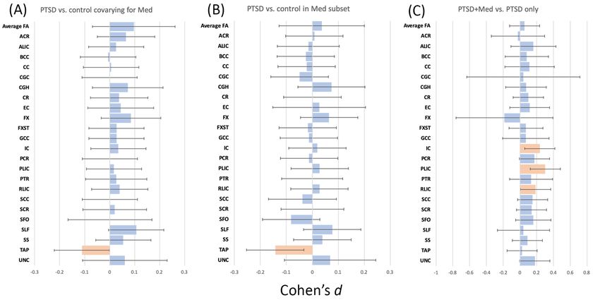
bioRxiv preprint first posted online Jun. 20, 2019; doi: http://dx.doi.org/10.1101/677153. The copyright holder for this preprint
     (which was not peer-reviewed) is the author/funder, who has granted bioRxiv a license to display the preprint in perpetuity.
                                  It is made available under a CC-BY-NC-ND 4.0 International license.

potential confounds in a single model. Details of these methods and results are provided in

Supplementary Note 5. Briefly, binary variables were created for depression, TBI, and medication

use. As depression was assessed using a variety of measures, we used published cut-offs to recode

the data as categorical depression (see Supplementary Note 2 for more details). Alcohol use

disorders and childhood trauma were coded as three-level ordinal variables based on evidence of

dose-dependent effects on brain structure and clinical severity, respectively 45,46: Alcohol use

disorders: 0=no alcohol use disorder, 1=alcohol abuse, 2=alcohol dependence, as measured by the

SCID or AUDIT 47; childhood trauma (as measured by the Childhood Trauma Questionnaire): 0=no

reported childhood trauma, 1=one type of childhood trauma exposure, 2=two or more types of

childhood trauma exposure; PTSD severity: To examine PTSD severity, we conducted linear

regressions on CAPS-4 score in the PTSD group for sites that collected CAPS-4. We examined linear

associations with CAPS-4 score covarying for childhood trauma, depression, alcohol use disorders,

TBI, and medication use, and we tested associations with CAPS-4 separately in military veterans and

civilians as well as males and females (see Supplementary Note 5 for more details).

Results

Group differences

       We found significantly lower FA in the PTSD group in the tapetum of the corpus callosum (d=-

0.12, p=0.0021) when comparing PTSD (n=1,397) and all controls (n=1,603). Post hoc analysis

revealed a larger effect in the left than in the right tapetum (left d=-0.14, p=0.00040; right d=-0.080,

p=0.038). Post hoc analysis also revealed higher RD in the tapetum in the PTSD group (bilateral

d=0.10, p=0.0085; left d=0.12, p=0.0016).

       In the analysis comparing participants with PTSD (n=1,339) to trauma-exposed controls

(n=1,481), we found lower FA and higher RD in the tapetum in PTSD, although the bilateral tapetum

was marginally significant (FA: bilateral d=-0.11, p=0.0044; left d=-0.14, p=0.00065; RD: bilateral

d=0.090, p=0.027; left d=0.12, p=0.0026) (see Table 2 and Figure 1, and see Figure 2 for site-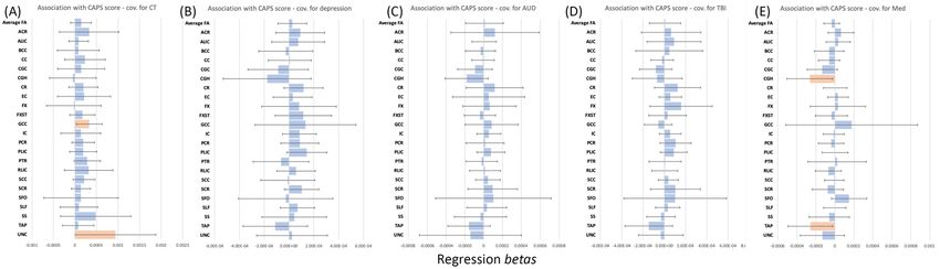
bioRxiv preprint first posted online Jun. 20, 2019; doi: http://dx.doi.org/10.1101/677153. The copyright holder for this preprint
     (which was not peer-reviewed) is the author/funder, who has granted bioRxiv a license to display the preprint in perpetuity.
                                  It is made available under a CC-BY-NC-ND 4.0 International license.

specific effects). PTSD participants from cohorts that only included trauma-unexposed controls were

not included.

       Comparing trauma-exposed (n=200) to trauma un-exposed controls (n=93) from 6 sites, we

found marginally lower FA in exposed controls in the tapetum, splenium of corpus callosum, and

fornix/stria-terminalis (d=-0.41, p=0.014; d=-0.48, p=0.0042; d=-0.36, p=0.019, respectively), along

with significantly higher MD and RD in the splenium (d=0.48, p=0.0017; d=0.56, p=0.00023,

respectively) and marginally higher RD in the tapetum (d=0.32, p=0.036).

Subgroups

       We examined military vs. civilian cohorts, and male vs. female participants separately. All

subgroups showed non-significant associations with PTSD, but there were marginal associations with

tapetum FA in the military-only and male-only subgroups separately (see Supplementary Note 3 and

Supplementary Figure 3 for more details). Results of group-by-sex and group-by-age interactions

were not significant and are shown in Supplementary Note 4 and Supplementary Figure 4.

Additional Covariates

       The role of potentially confounding variables on the association between PTSD and the

tapetum was tested in several post hoc analyses focused on left, right, and bilateral tapetum FA (see

Supplementary Figure 2). As these analyses were considered post hoc and limited to the tapetum,

we used a test-wise significance threshold of p
bioRxiv preprint first posted online Jun. 20, 2019; doi: http://dx.doi.org/10.1101/677153. The copyright holder for this preprint
(which was not peer-reviewed) is the author/funder, who has granted bioRxiv a license to display the preprint in perpetuity.
It is made available under a CC-BY-NC-ND 4.0 International license.
Including childhood trauma as a covariate (367 PTSD vs. 598 controls) did not yield any significant
results, but neither did the analysis in the reduced sample, suggesting that the sample reduction
impacted these results. To control for covariate- and cohort-dependent changes in sample size, each
analysis was repeated in a smaller sample that corresponded to omitting the relevant covariate. The
tapetum results remained consistent in nearly all reduced sample analyses - significant effects
survived covariate adjustment and effects that disappeared (such as with childhood trauma) were
also absent in the reduced sample. Thus, covariates had minimal impact beyond the reduction in
sample size (see Supplementary Note 5 and Supplementary Figures 5-9). A table showing how
many participants at each site had information on these potentially confounding variables may be
found in Supplementary Table 3.
PTSD Severity
PTSD symptom severity in the PTSD group (measured by the CAPS-4, N=979 from 18 sites)
was not associated with FA (Figure 3). Subgroup analyses yielded marginal associations between
CAPS-4 score and tapetum FA in the military cohorts, but no other subgroup. Results were similar
when potentially confounding variables were included, with no significant associations, although the
tapetum was marginally significant when psychotropic medication use was included. Detailed
analyses of PTSD severity and covariates within subgroups are in Supplementary Note 5 and
Supplementary Figures 10 and 11.
Discussion
We present DTI results from a multi-cohort study conducted by the PGC-ENIGMA PTSD
consortium. In a meta-analysis of 3,049 participants from 28 sites, we found lower FA and higher RD
in the tapetum among adults with PTSD (neuroanatomical figure - Supplementary Figure 12), which
remained after accounting for several potentially confounding factors. The tapetum is a major tract
within the corpus callosum that serves as a conduit between right and left hippocampus. Prior studies
of white matter disruption in PTSD have found alterations in other hippocampal tracts, but werebioRxiv preprint first posted online Jun. 20, 2019; doi: http://dx.doi.org/10.1101/677153. The copyright holder for this preprint
(which was not peer-reviewed) is the author/funder, who has granted bioRxiv a license to display the preprint in perpetuity.
It is made available under a CC-BY-NC-ND 4.0 International license.
generally hindered by small sample sizes leading to inconsistent findings across studies. Our results
add to the existing literature in identifying structural disruptions that compromise putative
hippocampal functions, which are known to play a central role in PTSD symptomatology 48,49.
The tapetum is a segment of the corpus callosum that connects the temporal lobes, in particular the
left and right hippocampus 50. It is one of the last corpus callosum segments to develop and
experiences rapid growth around age 14, which may make it vulnerable to the effects of trauma for a
longer period of time 51. Along with the inferior longitudinal fasciculus, cingulum, fornix, spino-limbic
tracts (not studied here), and the anterior commissure, the tapetum is a dominant hippocampal
pathway 50. Structural and functional alterations in the hippocampus are frequently reported in PTSD,
with smaller volumes 37, decreased activation, and disrupted functional connectivity with the medial
and lateral prefrontal cortices 52,53. Here we report microstructural evidence that structural connectivity
between the left and right hippocampus may also be disrupted in PTSD. While many studies have
reported that PTSD is associated with alterations in the cingulum bundle 9–13,16,18,21,23–29,31,32,36, which
has a hippocampal component, the tapetum has not yet emerged for several possible reasons. Many
prior studies took an ROI approach, which limited analyses to pre-determined regions that frequently
omitted the tapetum, a small region often grouped with other tracts such as the splenium or posterior
thalamic radiation. Critically, in 2013, an error was uncovered in the JHU atlas used as part of the
TBSS pipeline, with the uncinate incorrectly identified as the inferior fronto-occipital fasciculus, and
the tapetum incorrectly identified as the uncinate 54. Thus, the tapetum was simply not examined in
prior studies, with one very recent exception showing that tapetum abnormalities are associated with
lower major depressive disorder remission 55. Finally, the precise role of the tapetum in connecting
the left and right hippocampus was only recently elucidated by mapping the subcortical connectome
with exquisitely high-resolution mapping capable of discerning the intermingling of tapetum and other
corpus callosum fibers. Super-resolution DTI conducted by the Chronic Diseases Connectome
Project that acquired 1150-direction single-subject and 391-direction 94-subject data made this ultra-
structural mapping possible 50.bioRxiv preprint first posted online Jun. 20, 2019; doi: http://dx.doi.org/10.1101/677153. The copyright holder for this preprint
(which was not peer-reviewed) is the author/funder, who has granted bioRxiv a license to display the preprint in perpetuity.
It is made available under a CC-BY-NC-ND 4.0 International license.
Childhood trauma is the greatest single risk factor for future vulnerability to PTSD 56; numerous
studies show significant alterations in brain structure and function in individuals who had experienced
significant early life stress 46,57. Some of these alterations likely contribute to a higher risk for
psychopathology, but childhood trauma exposure did not explain the association between tapetum
white matter disruption and PTSD that we report here. Depression is frequently comorbid with PTSD
58
and is associated with disrupted white matter organization, although the affected tracts are broadly
distributed 59. Accounting for depression in group comparisons did not significantly alter our results,
suggesting that tapetum white matter disruption is specific to PTSD. Particularly in military
populations, which formed the majority of our sample, PTSD is often comorbid with traumatic brain
injury (TBI) 60. White matter is particularly vulnerable to TBI, which produces stretching and shearing
of axons and altered neurometabolism 61. Accounting for TBI also did not significantly change our
results, indicating that TBI was associated with white matter damage generally, but not specifically
within the tapetum. Psychotropic medications are another potential confound, given their neurotrophic
and neuroprotective effects 62. The result in the tapetum persisted after covarying for psychotropic
medication, indicating that our findings are unlikely to be explained by medication. Lastly, PTSD can
be comorbid with alcohol use disorders, which have a poorer clinical prognosis 63,64. alcohol use
disorders have been associated with significant changes in white matter organization 65,66 but did not
influence the present results.
We found a marginally significant association of PTSD with FA in the tapetum in male and
military subgroups separately. Although results were non-significant in female or civilian subgroups,
the effect size was slightly larger and in the same direction. The female and civilian subgroups were
smaller and therefore the analyses had lower power than in male and military subgroups. Most prior
dMRI studies in civilians report lower FA in PTSD 9,10,12–14,16,17,21–25,30–32,67. Studies of military cohorts
have been mixed, reporting higher FA 26–29,36, lower FA 8,15,18–20, and null results 33–35. This
discrepancy may be due to differences in age, chronicity, and type of trauma exposure, although
military personnel often also experience civilian trauma. Combat-related PTSD is often comorbid withbioRxiv preprint first posted online Jun. 20, 2019; doi: http://dx.doi.org/10.1101/677153. The copyright holder for this preprint
(which was not peer-reviewed) is the author/funder, who has granted bioRxiv a license to display the preprint in perpetuity.
It is made available under a CC-BY-NC-ND 4.0 International license.
TBI, which is also associated with white matter disruption, constituting a potentially confounding factor
for studies 68.
In the absence of longitudinal data, our analysis cannot make causal inferences about the
direction of the relationship between PTSD and tapetum white matter organization. Disrupted white
matter of the tapetum may represent a vulnerability that predates the onset of PTSD, or a
pathological response to trauma. In twins discordant for exposure to combat stress, the unexposed
twins of combat veterans with PTSD have smaller hippocampal volume than the unexposed twins of
combat veterans without PTSD 69. Individuals with two risk alleles of the FKBP5 gene have
demonstrated lower cingulum FA above and beyond the association of cingulum FA with PTSD 11,67.
These studies suggest that heritable differences in brain structure may influence risk of developing
PTSD. Evidence that alterations are caused by PTSD was observed in Israeli Defense Force recruits
with reduced structural connectivity between the hippocampus and ventromedial prefrontal cortex, but
only after exposure to military stress 70. With the varying developmental trajectories of brain structure,
function, and connectivity, along with the varying distribution of stress hormone receptors in the brain,
the complex question of vulnerability vs. consequence will require prospective longitudinal
neuroimaging studies.
Some evidence indicates that high FA is a marker of resilience to the effects of stress 71,72. A
putative marker of resilience is the ability to attenuate stress-induced increases in corticotropin-
releasing hormone and glucocorticoids through an elaborate negative feedback system, and to
modulate the expression of brain-derived neurotrophic factor (BDNF) 73,74. BDNF has myriad
functions including supporting neuronal differentiation, maturation, and survival 75,76. In particular,
hippocampal BDNF is implicated in the development of neural circuits that promote stress
adaptations 73. These stress adaptation circuits involve white matter in the fornix and other fronto-
limbic connections 77.
LimitationsbioRxiv preprint first posted online Jun. 20, 2019; doi: http://dx.doi.org/10.1101/677153. The copyright holder for this preprint
(which was not peer-reviewed) is the author/funder, who has granted bioRxiv a license to display the preprint in perpetuity.
It is made available under a CC-BY-NC-ND 4.0 International license.
Our study has several limitations. One limitation of TBSS studies is the inability to fully attribute
results to particular fiber bundles. Future studies may benefit by using tractography to more reliably
identify the affected bundles, but this is difficult across so many varied sites. Second, not all
participants classified as PTSD received clinician-administered interview (such as the CAPS) to
confirm diagnoses. Third, we could not reliably measure chronicity across different cohorts. Other
variables that we could not examine given the heterogeneity across sites include treatment effects,
symptom clusters, trauma types, and lifetime as opposed to current PTSD diagnosis. Although we
analyzed data from over 3,000 participants, we may have been underpowered to examine group-by-
sex interactions, as 55% of our sample came from cohorts including only males or only females or
samples that were >90% male. Diffusion metrics are not scanner invariant, and can vary even in
scanners of the same model. For this reason, we were limited to a meta-analytic approach which may
have lower power than mega-analysis. However, studies including both meta- and mega-analyses of
brain volume in other ENIGMA groups have found minimal differences 78,79.
Future studies should further investigate the tapetum using high spatial and angular resolution
tractography to replicate our findings. Future and existing studies with more in-depth phenotyping
than was possible here could examine how alterations in the tapetum vary with trauma type,
chronicity, treatment, and whether they are associated with specific symptom clusters. The current
study excluded pediatric cases, so additional research on white matter disruption in pediatric trauma
and PTSD is warranted. Lastly, while we considered comorbidities as potential confounding variables,
we did not examine their association with dMRI metrics. Future collaborations with the ENIGMA Brain
Injury, MDD, and Addiction working groups will provide opportunities to separate general
neuroimaging biomarkers of psychopathology and disorder-specific effects.
Conclusions
Here we presented results from the PGC-ENIGMA PTSD working group, reporting poorer
white matter organization in the tapetum in individuals currently suffering from PTSD. We present the
largest DTI study in PTSD to date and the first to use harmonized image processing across sites,bioRxiv preprint first posted online Jun. 20, 2019; doi: http://dx.doi.org/10.1101/677153. The copyright holder for this preprint
(which was not peer-reviewed) is the author/funder, who has granted bioRxiv a license to display the preprint in perpetuity.
It is made available under a CC-BY-NC-ND 4.0 International license.
increasing our power to detect subtle effects. While future studies need to confirm the involvement of
the tapetum specifically, our results add to the existing literature implicating the hippocampus as a
primary area of disruption in PTSD.bioRxiv preprint first posted online Jun. 20, 2019; doi: http://dx.doi.org/10.1101/677153. The copyright holder for this preprint
(which was not peer-reviewed) is the author/funder, who has granted bioRxiv a license to display the preprint in perpetuity.
It is made available under a CC-BY-NC-ND 4.0 International license.
Acknowledgements
K99NS096116 to Dr. Emily Dennis; CIHR, CIMVHR; Dana Foundation (to Dr. Nitschke); the University of
Wisconsin Institute for Clinical and Translational Research (to Dr. Emma Seppala); a National Science
Foundation Graduate Research Fellowship (to Dr. Daniel Grupe); R01MH043454 and T32MH018931 (to Dr.
Richard Davidson); and a core grant to the Waisman Center from the National Institute of Child Health and
Human Development (P30HD003352); Defense and Veterans Brain Injury Centers, the U.S. Army Medical
Research and Materiel Command (USAMRMC; W81XWH-13-2-0025) and the Chronic Effects of Neurotrauma
Consortium (CENC; PT108802-SC104835); Department of Defense award number W81XWH-12-2-0012;
ENIGMA was also supported in part by NIH U54EB020403 from the Big Data to Knowledge (BD2K) program,
R56AG058854, R01MH116147, R01MH111671, and P41EB015922 to Dr. Paul Thompson and Dr. Neda
Jahanshad; Department of Veterans Affairs via support for the National Center for PTSD, NIAAA via its support
for (P50) Center for the Translational Neuroscience of Alcohol, and NCATS via its support of (CTSA) Yale
Center for Clinical Investigation; DoD W81XWH-10-1-0925; Center for Brain and Behavior Research Pilot
Grant; South Dakota Governor’s Research Center Grant; F32MH109274; Funding from the Bill & Melinda
Gates Foundation; Funding from the SAMRC Unit on Risk & Resilience in Mental Disorders; German Research
Foundation grant to J. K. Daniels (numbers DA 1222/4-1 and WA 1539/8-2); German Research Society
(Deutsche Forschungsgemeinschaft, DFG; SFB/TRR 58: C06, C07); I01-CX000715 & I01-CX001542;
K01MH118428; K23MH090366; K2CX001772, Clinical Science Research and Development Service, VA Office
of Research and Development ; MH098212; MH071537; M01RR00039; UL1TR000454; HD071982;
HD085850; MH101380; NARSAD 27040; NARSAD Young Investigator, K01MH109836, Young Investigator
Grant, Korean Scientists and Engineers Association; NHMRC Program Grant # 1073041; R01AG050595,
R01AG022381; R01AG058822; R01EB015611; R01MH105355; R01MH105355, R01DA035484;
R01MH111671, R01MH117601, R01AG059874, MJFF 14848; R01MH111671; VISN6 MIRECC;
R21MH112956, Anonymous Women's Health Fund, Kasparian Fund, Trauma Scholars Fund, Barlow Family
Fund; South African Medical Research Council for the “Shared Roots” Flagship Project, Grant no. MRC-RFA-
IFSP-01-2013/SHARED ROOTS” through funding received from the South African National Treasury under its
Economic Competitiveness and Support Package. The work by Dr. Leigh van Heuvel reported herein was
made possible through funding by the South African Medical Research Council through its Division of
Research Capacity Development under the SAMRC Clinician Researcher (M.D PHD) Scholarship Programme
from funding received from the South African National Treasury; South African Research Chairs Initiative in
Posttraumatic Stress Disorder through the Department of Science and Technology and the National Research
Foundation.; the National Natural Science Foundation of China (No. 31271099, 31471004), the Key Research
Program of the Chinese Academy of Sciences (No. ZDRW-XH-2019-4); The study was supported by ZonMw,
the Netherlands organization for Health Research and Development (40-00812-98-10041), and by a grant from
the Academic Medical Center Research Council (110614) both awarded to Dr. Miranda Olff; Translational
Research Center for TBI and Stress Disorders (TRACTS), a VA Rehabilitation Research and Development
(RR&D) Traumatic Brain Injury Center of Excellence (B9254-C) at VA Boston Healthcare System; VA CSR&D
1IK2CX001680; VISN17 Center of Excellence Pilot funding; VA CSR&D 822-MR-18176 and Senior Career
Scientist Award; VA National Center for PTSD; VA RR&D 1IK2RX000709; VA RR&D 1K1RX002325;
1K2RX002922; VA RR&D I01RX000622; CDMRP W81XWH-08–2–0038; W81XWH08-2-0159 to Dr. Murray
Stein from the US Department of Defense. The views reflected here are strictly those of the authors and do not
constitute endorsement by any of the funding sources listed here.bioRxiv preprint first posted online Jun. 20, 2019; doi: http://dx.doi.org/10.1101/677153. The copyright holder for this preprint
(which was not peer-reviewed) is the author/funder, who has granted bioRxiv a license to display the preprint in perpetuity.
It is made available under a CC-BY-NC-ND 4.0 International license.
Conflicts of interest
Dr. Abdallah has served as a consultant, speaker and/or on advisory boards for FSV7, Lundbeck, Genentech
and Janssen, and editor of Chronic Stress for Sage Publications, Inc.; he has filed a patent for using mTOR
inhibitors to augment the effects of antidepressants (filed on August 20, 2018). Dr. Davidson is the founder and
president of, and serves on the board of directors for, the non-profit organization Healthy Minds Innovations,
Inc. Dr. Krystal is a consultant for AbbVie, Inc., Amgen, Astellas Pharma Global Development, Inc.,
AstraZeneca Pharmaceuticals, Biomedisyn Corporation, Bristol-Myers Squibb, Eli Lilly and Company,
Euthymics Bioscience, Inc., Neurovance, Inc., FORUM Pharmaceuticals, Janssen Research & Development,
Lundbeck Research USA, Novartis Pharma AG, Otsuka America Pharmaceutical, Inc., Sage Therapeutics,
Inc., Sunovion Pharmaceuticals, Inc., and Takeda Industries; is on the Scientific Advisory Board for Lohocla
Research Corporation, Mnemosyne Pharmaceuticals, Inc., Naurex, Inc., and Pfizer; is a stockholder in
Biohaven Pharmaceuticals; holds stock options in Mnemosyne Pharmaceuticals, Inc.; holds patents for
Dopamine and Noradrenergic Reuptake Inhibitors in Treatment of Schizophrenia, US Patent No. 5,447,948
(issued September 5, 1995), and Glutamate Modulating Agents in the Treatment of Mental Disorders, U.S.
Patent No. 8,778,979 (issued July 15, 2014); and filed a patent for Intranasal Administration of Ketamine to
Treat Depression. U.S. Application No. 14/197,767 (filed on March 5, 2014); US application or Patent
Cooperation Treaty international application No. 14/306,382 (filed on June 17, 2014). Filed a patent for using
mTOR inhibitors to augment the effects of antidepressants (filed on August 20, 2018). Dr. Jahanshad received
partial research support from Biogen, Inc. (Boston, USA) for research unrelated to the content of this
manuscript. Dr. Thompson received partial research support from Biogen, Inc. (Boston, USA) for research
unrelated to the topic of this manuscript. All other authors report no conflicts of interest.bioRxiv preprint first posted online Jun. 20, 2019; doi: http://dx.doi.org/10.1101/677153. The copyright holder for this preprint
(which was not peer-reviewed) is the author/funder, who has granted bioRxiv a license to display the preprint in perpetuity.
It is made available under a CC-BY-NC-ND 4.0 International license.
References
1 Atwoli L, Stein DJ, Koenen KC, McLaughlin KA. Epidemiology of posttraumatic stress disorder:
prevalence, correlates and consequences. Curr Opin Psychiatry 2015; 28: 307–311.
2 Fulton JJ, Calhoun PS, Wagner HR, Schry AR, Hair LP, Feeling N et al. The prevalence of
posttraumatic stress disorder in Operation Enduring Freedom/Operation Iraqi Freedom
(OEF/OIF) Veterans: a meta-analysis. J Anxiety Disord 2015; 31: 98–107.
3 Ferry F, Bunting B, Murphy S, O’Neill S, Stein D, Koenen K. Traumatic events and their
relative PTSD burden in Northern Ireland: a consideration of the impact of the ‘Troubles’.
Social Psychiatry and Psychiatric Epidemiology. 2014; 49: 435–446.
4 Galea S, Nandi A, Vlahov D. The epidemiology of post-traumatic stress disorder after
disasters. Epidemiol Rev 2005; 27: 78–91.
5 Basser PJ, Mattiello J, LeBihan D. MR diffusion tensor spectroscopy and imaging. Biophys J
1994; 66: 259–267.
6 Uno H, Eisele S, Sakai A, Shelton S, Baker E, DeJesus O et al. Neurotoxicity of
glucocorticoids in the primate brain. Horm Behav 1994; 28: 336–348.
7 Antonow-Schlorke I, Helgert A, Gey C, Coksaygan T, Schubert H, Nathanielsz PW et al.
Adverse effects of antenatal glucocorticoids on cerebral myelination in sheep. Obstet Gynecol
2009; 113: 142–151.
8 Bolzenius JD, Velez CS, Lewis JD, Bigler ED, Wade BSC, Cooper DB et al. Diffusion Imaging
Findings in US Service Members With Mild Traumatic Brain Injury and Posttraumatic Stress
Disorder. J Head Trauma Rehabil 2018; 33: 393–402.
9 Durkee CA, Sarlls JE, Hommer DW, Momenan R. White matter microstructure alterations: a
study of alcoholics with and without post-traumatic stress disorder. PLoS One 2013; 8:
e80952.
10 Fani N, King TZ, Jovanovic T, Glover EM, Bradley B, Choi K et al. White matter integrity in
highly traumatized adults with and without post-traumatic stress disorder.
Neuropsychopharmacology 2012; 37: 2740–2746.
11 Fani N, King TZ, Shin J, Srivastava A, Brewster RC, Jovanovic T et al. Structural And
Functional Connectivity In Posttraumatic Stress Disorder: Associations With FKBP5. Depress
Anxiety 2016; 33: 300–307.
12 Kim SJ, Jeong D-U, Sim ME, Bae SC, Chung A, Kim MJ et al. Asymmetrically altered integrity
of cingulum bundle in posttraumatic stress disorder. Neuropsychobiology 2006; 54: 120–125.
13 Kim MJ, Lyoo IK, Kim SJ, Sim M, Kim N, Choi N et al. Disrupted white matter tract integrity of
anterior cingulate in trauma survivors. Neuroreport 2005; 16: 1049–1053.
14 Koch SBJ, van Zuiden M, Nawijn L, Frijling JL, Veltman DJ, Olff M. Decreased uncinate
fasciculus tract integrity in male and female patients with PTSD: a diffusion tensor imaging
study. J Psychiatry Neurosci 2017; 42: 331–342.
15 Lepage C, de Pierrefeu A, Koerte IK, Coleman MJ, Pasternak O, Grant G et al. White matterbioRxiv preprint first posted online Jun. 20, 2019; doi: http://dx.doi.org/10.1101/677153. The copyright holder for this preprint
(which was not peer-reviewed) is the author/funder, who has granted bioRxiv a license to display the preprint in perpetuity.
It is made available under a CC-BY-NC-ND 4.0 International license.
abnormalities in mild traumatic brain injury with and without post-traumatic stress disorder: a
subject-specific diffusion tensor imaging study. Brain Imaging Behav 2018; 12: 870–881.
16 O’Doherty DCM, Ryder W, Paquola C, Tickell A, Chan C, Hermens DF et al. White matter
integrity alterations in post-traumatic stress disorder. Hum Brain Mapp 2018; 39: 1327–1338.
17 Olson EA, Cui J, Fukunaga R, Nickerson LD, Rauch SL, Rosso IM. Disruption of white matter
structural integrity and connectivity in posttraumatic stress disorder: A TBSS and tractography
study. Depress Anxiety 2017; 34: 437–445.
18 Sanjuan PM, Thoma R, Claus ED, Mays N, Caprihan A. Reduced white matter integrity in the
cingulum and anterior corona radiata in posttraumatic stress disorder in male combat veterans:
a diffusion tensor imaging study. Psychiatry Res 2013; 214: 260–268.
19 Santhanam P, Teslovich T, Wilson SH, Yeh P-H, Oakes TR, Weaver LK. Decreases in White
Matter Integrity of Ventro-Limbic Pathway Linked to Post-Traumatic Stress Disorder in Mild
Traumatic Brain Injury. J Neurotrauma 2019; 36: 1093–1098.
20 Schuff N, Zhang Y, Zhan W, Lenoci M, Ching C, Boreta L et al. Patterns of altered cortical
perfusion and diminished subcortical integrity in posttraumatic stress disorder: an MRI study.
Neuroimage 2011; 54 Suppl 1: S62–8.
21 Sun Y, Wang Z, Ding W, Wan J, Zhuang Z, Zhang Y et al. Alterations in white matter
microstructure as vulnerability factors and acquired signs of traffic accident-induced PTSD.
PLoS One 2013; 8: e83473.
22 Sun Y-W, Hu H, Wang Y, Ding W-N, Chen X, Wan J-Q et al. Inter-hemispheric functional and
anatomical connectivity abnormalities in traffic accident-induced PTSD: a study combining
fMRI and DTI. J Affect Disord 2015; 188: 80–88.
23 Wang H-H, Zhang Z-J, Tan Q-R, Yin H, Chen Y-C, Wang H-N et al. Psychopathological,
biological, and neuroimaging characterization of posttraumatic stress disorder in survivors of a
severe coalmining disaster in China. J Psychiatr Res 2010; 44: 385–392.
24 Hu H, Zhou Y, Wang Q, Su S, Qiu Y, Ge J et al. Association of abnormal white matter integrity
in the acute phase of motor vehicle accidents with post-traumatic stress disorder. J Affect
Disord 2016; 190: 714–722.
25 Abe O, Yamasue H, Kasai K, Yamada H, Aoki S, Iwanami A et al. Voxel-based diffusion tensor
analysis reveals aberrant anterior cingulum integrity in posttraumatic stress disorder due to
terrorism. Psychiatry Res 2006; 146: 231–242.
26 Aschbacher K, Mellon SH, Wolkowitz OM, Henn-Haase C, Yehuda R, Flory JD et al.
Posttraumatic stress disorder, symptoms, and white matter abnormalities among combat-
exposed veterans. Brain Imaging Behav 2018; 12: 989–999.
27 Averill CL, Averill LA, Wrocklage KM, Scott JC, Akiki TJ, Schweinsburg B et al. Altered White
Matter Diffusivity of the Cingulum Angular Bundle in Posttraumatic Stress Disorder. Mol
Neuropsychiatry 2018; 4: 75–82.
28 Bierer LM, Ivanov I, Carpenter DM, Wong EW, Golier JA, Tang CY et al. White matter
abnormalities in Gulf War veterans with posttraumatic stress disorder: A pilot study.
Psychoneuroendocrinology 2015; 51: 567–576.bioRxiv preprint first posted online Jun. 20, 2019; doi: http://dx.doi.org/10.1101/677153. The copyright holder for this preprint
(which was not peer-reviewed) is the author/funder, who has granted bioRxiv a license to display the preprint in perpetuity.
It is made available under a CC-BY-NC-ND 4.0 International license.
29 Davenport ND, Lim KO, Sponheim SR. White matter abnormalities associated with military
PTSD in the context of blast TBI. Hum Brain Mapp 2015; 36: 1053–1064.
30 Li L, Lei D, Li L, Huang X, Suo X, Xiao F et al. White Matter Abnormalities in Post-traumatic
Stress Disorder Following a Specific Traumatic Event. EBioMedicine 2016; 4: 176–183.
31 Weis CN, Belleau EL, Pedersen WS, Miskovich TA, Larson CL. Structural Connectivity of the
Posterior Cingulum Is Related to Reexperiencing Symptoms in Posttraumatic Stress Disorder.
Chronic Stress (Thousand Oaks) 2018; 2. doi:10.1177/2470547018807134.
32 Zhang L, Zhang Y, Li L, Li Z, Li W, Ma N et al. Different white matter abnormalities between
the first-episode, treatment-naive patients with posttraumatic stress disorder and generalized
anxiety disorder without comorbid conditions. J Affect Disord 2011; 133: 294–299.
33 Dretsch MN, Lange RT, Katz JS, Goodman A, Daniel TA, Deshpande G et al. Examining
Microstructural White Matter in Active Duty Soldiers with a History of Mild Traumatic Brain
Injury and Traumatic Stress. Open Neuroimag J 2017; 11: 46–57.
34 Maksimovskiy AL, McGlinchey RE, Fortier CB, Salat DH, Milberg WP, Oscar-Berman M. White
Matter and Cognitive Changes in Veterans Diagnosed with Alcoholism and PTSD. J Alcohol
Drug Depend 2014; 2: 144.
35 Morey RA, Haswell CC, Selgrade ES, Massoglia D, Liu C, Weiner J et al. Effects of chronic
mild traumatic brain injury on white matter integrity in Iraq and Afghanistan war veterans. Hum
Brain Mapp 2013; 34: 2986–2999.
36 Kennis M, van Rooij SJH, van den Heuvel MP, Kahn RS, Geuze E. Functional network
topology associated with posttraumatic stress disorder in veterans. Neuroimage Clin 2016; 10:
302–309.
37 Logue MW, van Rooij SJH, Dennis EL, Davis SL, Hayes JP, Stevens JS et al. Smaller
hippocampal volume in posttraumatic stress disorder: a multi-site ENIGMA-PGC study. Biol
Psychiatry 2017.
38 Jahanshad N, Kochunov PV, Sprooten E, Mandl RC, Nichols TE, Almasy L et al. Multi-site
genetic analysis of diffusion images and voxelwise heritability analysis: A pilot project of the
ENIGMA–DTI working group. Neuroimage 2013; 81: 455–469.
39 Kelly S, Jahanshad N, Zalesky A, Kochunov P, Agartz I, Alloza C et al. Widespread white
matter microstructural differences in schizophrenia across 4322 individuals: results from the
ENIGMA Schizophrenia DTI Working Group. Mol Psychiatry 2018; 23: 1261–1269.
40 Pauline Favre, Melissa Pauling, Jacques Stout, Franz Hozer, Samuel Sarrazin, Christoph Abé,
Martin Alda, Clara Alloza, Silvia Alonso-Lana, Ole A Andreassen, Bernhard T Baune,
Francesco Benedetti, Geraldo F Busatto, Erick J Canales-Rodríguez, Xavier Caseras, Tiffany
Moukbel Chaim-Avancini, Christopher R. K Ching, BA, Udo Dannlowski, Michael Deppe, Lisa
T Eyler, Mar Fatjo-Vilas, Sonya F Foley, Dominik Grotegerd, Tomas Hajek, Unn K Haukvik,
Fleur M Howells, Neda Jahanshad, Harald Kugel, Trine V Lagerberg, Stephen M Lawrie, Julia
O Linke, Andrew McIntosh, Elisa M. T Melloni, MS, Philip B Mitchell, Mircea Polosan, Edith
Pomarol-Clotet, Jonathan Repple, Pedro G. P Rosa, Raymond Salvador, Salvador Sarró,
Peter R Schofield, Mauricio H Serpa, Kang Sim, Dan J Stein, Jess E Sussmann, Henk S
Temmingh, Paul M Thompson, Norma Verdolini, Eduard Vieta, Michele Wessa, Heather C
Whalley, Marcus V Zanetti, Marion Leboyer, Jean-François Mangin, Chantal Henry, EdouardYou can also read