Rapid and iterative genome editing in the malaria parasite Plasmodium knowlesi provides new tools for P. vivax research
←
→
Page content transcription
If your browser does not render page correctly, please read the page content below
TOOLS AND RESOURCES

                                   Rapid and iterative genome editing in the
                                   malaria parasite Plasmodium knowlesi
                                   provides new tools for P. vivax research
                                   Franziska Mohring1, Melissa Natalie Hart1, Thomas A Rawlinson2, Ryan Henrici1,
                                   James A Charleston1, Ernest Diez Benavente1, Avnish Patel1, Joanna Hall3,
                                   Neil Almond3, Susana Campino1, Taane G Clark1, Colin J Sutherland1,
                                   David A Baker1, Simon J Draper2, Robert William Moon1*
                                   1
                                    Faculty of Infectious and Tropical Diseases, London School of Hygiene & Tropical
                                   Medicine, London, United Kingdom; 2The Jenner Institute, University of Oxford,
                                   Oxford, United Kingdom; 3Division of Infectious Disease Diagnostics, National
                                   Institute for Biological Standards and Control, Health Protection Agency,
                                   Hertfordshire, United Kingdom

                                   Abstract Tackling relapsing Plasmodium vivax and zoonotic Plasmodium knowlesi infections is
                                   critical to reducing malaria incidence and mortality worldwide. Understanding the biology of these
                                   important and related parasites was previously constrained by the lack of robust molecular and
                                   genetic approaches. Here, we establish CRISPR-Cas9 genome editing in a culture-adapted P.
                                   knowlesi strain and define parameters for optimal homology-driven repair. We establish a scalable
                                   protocol for the production of repair templates by PCR and demonstrate the flexibility of the
                                   system by tagging proteins with distinct cellular localisations. Using iterative rounds of genome-
                                   editing we generate a transgenic line expressing P. vivax Duffy binding protein (PvDBP), a lead
                                   vaccine candidate. We demonstrate that PvDBP plays no role in reticulocyte restriction but can
                                   alter the macaque/human host cell tropism of P. knowlesi. Critically, antibodies raised against the
                                   P. vivax antigen potently inhibit proliferation of this strain, providing an invaluable tool to support
*For correspondence:               vaccine development.
rob.moon@lshtm.ac.uk               DOI: https://doi.org/10.7554/eLife.45829.001

Competing interests: The
authors declare that no
competing interests exist.
                                   Introduction
Funding: See page 23               Malaria remains a serious health burden globally, with over 216 million cases annually (WHO, 2018).
Received: 06 February 2019         Plasmodium falciparum is responsible for 99% of estimated malaria cases in sub-Saharan Africa. Out-
Accepted: 28 May 2019              side Africa, P. vivax is the predominant parasite and causes ~ 7.4 million clinical cases annually.
Published: 17 June 2019            Despite extensive efforts, in 2016 the number of malaria cases were on the rise again for the first
                                   time in several years (WHO, 2018). Achieving global malaria eradication requires new tools and
Reviewing editor: Dominique
Soldati-Favre, University of
                                   approaches for addressing emerging drug resistance, relapsing P. vivax infections, and emerging
Geneva, Switzerland                zoonotic P. knowlesi infections, which represent significant causes of severe disease and death
                                   (Singh and Daneshvar, 2013; Hanboonkunupakarn and White, 2016; Menard and Dondorp,
   Copyright Mohring et al. This
                                   2017).
article is distributed under the
                                      Although P. vivax displays some distinctive features to P. knowlesi, including the formation of
terms of the Creative Commons
Attribution License, which         latent hypnozoites stages in the liver and restriction to reticulocytes in the blood, the two parasites
permits unrestricted use and       are closely related, occupying a separate simian parasite clade to P. falciparum (Pacheco et al.,
redistribution provided that the   2018). Host cell invasion by P. vivax and P. knowlesi relies on the Duffy binding proteins (DBP)
original author and source are     PvDBP and PkDBPa, respectively, both ligands for human red blood cell (RBC) Duffy antigen/recep-
credited.                          tor for chemokines (DARC) (Adams et al., 1990; Horuk et al., 1993; Singh et al., 2005;

Mohring et al. eLife 2019;8:e45829. DOI: https://doi.org/10.7554/eLife.45829                                                         1 of 29
Tools and resources                                                  Genetics and Genomics Microbiology and Infectious Disease

                                   Miller et al., 1975). The critical binding motif of the ligands is the cysteine-rich region 2 (DBP-RII)
                                   (Chitnis and Miller, 1994), with ~70% identity between PkDBPa and PvDBP (Ranjan and Chitnis,
                                   1999). Despite their similarity, PvDBP has also been implicated in both P. vivax reticulocyte restric-
                                   tion (Ovchynnikova et al., 2017) and as a host tropism factor preventing P. vivax from infecting
                                   macaques (Tachibana et al., 2015). PvDBP-RII is also the leading blood stage vaccine candidate for
                                   P. vivax (Ntumngia et al., 2012; Payne et al., 2017a; Singh et al., 2018), with antibodies targeting
                                   PvDBP-RII blocking parasite invasion in ex vivo P. vivax assays (Russell et al., 2011). P. knowlesi
                                   additionally contains two PkDBPa paralogues, namely DBPb and DBPg which share high levels of
                                   amino acid identity (68–88%) to PkDBPa but bind to distinct receptors via N-glycolylneuraminic acid
                                   - a sialic acid found on the surface of macaque RBCs, but absent from human RBCs (Dankwa et al.,
                                   2016).
                                       Due to the lack of a long-term in vitro culture system for P. vivax, vaccine development currently
                                   relies on recombinant protein assays, or low throughput ex vivo studies, primate infections or con-
                                   trolled human malaria infections (Russell et al., 2011; Arévalo-Herrera et al., 2005; Shakri et al.,
                                   2012; Payne et al., 2017b). Thus, higher throughput parasitological assays to assess antisera and
                                   antigens, prior to escalation to in vivo work, are desperately needed. The evolutionary similarity
                                   between P. vivax and P. knowlesi means the adaptation of P. knowlesi to long-term culture in human
                                   RBCs (Moon et al., 2013; Lim et al., 2013) provides unique opportunities to study DARC-depen-
                                   dent invasion processes in both species. While adaptation of the CRISPR-Cas9 genome editing sys-
                                   tem to the most prevalent malaria parasite, P. falciparum (Ghorbal et al., 2014), provided a
                                   powerful tool for studying parasite biology, scalable approaches for P. falciparum remain con-
                                   strained by inefficient transfection and very high genome AT-content (averaging 80.6%)
                                   (Gardner et al., 2002). P. knowlesi offers significant experimental advantages over P. falciparum
                                   including a more balanced genome AT-content of 62.5% and orders-of-magnitude-more-efficient
                                   transgenesis (Moon et al., 2013; Grüring et al., 2014; Kocken et al., 2002).
                                       Here, we establish CRISPR-Cas9 genome editing in P. knowlesi. Using an optimised and scalable
                                   PCR-based approach for generating targeting constructs we define critical parameters determining
                                   effective genome editing and apply the technique to introduce epitope/fluorescent protein tags to a
                                   variety of proteins with distinct cellular locations. We then use these tools to replace the P. knowlesi
                                   PkDBPa gene with its PvDBP orthologue, and delete the P. knowlesi DBP paralogues to create a
                                   transgenic P. knowlesi line reliant on the PvDBP protein for invasion of RBCs. The additional deletion
                                   of the PkDBP paralogues not only excludes interference through antibody cross-reactivity during
                                   growth inhibition assays, but also allows us to demonstrate that, in contrast to previous findings
                                   (Ovchynnikova et al., 2017), PvDBP plays no role in reticulocyte restriction, but has an effect on
                                   macaque/human host cell preference. Finally, we show that antibodies raised against the P. vivax
                                   antigen are potent inhibitors of P. knowlesi/PvDBP transgenic parasites, providing an invaluable tool
                                   to support P. vivax vaccine development. Thus, we have developed a robust and flexible system for
                                   genome editing in an important human malaria parasite and generated essential new tools to accel-
                                   erate both basic and applied malaria research.

                                   Results
                                   Homology mediated CRISPR-Cas9 genome editing is highly efficient in
                                   P. knowlesi
                                   Plasmodium parasites lack a canonical non-homologous end joining pathway, instead relying almost
                                   exclusively on homology-directed repair of double-stranded breaks (DSBs), such as those introduced
                                   by the Cas9 endonuclease. Effective CRISPR-Cas9 genome editing of malaria parasites therefore
                                   requires expression cassettes for the guide RNA and the Cas9 nuclease, and a DSB repair template
                                   (donor DNA) containing the desired change, flanked by two regions of homology to the genomic
                                   target.
                                      Whilst a variety of approaches have been used in P. falciparum, many of the earlier methods
                                   embed these elements into two plasmids, each expressing a different drug-selectable marker
                                   (Ghorbal et al., 2014; Crawford et al., 2017; Mogollon et al., 2016). This allows for selection of
                                   very rare events, but complicates construct design and is not ideal for multiple modifications of a
                                   given line – as both selectable markers must then be recycled. As transfection efficiency is

Mohring et al. eLife 2019;8:e45829. DOI: https://doi.org/10.7554/eLife.45829                                                        2 of 29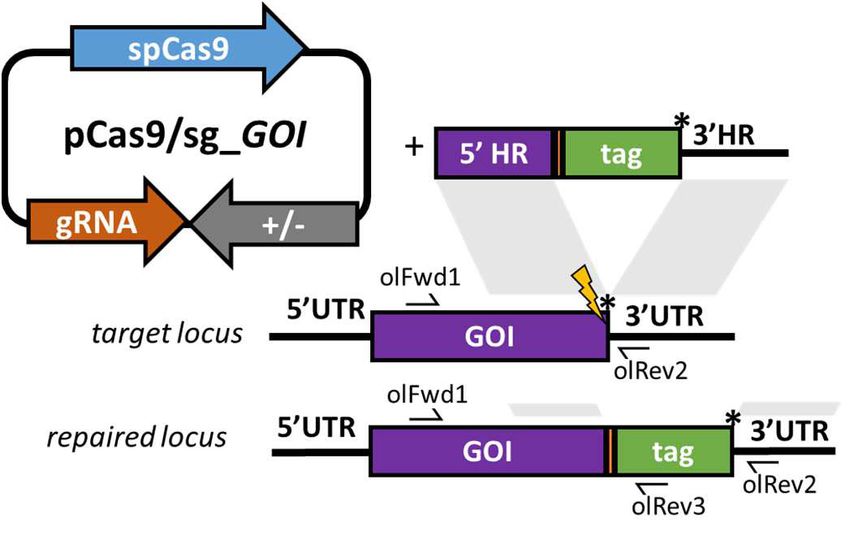
Tools and resources                                                  Genetics and Genomics Microbiology and Infectious Disease

                                   significantly higher in P. knowlesi than P. falciparum (Grüring et al., 2014), we reasoned that we
                                   may be able to use a single positive drug selectable marker to cover all the required components for
                                   editing. Pairing the guide and Cas9 cassette on a single ‘suicide’ plasmid (Lu et al., 2016) with posi-
                                   tive and negative selection cassettes would allow for indirect selection of a separate plasmid con-
                                   taining the repair template, as only parasites that took up the repair template as well as the Cas9
                                   plasmid would be able to repair the DSB. A similar approach to this has been used successfully in P.
                                   falciparum and has allowed the generation of lines entirely free of resistant cassettes after dilution
                                   cloning (Knuepfer et al., 2017). Supporting this approach, co-transfection of plasmids expressing
                                   eGFP or mCherry revealed that ~30% of P. knowlesi transgenic parasites took up both plasmids,
                                   although the proportion expressing both declined rapidly in the following days (Figure 1—figure
                                   supplement 1A). Our two-plasmid CRISPR-Cas9 system comprises one plasmid (pCas9/sg) that pro-
                                   vides Cas9, sgRNA (driven by the PkU6 promoter) and a hDHFR-yFCU fusion product for positive/
                                   negative selection, and a second plasmid (pDonor) providing the donor DNA with homology regions
                                   (HRs) flanking the DSB for repair by homologous recombination. To test this system, we designed
                                   constructs to integrate an eGFP expression cassette into the non-essential p230p locus (Figure 1A
                                   and Figure 1—figure supplement 1C). A 20 bp guide sequence targeting a seed sequence
                                   upstream of a protospacer adjacent motif (PAM) within the p230p gene was cloned into pCas9/sg
                                   (pCas9/sg_p230p), and a repair template plasmid was synthesized by including an eGFP expression
                                   cassette flanked by 400 bp HRs targeting either side of the PAM sequence (pDonor_p230p). Both
                                   plasmids (each 20 mg) were combined and introduced into P. knowlesi schizonts via electroporation
                                   (Moon et al., 2013) along with control transfections (pCas9/sg without guide sequence and repair
                                   template). To simplify synchronisation of parasites, the transfection procedure was altered to addi-
                                   tionally include a 2 hr incubation of purified schizonts with 1 mM of the schizont egress inhibitor com-
                                   pound 2, immediately prior to transfection. This compound reversibly inhibits the cGMP-dependent
                                   protein kinase (PKG) (Collins et al., 2013) and facilitates accumulation of the fully segmented forms
                                   required for transfection. Parasites were placed under selection with pyrimethamine for 5 days after
                                   transfection and successful integration monitored by PCR. Correct integration at the p230p locus
                                   was detectable by PCR within 3 days of transfection and only low levels of wild type DNA was
                                   detectable after day 11 (Figure 1B). Expression of eGFP was confirmed by live microscopy
                                   (Figure 1C). The eGFP positivity rate was calculated the day after transfection (day 1), to evaluate
                                   transfection efficiency (8.4%±2.1 SD). In P. knowlesi, expression of GFP can be detected in any para-
                                   sites that take up the eGFP cassette after transfection, regardless of whether on an episomal or line-
                                   arised construct. This indicates that 8.4% of parasites successfully take up the construct, but only a
                                   very small fraction of these are likely to be integrated at this stage. Only the plasmid pCas9/sg is
                                   selected for with pyrimethamine, and so the number of parasites with non-integrated linear donor
                                   DNA decreases rapidly over time. The eGFP positivity was assessed again once parasites reached
                                   0.5% parasitemia (day 12), indicating 83.3% (±1.8 SD) of the parasites had integrated the construct
                                   (Figure 1D); Figure 1—source data 1. Parasites transfected with pCas9/sg_p230p without providing
                                   pDonor_p230p were visible in culture several days after the integrated lines. An intact guide and
                                   PAM site was detected in these parasites, suggesting that a small population of parasites did not
                                   form DSB. Parasites transfected with pCas9/sg without a cloned sgRNA appeared in culture within a
                                   few days after transfection, with comparable growth rates to the eGFP plasmid, suggesting the Cas9
                                   expression without a targeting sgRNA is not toxic (Figure 1E); Figure 1—source data 1. Integrated
                                   lines were grown for one week before negative selection with 5-Fluorocytosine and subsequent limit-
                                   ing dilution cloning. Clones were identified using a plaque-based assay (Figure 1—figure supple-
                                   ment 1B) previously used for P. falciparum (Thomas et al., 2016), and 10/10 genotyped clone’s
                                   harboured correctly integrated, markerless eGFP (Figure 1F).

                                   A three-step PCR method enables rapid, cloning-free generation of
                                   donor constructs
                                   P. knowlesi readily accepts linearised plasmids for homologous recombination (Moon et al., 2013;
                                   Kocken et al., 2002; Moon et al., 2016), so we next tested whether we could use a PCR-based
                                   approach for scalable generation of repair templates. As no selectable marker is used within the
                                   repair template, this could be easily produced by using PCR to fuse 5’ and 3’ HRs with the region
                                   containing the desired insertion, dispensing with the need for a plasmid back-bone. Modifying a
                                   method used for homologous recombination in P. berghei (Ecker et al., 2006), we developed a

Mohring et al. eLife 2019;8:e45829. DOI: https://doi.org/10.7554/eLife.45829                                                        3 of 29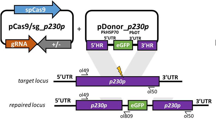
Tools and resources Genetics and Genomics Microbiology and Infectious Disease
QR'1$ &
SNFRQ*)3HS
S&DV
VJHS
H*)3SRVLWLYH
3DUDVLWHPLD
S&DV
VJBSS
S&DV
VJBSS
S'RQRUBSS
D\
D\
'D\VDIWHUWUDQVIHFWLRQ /RJ
'
'
Figure 1. CRISPR-Cas9 genome editing in P.knowlesi. (A) Schematic of CRISPR-Cas9 strategy. Integration of the eGFP expression cassette into the
target p230p locus via homologous recombination. Arrows indicating oligo positions for diagnostic PCRs. (B) Parasites transfected with pCas9/
sg_p230p and pDonor_p230p plasmids were analysed with diagnostic PCRs on consecutive days after transfection. PCR reactions detecting the wild
type locus (ol49 +ol50), integration locus (ol01 +ol50) and a control PCR targeting an unrelated locus (ol75 +ol76) using approximately 3 ng/ml genomic
DNA. For each day, three transfections are shown. (C) Representative live microscopy image of eGFP positive schizont transfected with pCas9/
sg_p230p and pDonor_p230p plasmids. Scale bar represents 5 mm. (D) Proportion of eGFP positive parasites (%) counted after transfection with pCas9/
sg_p230p and pDonor_p230p plasmids to show transfection efficiency on day one and integration efficiency after culture reached 0.5% parasitemia (day
12) (n = 3). Error bars denote ±1 SD. (E) Graph shows change in parasitemia (%) over time for parasite lines transfected with the dual plasmid Cas9
targeting vectors (pCas9/sg_p230p and pDonor_p230p), controls without an sgRNA (pCas9/sg), without homology repair template DNA (pCas9/
sg_p230p) or with no DNA. A fifth control reaction shows outgrowth of an episomal control plasmid (pkconGFPep) (n = 3). Parasites were placed under
drug selection on day 1. Error bars denote ±1 SD (F) Parasites transfected with pCas9/sg_p230p and pDonor_p230p plasmids were cloned by limiting
dilution and four clones analysed by diagnostic PCR.
DOI: https://doi.org/10.7554/eLife.45829.002
The following source data and figure supplement are available for figure 1:
Source data 1. Source data for graphs.
DOI: https://doi.org/10.7554/eLife.45829.004
Figure supplement 1. P.knowlesi dual plasmid uptake and plasmid map of pCas9/sg.
DOI: https://doi.org/10.7554/eLife.45829.003
three-step PCR scheme which first amplified the eGFP cassette and 400 bp HRs with eGFP cassette
adaptors separately, with the second and third reactions fusing each HR to the eGFP cassette in turn
(Figure 2A). The addition of nested primers for the second and third PCR step removed background
bands and improved robustness. The final PCR construct (HR1-eGFPcassette-HR2) was transfected
along with the pCas9/sg_p230p plasmid (Figure 2—figure supplement 1A), and resultant parasite
lines demonstrated integration by PCR (Figure 2B), and an eGFP positivity rate of 74% (±8 SD),
Mohring et al. eLife 2019;8:e45829. DOI: https://doi.org/10.7554/eLife.45829 4 of 29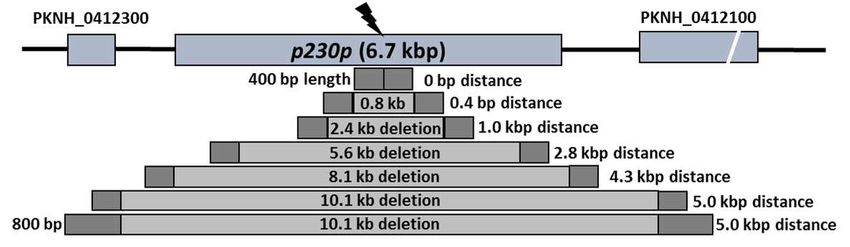
Tools and resources                                                                                                Genetics and Genomics Microbiology and Infectious Disease


                                                                                                                                                                         QV


                                                                                                                                      H*)3SRVLWLYH


                                                                                                                                                                LG

                                                                                                                                                                               5
                                                                                                                                                                          3&
                                                                                                                                                               P
                                                                                                                                                             DV
                                                                                                                                                           3O
                                                                                                                                                                                                                                                    QV   QV
                                                                                                                          QV

                                                                                 'D\VWR3DUDVLWHPLD
                                                                                                                                                                                       &


                                                                                                                                                                                          H*)33RVLWLYH

                                                                                                                                                                QV
                           ƉϮϯϬƉ ;ϲ͘ϳŬďƉͿ
                                                                                                                                                                         QV
                     ϱϬďƉ ůĞŶŐƚŚ    ϬďƉ ĚŝƐƚĂŶĐĞ

                    ϭϬϬďƉ ůĞŶŐƚŚ
                   ϮϬϬďƉ ůĞŶŐƚŚ

                 ϰϬϬďƉ ůĞŶŐƚŚ
                ϴϬϬďƉ ůĞŶŐƚŚ
          ϭϲϬϬďƉ ůĞŶŐƚŚ                                                                                        /HQJWKRIKRPRORJ\UHJLRQ ES                                                                        /HQJWKRIKRPRORJ\UHJLRQ ES
                                                                                                                                                             QV

                                                                             'D\VWR3DUDVLWHPLD
    '                                                                    ,                                                                                                               /


                                                                                                                                                                                            H*)33RVLWLYH


                                                                                                                                                                                                                                  QV                                      QV


                                                                                                                                                                    ES                                                               ES

                                                                                                                          'LVWDQFHIURP'6% NES                                                                                  'LVWDQFHIURP'6% NES

Figure 2. Fusion PCR based approach enables cloning-free production of homology repair templates and evaluation of key parameters for efficient
homology-driven repair. (A) Schematic of the nested PCR method to generate linear donor constructs for transfection. First, homology regions (HRs),
with eGFP adaptors in primers ol36 and ol37 and eGFP cassette were amplified by PCR with small overhangs (OH) and gel extracted. In a second
nested step 5’HR and eGFP cassette were fused and again in the third step the 5’HR-eGFP product was fused with 3’HR. (B) Parasites transfected with
pCas9/sg_p230p and PCR repair template (PCR donor), comprised of an eGFP cassette and 400 bp HRs, were analysed with diagnostic PCRs
amplifying the wild type p230p locus (ol49 +ol50), integration locus (ol01 +ol50) and a control targeting an unrelated locus (ol75 +ol76). (C) After
selection for integration, the proportion of eGFP positive parasites (%) was determined by fluorescent microscopy and compared between Cas9
transfections made with 400 bp HR plasmid (pDonor_p230p) or 400 bp HR PCR donor DNA. Data points represent the mean and error bars indicate ±1
SD of two biological independent experiments (n = 2). (D) The p230p locus was targeted using PCR donor DNA constructs using HRs with 50–1600 bp
length. The bar chart shows, for each of the constructs with HRs of 50 to 1600 bp length, (E) the number of days for transfections to reach 1%
parasitemia and (F) proportion of eGFP positive parasites (%) after selection. All transfections were carried out in two biological independent
experiments (n = 2). (G) The p230p locus was targeted using PCR donor DNA constructs with HRs placed at varying distance from the Cas9 induced
double strand break (DSB). For each construct based on distance to the DSB, the bar chart shows, (H) the number of days for transfections to reach 1%
parasitemia and (I) proportion of eGFP positive parasites (%) after selection. Data points represent the mean and error bars denote ±1 SD of two
biological independent experiments (n = 2). Results were all compared to the 400 bp HR construct at 0 kb from DSB as the control using a one-way
ANOVA with Dunnett’s multiple comparison of means. ns p>0.05, *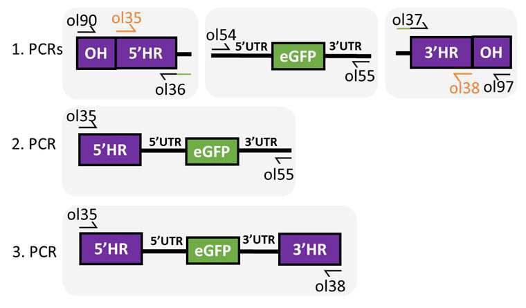
Tools and resources                                                 Genetics and Genomics Microbiology and Infectious Disease

                                   Longer HRs increase the integration efficiency and offsets DSB distance
                                   efficiency loss
                                   We next used this PCR approach to investigate the optimal parameters and limits of the Cas9 sys-
                                   tem in P. knowlesi. Varying the length of HRs targeting the same p230p locus (Figure 2D), allowed
                                   us to determine the effect on integration efficiency as well as the size limits of the PCR approach.
                                   The largest construct generated in this way was 6.1 kb in length (2  1.6 kb HRs flanking the 2.9 kb
                                   eGFP expression cassette). Attempts to generate a larger 9.3 kb construct (2  3.2 kb HRs) failed
                                   during the final PCR step. PCR yields were lower for larger constructs, with the 6.1 kb construct
                                   yielding half that of the 3.7 kb construct. PCR repair templates with HRs ranging from 50 to 1600 bp
                                   generated single specific bands with exception of the 400 bp HRs which contained an additional
                                   lower band, due to a primer additionally annealing to a repeat region in HR1 (Figure 2—figure sup-
                                   plement 1B). The PCR constructs were transfected together with the pCas9/sg_p230p plasmid and
                                   integration efficiency monitored. All 6 HR lengths produced evidence of integration by PCR, but the
                                   efficiency rapidly declined with HRs shorter than 400 bp (Figure 2—figure supplement 1D).
                                       Parasites transfected with 800 and 1600 bp HR constructs were the fastest to reach 1% parasite-
                                   mia on day 12 and 9 post transfection, respectively (Figure 2E); Figure 2—source data 1. For the
                                   50 and 100 bp HR constructs no eGFP positive parasites were detected by fluorescence microscopy
                                   suggesting very low targeting efficiencies. Constructs with HRs > 400 bp provided GFP positivity
                                   ranging from 79% and 81% (Figure 2F); Figure 2—source data 1, which taken together with PCR
                                   yields and transfection recovery time suggest an optimal HR length of at least ~800 bp.
                                       To undertake large gene deletion or replacement experiments, HRs may need to be placed at a
                                   distance from the Cas9-induced DSB, and it is well known in other systems that efficiency rapidly
                                   declines with distance to DSB (Byrne et al., 2015). To determine how distance from DSB affected
                                   efficiency of integration, we used the same p230p PAM site and moved our 400 bp HRs varying dis-
                                   tances away from the DSB, ranging from 0 to 5 kb (Figure 2G). PCR repair templates with HRs
                                   showed good yields, but again contained an additional lower band for the HRs furthest away from
                                   the double strand break (5 kb) (Figure 2—figure supplement 1C).
                                       Whilst all transfections were PCR positive for integration and reached 1% parasitemia at similar
                                   times (14–20 days) (Figure 2—figure supplement 1E, Figure 2H and Figure 2—source data 1), the
                                   integration efficiency declined with distance from DSB. This decline was surprisingly small, with HRs
                                   placed even 5 kb away from either side of the DSB yielding a 14% (±18 SD) integration efficiency
                                   (Figure 2I). Interestingly, we found that extending HR length to 800 bp restored integration efficien-
                                   cies to 54.8% (±8.7 SD) at a 5 kb distance from DSB (Figure 2I; Figure 2—source data 1). Thus, HR
                                   length can directly offset efficiency losses due to distance from DSB and this system can readily
                                   remove genes at least as large as 10 kb in size from a single PAM site, accounting for ~98% of genes
                                   in the P. knowlesi genome (Pain et al., 2008). All primer pairs for template generation and diagnos-
                                   tic PCRs are shown in Figure 2—source data 2 and primer sequences are listed in Figure 5—source
                                   data 2.

                                   Cas9-based PCR constructs enable rapid and flexible gene tagging in P.
                                   knowlesi
                                   Having demonstrated consistent performance of an sgRNA sequence in the sgRNA/Cas9 suicide
                                   vector and PCR constructs for targeting a single control locus, we next sought to determine how
                                   robust the system is for targeting a range of loci. We therefore used the PCR-based approach for
                                   fusion of fluorescent or epitope tags to proteins of interest (Figure 3A). For C-terminal tags, the
                                   PCR repair templates were generated by creating fusions of the tag with HRs targeting the 3’end of
                                   the gene and the 3’UTR. Similarly, N-terminal tag repair templates were created by flanking the tag
                                   with HRs targeting the 5’UTR and 5’end of the coding region. In each case a PAM site was selected
                                   so that the 20 bp guide sequence crossed the stop codon (for C-terminal) or start codon (for N-ter-
                                   minal) such that integration of the tag alone, with no other exogenous sequence, was sufficient to
                                   disrupt the guide sequence. For genes, such as the Chloroquine Resistance Transporter (CRT), where
                                   the PAM site preceded the stop codon, intervening sequences were recodonised when generating
                                   the 5’HR to disrupt the PAM site using silent mutations. We selected five genes with disparate sub-
                                   cellular locations and functions to test this approach: the micronemal protein apical membrane anti-
                                   gen 1 (AMA1) (Bannister et al., 2003), rhoptry neck protein 2 (RON2) (Cao et al., 2009), inner

Mohring et al. eLife 2019;8:e45829. DOI: https://doi.org/10.7554/eLife.45829                                                       6 of 29
Tools and resources                                                         Genetics and Genomics Microbiology and Infectious Disease


Figure 3. CRISPR Cas9 PCR repair templates enable rapid and flexible tagging of parasite proteins. (A) Schematic of CRISPR-Cas9 system for
C-terminal tagging. pCas9/sg plasmid with gene of interest (GOI) specific sgRNA, is combined with repair template generated by fusion PCR. Lightning
bolt indicates Cas9 induced double strand break, which is repaired by insertion of the desired tag. (B) Diagnostic PCRs specific to each GOI locus were
carried out to amplify the wild type locus (schematic positions olFwd1 +olRev2), integration locus (schematic positions olFwd1 +olRev3) and a control
targeting an unrelated locus (ol75 +ol76). Specific primers used for each GOI is shown in Figure 3—figure supplements 1A–E, 2. As no DNA is
removed in this process, the wild type specific locus primers also generate slightly larger amplicons in tagged lines, which can be seen as double bands
for both the Myosin A and K13 PCRs. (C) Representative immunofluorescence images of HA-tagged Apical membrane antigen-1 (AMA1-HA) and
Rhoptry neck protein 2 (RON2-HA) parasite lines, and live cell imaging of Chloroquine Resistance Transporter-eGFP (CRT-eGFP), Myosin A-eGFP
(MyoA-eGFP) and mCherry-Kelch13 (K13). Panel shows brightfield (BF), DNA stain (blue) and anti-tag antibodies/live fluorescence (green or red) of
schizont stage parasites from each line. Scale bars represent 2 mm.
DOI: https://doi.org/10.7554/eLife.45829.009
The following source data and figure supplements are available for figure 3:
Source data 1. Primer pairs and guide sequences for generation and analysis of tagged parasite lines.
DOI: https://doi.org/10.7554/eLife.45829.012
Figure supplement 1. CRISPR-Cas9 tagging of P.knowlesi proteins.
DOI: https://doi.org/10.7554/eLife.45829.010
Figure supplement 2. Comparison of P. knowlesi and P. falciparum 3D7 genes.
DOI: https://doi.org/10.7554/eLife.45829.011

                                     membrane complex protein myosin A (MyoA) (Baum et al., 2006), digestive vacuole membrane pro-
                                     tein involved in drug resistance CRT (Ehlgen et al., 2012), and a protein involved in artemisinin resis-
                                     tance in cytoplasmic foci Kelch13 (K13) (Birnbaum et al., 2017). A single sgRNA was selected for
                                     each, and repair templates were generated by fusion PCR to incorporate an eGFP, mCherry (both
                                     with 24 bp glycine linker) or a hemagglutinin (HA) tag (Figure 3—figure supplement 1A–E). An
                                     N-terminal tag was used for K13, as previous work in P. falciparum suggested that C-terminal tag-
                                     ging affected parasite growth (Birnbaum et al., 2017), and C-terminal tags used for all the other tar-
                                     gets. All lines grew up quickly after transfection, reaching 1% after between 8 and 15 days, and PCR

Mohring et al. eLife 2019;8:e45829. DOI: https://doi.org/10.7554/eLife.45829                                                                     7 of 29
Tools and resources Genetics and Genomics Microbiology and Infectious Disease
analysis indicated that correct integration had occurred (Figure 3B). All primers used for generating
repair templates and for diagnostic PCRs are shown in Figure 3—source data 1 and Figure 5—
source data 2.
Whilst it is, to our knowledge, the first time each of these proteins have been tagged in P. knowl-
esi, all demonstrated localisation patterns were consistent with previous reports for P. falciparum
(Figure 3C). AMA1, MyoA and K13 showed clear bands at the expected size on western blots. The
CRT-eGFP fusion protein showed a band at ~50 kDa, in line with work in P. falciparum which showed
CRT-eGFP migrates faster than its predicted size of 76 kDa (Figure 3—figure supplement 1F)
(Ehlgen et al., 2012). We were unable to visualise a band for RON2-HA most likely due to poor blot-
ting transfer of this 240 kDa protein. Together, these results demonstrate that the fusion PCR
approach can be used to tag P. knowlesi genes rapidly and robustly at a variety of loci. Analysis of
equivalent P. falciparum loci revealed only 2/5 had suitably positioned PAM sites, and equivalent
UTR regions had an average GC-content of only 11.8% (36% for P. knowlesi), suggesting a similar
approach would have been more challenging in P. falciparum (Figure 3—figure supplement 2). All
sgRNA sequences with predicted on- and off-target scores that successfully targeted a gene of
interest in this study are shown in Figure 3—source data 1.
Transgenic P. knowlesi orthologue replacement lines provide
surrogates for P. vivax vaccine development and DBP tropism studies
Having demonstrated the utility of this technique for rapidly manipulating genes of interest, we next
sought to use this system to study P. vivax biology. The orthologous RBC ligands PkDBPa and
PvDBP, mediate host cell invasion by binding to the DARC receptor on human RBCs in P. knowlesi
and P. vivax, respectively (Adams et al., 1990; Horuk et al., 1993; Singh et al., 2005; Miller et al.,
1975). PvDBP is currently the lead candidate for a P. vivax blood stage vaccine (Ntumngia et al.,
2012; Payne et al., 2017a; Singh et al., 2018), thus P. knowlesi could provide an ideal surrogate for
vaccine testing in the absence of a robust in vitro culture system for P. vivax. Whilst likely functionally
equivalent, the DBP orthologues are antigenically distinct (~70% amino acid identity in binding
region II) so we used genome-editing to generate transgenic P. knowlesi parasites in which DARC
binding is provided solely by PvDBP. The donor DNA constructs required to fully reconstitute the
DBP coding regions were large and UTR and coding sequences for each of the three PkDBP
paralogues highly similar at the nucleotide level. Therefore, HRs were amplified from genomic DNA
and cloned into a plasmid vector containing the recodonized PkDBPa or PvDBP genes rather than
using the 3-step PCR to generate repair templates (Figure 4—source data 2). This allowed us to
verify amplification of the correct DBP locus and avoid any chance of mutations within the 4.3 kb
sized template DNA. We first carried out an orthologue replacement (OR) of the full-length PkDBPa
with PvDBP in the P. knowlesi A1-H.1 line (PvDBPOR) – using a recodonised synthetic PvDBP gene
flanked by HRs targeting the 5’ and 3’UTRs of the PkDBPa gene (Figure 4—figure supplement 1A).
Once integrated, this deletes the PkDBPa gene and places the PvDBP gene under control of the
PkDBPa regulatory sequences, enabling a precisely matched expression profile. As a control we also
exchanged PkDBPa with a recodonised PkDBPa gene (PkDBPaOR) using the same sgRNA. Success-
ful integration was readily achieved and limiting dilution cloning resulted in 100% integrated clones
for PkDBPaOR and 40% for PvDBPOR (Figure 4A). The PkA1-H.1 line relies on the DARC receptor for
invasion of human RBCs (Moon et al., 2013) and PkDBPa is required to mediate this interaction
(Singh et al., 2005), thus the successful replacement indicates that the Pv orthologue can comple-
ment its role in DARC binding sufficiently well to maintain growth.
P. knowlesi contains two DBPa paralogues, DBPb and DBPg, which are highly homologous at the
nucleotide (91–93% identity) and amino acid (68–88% identity) levels, but are thought to bind to dis-
tinct sialic acid-modified receptors unique to macaque RBCs (Dankwa et al., 2016). The PkDBPa
sgRNA was carefully designed to be distinct to equivalent DBPb and DBPg target sequences (85%
identical to DBPg and 47.8% to DBPb), because, as in other systems, off-target Cas9-induced DSBs
are a major issue (Figure 4—figure supplement 1D) (Zischewski et al., 2017; Wagner et al.,
2014). We therefore sequenced the four most similar target sequences, including one in DBPg, in
the PvDBPOR lines (Figure 4—figure supplement 2) and did not detect any off-target mutations,
suggesting that as for other malaria parasites (Ghorbal et al., 2014) the absence of non-homolo-
gous end joining ameliorates the potential for off-target mutations. However, diagnostic PCRs for
DBPb failed, as well as PCRs in genes flanking the DBPb locus. Whole genome sequencing and
Mohring et al. eLife 2019;8:e45829. DOI: https://doi.org/10.7554/eLife.45829 8 of 29Tools and resources Genetics and Genomics Microbiology and Infectious Disease
QV QV
QV
QV QV QV
K5%&
QV QV
)ROGPXOWLSOLFDWLRQ
)ROGPXOWLSOLFDWLRQ
P5%&
QV
QV QV
3Y 3D 2 5'
% '
3Y 3D 2 5
% '
3N Y' D 25 7
3N Y' D 25 7
' J
' J
'J
'J
'J
'J
7
5 '
5 '
2
5
2
5 '
:
:
:
3 2
3 2
' %3 '
' %3 '
'
'
5
5
2
32
3 3
3 3
3D
%
%
%
'
'
%
%
'
'
%
'
3N
3N
'
3Y
3N
& '
:7
3N'%3D 25''J 3Y'%325''J
)ROGPXOWLSOLFDWLRQ
)ROGPXOWLSOLFDWLRQ
)ROGPXOWLSOLFDWLRQ
)\$ )\% )\$)\% )\$ )\% )\$)\% )\$ )\% )\$)\%
'XII\3KHQRW\SH 'XII\3KHQRW\SH 'XII\3KHQRW\SH
Figure 4. PvDBP expressing P. knowlesi line demonstrates preference for growth in human RBCs but no preference for different Duffy haplotypes. (A)
The P. knowlesi Duffy binding protein a (DBPa) gene was targeted for replacement with either a recodonised PkDBPa or P. vivax DBP repair template.
Sequencing revealed a loss of ~44 kb in chromosome 14, including loss of PkDBPb (PkDBPaOR/D14 and PvDBPOR/D14). These lines were then
subsequently modified to knockout PkDBPg (PkDBPaOR/D14Dg and PvDBPOR/D14Dg). Parasite lines were analysed using PCR reactions detecting the
wild type (WT) locus PkDBPa (ol186 +ol188), orthologue replacement (OR) locus of PkDBPaOR (ol186 +ol189) or PvDBPOR (ol186 +ol187), WT PkDBPb
locus (ol480 +481), WT locus of PkDBPg (ol483 +ol484), KO locus of PkDBPg (ol483 +ol258) and a control PCR targeting an unrelated locus (ol75 +ol76).
(B) The PkDBPaOR line was modified to knockout PkDBPb (PkDBPaOR/Db). Parasite lines including the transfection line (TF) and three clones were
analysed using PCR reactions detecting WT locus of PkDBPb (ol480 +ol481), KO locus of PkDBPb (ol284 +ol481) and a control PCR targeting an
unrelated locus (ol75 +ol76). (C) Bar chart showing mean fold replication of parasites lines in FACS-based multiplication assays over one growth cycle
(24 hr). Assays were carried out in eight biological independent experiments for human blood (hRBC) and three biological independent experiments for
Macaca fascicularis blood (mRBC). Data points represent mean growth rates and error bars denote ±1 SD. Replication rates of the parasite lines were
compared by using one-way ANOVA with Tukey’s multiple comparisons test of means. There are significant differences in fold multiplication rates of
WT against PkDBPaOR/D14Dg in hRBCs (pTools and resources Genetics and Genomics Microbiology and Infectious Disease
Figure 4 continued
DOI: https://doi.org/10.7554/eLife.45829.019
Figure supplement 1. Transgenic P. knowlesi DBP orthologue replacement, knockout design and genotypic analysis.
DOI: https://doi.org/10.7554/eLife.45829.014
Figure supplement 1—source data 1. Source data for graphs.
DOI: https://doi.org/10.7554/eLife.45829.015
Figure supplement 2. Off-target guide sequences for PkDBPa sgRNA.
DOI: https://doi.org/10.7554/eLife.45829.016
Figure supplement 3. Sequencing of PvDBPOR/D14Dg parasite line.
DOI: https://doi.org/10.7554/eLife.45829.017
mapping against the A1-H.1 reference genome revealed that the PkDBPaOR and PvDBPOR line have
a ~ 44 kb truncation at one end of chromosome 14 (Figure 4—figure supplement 3), which also har-
bours DBPb, therefore we renamed the lines PkDBPaOR/D14 and PvDBPOR/D14. The loss of the ~44
kb of chromosome 14 is also present in parasites that have been transfected simultaneously with
pCas/sg_p230p, suggesting that the 44 kb deletion occurred in the A1-H.1 parental parasite line
(wt/D14) prior to transfection and was not an artefact caused by targeting DBPa. Similar spontane-
ous deletions have been reported previously, including ~66 kb loss at the other end of chromosome
14 in the P. knowlesi A1-C line maintained in cynomolgus macaque blood that included the invasion
ligand NBPXa (Moon et al., 2016), and a deletion of DBPg in the PkYH1 line at the end of chromo-
some 13 (Dankwa et al., 2016). Furthermore, the PAM site of the DBPa targeting guide sequence
is absent in DBPb (Figure 4—figure supplement 1D) which makes it unlikely that the disruption of
DBPb was induced by Cas9 during DBPa targeting. To confirm this, another PkDBPaOR clonal line
was generated in an independent transfection using the A1-H.1 parental parasite line with intact
DBPb locus.
Having established accurate targeting of the PkDBPa locus, we investigated the role of the
paralogues in human and macaque red cell invasion and whether they could interfere with inhibitory
effects of test antibodies. Reasoning that as the D14 truncation may have provided a selective advan-
tage to the parasites and thus may well reoccur, we took advantage of the spontaneous DBPb loss
(PkDBPaOR/D14 or PvDBPOR/D14) and then used pCas9/sg plasmid recycling to additionally delete
the DBPg locus (Figure 4—figure supplement 1B), generating PkDBPaOR/D14Dg and PvDBPOR/
D14Dg. An overview of all generated DBP lines is depicted in Figure 4—figure supplement 1E).
The final PvDBPOR/D14Dg clonal line was subjected to whole genome sequencing to verify
changes at all three loci, and this confirmed precise targeting of the PvDBP allele swap into the
PkDBPa locus, and complete deletion of the DBPg open reading frame in the PkDBP g locus (Fig-
ure 4—figure supplement 3). Using the PkDBPaOR clonal line with the start of chromosome 14
intact, we were able to generate a Cas9 mediated DBPb knockout (PkDBPaOR/Db) (Figure 4B and
Figure 4—figure supplement 1C). FACS-based multiplication assays showed no growth effects of
PkDBPb knockout in human blood (Figure 4—figure supplement 1F).
Analysis of the wild type line, and the four transgenic lines (PkDBPaOR/D14, PvDBPOR/D14,
PkDBPaOR/D14Dg and PvDBPOR/D14Dg) revealed no difference in growth rate in human RBCs for all
lines except the PkDBPaOR/D14Dg which demonstrated significantly increased growth rate compared
to wild type (Figure 4C and Figure 4—source data 1). This confirmed that the P. vivax protein was
able to complement the role of its P. knowlesi orthologue. It also demonstrated, that not only are
PkDBPb and g proteins dispensable for multiplication in humans RBCs (Dankwa et al., 2016), but as
their loss leads to increased multiplication rate, they may actually impede invasion in human RBCs. A
further experiment comparing multiplication rates in 21 Duffy positive blood donations revealed sig-
nificant higher growth rates of the PkDBPaOR/D14Dg line (8.9 fold) compared to the 6.9 fold multipli-
cation of the wild type line and PvDBPOR/D14Dg line (pTools and resources Genetics and Genomics Microbiology and Infectious Disease
hypothesized that the additional invasion pathways provided by DBPb and DBPg are in part respon-
sible for the increased invasion efficiency in macaque RBCs (Dankwa et al., 2016). Interestingly, loss
of DBPb and DBPg in the PkDBPaOR/D14Dg line did not reduce the parasite multiplication rate in
macaque RBC but rather slightly increased it (Figure 4C), with the lines retaining a macaque prefer-
ence ratio (macaque fold growth/human fold growth) of 1.43, similar to both the wild type (1.26)
and PkDBPaOR/D14 (1.33). This demonstrates that both PkDBPb and PkDBPg are dispensable for
invasion of macaque RBCs, and PkDBPa is sufficient to retain full growth rate in macaque cells.
Unlike P. knowlesi, P. vivax is unable to infect macaques, and sequence differences between the
DARC receptor in the two hosts have been suggested to underlie this restriction (Tachibana et al.,
2015). Whilst in macaque RBCs the multiplication rates and host preference ratio (1.16) were unaf-
fected for the PvDBPOR/D14 line, the additional deletion of DBPg in the PvDBPOR/D14Dg line
resulted in a 40% reduction in macaque multiplication rates (Figure 4B) which caused a shift to
human RBC preference with a ratio of 0.75. This suggests that in the absence of redundant DBP
pathways, PvDBP is less effective at facilitating invasion of macaque cells than of human cells, but
nevertheless can support invasion of both host cell types.
The Duffy gene exists as three distinct alleles, Fya, Fyb and Fynull. Whilst the Fy null phenotype
has long been established to protect individuals from P. vivax/P. knowlesi infection (Miller et al.,
1975; Miller et al., 1976), previous studies have also shown stronger binding of recombinant
PvDBPRII to Fy(a b+) and Fy(a+b+) human RBCs compared to Fy(a+b ) as well as higher risks of clinical
episodes of P. vivax malaria in Fy(a b+). This suggests that P. vivax may invade to Fy(a+b ) RBCs less
efficiently (King et al., 2011; Fong et al., 2018; Kano et al., 2018). Whilst we have previously been
unable to detect any Fy subtype preference in P. knowlesi A1-H.1 (22), the PvDBPOR lines provided
an opportunity to see if multiplication rate was sensitive to the Duffy subtype when the parasites
used PvDBP for invasion. Schizonts of the wild type, PkDBPaOR/D14Dg and PvDBPOR/D14Dg lines
were added in duplicate to washed RBC from 21 volunteers of the three Duffy-positive phenotypes
(7x Fya, 9x Fyb, and 5x Fy(a+b+)) at 0.5% parasitaemia. Parasites were maintained in 96-well microtiter
plates for 24 hr and the parasitaemia monitored by FACS. For all three lines the average growth
rates in each of the three Fy subtypes were similar and there was no significant difference between
growth in Fya and Fyb RBCs (Figure 4E–G; Figure 4—source data 1).
Growth inhibition activity (GIA) assays revealed that all lines remained equally susceptible to inva-
sion inhibition by both an anti-DARC camelid nanobody CA111 and a polyclonal aPkMSP119 anti-
body (Figure 5A). In contrast, purified IgGs from polyclonal rabbit sera raised against PvDBP_RII,
demonstrated low-level GIA activity for wild type and PkDBPaOR/DbDg lines (~30% inhibition at 10
mg/ml) but a significantly stronger GIA activity against the PvDBPOR/D14 and PvDBPOR/DbDg lines,
reaching a maximum inhibition of ~75% at 10 mg/ml and around 50% at 4 mg/ml (Figure 5B and C).
At 2.5 mg/ml IgG the GIA activity against the PvDBP OR/D14 and PvDBPOR/DbDg lines were
29.6 ± 3.3 and 24.6 ± 2.6% and significantly higher compared to wild type and PkDBPaOR/DbDg lines
(pTools and resources Genetics and Genomics Microbiology and Infectious Disease
A
100
PkDBPaOR/D14
80
PvDBPOR/D14
PkDBPaOR/D14Dg
GIA (%)
60
PvDBPOR/D14Dg
40
20
0
L] L] L] L]
/m /m g/
m m
mg mg m g/
.5 [3 m
[1 .5 [5
C RC [2 G
A G Ig
AR D Ig
i-D ti- 1 19 119
an
t an SP SP
M k M
Pk ti-
P
ti- an
an
B C
PvDBPOR/D14 PvDBPOR/D14Dg
100 100
Anti-PvDBP_RII IgG Anti-PvDBP_RII IgG
Control IgG Control IgG
80 80
WT PkDBPaOR/D14Dg
Anti-PvDBP_RII IgG Anti-PvDBP_RII IgG
60 Control IgG 60 Control IgG
GIA (%)
GIA (%)
40 40
20 20
0 0
10 1 0.1 10 1 0.1
IgG (mg/mL) IgG (mg/mL)
D E
50 WT
**** 50 PkDBPaOR/D14Dg
**** ****
40 PvDBPOR/D14 40 PvDBPOR/D14Dg
****
GIA (%)
30
GIA (%)
**** 30
****
20 20
10 10
0 0
ol
ol
II
II
R
l
ol
II
R
tr
tr
C II
ro
R
P_
R
Pv ntr
P_
ti- Con
on
P_
Pv nt
P_
B
B
C
ti- Co
o
B
D
B
D
D
Pv
D
Pv
ti-
ti-
an
an
an
an
2.5 mg/mL IgG 2.5 mg/mL IgG
Figure 5. Transgenic P. knowlesi orthologue replacement lines provide surrogates for P. vivax vaccine development. (A) Graph showing growth
inhibition activity (GIA, %) of anti-DARC nanobody at 1.5 and 3 mg/ml and anti-MSP119 purified total rabbit IgG at 2.5 and 5 mg/ml on the parasite lines.
Data points represent the mean and error bars denote ±1 SD of triplicate test wells (n = 3). GIAs of each antibody were compared across the parasite
lines by using unpaired one-way ANOVA with Tukey’s multiple comparisons test of means. No significant changes were observed. (B) Graph shows the
Figure 5 continued on next page
Mohring et al. eLife 2019;8:e45829. DOI: https://doi.org/10.7554/eLife.45829 12 of 29Tools and resources Genetics and Genomics Microbiology and Infectious Disease Figure 5 continued % GIA of a dilution series of IgG purified from sera of PvDBP_RII (SalI)-immunized rabbits as well as control IgG from the pre-immunisation sera of the same rabbits against wild type (WT) and PvDBPOR/D14 transgenic P. knowlesi lines and (C) against PkDBPaOR/D14Dg and PvDBPOR/D14Dg lines. Data points represent the mean and error bars denote ±1 SD of five or six replicates. (D) Bar chart showing % GIA of 2.5 mg/ml IgG purified from sera of PvDBPRII (SalI)-immunized rabbits as well as control IgG from the pre-immunisation sera of the same rabbits against wild type (WT) and PvDBPOR/D14 transgenic P. knowlesi lines and (E) against PkDBPaOR/D14Dg and PvDBPOR/D14Dg lines. Bars represent the mean and error bars denote ±1 SD of five or six replicates and were compared by using one-way ANOVA with Tukey’s multiple comparisons test of means. ns p>0.05, *
Tools and resources                                                  Genetics and Genomics Microbiology and Infectious Disease

                                   reaching 1% at only 5 to 6 days suggesting significant differences in targeting efficiency at different
                                   loci and with different gRNAs. Whilst P. knowlesi grows slightly faster (with 3–4 fold per cycle equat-
                                   ing to around 9-fold in 48 hr) than P. falciparum, this still indicates orders of magnitude greater mod-
                                   ification rates than achieved for P. falciparum - which can take 2–6 weeks for transgenic parasites to
                                   be detected in culture depending on the transfection system used (Ghorbal et al., 2014;
                                   Mogollon et al., 2016; Lu et al., 2016; Knuepfer et al., 2017; Wagner et al., 2014). Despite the
                                   relatively fast recovery time it is also clear that as more than 8% of parasites are able to take up
                                   donor DNA during transfection it still takes more than a week for integrated parasites to grow out,
                                   indicating that efficiency of homology mediated repair is still the major bottleneck and the majority
                                   of Cas9 induced DSBs are not successfully repaired.
                                       We systematically tested key parameters associated with successful genome editing and found
                                   increasing HR length enhanced integration efficiency proportionately, a trend seen in both P. falcipa-
                                   rum and P. berghei (Collins et al., 2013; Wagner et al., 2014; MacPherson and Scherf, 2015).
                                   Whilst integration was detected with HRs as short as 50 bp, efficient editing was achieved with HRs
                                   between 200–800 bp. We were also able to examine how distance from the DSB affected editing
                                   efficiency. Whilst in other systems editing efficiency decreases rapidly as the DSB distance increases,
                                   we saw only a steady decline with distance, an effect which could be ameliorated by simply increas-
                                   ing HR length. The use of PCR products as repair templates is particularly well adapted for tagging
                                   and knockout approaches which tend to be relatively short. For relatively large or complex con-
                                   structs, such as those for gene replacement, or those targeting multigenic families conventional clon-
                                   ing may be preferable, as shown here for the PkDBP family.
                                       By applying these techniques to the P. knowlesi and P. vivax DBP family we have been able to
                                   examine the role of these genes in host and reticulocyte preference of the two species. Even after
                                   long-term adaptation to culture with human RBCs, P. knowlesi parasites can retain a strong prefer-
                                   ence for invasion of macaque RBCs (Dankwa et al., 2016; Moon et al., 2016). Both DBPg and DBPb
                                   have been shown to bind to proteins with a distinct sialic acid residue found in non-human primates,
                                   but absent in humans (Dankwa et al., 2016). Deletion of these genes led to a significant increase in
                                   growth rates in human RBCs, and interestingly also a similar, but non-significant, increase in growth
                                   rates in macaque RBCs. This suggests that PkDBPa alone is sufficient to retain the multiplication
                                   capacity and that the PkDBP paralogues are not responsible for the macaque cell preference
                                   retained in the A1-H.1 human adapted line. Whilst likely non-functional for invasion of human RBCs,
                                   both PkDBPg and DBPb would still compete for surface space and potentially some of the same
                                   interacting proteins as DBPa, which may account for the increased growth rate when they are
                                   deleted. The increased growth rate in macaque cells, would suggest that even here, where DBPg
                                   and DBPb are functional, PkDBPa is the preferred and more efficient pathway for invasion in
                                   macaque cells. In vivo, this advantage would be countered by the significant benefits provided by
                                   redundancy both to combat host blood cell polymorphisms and antibody responses. Importantly,
                                   this data suggests that loss of DBPg and DBPb could provide an adaptation route to increased
                                   growth/virulence within human infections. However, to do so the parasites would need to sacrifice
                                   redundancy within its primary macaque hosts – a situation only likely to occur with prolonged human
                                   to human transmission. Further work to analyse the effect of these mutations through long-term par-
                                   asite competition assays will enable us to determine the precise extent of this advantage and deter-
                                   mine how quickly such mutations could move to fixation within a parasite population.
                                       Despite being closely related to P. knowlesi and other macaque infecting species such as P. cyno-
                                   molgi, P. vivax cannot infect macaques and the PvDBP protein has been suggested to play a role in
                                   enforcing this tropism, as key interacting residues are missing within the macaque DARC protein
                                   (Tachibana et al., 2015). P. knowlesi parasites expressing PvDBP in the absence of DBP paralogues
                                   demonstrate a significant reduction in growth in macaque cells, resulting in an overall shift towards
                                   preference for human cells consistent with PvDBP binding macaque DARC less efficiently. Neverthe-
                                   less as the multiplication rate remained quite close to that seen for human RBCs it seems unlikely
                                   that the PvDBP protein alone represents a significant barrier to P. vivax infection of macaques.
                                       Previous work has shown that PvDBP_RII binds more strongly to RBCs expressing the Fyb allele of
                                   the Duffy antigen than Fya, with studies using recombinant protein demonstrating that the binding
                                   efficiency is 50% lower for Fya (Kano et al., 2018). This effect was shown to correlate with a 30–80%
                                   reduced risk of clinical vivax malaria amongst patients with the FyA phenotype (King et al., 2011). A
                                   similar effect of reduced DBP-FyA binding has also been demonstrated using recombinant PkDBPa

Mohring et al. eLife 2019;8:e45829. DOI: https://doi.org/10.7554/eLife.45829                                                       14 of 29
Tools and resources Genetics and Genomics Microbiology and Infectious Disease
(Fong et al., 2018) but experiments to examine this with human RBC culture adapted P. knowlesi
parasites failed to demonstrate any difference in multiplication rates in either Fyb+ or Fya+ RBC
(Moon et al., 2013). Here, we also did not observe a significant association between growth of the
PvDBPOR/D14Dg line and Duffy haplotype. Whilst the size of our dataset cannot rule out relatively
subtle effects on parasite growth rate, which over the course of an infection could become clinically
relevant, it does rule out major growth differences and demonstrates that the invasion process is
very tolerant of even quite large changes in receptor-ligand affinity. Notably, the study first identify-
ing a role for Duffy polymorphisms in P. vivax clinical susceptibility also demonstrated that the Fya+
RBCs demonstrate increased sensitivity to invasion-blocking antibodies, so protective effects may
stem from this rather than a direct effect on invasion efficiency (King et al., 2011; Moon et al.,
2013). Subsequent experiments using these lines could readily test this hypothesis.
Another key difference between the two species is that unlike P. knowlesi, P. vivax has a strict
restriction to invasion of reticulocytes. A second family of RBC binding proteins, known as the reticu-
locyte binding-like proteins (RBPs) have previously been implicated in this tropism. More recently,
the PvDBP protein itself has been implicated with work using recombinant PvDBP_RII suggesting
that whilst DARC is present on both reticulocytes and mature normocytes, changes during red cell
maturation mean that DARC is only accessible to PvDBP binding in young reticulocytes
(Ovchynnikova et al., 2017; Haynes et al., 1988). Here we show that transgenic P. knowlesi para-
sites using PvDBP for invasion have no such restriction, invading human RBCs (which typically contain
less than 0.5% reticulocytes) with the same efficiency as those expressing PkDBPa – thus providing
compelling evidence that PvDBP plays no role in the reticulocyte tropism. Further, recent work
determining that PvRBP2b, which lacks an orthologue in P. knowlesi, binds to the reticulocyte spe-
cific marker CD71 further asserts the RBPs as the key to reticulocyte tropism. Importantly, the ability
to compare and contrast activity of Pk/Pv DBP family members in parasitological assays will provide
a vital new tool to test hypotheses and models arising from studies that have until now relied on
assays using recombinant protein fragments.
Using a combination of binding assays, mutagenesis and structural studies with recombinant
PvDBP_RII, previous work has identified residues involved in DBP-DARC interactions (Choe et al.,
2005; Hans et al., 2005; VanBuskirk et al., 2004), as well as a DBP dimerization domain thought to
drive the stepwise engagement with DARC (Batchelor et al., 2014; Batchelor et al., 2011). Efforts
to develop a P. vivax vaccine to elicit antibodies against the lead candidate PvDBP have predomi-
nantly relied on using ELISA-based assays, which assess the ability of antibodies to block recombi-
nant PvDBP_RII binding to DARC (Shakri et al., 2012). This has successfully identified a range of
binding inhibitory antibodies mapped to residues involved in DARC binding, dimerization as well as
subdomain 3, which is distant from the DARC binding site, demonstrating that multiple mechanisms
of inhibition may be important (Chen et al., 2016; Chootong et al., 2010). However, validation of
these findings in a parasitological assay is critical. Some epitopes identified in recombinant protein
assays may be inaccessible in the context of invasion and it is also possible that not all inhibitory anti-
bodies block receptor binding. DARC-DBP binding is only one step in the multi-step invasion pro-
cess, with subsequent conformational changes and potential downstream signalling roles for the
protein (Batchelor et al., 2011). The full-length DBP antigen is 140 kDa which contains a C-terminal
transmembrane domain and as such structural and biochemical analysis of the protein has almost
exclusively focused on the PvDBP_RII fragment alone. Whilst efforts to standardise ex vivo P. vivax
assays have been successful (Russell et al., 2011), they remain hugely challenging, low throughput
and rely on genetically diverse P. vivax clinical isolates, that are maintained in culture for a only a sin-
gle cycle of RBC invasion. The P. knowlesi PvDBPOR line thus provides an opportunity to interrogate
the function of the full-length protein in an accessible and scalable manner.
Whilst the relationship between in vitro GIA and in vivo protection against malaria challenge has
not always been clear, previous studies have shown that there is a strong correlative link between
activity in the in vitro assay of GIA and in vivo protection from challenge of two different species of
human malaria (P. falciparum and P. knowlesi) in two different non-human primate models (Aotus
monkeys and rhesus macaques) targeting three different merozoite proteins (PfAMA1, PfRH5,
PfMSP1) (Douglas et al., 2015; Mahdi Abdel Hamid et al., 2011; Singh et al., 2006). Importantly,
Douglas et al. in 2015 determined a threshold value for in vitro GIA assay which must be reached
before protection from malaria challenge is afforded (Douglas et al., 2015). Thus, the ability to
Mohring et al. eLife 2019;8:e45829. DOI: https://doi.org/10.7554/eLife.45829 15 of 29You can also read