Omega-3 Versus Omega-6 Polyunsaturated Fatty Acids in the Prevention and Treatment of Inflammatory Skin Diseases - MDPI
←
→
Page content transcription
If your browser does not render page correctly, please read the page content below
International Journal of
            Molecular Sciences

Review
Omega-3 Versus Omega-6 Polyunsaturated Fatty
Acids in the Prevention and Treatment of
Inflammatory Skin Diseases
Anamaria Balić 1 , Domagoj Vlašić 2 , Kristina Žužul 3 , Branka Marinović 1 and
Zrinka Bukvić Mokos 1, *
 1    Department of Dermatology and Venereology, University Hospital Centre Zagreb, School of Medicine
      University of Zagreb, Šalata 4, 10 000 Zagreb, Croatia; jovicanamaria@gmail.com (A.B.);
      branka.marinovic@kbc-zagreb.hr (B.M.)
 2    Department of Ophtalmology and Optometry, General Hospital Dubrovnik, Ulica dr. Roka Mišetića 2,
      20000 Dubrovnik, Croatia; domagojvlasic@yahoo.com
 3    School of Medicine, University of Zagreb, Šalata 3, 10000 Zagreb, Croatia; zuzulkristina@gmail.com
 *    Correspondence: zrinka.bukvic.mokos@gmail.com

 Received: 31 December 2019; Accepted: 21 January 2020; Published: 23 January 2020

 Abstract: Omega-3 (ω-3) and omega-6 (ω-6) polyunsaturated fatty acids (PUFAs) are nowadays
 desirable components of oils with special dietary and functional properties. Their therapeutic
 and health-promoting effects have already been established in various chronic inflammatory and
 autoimmune diseases through various mechanisms, including modifications in cell membrane
 lipid composition, gene expression, cellular metabolism, and signal transduction. The application
 of ω-3 and ω-6 PUFAs in most common skin diseases has been examined in numerous studies,
 but their results and conclusions were mostly opposing and inconclusive. It seems that combined
 ω-6, gamma-linolenic acid (GLA), and ω-3 long-chain PUFAs supplementation exhibits the highest
 potential in diminishing inflammatory processes, which could be beneficial for the management
 of inflammatory skin diseases, such as atopic dermatitis, psoriasis, and acne. Due to significant
 population and individually-based genetic variations that impact PUFAs metabolism and associated
 metabolites, gene expression, and subsequent inflammatory responses, at this point, we could not
 recommend strict dietary and supplementation strategies for disease prevention and treatment that
 will be appropriate for all. Well-balanced nutrition and additional anti-inflammatory PUFA-based
 supplementation should be encouraged in a targeted manner for individuals in need to provide better
 management of skin diseases but, most importantly, to maintain and improve overall skin health.

 Keywords: nutraceuticals; polyunsaturated fatty acids; supplementation; eicosapentaenoic acid;
 docosahexaenoic acid; gamma-linolenic acid; inflammatory skin diseases; atopic dermatitis;
 acne; psoriasis

1. Introduction
      Since 1929, when Burr and Burr first described a syndrome caused by stringent fat reduction in a
diet (which manifested mostly as cutaneous symptoms, such as erythema with scaling, hair loss, itch,
and increased water loss), it became clear that particular fat pay an essential role in skin structure [1,2].
The word “essential” best describes these fats because of the inability of the human organism to
synthesize them, which means they can only be provided through dietary intake. The term essential
fatty acids (EFAs) was formed and referred to two polyunsaturated fatty acids (PUFAs), linoleic acid
(LA) and α-linolenic acid (ALA), initiating acids for the cascade of elongation to very long-chain
PUFAs (more than 22 C-atoms). PUFAs are divided into two families, omega-3 (ω-3) and omega-6

Int. J. Mol. Sci. 2020, 21, 741; doi:10.3390/ijms21030741                            www.mdpi.com/journal/ijms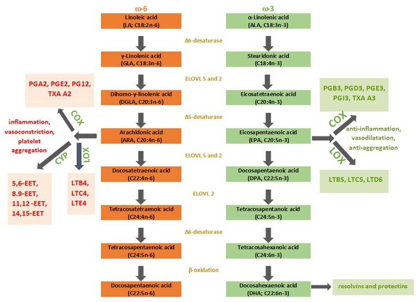
Int. J. Mol. Sci. 2020, 21, 741                                                                                 2 of 26

(ω-6). ω-3 fatty acids (FAs) have in common a terminal carbon-carbon double bond in the omega
three-position, the third bond from the methyl end of the acid, whereas, ω-6 acids have it in the omega
six-position, the sixth bond from the methyl end of the fatty acid, respectively. LA is a member of the
ω-6 family, whereas, ALA is classified as ω-3 PUFA. The double bonds in these EFAs are always in
cis-configuration, which means there are two hydrogen atoms on the same side of the double bond [3].
Salubrious effects of PUFAs might be mediated through various mechanisms, including modifications
in cell membrane lipid composition, gene expression, cellular metabolism, and signal transduction [4].
However, ω-3 and ω-6 FAs have antagonistic effects on metabolic functions in the human organism.
      PUFAs are nowadays desirable components of “specialty oils”, oils with special dietary,
and functional properties that are used as nutraceuticals or cosmeceuticals. Due to the better
understanding of their biological and functional properties, and their health benefits, PUFAs,
specially ω-3 PUFAs are of great importance for health system, due to their potential applications in
disease prevention, but also treatment of the most common chronic inflammatory diseases, including
inflammatory skin diseases, such as atopic dermatitis (AD), psoriasis, and acne. Nowadays, clinicians
have at the disposal useful tools like lipidomics and nutrilipomics that guide them to provide the most
appropriate and individualized FAs supplementation in the treatment of their patients, but also the
prevention of disease in various clinical fields, as well as the field of dermatology [5].

2. Sources of PUFAs
      High dietary intake of ω-6 is typical to the Western diet, which is loaded with processed food
and lacks fish meals as opposed to high red meat dietary intake [6]. Most seed and vegetable oils
(safflower, grape seed, rapeseed, poppy seed, sunflower, palm, hemp, corn, wheat germ, cottonseed,
soybean) which are used in the kitchen are significant sources of ω-6 PUFAs in the form of LA with
low proportions of ω-3 FAs, first to mention—ALA. In contrast to ω-6 FAs, the intake of ω-3 is usually
insufficient because of limited sources. ALA is found in green leafy vegetables, flaxseed, walnuts,
soybean, and canola oils. Their derivatives, eicosapentaenoic acid (EPA) and docosahexaenoic acid
(DHA), are obtained through breast milk and the fish oils, such as salmon, mackerel, sardines, anchovies,
herring, and rainbow trout, but also algae [7–10]. Cold-water marine (wild) fishes are abundant in ω-3
PUFAs, since most of them feed on phytoplankton and zooplankton, a rich ω-3 PUFA source. ω-6
PUFAs, gamma-linolenic acid (GLA) that is typically consumed as part of dietary supplementation,
is found in human milk and some botanical seed oils as borage oil, blackcurrant and evening primrose
oil (EPO), while arachidonic acid (ARA) is obtained from diet rich in organ meats, poultry, and eggs. In
typical Western diets, the ω-6/ω-3 ratio is 15/1 to 16.7/1, although the recommended ratio varies from 1:1
up to 4:1, as recommended by authoritative bodies in Japan (Table 1). In plants and animal-based food
majority of PUFAs are found in the form of triacylglycerols (TAG), phospholipids (PL), diacylglycerols
(DAG), and cholesterol esters (CE). PLs are the most bioavailable because of their aliphatic characteristics,
which lead to better water dispersibility and greater susceptibility [11]. Therefore, krill oils obtained
from Antarctic krill (Euphausia superba), which are fulfilled of ω-3 PUFAs in PL form, are becoming
more popular as a ω-3 supplement. In addition to food supplements, we can now find many new
conventional foods and infant feeding formulas fortified with microalgae and fish DHA [12].

                Table 1. Contents of ω-3 and ω-6 fatty acids in selected plant and animal-based foods.

                                                        ω-3                         ω-6
                             Food Source                                                             Reference
                                              ALA *    EPA *    DHA *     LA *     ARA *    DPA *
                                corn           0.6       -         -      49.83      -         -
                             sunflower        0.33       -         -      49.89      -         -
                                                                                                         [13]
                              soybean         7.6        -         -      51.36      -         -
                             wheat germ       5.3        -         -       55.1      -         -
          Oil
                                   Canola     9.15       -         -      18.65      -         -
                                  Safflower    0.1       -         -      12.72      -         -         [14]
                                  Flaxseed    53.37      -         -      14.33      -         -
Int. J. Mol. Sci. 2020, 21, 741 3 of 26
Table 1. Cont.
ω-3 ω-6
Food Source Reference
ALA * EPA * DHA * LA * ARA * DPA *
Salmon - 13.3 18.23 - - 2.99
Sardine - 10.15 10.66 - - 1.97
Fish oil [14]
Herring - 6.28 4.21 - - 0.62
menhaden - 13.18 8.56 - - 4.92
lettuce, raw 0.15 - - 0.06 - -
Vegetables green broccoli, raw 0.11 - - 0.03 - - [13]
brussels sprouts, raw 0.17 - - 0.08 - -
salmon, raw 0.09 0.89 1.19 0.15 0.05 -
herring, raw 0.19 1.09 1.01 0.22 0.1 -
Fish sardine, raw - 0.51 1.16 0.06 0.04 - [13]
trout, raw 0.1 0.15 0.5 0.37 0.05 -
cod, dried - 0.02 0.62 0.03 0.12 -
lamb, lean meat 0.11 - - 0.11 - -
pork, fat and lean meat,
- - - 1.63 0.03 -
Meat without visible fat [13]
beef, veal, 4 months,
lean meat, without 0.08 - - 0.13 - -
visible fat
chia, dried 17.83 - - 5.84 - - [14]
walnuts, dried 6.64 - - 34.02 - -
Seeds [13]
hazelnuts, dried 0.11 - - 5.09 - -
almond, dried 0.3 - - 10.54 - -
* g/100g; ALA, α-linolenic acid; ARA, arachidonic acid; DHA, docosahexaenoic acid; DPA, docosapentaenoic acid;
EPA, eicosapentaenoic acid; LA, linoleic acid; ω-3, omega-3 fatty acids; ω-6, omega-6 fatty acids.
3. Metabolism of PUFAs
The ω-6 and ω-3 FAs, when consumed in food, are mostly assimilated into TAG and undergo
digestion in the small intestine. The destruction of TAG structure and liberation of FAs allows
absorption and transport in the bloodstream to the tissues where they are incorporated in their
structure. The FAs can take three different metabolic pathways: (a) esterification into cellular lipids as
PL, TAG, CE; (b) beta-oxidation to provide energy for ATP formation; (c) being an initiating structure
for the process of elongation and desaturation through enzymatic reactions to create long-chain PUFAs.
The distribution of FAs into PL is nowadays considered to be the most important factor that drives the
cell membrane composition and consequent cell membrane homeostasis maintenance obtained by
the membrane balance of FAs, but also FAs detachment of the PL for becoming signaling mediators.
Nutrition, metabolism, external and internal stressors with consequent oxidative stress, and lifestyle
factors all cause FAs changes that result in the impaired cell membrane homeostasis. This is why
FAs are nowadays considered as one of the biomarkers of pathological conditions and why it is
important to address the role of fatty acid-based membrane lipidomics, a powerful diagnostic tool for
determining quantitative and qualitative changes of FAs, but also the follow-up of the membrane FAs
remodeling associated with different physiological and pathological conditions [15]. The creation of the
long-chain PUFAs occurs mostly in the liver. Other tissues are active in this process in an insignificant
amount [3]. The most prominent and metabolically significant PUFAs derived from LA are GLA,
dihomo-gamma-linolenic acid (DGLA), and ARA. On the other hand, the ω-3 family is derived from
ALA, and the most notable among them are EPA and DHA. In the enzymatic cascades, eicosanoids are
produced from the latter PUFAs (Figure 1). Eicosanoids are defined as bioactive signaling molecules
with pro-inflammatory and anti-inflammatory characteristics, which are derived from ARA, DGLA,
EPA, and DHA through reactions led by cyclooxygenase (COX-1 and COX-2), lipoxygenase (5-LOX
and 15-LOX) and epoxygenases (cytochrome P450 or CYP). LA and ALA compete in the synthesis of
eicosanoids by targeting the same enzymes, precisely desaturase and elongase enzymes, to become C20
FAs, which are the precursors of eicosanoids—oxygenated derivates of C20 FAs made by oxygenaseInt. J. Mol. Sci. 2020, 21, 741 4 of 26 enzymes. Higher intake of ALA results in the increased production of anti-inflammatory eicosanoids because ω-3 FAs are more favored substrates for desaturase and elongase enzymes than ω-6 FAs in eicosanoid synthesis. LA can be converted to ALA in the majority of higher plants, algae, and fungi because they possess ∆12- and ∆15-desaturase [16]. Nevertheless, in mammals, these enzymes are absent for genetic reasons, which makes humans unable to convert oleic acid (OA) to ALA and LA. This enzyme deficit makes the latter PUFAs essential. The level of EPA and DPA in the organism is mostly determined by direct dietary consumption because most of dietary ALA undergo beta-oxidation in the mitochondria [3]. Desaturation led by ∆6-fatty acid desaturase (FADS) makes the first step in long-chain PUFA synthesis by adding a double bond at the 6th C-C position from the -COOH of LA and ALA generating GLA and stearidonic acid (SDA), respectively. ∆6-FADS is a crucial enzyme in the long-chain (LC)-PUFAs synthesis because it is rate-limiting in animals and humans. The next step is elongation, and DGLA and eicosatetraenoic acid (ETA), respectively, are synthesized. Finally, ∆5-FADS adds a new double bond to the 5th C-C bond from carboxy-end, which results in the generation of ARA and EPA, respectively. Desaturation on the 17th C-C position results in GLA, DGLA, and ARA conversion to SDA, ETA, and EPA, respectively. FADS, crucial enzymes of rate-limiting steps in the LC-PUFAs biosynthesis pathway, are encoded by fatty acid desaturase 1 (FADS1) and 2 (FADS2) genes located on chromosome 11 (FADS cluster). A mammalian organism can synthesize DHA through three steps (two elongations and beta-oxidation) from EPA. This synthesis is also known as the Sprecher pathway [17]. The EPA and DHA formation take place in human hepatoma cells at the highest rate when the ratio 1:1 = ALA: LA is present [18]. The recorded conversion rates were 16% for EPA and 0.7% for DHA, which leads to the conclusion that DHA supplementation is the most effective way to improve body DHA levels. Biosynthesised PUFAs (ARA, DGLA, EPA, DHA) are stored in esterified form in PL or as neutral glycerides and can be mobilized when needed by phospholipase A2 as free (unesterified) FAs to form eicosanoids or other autacoids by oxygenase enzymes [19]. COX-2 forms series two prostaglandins (PG) from ARA, whereas, lipoxygenase (5-LOX), forms series four leukotrienes (LT) (B4, C4, E4). On the other hand, EPA is metabolized to series three (B3, D3, E3, I3) PG and series five LT (B5, C5, and D6) with their potent anti-inflammatory, vasodilatory, and anti-aggregative functions [20]. Protectins, D-series resolvins, and maresins are autacoids, which are the product of DHA metabolism. To some extent, we can say that ω−6 PUFAs derived eicosanoids are pro-inflammatory, whereas, ω−3 PUFAs derived eicosanoids have an anti-inflammatory role; the ratio ω−6/ω−3 PUFAs in a diet mainly induces the production of pro-inflammatory and anti-inflammatory eicosanoids which regulate homeostatic and inflammatory processes connected with infection, inflammation and cancer formation [19,21]. Although ω−6 FAs and their derivates are in general considered as “bad” omegas mainly because of the ARA and its products that enhance inflammation in numerous cell types and disease states, ARA’s substrate DGLA (ARA is synthesized from DGLA via ∆5-FADS) has long been considered as potent anti-inflammatory PUFA due to the oxygenated derivates—series-1 PGs, particularly PGE1 and 15-hydroxyeicosatrienoic acid (15-HETrE) that both antagonize the synthesis of ARA-derived pro-inflammatory eicosanoids [22,23].
formation [19,21]. Although ω−6 FAs and their derivates are in general considered as ‘‘bad’’ omegas
mainly because of the ARA and its products that enhance inflammation in numerous cell types and
disease states, ARA’s substrate DGLA (ARA is synthesized from DGLA via Δ5-FADS) has long
been considered as potent anti-inflammatory PUFA due to the oxygenated derivates—series-1 PGs,
particularly PGE1 and 15-hydroxyeicosatrienoic acid (15-HETrE) that both antagonize the synthesis
Int. J. Mol. Sci. 2020, 21, 741                                                                5 of 26
of ARA-derived pro-inflammatory eicosanoids [22,23].

     Figure 1.
             1. Schematic
                Schematicpresentation
                           presentationofofthe
                                             the PUFAs
                                               PUFAs    pathway.
                                                     pathway.  ω-3,ω-3, omega-3
                                                                    omega-3  fattyfatty
                                                                                   acids;acids;
                                                                                           ω-6, ω-6,  omega-6
                                                                                                omega-6   fatty
     fatty
     acids;acids;
            COX, COX,   cyclooxygenase;
                  cyclooxygenase;          CYP, cytochrome
                                   CYP, cytochrome          P450;
                                                     P450; EET,    EET, epoxyeicosatrienoic
                                                                epoxyeicosatrienoic             acid; elongase;
                                                                                      acid; ELOVL,     ELOVL,
     elongase;  LOX, lipoxygenase;
     LOX, lipoxygenase;             LT, leukotriene;
                          LT, leukotriene;           PG, prostaglandin;
                                           PG, prostaglandin;            TXA, thromboxane.
                                                              TXA, thromboxane.

4. PUFAs as Gene Expression Regulators
4. PUFAs as Gene Expression Regulators
      PUFAs are found to be significant gene modulators that regulate the expression of proteins
      PUFAs are found to be significant gene modulators that regulate the expression of proteins
related to inflammation and lipid metabolism [3]. Depending on the specific cell/tissue context
related to inflammation and lipid metabolism [3]. Depending on the specific cell/tissue context and
and target gene, PUFAs and their oxidized metabolites might use different routes to regulate
target gene, PUFAs and their oxidized metabolites might use different routes to regulate
transcription and consequent cellular activities via nuclear and cellular receptors [22,24]. According to
transcription and consequent cellular activities via nuclear and cellular receptors [22,24]. According
Deckelbaum et al. [25], the PUFAs affect sterol regulatory element-binding protein (SREBP)-depended
to Deckelbaum et al. [25], the PUFAs affect sterol regulatory element-binding protein
gene expression. Namely, by activating the cellular cascade in state of sterol deprivation,
(SREBP)-depended gene expression. Namely, by activating the cellular cascade in state of sterol
a transcriptionally active amino-terminal fragment of SREBP (n-SREBP) is released and binds to SRE
deprivation, a transcriptionally active amino-terminal fragment of SREBP (n-SREBP) is released and
in the promoter region of many genes of lipid metabolism. The cascade begins in the endoplasmatic
reticulum (ER). Cholesterol and oxysterols are critical regulators of this process as they act as
end-product feedback inhibitors [26]. EPA, DHA, and ARA affect this process by decreasing the
affinity of cholesterol for PL, resulting in enhancing its transfer from cholesterol-abundant regions
(for example, cell membrane) to cholesterol-lacking regions (such as ER) [27]. This indirect inhibition
orchestrated by PUFAs results in decreased SREBP transport out of ER to Golgi and, consequently,
the absence of n-SREBP release. The other way how PUFAs can regulate gene expression includes
activation of transcription factors via peroxisome proliferator-activated receptors (PPARs). PPARs
are present as three types: PPAR-α, PPAR-β/δ, and PPAR-γ with its three isoforms: γ1, γ2, γ3. They
are the members of the nuclear receptor family with tissue-specific expression and ligand-specific
activation which pairs with the retinoic acid X receptor (RXR) and bind to specific regions on the
DNA of target genes to achieve their comprehensive actions—increasing transcription of specific
genes and decreasing transcription of others involved in the regulation of cellular differentiation,
development, carbohydrate, lipid and protein metabolism, and tumorigenesis. The function of PPARs
is orchestrated by the precise shape of their ligand-binding domain induced by ligand binding and by
Int. J. Mol. Sci. 2020, 21, 741 6 of 26
a vast number of proteins, coactivators, and corepressors, responsible for the stimulation of receptor
function, respectively [28]. Although they have been used in endocrinology as lipid and insulin
regulators for a long time, their use in dermatology is still modest. This diffident use in treating skin
diseases is about to change because it has been discovered, regarding the skin tissue, that keratinocytes,
Langerhans cells (LHCs) and melanocytes express all three PPAR isoforms [29–31] The most interesting
among them in terms of use in dermatology is PPAR-γ, due to its potent anti-inflammatory properties.
The best-known PPAR-γ agonists are thiazolidinediones, a group of drugs known as glitazones that
are being used in endocrinology for decades. Also, many naturally occurring agents directly bind
with and activate PPAR-γ; PUFAs and their metabolites, eicosanoids, are one of them. Binding of
PPAR-γ to its coactivators also appears to reduce the levels of these coactivators available for binding to
pro-inflammatory transcription factors, such as nuclear factor kappa B (NF-κB) which results in PPAR-γ
transpression, repression of key inflammatory transcription factors of various pro-inflammatory
genes, including tumor necrosis factors (TNF) and interleukins (IL). Activation of PPAR-γ inhibits
TNF-alpha (TNF-α) formation from adipocytes [32]. It has also been found that in skin PPAR-γ
has an inhibiting effect on macrophage-induced interferon-gamma (IFN-γ) production [33,34] and
secretion of IL-12 from dendritic cells (DCs) [35,36]. The researches also proved PPAR-γ as an inhibitor
of the LOX activity [37]. The latter mentioned anti-inflammatory properties of PPAR-γ make it an
exciting candidate for psoriasis, AD, acne, and suppurative hidradenitis treatment. ω-3 PUFAs, EPA
and DHA, being natural PPAR ligands, serve as natural mediators between PPAR signaling, SREBP
expression, and liver-X-receptor [38–40]. These two ω-3 PUFAs are natural PPAR ligands. Their ability
to inhibit inflammation and suppress genes related to lipid metabolism makes them an excellent and
valuable agent in dyslipidemia, type 2-diabetes, cardiovascular, and inflammatory skin diseases. This
mechanism has been proved when EPA and DHA manifested similar regulatory effects as glitazone on
numerous genes and transcription factors [41,42].
5. Toll-Like Receptors and PUFAs—Modulating Immune Response
The recent findings have shown the importance of immune response modulation by
activating/inhibiting Toll-like receptors (TLRs), a group of membrane-spacing protein receptors
referred to as pattern-recognition receptors (PRRs), involved in the first-line recognition of pathogens
that are also considered as critical proteins linking innate and acquired immunity [43,44]. The TLRs
are present on a vast number of cells in the human body, including monocytes, macrophages, DCs,
LHCs, natural killer cells (NKCs), granulocytes, and non-immune cells as endothelial and epithelial
cells, as well as fibroblasts and adipocytes. Regarding the skin, keratinocytes, monocytes, sebocytes,
LHCs, and dermal DCs all exhibit TLRs [45]. There are minimal 10 TLRs present in humans [45,46].
They are divided into three categories: lipopeptide-sensing TLRs (1, 2, 4, and 6), protein-sensing TLRs
(5), and nucleic acid-sensing TLRs (3, 7, 8, and 9) [46]. The most important epidermal cells expressing
TLRs include keratinocytes, which express TLRs receptors 1-6 and 9, and LCs that can express all
TLRs, particularly TLRs 1, 2, 3, 5, 6, and 10 [47]. TLRs can distinct signals from recognized commensal
organisms, where tolerance has been formed upon them through evolution, or when danger is sensed,
start inflammatory response leading to DCs maturation and adaptive immuno-response activation. The
intracellular transmembrane component of TLR is homologous to the IL-1 receptor, which leads to the
activation of the NF-κB pathway [48]. Besides the best-known NF-κB pathway, in TLRs signaling, there
is the activation of some other kinases, such as extracellular signal-regulated kinase (ERK) 1 and 2, p38
mitogen-activated protein kinases (MAPKs) and c-Jun-NH2-terminal kinases. By activating the TLRs
pathway, pro-inflammatory cytokines and antimicrobial peptides are produced. As we mentioned
before, besides their critical role in innate immunity, TLRs control, to some extent, B and T cell adaptive
immune responses, as well as humoral ones. TLRs exhibit their actions related to the adaptive immune
responses through the promotion of upregulation of CD80, CD40, and CD86, and the production of
interleukin IL-12 by DCs and monocytes which participates in the differentiation of naive T cells into T
helper 1 (Th-1) cells, inducing Th-1 cell-mediated immune responses. DHA and EPA, as natural TLRsInt. J. Mol. Sci. 2020, 21, 741 7 of 26
ligands, can reduce the pro-inflammatory signals from TLR-2 in human monocytes [41]. Furthermore,
both DHA and EPA inhibit TLR-4, the receptor for lipopolysaccharide (LPS), both exhibiting inhibitory
effect on LPS-induced NF-κB activation and subsequent production of inflammatory cytokines (TNF-α,
IL-1, IL-6, IL-8, and IL-12), CPX-2, and inducible nitric oxide synthase (iNOS) in various cells, due to
decreased MAPKs activation via G-protein coupled receptor (GPR) 120 which results in decreased IκB
phosphorylation [46,49]. Results of Mobraten et al., who examined the LC-PUFAs in the treatment of
inflammatory bowel disease (IBD), showed that both ω-3 (EPA, DHA) and ω-6 PUFAs (ARA), natural
GPR120 ligands, induce the same GPR120-mediated anti-inflammatory signaling events, but with
different kinetics and efficiency in intestinal epithelial Caco-2 cells [50]. Since TLRs expression has
been demonstrated in the skin where host-pathogen interactions are of great importance, they play
an important role in the pathophysiology of the most common inflammatory skin diseases likeAD,
psoriasis, acne, as well as skin infections [51,52]. At the same time, this is an important field where
beneficial properties of PUFAs could be implemented.
6. Skin and PUFAs
The production of LC-PUFAs derivatives is minimal in the skin, due to the absence of ∆5- and
∆6-FADS enzymes. These enzymes are crucial in synthesis cascade, which results in the biosynthesis of
inflammatory modulators, eicosanoids, and autacoids. Besides being insufficient, they are sensitive to
external influences. Namely, trans-fats, such as fat-free and glucose-rich diets, alcohol, corticosteroids,
hypoinsulinemia, hypothyroidism, age, smoking, vitamin B6 and zinc deficiency, reduce the activity of
the desaturase enzymes. These facts lead to a low conversion rate of ALA and LA [18]. The epidermis
is composed of cells and lipid-rich extracellular matrix (ECM), with LA being the most prominent
FA. It is a precursor to ceramides, a significant construction component of the matrix, which forms
a stratum corneum permeability barrier (SCPB). The SCBP consists of three principal components:
The extracellular lipid matrix, the cornified envelop, and thick keratin fibrils aggregated by filaggrin
protein [53]. The extracellular lipid matrix consists of 50% ceramides, 25% cholesterol, and 15%
free FAs [54]. In between lamellar bodies in upper layers of the epidermis [stratum spinosum and
granulosum] lipids, enzymes, and antimicrobial peptides can be found [53]. The robust watertight
lamella is formed by an extracellular lipid matrix overlaying the cornified envelop proteins. SCBP
constructional defects are a result of mutations of the genes which code proteins and enzymes or in lipid
deprivation state [54]. The epidermis can synthesize monounsaturated and saturated FAs, in contrast
to PUFAs, which have to be acquired from the diet, due to the absence of ∆6- and ∆5-elongase [55].
The experiments discovered proteins that are responsible for the transport of dietary FAs and cholesterol
to the stratum corneum. These FA-transport proteins are present in keratinocytes [56–61]. Furthermore,
it has been proved that they are more selective for PUFAs than monounsaturated FAs.
7. Atopic Dermatitis and PUFAs
AD is the most common chronic inflammatory skin disease with increasing prevalence,
characterized by strong genetic background, chronic or chronically relapsing course, pruritus,
and typical eczematous morphology with age-specific distribution patterns [62]. The pathophysiology
of AD is complex and multifactorial, but in general, can be divided into three major categories which
are each modulated by genetic and environmental factors—epidermal barrier dysfunction, immune
dysregulation, and alteration of the microbiome [63]. Loss of function mutations in filaggrin has
been implicated in severe aAD, due to a potential increase in trans-epidermal water loss (TEWL), pH
alterations, and dehydration. Other genetic changes have also been identified, which may alter the
skin’s barrier function, resulting in an ADphenotype. [64]. Filaggrin is considered essential for the
aggregation of keratin filaments in the stratum corneum. The adequate ceramide production and
consequently rigid SCPB construction are depended upon the delivery of acid derivatives. Besides
physiological filaggrin structure, an effective permeability barrier is the result of synchronized enzyme
reactions. Although a defect barrier in AD makes an ideal entry point for continual antigen invasionInt. J. Mol. Sci. 2020, 21, 741 8 of 26
and inflammation, patients with inherited filaggrin defects in ichthyosis vulgaris do not experience
high inflammation levels, showing the importance of immunity in AD, both innate and adaptive
immune systems and their dynamic, interrelated roles [65]. We can say that AD is an inflammatory
disease spectrum driven not by a single polar immune pathway, but multiple immune pathways
responsible for different clinical features. There is a predominance of the Th2 inflammatory pathway in
acute AD lesions characterized by elevated IL-4, IL-5, IL-13, IL-31, subsequent activation of mastocytes
and eosinophils, and the production of allergen-specific IgE. Parallel to the chronicity of lesions, Th2
cytokine profile changes to Th1 and Th 22 profile, but also variable levels of Th17 cytokines in both
acute and chronic inflammation that are important in the regulation of innate immunity, specifically
neutrophil recruitment [66]. In analyses of lipids related to AD, central position was given to FAs, with
double bonds in the natural cis configuration. As oxidative stress is also documented in inflammation
of AD, Ferreri et al. examined cis-trans isomerization of membrane unsaturated lipids catalyzed by free
radicals in AD/eczema syndrome, and showed that trans FAs can arise from isomerization of natural
cis lipids under conditions of radical stress—giving the trans FAs a role in perturbation of membrane
properties, but also a role as markers of cellular stress [67].
As part of the “atopic march”, AD is often accompanied by other atopic disorders, such as
asthma, allergic rhinoconjunctivitis, food allergies, and eosinophilic esophagitis which may appear
simultaneously with the onset of AD or later with its progression. Given that progression of atopic
disease starts with skin inflammation, AD, management of atopic patients should not be concentrated
just on the treatment of acute flares, but also be directed towards ameliorating epidermal barrier
dysfunction and preventing active inflammation via maintenance therapy and preventive measures.
7.1. Supplementation with ω-3PUFAs in Atopic Dermatitis
Given fish oil, which contains EPA and DHA, is reported to be useful for improving dermatitis
symptoms, due to the ω-3 PUFAs overall suppressive, anti-inflammatory effects—inhibition of
lymphocyte proliferation, cytokine and antibody production, adhesion molecules expression, NKCs
activity and triggering apoptosis [68,69]. For instance, fish oil supplementation during pregnancy
was associated with improvements in clinical severity in AD in the first year [70]. The early nutrition
is crucial for the development of the infant’s immune system. Many studies have shown that
breastfeeding has a protective effect on the development of AD [71,72]. An increased incidence of AD
was found in infants consuming breast milk rich in saturated fat and low in ω-3 fats [73]. This fact
emphasizes the importance of DHA and EPA in early infant development. Therefore, the US Food
and Drug Administration included the LC-FAs, DHA, and ARA in infant formulas. The cutaneous
dryness and pruritus were reduced by oral supplementation of fish oil in rats [74]. Dry skin is a
consequence of the epidermal water loss, due to SCBP defect [75]. After 60 days of fish oil consumption,
a 30% increase in cutaneous hydration was detected, and the itch-related scratching behavior was
eliminated. The 90-day supplementation resulted in increased uptake of DHA, EPA, and DPA into the
skin. The application of PPAR activators in AD divides opinion. However, topical PPAR-α therapy
shows a positive outcome in AD treatment. Few described mechanisms result in the maturation
of SCPB. The most prominent are higher epidermal FAs, cholesterol, sphingomyelin production,
and β-glucocerebrosidase activity [76]. DHA, EPA, and other natural PPAR agonists (such as cis-9,
trans-11 conjugated linoleic acid (CLA) and luteolin, rosmarinic acid, biochanin A found in red clover)
may be used as the anti-inflammatory agents in AD treatment [77]. These nutrients are known
as “reversible agonists” of PPAR-γ and are dependent upon higher concentrations to activate the
receptor. For instance, CLA is endogenously produced by the probiotic bacteria in the colon from LA.
Therefore, the therapeutic effect of probiotics in AD may be a result of CLA production [78]. In the
recent decade, endogenous lipid mediators, resolvins, attract scientists’ attention. These mediators
are generated during the acute inflammation resolution phase from n-3 PUFAs. Resolvin E1 (RvE1;
5S,12R,18R-trihydroxy-eicosapentaenoic acid) is a member of the E series of resolvins, which is isolated
from EPA [79–82]. RvE1 significantly reduces DCs migration, interleukin IL-12 production, neutrophilInt. J. Mol. Sci. 2020, 21, 741 9 of 26
transendothelial migration, and dermal inflammation in complex disease models [83–85]. In another
animal model, NC/Nga mice, RvE1 reduced the development of AD-like skin lesions induced by
2,4-dinitrofluorobenzene (DNFB) treatment by suppressing the production of IL-4 and IFN-γ by
activated CD4+T cells, and by decreasing lesional infiltration by CD+4, CD8+ T cells, as well as the
mast cells and eosinophils, in addition to suppression of total serum IgE levels [86]. In the treatment of
AD, the use of DHA and EPA may also ameliorate inflammation by modulation of TLR-2 and TLR-4
and subsequent signaling [53]. Recent research led by Nishi et al. showed that docosahexaenoyl
ethanolamide (DHEA), an endogenously produced DHA metabolite, mitigates IgE-mediated allergic
reactions in vitro and in vivo by inhibition of mast cell degranulation which indicated that DHEA could
be a promising anti-allergic agent [87]. To summarize, ω-3 PUFAs, ALA and corresponding derivates,
have numerous beneficial effects for the preventive or therapeutic use in AD patients, such as barrier
function maintenance, stratum corneum maturation and differentiation, lamellar body formation, LOX
and proinflammatory eicosanoid inhibition, cytokine suppression, inhibition of mast cell degranulation
and modulation of other immune cells [68,87,88].
7.2. Supplementation with ω-6PUFAs in Atopic Dermatitis
As from Burr and Burr findings and further attempts to substitute deficient EFAs with relatively
high daily oral doses of LA oils in patients with AD/eczema, ω-6 PUFAs supplementation, primarily
GLA dietary supplementation and its capacity to relieve signs and symptoms of AD were investigated
in numerous studies with contradictory outcomes [89,90]. Early studies showed that GLA could be
a dietary supplement to ameliorate dry skin and AD by modifying FAs metabolism and improving
SCPB, however, more recent meta-analyses and reviews raised doubts about earlier findings and the
effectiveness of GLA-rich supplementation (mostly EPO, borage oil that contains three times more
GLA than EPO) in the therapeutic management of AD [22,90–92]. Because of the skin’s inherent
requirement for GLA and its inability to synthesize it, GLA supplementation via ingestion of GLA-rich
oils or its topical administration are becoming more popular among healthy adults, as well as patients
with impaired skin barrier, because of its beneficial effects on the biophysical skin parameters—skin
moisture, TEWL, firmness, roughness, elasticity [91,93,94]. One clinical study involved 130 subjects
with mild AD who were administered GLA-rich oil into a diet for oral consumption [95]. In a four
week period, which is the minimum period for middle-aged epidermis to turn over and FAs to reach
new tissue concentrations, the GLA group showed lower TEWL and a higher stratum corneum index
compared to the control. The review of Foster et al. identified 12 clinical trials of topical or oral borage
oil for treatment of AD and one preventive trial with highly variable results and the conclusion that
supplementation with borage oil could be of benefit in some patients with less severe AD as additional
treatment, but could not be used as monotherapy, due to the lack of greater clinical effect, especially in
patients with more severe AD [90]. The possible generation of anti-inflammatory metabolites from
GLA might be a reasonable explanation of the mechanism of skin barrier recovery. GLA enters the
ω-6 biosynthesis pathway distal to the ∆6-FADS enzymatic step and is rapidly converted by FAs
elongase 5 (ELOVL5) to measurable levels of DGLA, which can be further converted to metabolites
with predominantly an anti-inflammatory role (PGE1 and 15-HETrE) [23]. Both PGE1 and 15-HETrE
antagonize the synthesis of AR-derived pro-inflammatory eicosanoids, but only PGE1 is involved in
T-cell maturation and differentiation; thus, IgE production may be influenced by PGE1, and alleviation
of AD may be attained. One would raise the question of how does not GLA supplementation lead to a
pro-inflammatory state, due to the increase in pro-inflammatory ARA, which is further synthesized from
DGLA utilizing ∆5-FADS enzymatic activity. That is because of differential expression of enzymatic
activities within organs, tissues and inflammatory cells (i.e., human neutrophils contain ELOVL5, but
not FADS1), but also population/ethnicity-based genetic and epigenetic variations within the FADS
cluster which are responsible for the amount of tissue and circulating LC-PUFAs levels [22,96]. Skin
also appears to have high ELOVL5 activity comparing to ∆5-FADS activity [97].Int. J. Mol. Sci. 2020, 21, 741 10 of 26
As GLA supplementation shows positive effects on the reduction of lipid pro-inflammatory
mediators and amelioration of clinical symptoms of chronic inflammatory disorders, not only AD, there
are promising strategies to maintain these anti-inflammatory effects without a consequent marked
increase in ARA with its potentially harmful effects. These supplementation strategies are based on the
addition of ω-3 LC-PUFAs, EPA, and DHA, to GLA-enriched supplement [98]. There are few rationales
for combined ω-3 LC-PUFAs, and GLA enriched supplementation: (a) ω-3 LC-PUFAs inhibit the
conversion of GLA metabolite DGLA to ARA; (b) this combination inhibits LT production, as well as
the genes for pro-inflammatory cytokines; (c) addition of ω-3 LC-PUFAs enriches cells and tissues with
EPA, DPA, and DHA and their potent anti-inflammatory metabolites [22]. Combinations of botanical
oils rich in ω-6 GLA (like borage oil, EPO) and ω-3 PUFAs ALA and SDA (like linseed oil, flaxseed oil),
also significantly increase levels of DGLA without increase on circulating ARA levels. This supports
prior findings that not only conversion of GLA to DGLA is enhanced, but also further conversion
of DGLA to ARA is inhibited when providing combined ω-3 and ω-6 supplementation. One of the
most valuable oils is Echium oil extracted from the seeds of Echium plantagineum which contains high
amount of GLA (19%), but also ALA (10%) and SDA (13%) which when consumed and being further
metabolized increases plasma concentrations of EPA, DPA, and DGLA without increasing the ARA
levels [99]. This oil is a good alternative for people who cannot tolerate fish oil or for vegetarians, to
benefit from enhanced anti-inflammatory effects of ω-6 and ω-3 PUFAs.
Despite the confusing pathophysiological pathway, lipid and other nutritional treatments may help
the maturation of the SCPB and reduce the risk of AD. Nutritional modulation begins already during
gestation. Low levels of ARA in cord blood have been associated with the risk of dermatitis [100–102].
Furthermore, ARA levels were in a positive correlation with gestational length. ARA is the precursor
to pro-inflammatory eicosanoids, series-4 LT, and lipoxin. On the other hand, ARA is a precursor to
PGE2, which is involved in the maturation of regulatory T cells, which are essential for inflammatory
response reduction [103]. Trans-FAs, found in margarine and baked goods, inhibit the ∆5- and ∆6-FADS
enzymes, which are crucial for the LA and ALA conversion to LC products. Concentrations of cis-
and trans- PUFA isomers in cord blood are equated to maternal serum concentrations. However,
there is an inverse relation between infant trans-FAs, ARA, and DHA concentrations in serum [104].
Trans-FA intake deprivation during pregnancy may help reduce the risk of AD by increasing the
concentration of ARA in infant serum. Also, an extended gestational period likely gives the epidermis
more time to mature. Infants can obtain adequate levels of ARA from infant formulas (Mucor javanicus
is used as the source), as well as egg lecithin [12]. In a recent paper, Blaess et al. emphasized the fact
that despite new targeted-oriented anti-inflammatory drugs, we still lack personalized therapy and
prophylaxis that would be a valuable complementary treatment option to restore the skin, enhance the
SCPB, and ameliorate dry skin and associated pruritus. They propose a new safe topical therapeutic
combination of LA and amitriptyline to treat dry skin, as well as mild to moderate AD, which would
also prevent the AD relapses [105].
8. Psoriasis and PUFAs
Psoriasis is a systemic inflammatory disease of multifactorial improve, in which an increased
release of proinflammatory cytokines and chronic activation of the immune system cause damage
to various tissues and organs [106]. It is a T-cell mediated disease affecting 2–3% of the population,
with chronic inflammation of the skin, but not limited to it, caused by increased levels of lesional
and circulating inflammatory Th17, Th22, and Th1 cells which elaborate IL-17, IL-23, IL-22, and
IFN-γ cytokines, respectively [107]. There is high TLRs expression detected in tissues of psoriasis
patients, as well as in peripheral blood mononuclear cells (PBMCs), which activation has been related
to resistance to pathogenic microorganisms, but also exacerbation of the disease. This is demonstrated
by the reaction seen after application of topical TLR-7/8 agonist, imiquimod, which is a potent immune
activator that exhibits its pro-psoriatic actions via the well-known IL-23/IL-17 axis [108,109].Int. J. Mol. Sci. 2020, 21, 741 11 of 26
Psoriasis is characterized by erythematous, scaling lesions, with distinct clinical phenotypes:
vulgar, inverse, guttate, erythrodermic, and pustular [110]. The gold standard in assessing psoriasis
severity is the Psoriasis Area and Severity Index (PASI) score [111]. Although the etiology of psoriasis
is still not completely understood, various risk factors have been recognized, including a strong
genetic component and environmental risk factors, such as diet, obesity, medications, smoking, trauma,
and stress [112]. It is estimated that 73% of psoriasis patients have at least one comorbidity. Studies
have demonstrated the association of psoriasis with psoriatic arthritis, inflammatory bowel disease,
cardiovascular diseases, depression, obesity, type 2 diabetes, and metabolic syndrome [113]. Systemic
inflammatory state led by IL-23/IL-17 pathway, a central player in immune regulation, and the related
Th17 cells seem to be the common denominator for all these comorbidities, as it is for many other
chronic inflammatory diseases [114,115].
Psoriasis patients may have disrupted lipid and amino acid metabolism [116]. The disease is
characterized by abnormal keratinocyte hyperproliferation, which arises due to the activation of T
cells, subsequently leading to the production of ARA, which leads to the generation of various
pro-inflammatory mediators. These include PG, LT, cytokines, and adhesion molecules [117].
In comparison to healthy skin, psoriatic plaques show higher levels of ARA and its metabolites—LTB4
and 12-hydroxyeicosatetraenoic acid (HETE), which are chemotactic for neutrophils [118]. Significant
advancements in the understanding of psoriasis immunopathogenesis in recent years led to the
development of various therapeutic options for this disease. Topical therapy does not affect systemic
inflammation present in psoriasis, and all the currently available treatments have to be administered
continuously, which increases the risk of side-effects. Therefore, new treatment modalities of high
efficacy, low probability of adverse events, and low cost are necessary. Only a few studies are
exploring the influence of lifestyle factors on psoriasis pathogenesis, and no guidelines regarding
dietary recommendations. Evidence is accumulating that by lifestyle modifications and incorporating
food components, such as EFAs it is possible to produce standalone benefits among sufferers and
improve psoriasis severity [119].
Psoriasis is not common in Africans, probably partly, due to genetic factors. However, the dietary
habits may provide another explanation, since diet in most parts of Africa is high in LA, but low in
other PUFAs and riboflavin [120]. In psoriasis patients, ALA derivatives, DHA and EPA, can modulate
the epidermal immune response by influencing T lymphocytes, acting on TLRs (TLR-2 and -4), and
activating caspase cascades that have an impact on various inflammatory dermatoses, such as prior
mentioned AD [53]. LA, the precursor of PGE2, and its high intake, especially in the absence of other
PUFAs and riboflavin, results in high tissue production of PGE2, which is known to suppress cellular
immunity, resulting in decreased expression of psoriasis [120]. Similarly, a low prevalence of psoriasis
in Eskimos has been attributed to the high dietary intake of EPA from fish and marine mammals.
However, even when on a Western diet, Eskimos have plasma ARA levels far below those seen in
Europeans, while DGLA levels are higher in Eskimos. That seems to be due to a genetic abnormality in
FAs desaturation, since they are found even when EPA intakes are low [121].
Partly due to the hypothesis that overproduction of pro-inflammatory eicosanoids from ARA are
central to the pathogenesis of psoriasis, and partly due to the observation that Eskimos have a low
incidence of the disease, a number of studies have examined the effect of dietary supplementation
with fish oils [55], since fish oil is rich in LC ω-3 PUFAs, particularly EPA and DHA [122]. Ω-3
PUFAs exert multiple functions in humans and are crucially involved in limiting and resolving
inflammatory processes. They have been intensively studied for their ability to improve morbidity
and mortality in patients with cardiovascular disease [123], possibly from reductions in platelet
aggregation, blood viscosity, thrombotic tendency, serum triglycerides, and cholesterol [124]. Due to
their anti-inflammatory and anti-chemotactic properties, PUFAs have been used as a safe, adjunct
therapy in various skin diseases [125]. Inhibition of inflammation by ALA and the derivatives are based
on barrier function maintenance, stratum corneum maturation and differentiation, proinflammatory
eicosanoid inhibition, lamellar body formation, lipoxygenase inhibition, and cytokine suppression [126].Int. J. Mol. Sci. 2020, 21, 741 12 of 26
ω-3 PUFAs attenuate cutaneous inflammation by competing with the inflammatory ARA and inhibiting
pro-inflammatory eicosanoid production [88]. The mechanism of action of fish oil supplementation
in psoriasis treatment is based on the alteration of serum, epidermal- and blood cell-membrane lipid
composition [127]. Exogenous supplements of ω-3 PUFAs are incorporated into cell membranes where
they compete with ω-6 PUFAs as substrates for the same 5-, 12-, and 15-LOX and COX pathways [124].
The metabolites of ω-3 PUFAs are far less potent inflammatory mediators than the degradation
products of ARA [127], and they are involved in the feedback regulation of IL-1 [124]. ω-3 PUFAs
suppress the production of pro-inflammatory cytokines, such as IL-6 and TNF-α [128], which are
known to be increased in psoriasis patients [129].
Using a fat-1 transgenic mouse model, Quin et al. investigated the mechanisms involved in
preventing inflammation in psoriasis-like mice by ω-3 PUFAs. The results showed that ω-3 FAs
acted on Th17 cells to produce lower levels of inflammatory factors, including IL-17, IL-22, IL-23,
and stimulated Treg cells to produce higher anti-inflammatory factors, such as Foxp3 [108]. A study
from 2018 [130], which investigated the effects of a ω-3 derived metabolite resolvin E1 (RvE1) on
psoriatic dermatitis, using an imiquimod-induced mouse psoriasis model, showed that RvE1 potently
suppressed the inflammatory cell infiltration and epidermal hyperplasia in the psoriatic skin. RvE1
has shown to inhibit IL-23 production by DCs in vitro, as well as the migration of cutaneous DCs and
IL-17-producing cells. These suppressive, antipsoriatic effects of RvE1 are as well mediated through
inhibition of LTB4 and its receptor BLT1, which is also known to be expressed on neutrophils, the cells
that are increased in psoriatic skin lesions. Resolvins regulate neutrophil migration in both animals
and humans so that could be one of the main anti-inflammatory effects of ω-3 FAs on psoriasis [130].
Although animal studies have supported the therapeutic effect of ω-3 PUFAs on psoriasis-like lesions,
investigations on humans have shown contradictory results [53]. Studies show that adding fish oil
to the diet of psoriasis patients leads to an increase in the plasma and platelet EPA-to-ARA ratios
and, by in vitro studies, to a significant decrease in LTB4 synthesis by neutrophils [131]. Also, dietary
intake of very LC ω-3 FAsmay suppress the expression of CD25 positive lymphocytes, which may
partly account for their anti-inflammatory effect [132]. Depending on the dose, supplementation
with ω-3 FAs results in inhibition of various pro-inflammatory mediators [117]. In 2017, the first
systematic review of the effects of ω-3 PUFA intervention on psoriasis severity was published by
Upala et al. [125]. Based on the data of 12 relevant studies, the results of this systematic review
were inconclusive regarding improvements in PASI score, erythema, scaling, itching, and infiltration.
Due to the potential to improve psoriasis, but controversial earlier findings, Clark et al. [117] have
recently conducted a meta-analysis to evaluate the efficacy of ω-3 FAsas a monotherapy in treating
psoriatic patients. The results showed that ω-3 PUFA supplementation, as a monotherapy, elicited
significant reductions in PASI score, erythema, and scaling. In a subgroup analysis, higher dosages
of >1800 mg/day andInt. J. Mol. Sci. 2020, 21, 741 13 of 26
EPO has shown beneficial effects in psoriasis. It contains a very high percentage of LA (70–74%)
and GLA (8–10%), which are precursors of anti-inflammatory eicosanoids, such as the series 1 PGs
and 15-HETrE. 15-HETrE blocks the conversion of ARA to LTB4 by direct inhibition of 5-LOX, while
GLA suppresses inflammatory cytokines—IL-1β, IL-6, and TNF-α [137], which mediate systemic
inflammation and are increased in serum of psoriatic patients [138].
PPARs have an important role in psoriatic patients, since they exhibit antiproliferative and
immunomodulatory functions in the skin [139]. In human keratinocytes, expression of PPAR subtypes
α and γ is reduced, while the expression of β subtypes is increased [140]. In animal models, activators of
these receptors, such as OA and LA, accelerate the development of skin barrier and maturity of stratum
corneum [141]. These findings were supported in humans by an improvement of psoriatic plaques
in patients treated with troglitazone [140]. When pioglitazone—a PPAR-γ agonist was evaluated
in patients with psoriatic arthritis, improvement of swollen joints and a 38% reduction of PASI was
observed, as well as adverse events, such as limb edema and weight gain [142]. Although systemic
administration has shown success, the topical application of PPAR agonists was not effective in psoriatic
patients [53]. Therefore, to be beneficial, the activation of PPARs must be systemic and not local [139].
It seems that in psoriasis treatment, high-dose EFA therapy may be a safer alternative to PPAR-γ
agonists [53].
With regard to topical FAs application, a topical linoleic acid-ceramide moisturizer was evaluated
in a randomized control trial, and results show that it can alleviate psoriasis, and could be useful
for the treatment and prevention of psoriasis [143]. A study aimed to investigate the in vitro and
in vivo efficacy of petroleum ether extract of Annona squamosa seeds (ASO) as an antipsoriatic agent
was conducted recently [144]. The extract predominantly consists of PUFAs (LA and OA) and shows
higher inhibition of keratinocyte proliferation compared to corticosteroid clobetasol propionate (CP) in
mice. After the application of ASO, there was a decrease of lesional inflammatory cytokines (IL6, IL17,
TNF-α, INF-γ, GM-CSF), and no adverse events were noted, which warrants further investigations of
this novel topical antipsoriatic agent for therapy in humans.
PUFA supplementation may lead to improved psoriasis management and the prevention of
comorbidities. Even a mild improvement in psoriasis severity mediated by PUFA could decrease the
dosage of currently available therapies and reduce the risk of their side effects. Therefore, further
clinical research and development of new treatment options and guidelines based mostly on prior
mentioned beneficial supplementation with both ω-3 and ‘’good” ω-6 PUFAs are needed to optimize
the therapeutic approach in patients with psoriasis.
9. Acne and PUFAs
Acne vulgaris is a chronic inflammatory skin disease affecting pilosebaceous follicles. It has been
estimated that approximately 85% of adolescents have acne; however, this dermatosis may occur
in childhood and can persist beyond adolescence. Clinically, it is characterized by open and closed
comedones and inflammatory lesions (papules, pustules, or cysts), located on the skin areas rich in
sebaceous glands, including the face, chest, and back [145]. Acne is related to significant psychological
morbidity, which may occur not only at the acute phase of the disease, but also in adulthood because
of permanent scarring. Therefore, adequate and early treatment is necessary to prevent scarring and
psychological sequelae [146].
Pathophysiology of acne involves the interplay between four main pathogenic factors, including
overproduction of sebum, altered follicular keratinization, follicular colonization with Cutibacterium
acnes (formerly Propionibacterium acnes) and inflammation that engages both innate and acquired
immunity [147]. Moreover, several additional factors are implicated in the pathogenesis of acne,
including genetic factors, neuroendocrine mechanisms, and diet [148,149].
An association between diet and acne has been hypothesized for decades. The epidemiological
studies have shown that the incidence of acne is significantly lower in non-industrialized societies than
in westernized populations [150]. For example, acne is extremely rare among the Inuit, Okinawan,Int. J. Mol. Sci. 2020, 21, 741 14 of 26
and Kitavian Islanders, and Ache hunter-gatherers. Moreover, several studies have noted that, as these
populations made their transition to modern life, either through a local cultural change or relocation,
adopting a Western diet, the prevalence of acne increased to similar ranges as in Western societies [151].
Typically, the western diet consists of a low ratio of ω-3 to ω-6 FAs, as compared with
non-westernized food. In standard western diet, the ω-6 content is almost ten times greater than ω-3
content, mostly because of the low intake of fish, wild plants, and wild game. Additionally, ω-3 fats are
usually lost or oxidized during food processing and cooking. Jung JJ et al. investigated the influence of
dietary patterns on acne in Koreans. They found that patients with acne consumed less fish and more
junk food when compared to healthy individuals [152]. Similarly, the study of an Italian population
demonstrated that fish consumption had a protective effect in means of moderate and severe acne
development [153]. The additional food types that promote acne are milk and dairy products and high
glycaemic index carbohydrates [154].
The anti-inflammatory activity of ω-3 FAs have been recognized for decades, and their therapeutic
effect in acne vulgaris occurs because the inflammation is one of the most important pathogenetic
factors in acne. Since there was never any doubt concerning the role of inflammation in the late stages
of acne, consistent with the occurrence of inflammatory lesions, later studies have shown that the
inflammatory process is one of the earliest events in acne pathogenesis [154]. The latter has been
confirmed by both increased infiltration of the perifollicular and papillary dermis by CD3+ and CD4+
T cells in uninvolved skin in acne patients and the effectiveness of the anti-inflammatory agents,
such as topical benzoyl peroxide, dapsone, and antibiotics, in reducing non-inflammatory lesions
(i.e., comedones) in acne patients [155].
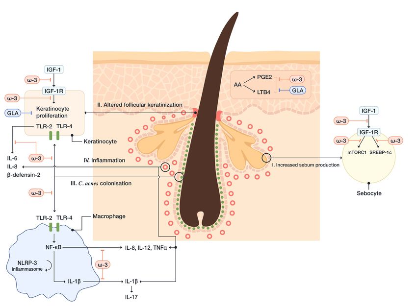
ω-3 FAs, particularly DHA, inhibit dimerization of TLR-1 and TLR-2 signaling [156]. C. acnes
increases the expression of TLRs on both keratinocytes and macrophages, which leads to
hyperproliferation of keratinocytes and the initiation of inflammatory reaction. The activation of
keratinocyte TLR-2 and TLR-4, induced by C. acnes, leads to activation of NF-κB and MAPK pathways,
subsequently, IL-1, IL-6, IL-8, TNF-α, human β-defensin-2, granulocyte-macrophage colony-stimulating
factor (GM-CSF), and matrix metalloproteinase (MMP) production [157–159]. Human β-defensin-2
belongs to the family of antimicrobial peptides (i.e., β-defensins), that are involved in the development of
inflammation in acne. The β-defensins induce the release of proinflammatory cytokines and chemotaxis
of immunocompetent cells and modulate cell maturation and migration [155]. C. acnes-induced
activation of monocyte TLR-2 leads to the production of proinflammatory cytokines, including IL-1β,
TNF-α, IL-8, and IL-12 [160]. Thus, DHA and EPA, inhibiting activation of TLR-signaling pathways,
may decrease the inflammatory response in patients with acne.
Furthermore, ω-3 FAs inhibit the activation of the NLRP3-inflammasome in antigen-presenting
cells. The activation of the NLRP3-inflammasome is triggered by C. acnes, which leaks out of
the pilosebaceous follicle after the disruption of the follicular epithelium and gets into the contact
with dermal macrophages. Subsequently, caspase-1 is activated, which leads to the release of
monocytes-derived IL-1β [161]. IL-1β promotes Th17 activation, which leads to IL-17-mediated
inflammation and keratinization. This process is regulated by reactive oxygen species (ROS) and
proteases [157]. Snodgrass RG et al. have demonstrated that DHA inhibits the expression of pro-IL-1β
and secretion of mature IL-1β in monocytes [156].
Additionally, it has been shown that EPA inhibits NF-κB activation. NF-κB transcribes
proinflammatory cytokines, including IL-1, IL-6, IL-8, upon its activation by the interaction of
C. acnes and TLR-2. EPA can also inhibit the expression of MMP-9, an endopeptidase that can degrade
the components of the ECM and expand the inflammation [162].
Insulin-like growth factor-1 (IGF-1) signaling is one of the most critical pathways in acne
development, which is sufficient to induce pro-inflammatory cytokine (IL-1β, IL-6, IL-8, TNF-α) and
MMPs expression in human sebocytes [163]. Several studies have shown that ω-3 FAs decrease
serum levels of IGF-1 and increase insulin-like growth factor binding protein-3 (IGFBP-3), thereby
preventing IGF-1 from binding to its receptors on sebocytes and acroinfundibular keratinocytes [164].You can also read