Inhibition of vascular calcification by inositol phosphates derivatized with ethylene glycol oligomers - Nature
←
→
Page content transcription
If your browser does not render page correctly, please read the page content below
ARTICLE
                  https://doi.org/10.1038/s41467-019-14091-4                 OPEN

                  Inhibition of vascular calcification by inositol
                  phosphates derivatized with ethylene glycol
                  oligomers
                  Antonia E. Schantl1,8, Anja Verhulst2,8, Ellen Neven2, Geert J. Behets 2, Patrick C. D’Haese2, Marc Maillard 3,
                  David Mordasini3, Olivier Phan3, Michel Burnier3, Dany Spaggiari4, Laurent A. Decosterd 4,
                  Mark G. MacAskill 5, Carlos J. Alcaide-Corral5, Adriana A.S. Tavares5, David E. Newby5, Victoria C. Beindl1,
1234567890():,;

                  Roberto Maj6, Anne Labarre7, Chrismita Hegde7, Bastien Castagner7, Mattias E. Ivarsson6,9* &
                  Jean-Christophe Leroux 1,9*

                  Myo-inositol hexakisphosphate (IP6) is a natural product known to inhibit vascular calcifi-
                  cation (VC), but with limited potency and low plasma exposure following bolus adminis-
                  tration. Here we report the design of a series of inositol phosphate analogs as crystallization
                  inhibitors, among which 4,6-di-O-(methoxy-diethyleneglycol)-myo-inositol-1,2,3,5-tetrakis
                  (phosphate), (OEG2)2-IP4, displays increased in vitro activity, as well as more favorable
                  pharmacokinetic and safety profiles than IP6 after subcutaneous injection. (OEG2)2-IP4
                  potently stabilizes calciprotein particle (CPP) growth, consistently demonstrates low
                  micromolar activity in different in vitro models of VC (i.e., human serum, primary cell cul-
                  tures, and tissue explants), and largely abolishes the development of VC in rodent models,
                  while not causing toxicity related to serum calcium chelation. The data suggest a mechanism
                  of action independent of the etiology of VC, whereby (OEG2)2-IP4 disrupts the nucleation
                  and growth of pathological calcification.

                  1 Institute of Pharmaceutical Sciences, Department of Chemistry and Applied Biosciences, ETH Zurich, Zurich, Switzerland. 2 Laboratory of

                  Pathophysiology, University of Antwerp, Antwerp, Belgium. 3 Service of Nephrology and Hypertension, Lausanne University Hospital,
                  Lausanne, Switzerland. 4 Division of Clinical Pharmacology, Lausanne University Hospital, Lausanne, Switzerland. 5 University-BHF Centre for
                  Cardiovascular Science, Queen’s Medical Research Institute, University of Edinburgh, Edinburgh, UK. 6 Inositec Inc., Zurich, Switzerland. 7 Department of
                  Pharmacology & Therapeutics, McGill University, Montreal, Canada. 8These authors contributed equally: Antonia E. Schantl, Anja Verhulst. 9These authors
                  jointly supervised this work: Mattias E. Ivarsson, Jean-Christophe Leroux. *email: mattias@inositec.com; jleroux@ethz.ch

                  NATURE COMMUNICATIONS | (2020)11:721 | https://doi.org/10.1038/s41467-019-14091-4 | www.nature.com/naturecommunications                                1
ARTICLE                                                                  NATURE COMMUNICATIONS | https://doi.org/10.1038/s41467-019-14091-4

V
        ascular calcification (VC) is the consequence of patholo-              micro-calcification34. Subsequently, larger confluent deposits
        gical deposition of calcium phosphate mineral in soft                 occur, which are visible on clinical computed tomography scan-
        tissues1. Symptoms and associated risks vary depending                ning35. Macrocalcifications negatively impact cardiovascular tis-
on the location (artery vs. valve, tunica media vs. intima) at which          sue (e.g., arterial and valvular stiffening, intima-media
calcification occurs, as well as on its nature (nano- vs. micro- vs.           thickening) and hemodynamic parameters (e.g., pulse wave
macrocalcification)2,3. Clinical consequences may include aortic               velocity, aortic valve pressure gradient). As a consequence, the
stiffening4, occlusive lesions (e.g., atherosclerotic plaques)5, or           risk for atherosclerotic events, heart failure, and cardiovascular as
aortic valve stenosis6,7, ultimately resulting in an increased risk           well as all-cause mortality increases35–38.
for cardiovascular morbidity and mortality8.                                     Myo-inositol hexakisphosphate (IP6) is a ubiquitously
   In chronic kidney disease (CKD), mineral homeostasis is dis-               occurring natural product and endogenous intracellular co-
turbed due to impaired renal function9, and cardiovascular                    factor in mammalian cells with putative functions in the areas
complications are the leading cause of death in these patients10,11.          of vesicle recycling and nuclear transport39. It inhibits the
Furthermore, VC is also a hallmark of a number of orphan dis-                 formation and growth of hydroxyapatite microcrystals in soft
eases, such as pseudoxanthoma elasticum and generalized arterial              tissues when administered parenterally40,41. Completed phase
calcification of infancy12,13. To date, there are no approved                  II studies in hemodialysis patients with calciphylaxis or cor-
pharmacological interventions for the direct prevention or                    onary artery calcifications, respectively, showed good safety,
treatment of VC. The etiology of VC is complex and multi-                     tolerability, as well as encouraging efficacy42–44. IP6 is injected
factorial. Several factors govern the calcification process in the             intravenously through the dialysis machine during hemodia-
vessel wall microenvironment, acting to promote or inhibit cal-               lysis sessions to allow it to reach therapeutic exposure despite
cification in conjunction with local precipitation of calcium and              its modest potency and short plasma half-life45. Both factors
phosphate8. The most important endogenous calcification inhi-                  complicate its development as a chronic ambulatory therapy46.
bitors include serum fetuin-A14, matrix Gla protein15, and pyr-               Given the paucity of data on the properties of IP6 analogs in
ophosphate (PPi)16. Most drug candidates currently under clinical             this context, this study investigates whether the chemical
investigation for VC (e.g., bisphosphonates, phosphate-binders,               modification of IP6 could produce more potent inhibitors with
vitamin K, or magnesium) act by correcting the imbalance of                   improved pharmacokinetic (PK) properties that may enable its
calcification promoters (i.e., hyperphosphatemia) and inhibitors               administration by a more convenient route. In previous work
in VC-affected individuals. However, these drug candidates have               from our group, where calcium phosphate nanoparticles for
yet to prove their efficacy in cardiovascular disease patients17,18.           gene delivery were developed, it was shown that coating of these
   Under non-pathological conditions, surges of plasma phos-                  particles with conjugates of oligo(ethylene glycol) (OEG)
phate or calcium, or local drops in the concentration of circu-               and myo-inositol pentakisphosphate prevent the rapid size
lating calcification inhibitors are buffered by acidic serum                   increase of calcium phosphate nanoparticles47,48. In the present
proteins, notably fetuin-A, via the formation of colloidal calci-             study, structurally related conjugates are tested in the context of
protein particles (CPPs)19. Primary CPPs (CPP1) are spherical                 VC inhibition49. Several OEGylated myo-inositol phosphate
complexes of amorphous calcium and phosphate with fetuin-A                    derivatives are synthesized and screened for their ability to
with a hydrodynamic radius (Rh) of less than 100 nm20. The                    inhibit calcification, as well as for their resistance towards
nanoparticles formed prevent precipitation of calcium and                     hydrolysis. The lead candidate is characterized for its efficacy
phosphate (i.e., hydroxyapatite formation)21. In the pathology of             in primary human vascular smooth muscle cells (VSMCs) and
VC, the system becomes depleted of factors delaying calcification              calcified human cardiovascular tissue explants, its biopharma-
propensity and the balance between CPP1 formation and                         ceutical properties, as well as its in vivo activity in rat models
absorption is distorted22. As a consequence, larger (Rh > 100 nm),            of VC50. This work identifies a potent OEG-derivatized inositol
crystalline, and spindle-shaped CPPs (secondary CPPs, CPP2s)                  phosphate, (OEG2)2-IP4, with a distinct mechanism of action
emerge20,23. Primary and secondary CPPs are predominantly                     that may find clinical utility for the prevention and/or
cleared by liver sinusoidal endothelial cells and liver Kupffer cells,        treatment of VC.
respectively. Scavenger receptor A is a major endocytosis receptor
for CPP2s but not for CPP1s, indicating separate clearance
pathways24. Native CPPs in fresh plasma are smaller and lower in              Results
density than synthetic CPP1 and CPP229. In CKD, CPPs are                      Design and synthesis of inhibitors of VC. Initially, OEG12-IP5, a
likely involved in the pathology of VC25,26. Serum CPP levels                 low molecular weight derivative of the chelator previously used by
increase with decline of renal function and correlate with cor-               our group to stabilize calcium phosphate nanoparticles for
onary artery calcification score, arterial stiffness, serum phos-              gene delivery purposes47, was synthesized in three steps starting
phate, inflammation, and procalcific factors27,28. Moreover, in                 from 1,3,5-O-methylidyne-myo-inositol (Supplementary Infor-
patients with advanced CKD, the Rh of CPP2 was associated with                mation 3.1.1.). It was purified by size-exclusion chromatography
VC29. The fact that CPP2s have the ability to induce VSMC                     and characterized by proton-1 nuclear magnetic resonance (1H-
calcification in vitro has raised the hypothesis that CPP2s may                NMR) and phosphorus-31 nuclear magnetic resonance (31P-
present a valid therapeutic target in VC30.                                   NMR) spectroscopies (Supplementary Information 5.), and elec-
   In patients with VC, a continuum of calcification typically                 trospray ionization-mass spectrometry (ESI-MS) (Supplementary
occurs with evidence of nano-, micro-, and macrocalcification,                 Table 1). Starting from OEG12-IP5, other inhibitors were designed
which follows a common pathophysiology. Scanning electron                     as follows: (i) increasing the number of linked OEG units while
microscopy of various diseased human aortic valve and arterial                decreasing that of phosphates (e.g., (OEG)2-IP4 and (OEG)3-IP3)
tissues reveals the presence of spherical, crystalline hydro-                 to study the trade-off between lower charge and increased steric
xyapatite particles ranging in size from 100 nm to 5 µm31, sug-               hindrance, (ii) replacing phosphates by sulfate groups (i.e., OEG11-
gesting that VC may follow a common process independent of                    IP2S3) to investigate chemical stability towards hydrolysis and to
the etiology31,32. Further evidence for the presence of micro-                reduce chelation of soluble Ca2+51, and (iii) reducing OEG
calcifications comes from positron emission tomography (PET)                   chain length (OEG12, OEG11, OEG7, and OEG2) to study the
imaging of high-risk coronary atherosclerotic plaque33 employing              influence of the latter on activity (Fig. 1a). Ten compounds,
[18F] sodium fluoride (18F-NaF), a marker of active vascular                   covering a broad range of chemical modifications, were produced

2                 NATURE COMMUNICATIONS | (2020)11:721 | https://doi.org/10.1038/s41467-019-14091-4 | www.nature.com/naturecommunications
NATURE COMMUNICATIONS | https://doi.org/10.1038/s41467-019-14091-4 ARTICLE
a OPO32– b OPO32– OPO32– OPO32–
OPO32– O OPO32–
O OPO32– OPO32– OH
n OPO32– OPO32–
2–
O3PO OPO32– 2–O PO OPO32–
3 2–
O3PO OPO32– 2– OPO32–
OPO32– OPO32– O3PO
OH OH
OEGn-IP5 Ins(1,2,3,4,5,6)P 6 Ins(1,2,3,5,6)P 5 Ins(2,3,5,6)P 4
(IP6) (IP5) (IP4)
OPO32– OH OPO32– OH
OPO32– O OPO32– OH OH
O OH OH OH
n
2– O PO OPO32– HO OPO32– HO OH HO OH
3
O OPO32– OPO32– OH
O
n
Ins(1,4,5)P 3 Ins(2,4)P 2 myo -Inositol
(OEGn)2-IP4 (IP3) (IP2)
–
O O O
–
OPO32– OPSO22–
OSO3– O O P P O O O
O OPSO22–
n OPSO22– O O P P
–O O–
–O SO
3 OSO3– 2– OPSO22–
O
OPO32–
O2SPO O– O–
OPSO22– –
O O–
O O
OEGn-IP2S3 O P P O PPi
IT6
O O O O
O P P
O –
O n –O O O
OPO32– O
O
n ITPP
O O O O
2–
O3PO OPO32– HO OH HO OH
O O
P P HO OH P P
O – OH –
O OH H
O NH2 P P O– N O
O –O
HO O
n HO 45
HO
(OEGn)3-IP3 O
Alendronate Etidronate PEG-alendronate
Fig. 1 Structures of functionalized inositol phosphates and control compounds. a Structure of the series of calcification inhibitors that were synthesized,
with n = 2, 7, 11, and 12 for OEGn-IP5, n = 2, 7, and 11 for (OEGn)2-IP4, n = 11 for OEGn-IP2S3, and n = 2 and 7 for (OEGn)3-IP3. Represented in red is the
backbone structure of the lead inhibitor of this study. b Structures of compounds used as controls, including dephosphorylated (IP5, IP4, IP3, IP2, myo-
inositol) and phosphorylated (myo-inositol trispyrophosphate (ITPP)) IP6 derivatives, myo-inositol hexathiophosphate (IT6), pyrophosphate (PPi), as well
as alendronate and etidronate (non-hydrolyzable analogs of PPi). PEG45-alendronate has been described previously to stabilize calcium phosphate
nanoparticles in vitro48.
or purchased, and details on their synthesis and characterization and (iii) longer OEG chains increase molar activity but reduce
are provided in the Supplementary Methods. These compounds activity by unit mass. Moreover, molar activity of (OEG2)2-IP4
were compared to 12 controls (Fig. 1b). was approximately six- and twofold higher than pyrophosphate
(PPi) (a strong endogenous inhibitor of VC) and etidronate (a
Drug candidate screening assay. The compounds were ranked hydrolysis-resistant analog of PPi), respectively (Table 1, Sup-
for calcification inhibition activity using a nanoparticle-based test, plementary Fig. 3).
developed for risk stratification, which measures the propensity
for calcification in patient serum22. The transformation of
amorphous CPP1s to larger crystalline CPP2s in pooled human In vitro characterization. To confirm the screening assay data,
serum was monitored in the presence of inhibitor, and artificially the hydrodynamic diameter of the CPPs was measured by
elevated calcium and phosphate concentrations at 37 °C. The dynamic light scattering (DLS). In the absence of inhibitors or in
time, after which 50% of CPP phase transition occurred (T50), was the presence of IP6 (up to 100 µM), the CPP1 size grew from
assessed by time-resolved spectrophotometry at 550 nm (Sup- roughly 150 nm after 1 h to ~1 µm after 3 h of incubation in
plementary Fig. 1a). To rank the compounds’ activity, a T50 vs. serum (Supplementary Fig. 4c, f), indicating transition to CPP2.
compound concentration plot was generated, and the con- The CPP2 size deduced by DLS is slightly larger than the size
centration necessary to delay T50 to 350 min (c350, at which IP6’s derived from transmission electron microscopy (TEM) (Supple-
activity was at half-maximum) was derived from nonlinear curve mentary Figs. 4c and 7a), the latter being in line with previous
fitting (Fig. 2a, Supplementary Figs. 1–3). (OEG11)2-IP4 displayed work52. (OEG2)2-IP4, added to CPP1s at a final concentration of
an almost 10-fold greater molar activity in delaying serum cal- 30 µM, stabilized particle growth (Supplementary Fig. 4h)29. TEM
cification propensity (c350 = 3.8 ± 0.3 µM) than IP6 (c350 = revealed the presence of spherical-shaped CPP1s in samples
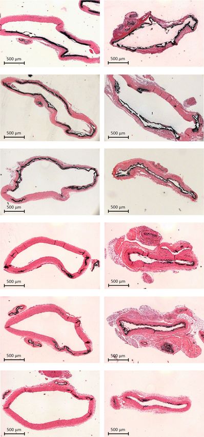
35.0 ± 9.9 µM). Further, as seen in Table 1, (OEG11)2-IP4 was the incubated with 100 µM (OEG2)2-IP4 vs. spindle-shaped CPP2s in
most active molecule of all screened functionalized inositol samples containing 100 µM IP6 (Fig. 2b). Corresponding selected
phosphates and tested controls. However, looking at activity by area electron diffraction analysis of CPP with 100 µM IP6 showed
unit mass, (OEG2)2-IP4 had an apparent 32% stronger inhibition bright spots and well-defined rings, indicating crystalline mate-
than (OEG11)2-IP4 due to its lower molecular weight—an rial, whereas the (OEG2)2-IP4-stabilized CPP1s were largely
important consideration for dosing purposes (Table 1). These amorphous. The derived interplanar- or d-spacings for CPP
data indicate that (i) replacement of two, rather than one or three incubated with IP6 were: d1 = 0.287 nm*, d2 = 0.369 nm†, and
phosphates of IP6 by OEG is optimal, (ii) replacement of phos- d3 = 0.403 nm, which are in line with reported literature d-spaces
phates by sulfates in the OEGylated molecules reduces activity, for hydroxyapatite: 0.28 nm*, 0.32 nm, 0.34 nm†, 0.47 nm, and
NATURE COMMUNICATIONS | (2020)11:721 | https://doi.org/10.1038/s41467-019-14091-4 | www.nature.com/naturecommunications 3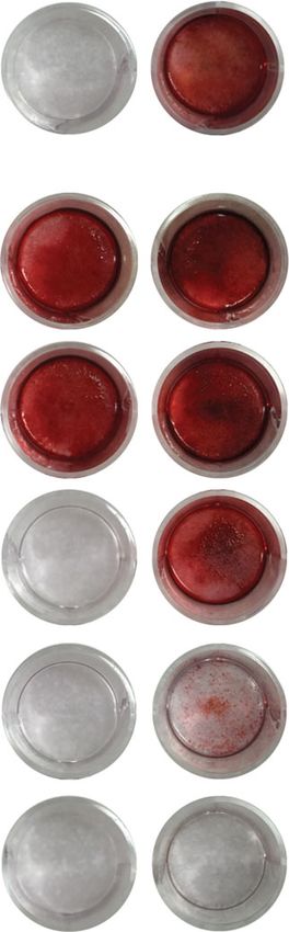
ARTICLE NATURE COMMUNICATIONS | https://doi.org/10.1038/s41467-019-14091-4
a 900 b
(OEG2)2-IP4
(OEG2)2-IP4 IP6
800 IP6
myo-Inositol
700
600
T50 (min)
500
400
300
200
100
0.1 1 10 100
[Compound] (μM)
c (OEG2)2-IP4, IC50 = 0.81 μM d e
IP6, IC50 = 3.96 μM OEG2-lP5 (OEG2)2-IP4
100
150 Alendronate, IC50 = 0.90 μM OEG11-lP5 (OEG11)2-IP4
60
IP6
50
50
ΔT50 (min)
Hydrolyzed (%)
Binding (%)
100 40 0
30
–50
50 20
10 –100
6
P4
4
0 0
IP
-IP
2 -I
)2
0 1 2 3 4 5 6 7 8
2)
10–1 100 101 102 103 104 105 106
11
EG
EG
Time (h)
(O
[Compound] (nM)
(O
Fig. 2 In vitro activities of functionalized inositol phosphates. a Compounds were tested in an in vitro serum calcification propensity assay. T50 is defined
as the time at which 50% of CPP1 is transformed to CPP2. The concentration necessary to delay T50 to 350 min (dotted line) was used to compare the
compounds’ activities (n = 4 for myo-inositol, n = 6 for (OEG2)2-IP4, and n = 26 for IP6). b Representative TEM images and corresponding selected area
electron diffraction patterns of CPPs, showing the predominant presence of spherical-shaped CPP1s and spindle-shaped CPP2s after incubation with
100 µM (OEG2)2-IP4 and IP6, respectively for 14 h at 37 °C (scale bar = 50 nm). c Competitive 18F-NaF-binding assay to characterize interaction of
compounds with CPPs (n = 3). d Phytase hydrolysis of compounds (n = 3). e Activity in the serum calcification propensity assay after incubation in
enzymatically active human serum for 4.5 h at 37 °C (n = 4). All data are expressed as mean ± s.d. from n independent experiments. Statistical significance
was calculated from a paired Student’s t-test, ***p < 0.001 vs. baseline T50.
0.52 nm53 (matching d-spaces between samples are indicated with side effects55, alendronate was not further investigated in
* and †) and suggest identical material. this study.
A 18F-NaF-based competitive binding assay was developed to Since IP6 is highly susceptible to degradation to lower inositol
indirectly assess the inhibitor affinity (IC50) to CPPs. The phosphates due to endogenous phosphatase activity39, the in vitro
uptake mechanism of 18F-NaF, a marker of micro-calcification stability of selected OEGylated inositol phosphates after exposure to
activity by chemisorption onto hydroxyapatite crystals, is 3-phytase and incubation in enzymatically active human serum was
contingent on the rapid exchange of hydroxyl (OH−) groups investigated. Enzymatic hydrolysis of inositol phosphates in buffer
present on the hydroxyapatite surface (Ca5(PO4)3(OH)) by 18F− was reduced and delayed by the presence of OEG moieties
to form fluoroapatite (Ca5(PO4)318F)34,54. Similarly to 18F-NaF, compared to IP6 (Fig. 2d), likely due to steric hindrance hampering
IP6 and (OEG2)2-IP4 are expected to be adsorbed to the enzymatic activity. Protection from phosphate hydrolysis increased
hydroxyapatite core of CPPs. (OEG2)2-IP4 reduced 18F binding with number, but not with length of conjugated OEG. OEGylated
to CPP more efficiently than IP6 (IC50 = 0.81 µM, 95% CI [0.48, inositol phosphates retained complete activity in the calcification
1.37] vs. 3.96 µM, 95% CI [2.03, 7.75]), despite the lower propensity assay after a 4.5-h incubation in enzymatically active
negative charge density, and thus lower Ca2+ chelating ability serum, as opposed to IP6, with which T50 decreased by 73 min from
of (OEG2)2-IP4 compared to IP6 (Fig. 2c), hence supporting the baseline (Fig. 2e).
hypothesis that the OEG moiety contributes to the activity of All subsequent testing was performed with (OEG2)2-IP4
(OEG2)2-IP4. Alendronate showed a comparable CPP-binding because it demonstrated comparable stability and molar activity
affinity (IC50 = 0.90 µM, 95% CI [0.54–1.50]) while having a to (OEG11)2-IP4, but would eventually be administered in vivo at
twofold lower activity in the calcification propensity assay a lower dose in mass, given its lower molecular weight.
(c350 = 9.9 ± 1.0 µM). Due to the electrostatic nature of the
interaction of (OEG2)2-IP4 with hydroxyapatite, the binding is Inhibition of human VSMC calcification. A postulated driving
likely reversible. Because of these results and the fact that force for VC at the cellular level are VSMCs, the main cell type of
clinical trials using bisphosphonates in VC reported important the tunica media. VSMCs can transdifferentiate towards an
4 NATURE COMMUNICATIONS | (2020)11:721 | https://doi.org/10.1038/s41467-019-14091-4 | www.nature.com/naturecommunications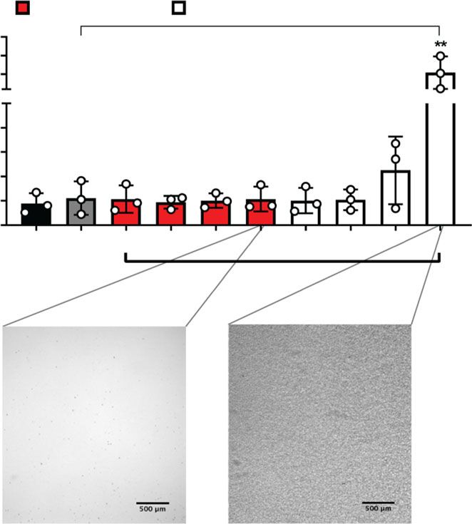
NATURE COMMUNICATIONS | https://doi.org/10.1038/s41467-019-14091-4                                                                      ARTICLE

 Table 1 Activities of compounds in the screening assay.                          macrophages were exposed to CPP2-supplemented medium.
                                                                                  CPP2 prepared from human serum as described previously61
                                                                                  (Supplementary Figs. 6 and 7), induced a pronounced VSMC
 Compound                         Concentration at T50 = 350 min
                                  mean ± s.d. (n)
                                                                                  monolayer calcification after 48 h, which was decreased by
                                                                                  (OEG2)2-IP4 in a dose-dependent fashion, and the effect became
                                  µM                       mg/L                   significant at 10 µM (Fig. 3e). These results were supported by
 Inositol phosphate derivatives                                                   Alizarin Red staining (Supplementary Fig. 8). (OEG2)2-IP4
  OEG2-IP5                          7.9 ± 2.7    (3)           7.1 ± 2.4   (3)    treatment did not reduce VSMC viability in this setup (Fig. 3f).
  OEG7-IP5                         6.7 ± 0.9     (3)           7.6 ± 1.0   (3)    Incubating VSMCs with CPP1 increased calcium deposition vs.
  OEG11-IP5                         5.3 ± 0.7    (3)          6.8 ± 0.9    (3)    control, but not significantly, and (OEG2)2-IP4 treatment starting
  OEG12-IP5                        4.9 ± 0.6     (5)          6.6 ± 0.9    (5)    at 10 µM reduced calcium deposition to the level of control
  (OEG2)2-IP4                      4.9 ± 0.8     (6)          4.3 ± 0.7    (6)
                                                                                  (Fig. 3g). Both H2O2 (proxy for oxidative stress) and tumor
  (OEG7)2-IP4                      4.5 ± 0.4     (3)          6.0 ± 0.6    (3)
  (OEG11)2-IP4                      3.8 ± 0.3    (3)          6.3 ± 0.6    (3)
                                                                                  necrosis factor-alpha (TNF-α) (proxy for inflammatory response)
  OEG11-IP2S3                      18.6 ± 1.9    (5)         22.9 ± 2.3    (5)    levels were assayed in the VSMC culture supernatants subsequent
  (OEG2)3-IP3                       7.1 ± 0.9    (3)          5.4 ± 0.7    (3)    to CPP2 treatment with and without compounds30. However, no
  (OEG7)3-IP3                      9.4 ± 0.6     (3)         14.3 ± 1.0    (3)    increase in H2O2 nor TNF-α compared to control could be
 Inositol controls                                                                detected in either assay. For the CPP-induced VSMC calcification
  myo-Inositol                          >100     (4)              >18.0    (4)    assay (Fig. 3e) a roughly 10-fold higher concentration of
  IP2                             35.0 ± 7.7     (3)          15.0 ± 3.3   (3)    (OEG2)2-IP4 was necessary to achieve similar activity as in the
  IP3                              18.6 ± 3.1    (3)          10.2 ± 1.7   (3)    CaP-induced VSMC calcification assay (Fig. 3b). This may be
  IP4                             19.5 ± 0.7     (3)          13.2 ± 0.5   (3)    explained by a more potent induction of calcification in the CPP-
  IP5                              13.2 ± 1.3    (3)          10.5 ± 1.0   (3)
  IT6                             14.4 ± 0.3     (3)       36.14 ± 0.24    (3)
                                                                                  induced assay, requiring higher (OEG2)2-IP4 concentration to
  ITPP                                  >100     (4)              >60.6    (4)    counteract the procalcific environment. In the CaP assay, low
 Known inhibitors of calcification                                                 inhibitor concentration may be sufficient to inhibit crystal
  IP6                             35.0 ± 9.9     (26)        30.9 ± 8.7    (26)   nucleation, whereas for the CPP assay higher inhibitor concen-
  Magnesium citrate                     >100     (3)             >21.4     (3)    trations may be needed to achieve sufficient particle stabilization
  PPi                             27.4 ± 2.8     (3)          7.3 ± 0.8    (3)    by chemisorption of inhibitor onto CPPs. Similar results have
  Etidronate                        10.5 ± 1.1   (4)          2.6 ± 0.3    (4)    been reported for hydrogen sulfide in the context of VC62,64. In
  Alendronate                       9.9 ± 1.0    (6)          3.2 ± 0.3    (6)    both the VSMC and macrophage assays, precipitation in culture
 Others                                                                           medium occurred in treatment groups containing high concen-
  PEG-alendronate                 12.3 ± 3.0     (3)          28.1 ± 6.9 (3)
                                                                                  trations of IP6 (100 µM) but not with (OEG2)2-IP4 at the same
                                                                                  concentrations (Fig. 3h microscopy images and Supplementary
                                                                                  Fig. 9). This effect is likely due to the strong chelating potential of
osteoprogenitor cell type under prolonged mineral exposure or                     IP6 with free ionized serum calcium. Indeed, the combined
uremic stress, and release vesicles packed with high concentra-                   treatment of 100 µM IP6 and CPP2 (i) resulted in precipitation
tions of calcium and phosphate serving as nidi for extracellular                  visible to the naked eye in the treatment medium, (ii) significantly
matrix calcification56–60.                                                         reduced macrophage viability by 24% (Fig. 3i), and (iii) TNFα
   Incubating primary human VSMCs with (OEG2)2-IP4                                (indicator for inflammatory response) release from macrophages
(0.01–100 µM) did not lead to any changes in cell viability                       increased 49-fold (Fig. 3h). An increase in the TNFα levels in the
(Fig. 3a). The activity of (OEG2)2-IP4 in inhibiting calcification of              cell culture supernatants was not observed for (OEG2)2-IP4 with
VSMCs was then examined using a high-CaP (2.7 mM calcium,                         (Fig. 3h) and without (Supplementary Fig. 10) combined CPP2
2.5 mM phosphate) medium (Supplementary Fig. 5). Incubation                       treatment.
of VSMCs in high-CaP medium induced cell monolayer
calcification after 5 days (Fig. 3b). Concurrent incubation with
                                                                                  Target engagement in calcified human explants. To investigate
(OEG2)2-IP4 (0.01–100 µM) dose-dependently prevented calcifi-
                                                                                  (OEG2)2-IP4 interaction with calcified tissues in a clinically
cation as evidenced from the quantitative measurement of the
                                                                                  relevant context, a competitive binding assay using 18F-NaF was
calcium content (Fig. 3b), and qualitative staining for calcium
                                                                                  applied on severely calcified human femoral arteries and native
with Alizarin Red (Fig. 3c). The effect became significant from
                                                                                  aortic valves ex vivo. The tissue explants were incubated with
0.1 µM (OEG2)2-IP4 on, where a 54% reduction in calcification
                                                                                  (OEG2)2-IP4 at a concentration inhibiting 90% 18F-NaF binding
was observed compared to untreated high-CaP medium-exposed
                                                                                  (IC90 = 8.6 μM), as assessed in Fig. 2c. First, 18F-NaF uptake was
cells. Calcium deposition was almost completely abrogated above
                                                                                  measured on tissue samples treated with vehicle (human serum)
1 µM (OEG2)2-IP4. IP6 reduced VSMC mineralization by
                                                                                  and thereafter, using the same tissue samples, treated with
~50–70% for all doses studied (up to 100 µM). The less efficient
                                                                                  (OEG2)2-IP4 in serum. Compared to the vehicle controls, results
inhibition in calcification may be due to the limited IP6 solubility
                                                                                  showed a significant reduction of 18F-NaF uptake in calcified
in high calcium medium51 or potential extracellular effects (e.g.
                                                                                  human femoral arteries (−38%) (Fig. 3j, l) as well as in calcified
protein binding)39. In the present model, cell viability seemed to
                                                                                  leaflets from native human aortic valves (−33%) (Fig. 3k, m).
be slightly decreased in low-dose treatment groups (0.01 and 0.1
µM (OEG2)2-IP4) vs. high-CaP medium control but this effect
disappeared above 1 µM for (OEG2)2-IP4 and 10 µM for IP6                          OEGylation improves the PK profile of inositol phosphates.
(Fig. 3d).                                                                        The plasma half-life (t1/2) of IP6 is short (8 min) after intravenous
   CPPs have been implicated in the mineralization paradox of                     (i.v.) bolus administration in rats45,65, and values of 27 and 54
CKD, and previous in vitro work using synthetic CPP2s suggests                    min after a 4-h i.v. infusion have been reported in hemodialyzed
a role in promoting VSMC calcification25,30,61,62 and in reducing                  patients and healthy volunteers, respectively43. Therefore, the PK
hydroxyapatite mineral stress in macrophages63. Macrophage                        profile of (OEG2)2-IP4 was assessed in healthy and uremic rats
accumulation and inflammation coincide with the initial                            and compared to that of IP6. At 10 mg/kg, (OEG2)2-IP4 displayed
phase of calcification3. To this end, VSMCs and THP-1-derived                      an almost 10-fold increase in plasmatic t1/2 vs. IP6 after i.v. bolus

NATURE COMMUNICATIONS | (2020)11:721 | https://doi.org/10.1038/s41467-019-14091-4 | www.nature.com/naturecommunications                                5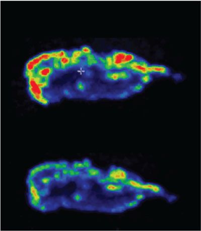
ARTICLE NATURE COMMUNICATIONS | https://doi.org/10.1038/s41467-019-14091-4
a b c
(OEG2)2-IP4 IP6 (OEG2)2-IP4 IP6
150 150
CaP
Calcium deposition
VSMC viability
(% of control)
(% of CaP)
100 100
Control CaP
50 50
0.01 μM
0 0
0.01 0.1 1 10 100 0.01 0.1 1 10 100 Ctrl. CaP 0.01 0.1 1 10 100 0.01 0.1 1 10 100
[Compound] (μM)
[Compound] (μM)
0.1 μM
d (OEG2)2-IP4 IP6 e (OEG2)2-IP4 IP6
150 150
CaP CPP2
Calcium deposition
VSMC viability
(% of CPP2)
(% of control)
100 100
1 μM
50 50
0 0
10 μM
CaP 0.01 0.1 1 10 100 0.01 0.1 1 10 100 Ctrl. CPP2 1 3 10 30 100 1 3 10 30 100
[Compound] (μM) [Compound] (μM)
f g (OEG2)2-IP4 IP6
#, §
100 μM
(OEG2)2-IP4 IP6
150 CPP1
CPP2 200
ns
Calcium deposition
VSMC viability
150
(% of control)
(% of CPP2)
100
CaP+ CaP+
100
(OEG2)2-IP4 IP6
50
50
0
l
0
CPP2 1 3 10 30 100 1 3 10 30 100 Ctrl. CPP2 CPP1 1 3 10 30 100 3 10 30 100 Vehicle (OEG2)2-IP4
[Compound] (μM) [Compound] (μM)
h i (OEG2)2-IP4 IP6
(OEG2)2-IP4 IP6 150
CPP2
Cell viability (% of control)
15,000 CPP2
10,000
5000 100
(pg/mg protein)
500
TNFα
400
50
300
200
100 0
0
CPP2 3 10 30 100 3 10 30 100
m
Control CPP2 3 10 30 100 3 10 30 100 [Compound] (μM) Vehicle
[Compound] (μM)
j 1500
k 1500
Maximum average binding
to native valves (kBq/mL)
Maximum average binding
to femoral artery (kBq/mL)
1000 1000
(OEG2)2-IP4
500 500
0 0
le
4
le
4
ic
-IP
-IP
ic
h
h
Ve
Ve
)2
)2
2
2
EG
EG
(O
(O
injection into male rats (t1/2 = 78 ± 32 vs. t1/2 = 8 ± 2 min, 45 ± 20 min) as well as i.v. (30 ± 3 vs. 14 ± 6 min) administrations
respectively) (Fig. 4a, b). The apparent volumes of distribution (Supplementary Fig. 11).
were low with 234 ± 32 vs. 958 ± 405 mL/kg for (OEG2)2-IP4 and No significant accumulation occurred following repeated s.c.
IP6, respectively, which is generally expected for hydrophilic com- administration of 50 mg/kg (OEG2)2-IP4 for 7 days (Fig. 4e).
pounds. The maximum plasma concentration (Cmax) increased Moreover, the systemic exposure of the compound after s.c.
fourfold, the area under the concentration vs. time curve (AUC0–t) administration was assessed in rats with VC (Fig. 4f, Supple-
increased sevenfold and the plasma clearance (CL) decreased mentary Table 3). In the presence of cardiovascular calcification,
ninefold compared to IP6 (Fig. 4b). Subcutaneous (s.c.) adminis- the plasma exposure of (OEG2)2-IP4 was significantly higher at 4
tration of 10 mg/kg (OEG2)2-IP4 resulted in an AUC comparable to and 6 h after dosing, but the AUC0–1440 was not statistically
that of 100 mg/kg IP6 (AUC0–360 = 1197 ± 36 vs. AUC0–90 = different to the value obtained before the induction of calcifica-
1165 ± 76 µM min), despite the 10-fold difference in doses (Fig. 4c, tion (AUC0–1440 = 9077 ± 2455 vs. 6049 ± 799 µM min).
d, Supplementary Table 2)45. The mean residence time (MRT) For many drugs, the PK profile depends on the patient’s renal
was longer for (OEG2)2-IP4 compared to IP6 after s.c. (78 ± 5 vs. status. Therefore, (OEG2)2-IP4 PK profile was also determined in
6 NATURE COMMUNICATIONS | (2020)11:721 | https://doi.org/10.1038/s41467-019-14091-4 | www.nature.com/naturecommunications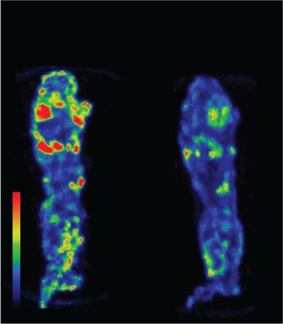
NATURE COMMUNICATIONS | https://doi.org/10.1038/s41467-019-14091-4                                                                       ARTICLE

Fig. 3 (OEG2)2-IP4 abrogates in vitro VSMC calcification and 18F-NaF uptake onto calcified human explants. a, d, f VSMC viability as determined by the
MTS assay compared to medium control. b, c VSMC calcification was induced by high-CaP medium and the effect of cell treatment with IP6 or (OEG2)2-
IP4 was investigated quantitatively by colorimetric analysis of cellular calcium deposition after 5 days control (calcium deposition in mass units in Ctrl.
ranged between 81 and 622 µg Ca/mg protein) (b) and qualitatively by Alizarin Red staining for calcium (c) in a 24-well plate setup compared to medium
control. e, g Same as b but calcium deposition was induced by CPP2- (e) and CPP1- (g) supplemented medium (50 µg Ca/mL), respectively (calcium
deposition in mass units in Ctrl. ranged between 189 and 734 µg Ca/mg protein in e). h TNFα concentration in human macrophage supernatants after
exposing cells to CPP2 (50 µg Ca/mL) and compounds as assessed by immunoassay. High concentrations of IP6, but not (OEG2)2-IP4, precipitated in the
treatment medium as shown in the insets and resulted in significantly increased TNFα levels in supernatants (scale bar = 500 µm). i Macrophage viability
as determined by the MTS assay compared to medium control. j, k, l, m Incubation of severely calcified human femoral arteries (j, l) or human aortic valves
(k, m) with (OEG2)2-IP4 and without (vehicle) resulted in a reduction of 18F-NaF uptake in (OEG2)2-IP4 samples compared with baseline vehicle studies as
evident from the maximum average 18F binding determined by 18F-NaF-PET imaging (j, k) and the maximum intensity projections of collected 18F-NaF-PET
data (l, m). All data are expressed as mean ± s.d. n = 7 (j) and n = 3 (k) biologically independent samples, and from n = 4 (e) and n = 3 (all others)
independent cell experiments. Statistical significance was calculated from an ordinary one-way ANOVA with Tukey’s multiple comparisons test or from an
unpaired Student’s t-test to determine significance between two data sets with *p < 0.05, **p < 0.01, ***p < 0.001 vs. gray bar, #p < 0.001 vs. CPP1, and
§p < 0.001 vs. 100 µM (OEG ) -IP4.
                             2 2

an adenine and high-phosphate-diet-induced rat model of                          spectrometry (Fig. 5a), but the decrease in abdominal aorta cal-
uremia, in which the compound was administered after serum                       cification was not significant (Supplementary Fig. 16). IP6-treated
creatinine levels had increased by fourfold relative to baseline                 animals had to be sacrificed ahead of treatment schedule on day
(Fig. 4g, h). The Cmax after i.v. injection of 10 mg/kg (OEG2)2-IP4              10 vs. day 12, due to the appearance of necrotic skin lesions
was almost identical to that of healthy controls (43 ± 12 vs. 35 ±               (despite maintaining formulation osmolarity and pH within
9 μM) but elimination appeared to be modestly slower in the                      physiological ranges) (Supplementary Fig. 17). However, calcifi-
uremic rats (1.6-fold longer t1/2 and twofold longer MRT)                        cation levels in IP6-treated animals at day 10 were not lower than
(Supplementary Table 4), thus resulting in a higher systemic                     those of (OEG2)2-IP4-treated animals at day 12 (Fig. 5a). Inter-
exposure to the compound (Fig. 4i). Similar results were observed                estingly, in this model (OEG2)2-IP4 treatment led to a beneficial
following s.c. administration (Supplementary Table 5). Forty                     impact on the renal function of vitamin D3-treated animals, as
percent of the intact (OEG2)2-IP4 dose was recovered in the urine                evident from plasma creatinine levels (Fig. 5c), which occurred in
of healthy rats, suggesting additional clearance modalities (Fig. 4j).           conjunction with a reduction of renal calcium content (Fig. 5b).
The protein-bound fraction of (OEG2)2-IP4 in healthy human                       Creatinine levels were significantly decreased in animals receiving
plasma was 58% and 42%, respectively, calculated over total                      (OEG2)2-IP4 (39.8 ± 6.6 μM) compared to vehicle controls (59.0 ±
measured concentrations of 2.8 and 27.5 µg/mL (i.e., 4 and                       7.9 μM), and remained in the healthy range for rats. Plasma cal-
39.5 µM, respectively), suggesting a potential saturation of the                 cium and phosphate levels were not impacted by treatment with
binding sites at high concentrations.                                            (OEG2)2-IP4 (Supplementary Table 6). Given the severity and
   One potential side effect of inositol phosphate-based drugs is                resultant variability in calcification burden in this vitamin D3
toxicity due to chelation of ionized serum calcium. In a previous                model, the activity of (OEG2)2-IP4 was assessed in a modified
study, a reduction in serum calcium levels by 17% was observed                   setup, where VC was induced by oral and less frequent adminis-
in uremic rats after 4 h i.v. infusion of 50 mg/kg IP645. No                     tration of vitamin D3 together with a warfarin-supplemented diet.
significant lowering of serum calcium was evident after admin-                    This model resulted in a robust but milder and less variable
istration of 50 mg/kg (OEG2)2-IP4 in either the acute or sub-                    induction of VC, and therefore allowed for a better investigation
chronic setting (7 days), measured at the tmax of 30 min (Fig. 4k).              of the (OEG2)2-IP4 dose–response. Starting from day 1 of the
In a separate in vitro test using human serum, a significant                      vitamin D3 oral administration, animals were treated with s.c.
difference in decrease of human serum-ionized calcium levels                     injections of increasing doses of (OEG2)2-IP4 administered once
between (OEG2)2-IP4 and IP6 became evident starting from                         or twice daily until day 7. Due to the severe local inflammation
600 µM (Fig. 4l). At 100 µM (OEG2)2-IP4, 96 ± 2% of control                      induced by the repeated s.c. injection of IP6 in the previous
serum-ionized calcium was measured.                                              experiment, this control was excluded from the present experi-
   The potential off-target activity of 10 µM (OEG2)2-IP4 was                    ment. In two dosing groups, rats lost significantly more weight
assessed against a panel of 87 enzymes, receptors, or ion channels               than the others compared to day 1, but it was not dose-related
that are used in standard safety pharmacology screens. The                       (Supplementary Fig. 18). Mortality rate in the vehicle group was
compound did not significantly inhibit or stimulate any of the                    38% (15/40) and declined to 0% (0/18) in the 2 × 25 mg/kg/day
respective biochemical assays (Supplementary Table 11).                          dose group, at which the effect was statistically significant
                                                                                 (Fig. 5d). (OEG2)2-IP4 dose-dependently prevented VC in the
                                                                                 abdominal aorta, as evident from total calcium measurement
Efficacy of (OEG2)2-IP4 in a vitamin D-induced model of VC.                       (Fig. 5e) and von Kossa staining (Fig. 5f, g). The effect was sta-
Activity of (OEG2)2-IP4 was assessed in an animal model of acute                 tistically significant starting at 2 × 25 mg/kg/day and 1 × 12.5 mg/
and severe calcification induced by vitamin D3 overdose, accom-                   kg/day with regard to total calcium content and % von Kossa-
panied by hypercalcemia, hyperphosphatemia, and increased                        positive area, respectively. Moreover, total calcium (Fig. 5h) and %
plasma creatinine (Supplementary Fig. 12), and reduced body                      von Kossa-positive area (Fig. 5i) in the thoracic aorta was sig-
weight (Supplementary Fig. 13), This model resulted in massive                   nificantly reduced starting at 2 × 50 and 2 × 25 mg/kg/day,
VC, which was observed primarily along the entire aorta (Sup-                    respectively. Intriguingly, and in contrast to the abdominal aorta,
plementary Fig. 14) in the tunica media (Supplementary Fig. 15).                 the thoracic aorta did not calcify extensively in all animals, making
(OEG2)2-IP4, dosed s.c. at 30 mg/kg twice daily for 11 days, sig-                differences between treatment groups less evident. A significant
nificantly decreased formation of carotid VC as evidenced from                    reduction in bulk calcium in the 2 × 25 mg/kg/day group was also
calcium quantification by inductively coupled plasma mass                         evident in more peripheral arteries, i.e., carotid artery (Fig. 5j) and

NATURE COMMUNICATIONS | (2020)11:721 | https://doi.org/10.1038/s41467-019-14091-4 | www.nature.com/naturecommunications                                  7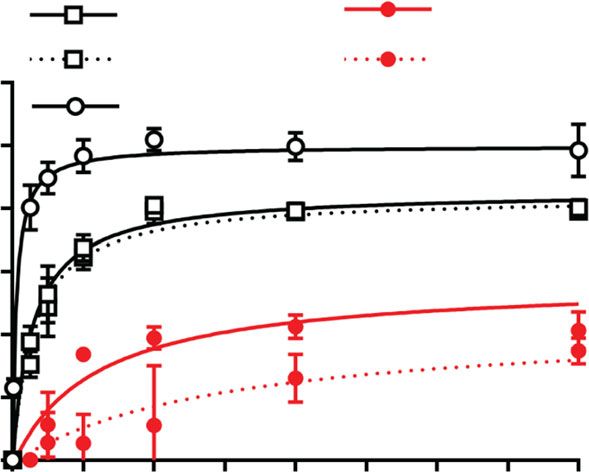
ARTICLE NATURE COMMUNICATIONS | https://doi.org/10.1038/s41467-019-14091-4
a b c
PK parameters, i.v. injection
80 100
Plasma concentration (μM)
Plasma concentration (μM)
(OEG2)2-IP4 IP6 10 mg/kg (OEG2)2-IP4
10 mg/kg (OEG2)2-IP4
10 mg/kg IP6 80 50 mg/kg (OEG2)2-IP4
60 Dose (mg/kg) 10 10
100 mg/kg IP6
60
40 t1/2 (min) 78 ± 32 8±2
40
Cmax (μM) 52 ± 13 13 ± 6
20
20
AUC0-t (μM·min) 1565 ± 289 226 ± 90
(t = 360 min) (t = 30 min)
0 0
0 20 40 60 80 100 120 Cl obs (mg/min/kg) 7.6 ± 0.5 68.5 ± 21.2 0 1 2 3 4 5 6
Time (min) Time (h)
Means ± s.d.
d e f
PK parameters, s.c. injection
(OEG2)2-IP4 IP6 100 60
Plasma concentration (μM)
Plasma concentration (μM)
Acute No calcification
80 50
Dose (mg/kg) 10 100 Sub-chronic Calcification
40
t1/2 (min) 47 ± 1 29 ± 15 60
30
t max (min) 25 ± 8 12 ± 6 40
20
Cmax (μM) 12 ± 1 33 ± 10 20 10
AUC0-t (μM·min) 1197 ± 36 1165 ± 76
0 0
(t = 360 min) (t = 90 min)
0 1 2 3 4 5 6 7 8 0 1 2 3 4 5 6
Means ± s.d. Time (h) Time (h)
g h i Healthy
60 20
Plasma concentration (μM)
4000 Uremic
Plasma concentration (μM)
Healthy Healthy
ns
Uremic Uremic
AUC0-t (μM • min)
15 3000
40
10 2000
20
5 1000
0 0 0
0 1 2 3 4 0 1 2 3 4 5 6 i.v. s.c.
Time (h) Time (h)
j k Predose l
60 30 min postdose
4 100 (OEG2)2-IP4
Recovery in urine (%)
Ionized serum calcium
Plasma calcium (mM)
ns ns
IP6
3 90
(% of control)
40
2 80
20
1 70
0 0 60
2 8 24 Acute Sub-chronic 0 250 500 750 1000
Time after administration (h) [Compound] (μM)
Fig. 4 PK studies and serum calcium studies with (OEG2)2-IP4. a, b Plasma concentration profiles (a) and PK parameters (b) of (OEG2)2-IP4 (n = 3) and
IP6 (n = 6) in healthy rats following the bolus i.v. injection of 10 mg/kg of compound. c, d Plasma concentration profiles (c) and pharmacokinetic
parameters (d) of (OEG2)2-IP4 (n = 3) and IP6 (n = 4) in healthy rats following s.c. administration. Dosing of 10 mg/kg IP6 s.c. resulted in plasma
concentrations below the lower limit of quantification (i.e., 772 nM). e Plasma concentrations after acute and sub-chronic treatment (7 days of
administration) with s.c. injection of 50 mg/kg/day (OEG2)2-IP4 (n = 4). f Plasma exposures following the s.c administration of 50 mg/kg/day (OEG2)2-
IP4 in a rat model of vitamin D and warfarin-induced VC (n = 4). A significant difference was evident after 4 and 6 h. g, h Plasma concentration profiles of
(OEG2)2-IP4 in healthy (non-uremic, n = 6) and adenine and high-phosphate-diet-induced calcification rats (n = 6) following the i.v. bolus (g) and s.c. (h)
administration of 10 mg/kg of compound. i Comparison of the AUC0–t of (OEG2)2-IP4 after administering into healthy (n = 6) and adenine and high-
phosphate-diet-induced calcification groups (n = 6) with t = 360 min for s.c. and t = 240 min for i.v. groups. j Cumulative recovery of (OEG2)2-IP4 from rat
urine after s.c. administration of 10 mg/kg (OEG2)2-IP4 into healthy rats (n = 5 at 2 h, n = 6 at 8 and 24 h). k Plasma calcium before and 30 min after s.c.
administration of 50 mg/kg (OEG2)2-IP4 in an acute and sub-chronic (7 days) setting in healthy rats (n = 4). l In vitro-ionized human serum calcium with
(OEG2)2-IP4 or IP6 (n = 3). All data are expressed as mean ± s.d. from n animals. Level of significance was derived from unpaired Student’s t-test in l and
non-parametric Mann–Whitney test in i, k; *p < 0.05 vs. no calcification (f) and IP6 (l), respectively; ns = not significant.
femoral artery (Supplementary Fig. 19). Calcification in the heart calcemic status of the treated animals. Serum levels of ionized
and the left kidney was low (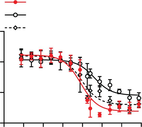
NATURE COMMUNICATIONS | https://doi.org/10.1038/s41467-019-14091-4 ARTICLE
a b 100 c
200 100 ns
Plasma creatinine (μM)
80
Calcium carotid artery
Calcium kidney
80
(mg/g tissue)
150
(mg/g tissue)
60
60
100 40
40
50 20 20
0 0 0
Vehicle (OEG2)2-IP4 IP6 Vehicle (OEG2)2-IP4 Sham Vehicle (OEG2)2-IP4
d e f
Thoracic aorta Abdominal aorta
Surviving animals per group (%)
40
100
Calcium abdominal aorta
80 Vehicle 30
Vehicle
(mg/g tissue)
1 × 12.5
60
1 × 25 20
40 1 × 50
2 × 25 10
20
1 × 12.5 mg/kg
2 × 50
0 0
0 14 5 6 7 8 Vehicle 1 × 12.5 1 × 25 1 × 50 2 × 25 2 × 50
Time (days) (OEG2)2-IP4 (mg/kg/day)
g h
1 × 25 mg/kg
50 70
area abdominal aorta (%)
Calcium thoracic aorta
60
Von Kossa-positive
40
(mg/g tissue)
50
30 40
20 30
20
2 × 25 mg/kg
10
10
0 0
Vehicle 1 × 12.5 1 × 25 1 × 50 2 × 25 2 × 50 Vehicle 1 × 12.5 1 × 25 1 × 50 2 × 25 2 × 50
(OEG2)2-IP4 (mg/kg/day) (OEG2)2-IP4 (mg/kg/day)
1 × 50 mg/kg
i 50 j 80
area thoracic aorta (%)
Calcium carotid artery
Von Kossa-positive
40
60
(mg/g tissue)
30
40
20
2 × 50 mg/kg
20
10
0 0
Vehicle 1 × 12.5 1 × 25 1 × 50 2 × 25 2 × 50 Vehicle 1 × 12.5 1 × 25 1 × 50 2 × 25 2 × 50
(OEG2)2-IP4 (mg/kg/day) (OEG2)2-IP4 (mg/kg/day)
Fig. 5 (OEG2)2-IP4 prevents calcification in a vitamin D-induced rat model of VC. Calcification was induced by either s.c. administration of 300,000 IU/
kg/day vitamin D3 for 5 days combined with high-phosphate diet (a–c) or by oral administration of 100,000 IU/kg/day vitamin D3 for 4 days combined
with a warfarin-supplemented diet (d–j). Bulk calcium in the carotids (a) and kidneys (b), and plasma creatinine (c) of vehicle (n = 8 (b), n = 10 (a, c)),
groups treated with twice daily s.c. injections of 30 mg/kg (OEG2)2-IP4 (n = 11) or IP6 (n = 5), and sham (n = 7). One rat in the vehicle group died several
hours after first vitamin D3 injection without explanation. Animals in the IP6 group had to be sacrificed 2 days ahead of schedule and no direct comparison
can be made. d Kaplan–Meier survival plot. e, g Calcium quantification (e) and von Kossa-positive area quantitation (g) in the abdominal aorta.
f Representative images of aortic calcifications stained with von Kossa (black) (scale bar = 500 µm). Calcium (h) and von Kossa-positive area (i)
quantification in the thoracic aorta. Calcium quantification in the carotid artery (j). In d, n = 40 for vehicle, n = 12 for 1 × 12 and 1 × 25 mg/kg/day, n = 18
for 2 x 25 mg/kg/day, and n = 19 for 1 × 50 and 2 × 50 mg/kg/day dose groups. In e–j, n = 7 (e–h) and n = 6 (j) for 1 × 12.5 mg/kg/day, n = 8 for 1 ×
25 mg/kg/day, n = 14 (i) and n = 15 (e–g, j) for 1 × 50 mg/kg/day, n = 18 for 2 × 25 mg/kg/day, n = 16 for 2 × 50 mg/kg/day dose groups, n = 24 (g) and
n = 25 (e, h–j) for vehicle. All data are presented as mean ± s.d. from n animals. Statistical significance was derived from non-parametric Kruskal–Wallis
and multiple comparison was performed by Mann–Whitney test with Bonferroni correction, with *p < 0.05, **p < 0.01 and ***p < 0.001 vs. vehicle.
Comparison of survival was performed by Mantel–Cox test.
Efficacy of (OEG2)2-IP4 in an adenine-induced model of VC. high-phosphate-diet. (OEG2)2-IP4 was administered via a s.c.-
Uremia increases the risk for developing VC. Therefore, the effect implanted osmotic pump at doses of 5, 15, or 50 mg/kg/day for
of (OEG2)2-IP4 on the development of VC was investigated in a 4 weeks, and treatment was initiated 1 week after the start of
previously established adenine and high-phosphate-diet-induced adenine supplementation. As opposed to the vitamin D-induced
rat model of VC that results in uremia and chronic renal failure66. calcification model, mortality in the adenine and high-phosphate-
Renal failure, and subsequent calcification, was induced by diet-induced VC model was low and did not differ significantly
feeding a high-phosphate-diet regime for a total of 7 weeks, which between treatment groups (Supplementary Table 7). Animals in
also had a low (2.5%) protein content and was supplemented with this model developed severe CKD (Supplementary Fig. 20), which
0.75% adenine for a period of 4 weeks, starting 2 weeks into the was accompanied by substantial weight loss (Supplementary
NATURE COMMUNICATIONS | (2020)11:721 | https://doi.org/10.1038/s41467-019-14091-4 | www.nature.com/naturecommunications 9
ARTICLE NATURE COMMUNICATIONS | https://doi.org/10.1038/s41467-019-14091-4
a 50
b c Thoracic aorta
Calcium abdominal aorta 0.20
40
0.15
(mg/g tissue)
Calcium heart
Vehicle
(mg/g tissue)
30
0.10
20
0.05
10
0
5 mg/kg/day
0.00
Vehicle 5 15 50 Vehicle 5 15 50
(OEG2)2-IP4 (mg/kg/day) (OEG2)2-IP4 (mg/kg/day)
d 50 e 50
Calcium carotid artery
area thoracic aorta (%)
40 40
Von Kossa-positive
15 mg/kg/day
(mg/g tissue)
30 30
20 20
10 10
50 mg/kg/day
0 0
Vehicle 5 15 50 Vehicle 5 15 50
(OEG2)2-IP4 (mg/kg/day) (OEG2)2-IP4 (mg/kg/day)
f 50 g 1000 h 500
Calcium femoral artery
Serum CPP (μg/mL)
40 800
400
(mg/g tissue)
T50 (min)
30 600
300
20 400
200
10 200
100
0 0
0
Vehicle 5 15 50 Vehicle 5 15 50 Vehicle 5 15 50
(OEG2)2-IP4 (mg/kg/day) (OEG2)2-IP4 (mg/kg/day) (OEG2)2-IP4 (mg/kg/day)
Fig. 6 (OEG2)2-IP4 dose-dependently prevents calcification in an adenine diet-induced rat model of VC. a, b, e, f Quantification of total calcium in the
abdominal aorta (a), the heart (b), the carotid arteries (e), and the femoral arteries (f). c Representative pictures of calcification in the thoracic aorta
stained positive with von Kossa (black) in the different treatment groups (scale bar = 500 µm). d Von Kossa-positive area quantitation in the thoracic
aorta. g Calcium content in CPPs collected from animal sera at sacrifice. h T50 derived from animal sera collected at sacrifice. Results are presented as
mean ± s.d. from n animals, with n = 11 for vehicle, n = 12 for the 5 mg/kg/day dose group, and n = 11 for the 15 and 50 mg/kg/day groups. N = 10 for
vehicle in e and h, and for 50 mg/kg/day in e. Statistical significance was derived from non-parametric Kruskal–Wallis test and multiple comparison was
performed by Mann–Whitney test with Bonferroni correction; *p < 0.05 vs. vehicle, #p < 0.05, ##p < 0.01 vs. 5 mg/kg/day, and §p < 0.05, §§p < 0.01 vs.
15 mg/kg/day.
Fig. 21). No significant difference in the level of renal failure and 50 mg/kg/day also prevented calcification in the carotid artery
body weight loss was observed between the different treatment compared to the 5 and 15 mg/kg/day groups (Fig. 6e). In the
groups, with the exception of creatinine at week 3 and serum femoral artery, reduction in bulk calcium was insignificant for all
phosphate at sacrifice in the 15 mg/kg/day group (Supplementary tested groups, likely due to the low level of calcification detected
Fig. 20). in the vehicle control (Fig. 6f, Supplementary Table 9). In
There was a trend in a dose-dependent reduction of bulk agreement with the results of the PK study and the vitamin D-
calcium in (OEG2)2-IP4-treated animals compared to vehicle in induced VC model, the calcemic status of the animals was not
the abdominal aorta, which is especially prone to calcify in this influenced by the administration of (OEG2)2-IP4 50 mg/kg/day
model, with a significant effect when comparing the highest dose for 4 weeks (Supplementary Fig. 20c). The calcium content
(50 mg/kg/day) to the lowest dose (5 mg/kg/day) (Fig. 6a). The derived from isolated serum CPP at sacrifice (Fig. 6g), CPP2
inhibitory effect of (OEG2)2-IP4 on heart calcifications was hydrodynamic diameter (Supplementary Fig. 22), and the serum
significant starting at 15 mg/kg/day (Fig. 6b). The compound’s calcification propensity (T50) measured in sera collected at
activity was also evident following von Kossa staining (Fig. 6c) on sacrifice (Fig. 6h) did not differ between treatment groups.
the thoracic part of the aorta, and quantification of the von Tibial bone histomorphometric analysis revealed an unchanged
Kossa-positive area (Fig. 6d). Treatment with (OEG2)2-IP4 at mineralized trabecular bone area for the various (OEG2)2-IP4
50 mg/kg/day significantly reduced calcification compared to doses tested (Supplementary Fig. 23). However, a distinct increase
vehicle (2.72 ± 5.97 vs. 14.50 ± 15.11% von Kossa-positive area), in the osteoid area was observed in the treatment groups, which,
and also compared to 5 and 15 mg/kg/day groups (Supplemen- in the presence of an intact osteoblast number/activity, points
tary Table 8). In line with this, treatment with (OEG2)2-IP4 at towards an imbalance between osteoid deposition and subsequent
10 NATURE COMMUNICATIONS | (2020)11:721 | https://doi.org/10.1038/s41467-019-14091-4 | www.nature.com/naturecommunicationsNATURE COMMUNICATIONS | https://doi.org/10.1038/s41467-019-14091-4 ARTICLE
mineralization (Supplementary Fig. 24). As both osteoblast and clinical trials, given the growing evidence of its value as a
osteoclast morphology were not impacted (Supplementary Fig. predictive marker for calcification progression33,34,69.
25), this observation suggests that (OEG2)2-IP4 may not exert The differences in activity between (OEG2)2-IP4 and IP6 are
cellular toxicity, and that the effect on mineralization of growing unlikely to be mediated solely by steric hindrance of the OEG
bone is probably due to a passive adsorption effect. In order to moieties, as the ethylene glycol chain length is short compared to
investigate the 3D bone structure, micro-computed tomography what is typically used for nanoparticle coating (0.1 vs. 2–5 kDa)47.
(µCT) analysis was performed, revealing the mineralized The observation that IP6 is a stronger chelator of soluble ionic
trabecular bone structures to be better preserved in the calcium than (OEG2)2-IP4, but a weaker inhibitor of calcification,
(OEG2)2-IP4-treated animals compared to vehicle-treated ani- leads to the hypothesis that an equilibrium exists between binding
mals (Supplementary Fig. 26, Supplementary Table 10). The low to soluble and solid forms of calcium. Therefore, the reduced
volume of distribution of (OEG2)2-IP4 observed in the PK study charge density of (OEG2)2-IP4 may decrease its binding to soluble
also suggests that only little compound leaves the circulation, and ionic calcium (present in large excess in biological fluids)
hence is available to interact with bone. rendering it more available for binding to solid forms of calcium.
The OEG chains may prevent stacking and precipitation of
inositol phosphate–calcium complexes and hence further con-
Discussion. Cardiovascular disease remains the leading cause of tribute to reducing mineral accretion.
death in the world despite significant advances in the develop- This mechanism of action is possibly similar to that of
ment of therapies addressing classical risk factors such as high extracellular matrix and serum proteins that contain acidic and
cholesterol, hypertension, and diabetes, combined with improved phosphorylated residues in order to function as exquisite
awareness of the negative impacts of obesity and smoking. This regulators of biomineralization32,70. Our observation is in line
highlights the importance of investigating different strategies to with the structural basis of calcification inhibition by fetuin-A,
combat cardiovascular diseases. One largely unexplored target for where surface binding of calcium is mediated by four negatively
pharmacological intervention is VC, despite the fact that it has charged amino acids on the extended β-sheet of domain D1 that
consistently been linked to poor clinical outcomes8. In fact, the occupy phosphate positions on the face of apatite crystals71.
coronary calcification score is the strongest predictor of coronary It can be hypothesized that (OEG2)2-IP4 acts by stabilizing
events in asymptomatic patients67. Certain high-risk populations CPP2 particles and reducing their uptake by VSMCs and
particularly prone to VC have been identified, such as CKD subsequent calcium overload, which may cause downstream
patients for which the extent of VC and risk of cardiovascular procalcific and pro-inflammatory events24,30. Chronic exposure
disease increase dramatically as renal function declines68. to CPPs, e.g., when clearance becomes overwhelmed as in CKD, is
In the present study, a series of inositol phosphate derivatives a likely driver of VC24. CPPs were found to be present in serum
were examined for their potential to inhibit VC. Screening for from high-phosphate-diet-fed rats with adenine-induced renal
modifications both in the number and size of OEG moieties on failure72. By stabilizing CPPs, (OEG2)2-IP4 may aid mineral
myo-inositol phosphates led to the identification of (OEG2)2-IP4 clearance from the circulation before VC can ensue. No delay in
as a potent inhibitor of VC. (OEG2)2-IP4 displayed activity in the T50 was observed when serum samples were tested from the
high nanomolar range in the competitive 18F-NaF CPP-binding adenine-induced VC rat model, as (OEG2)2-IP4 was likely to be
assay, and prevented the growth of nascent CPPs, their transition already eliminated or bound to existing calcified deposits at the
into the crystalline phase, and calcification of VSMC monolayers time of sampling.
in the low micromolar range. Conversely, IP6 was associated with Although PEGylation of small molecules with relatively long
mid-micromolar activity or a lack of dose–response in these PEG chains (>2 kDa) has previously been reported to improve
assays. As opposed to (OEG2)2-IP4, IP6 did not stabilize CPP in PK, it was unexpected to discover that conjugation with only two
their primary state and led to the formation of CPP2s, which are ethylene glycol repeating units was sufficient to increase systemic
hypothesized to initiate pathological response29,30. exposure73. The prolonged plasma half-life of (OEG2)2-IP4
In the case of the CaP-induced VSMC calcification model, the compared to IP6, observed in healthy animals, is likely due to
(OEG2)2-IP4 possibly acts in multiple ways to prevent calcifica- the enhanced metabolic stability of (OEG2)2-IP4. Forty percent of
tion. Matrix vesicles secreted by VSMCs under pro-calcifying the intact compound was recovered in the urine, suggesting
conditions form the first nidus for mineralization and chemi- additional routes of elimination, such as hepatic clearance and
sorption of (OEG2)2-IP4 onto such nidi may result in crystal degradation in plasma that may result in dephosphorylated
growth inhibition57. Potential intracellular effects, although metabolites of (OEG2)2-IP4. Plasma exposure of (OEG2)2-IP4 was
unlikely due to the compound’s hydrophilicity and charge somewhat extended in adenine and high-phosphate-diet-fed
making it difficult to diffuse through cell membranes, cannot be animals compared to healthy controls. This effect is seen for
excluded and should be further investigated. many active pharmaceutical ingredients, and can be rationally
(OEG2)2-IP4 was efficacious in both adenine and high- explained by the decreased renal clearance. In contrast, the
phosphate-diet-induced, and vitamin D-induced rat models of plasma level of IP6 has been reported to be decreased in uremic
VC, and displayed dose–response in vitro as well as strong in vivo rats45, possibly due to increased enzymatic metabolism in the
activity without affecting serum calcium levels. Importantly, state of uremia, which for (OEG2)2-IP4 may be counterbalanced
(OEG2)2-IP4 exhibited superior activity and prolonged plasma by its higher stability.
exposure compared to IP6 following bolus i.v. and s.c. admin- Since the OEG polymer shows little toxicity, is highly water
istration, the latter of which opens the possibility of therapeutic soluble and eliminated from the body intact via the kidneys
use in a chronic and ambulatory setting. The PK data show that (PEGs < 30 kDa), and IP6 has previously been administered to
after s.c. injection plasmatic concentrations above the IC50 were patients displaying acceptable safety and tolerability43, the
achieved for at least 4 h vs. 12 min for IP6 (Fig. 4c), which partly likelihood of observing serious toxic side effects with this
explain the higher efficacy achieved with (OEG2)2-IP4. Moreover, chemical entity is low.
to our knowledge, this is the first study showing that a In the present study, we observed an effect of (OEG2)2-IP4 on
pharmacological treatment can impact the 18F-NaF-PET signal bone osteoid area. However, it was seen in a CKD/VC rat model
in the context of VC, and it may therefore serve as a useful tool in young animals with open growth plates and rapid bone growth
for pharmacodynamic read-outs in pre-clinical and early stage that presents a dramatically increased bone turnover with chaotic
NATURE COMMUNICATIONS | (2020)11:721 | https://doi.org/10.1038/s41467-019-14091-4 | www.nature.com/naturecommunications 11You can also read