Photobiomodulation of the Visual System and Human Health - MDPI
←
→
Page content transcription
If your browser does not render page correctly, please read the page content below
International Journal of
            Molecular Sciences

Review
Photobiomodulation of the Visual System and
Human Health
John Buch 1, *       and Billy Hammond 2
 1    Johnson & Johnson Vision, Research & Development, Jacksonville, FL 32256, USA
 2    Department of Psychology, University of Georgia, Athens, GA 30602, USA; bhammond@uga.edu
 *    Correspondence: jbuch@its.jnj.com; Tel.: +1-904-443-1707

 Received: 30 September 2020; Accepted: 27 October 2020; Published: 28 October 2020

 Abstract: Humans express an expansive and detailed response to wavelength differences within
 the electromagnetic (EM) spectrum. This is most clearly manifest, and most studied, with respect
 to a relatively small range of electromagnetic radiation that includes the visible wavelengths with
 abutting ultraviolet and infrared, and mostly with respect to the visual system. Many aspects of our
 biology, however, respond to wavelength differences over a wide range of the EM spectrum. Further,
 humans are now exposed to a variety of modern lighting situations that has, effectively, increased
 our exposure to wavelengths that were once likely minimal (e.g., “blue” light from devices at night).
 This paper reviews some of those biological effects with a focus on visual function and to a lesser
 extent, other body systems.

 Keywords: electromagnetic radiation; photobiomodulation; action spectrum; phototoxicity;
 visual system

1. Introduction
      James Maxwell, building on postulates from Gauss, Riemann, and Faraday, proposed in 1864
that an electromagnetic “disturbance” exists that travels in free space with the velocity of light [1].
His theory was later confirmed by Hertz around 1888 [1] and thus began our understanding of
electromagnetic (EM) radiation. It is now accepted that EM radiation is the flow of photons that behave
as both particles and waves, and is divided into bands depending on the propagating wavelength.
To illustrate, gamma waves have wavelengths measured at the atomic level (700 nm), 42.3% is visible
(400–700 nm), 6.3% is ultraviolet (UV) A (320–400 nm), 1.5% is UVB (290–320 nm), and 0.5% is UVC
(
Int. J. Mol. Sci. 2020, 21, 8020 2 of 22
constant and close proximity to humans. As such, there has been concern that chronic exposure to this
seemingly insignificant energy source (around 3 kHz–300 GHz) carries the potential for tissue damage.
Reflecting such concerns [4], the National Toxicology Program published extensive results [5] on the
effects aggregated over a lifespan of about 2 years, including in utero, of radiofrequency exposures
on the brains and hearts of rats and mice. The study found significant pathological changes, such
as DNA damage, using similar frequencies as those used by the telecommunications industry (900
and 1900 MHz), but the study used much higher energy exposures. Mice in the study, for instance,
were exposed to intensities of up to 10 Watts/Kg (analogous to ~500 Watts for a 110-pound human)
for around 9 h per day. Cell phone energy varies from about 2 Watts to around 0.2 Watts during a
call. The relatively recent release of 5G cellular networks has its own health controversies [6,7], and of
course, some individuals keep their cell phones within close proximity constantly.
Hence the conundrum when considering the interaction of EM radiation and biology: how are
the relatively intense, acute, and easily quantifiable exposures used in animal models comparable to
the relatively weak exposures that humans experience over a lifetime? Human behavior is complex.
How does one link any complex outcome to any single (often weak) input with dozens of significant
and confounding correlates? Not surprisingly, for example, epidemiological studies on the relation of
cancer to cell phone usage are inconsistent at best [5].
Similar challenges are encountered when relating light damage to chronic ocular disease. Intense
acute studies on animal models tend to show very clear results with easily described underlying
mechanisms (for example, Barker et al. studied the protective effects of xanthophylls on short-wave
light-induced retinal damage [8]). Long-term chronic studies tend to be inconsistent [9] and depend
strongly on the nature of the disease. With respect to the latter, for instance, easily quantified (e.g., eyelid
malignancies) or relatively acute conditions (e.g., photokeratitis) show a much more straightforward
relation to actinic light than more difficult to quantify retinal diseases such as macular degeneration [10].
As a consequence, some researchers have decided to focus less on chronic degenerative diseases,
which are characterized by long latencies and complex etiologies, and more on acute biological effects.
If a stressor causes effect X, this is both meaningful in its own right and likely has implications for
long-term disease. For example, cells phones (radio frequency detectors) are often carried around
the waist and reproductive germ cells divide rapidly (spermatogenesis/oogenesis), which are easy to
quantify in terms of cell counts and dynamics. There is good evidence that mobile phone exposure
influences oxidative stress, sperm mobility, and fertilization [11]. Implantation of a UV-transmitting
intraocular implant in one eye and a UV-blocking implant in the other causes loss of S-cone sensitivity
in the UV-transmitting eye over a span of five years [12], not the same as AMD but S-cone loss is
significant in its own right, a likely good indicator of approaching retinal disease [13].
Taking such a direct approach—light exposure vs biological response—allows a number of
answerable questions to be posed. The first is methodological: How does one relate wavelengths,
throughout the EM spectrum, to human biological response? This methodology was perhaps best
worked out by plant photobiologists in the form of action spectra. Action spectroscopy is characterized
by the measurement of biological effects that occur as a function of wavelength [14]. The Commission
Internationale de l’Eclairage (CIE) defines the photobiologically active range from UVC (100 nm) to
IRC (1,000,000 nm) [15]. The origin of this technology began with the obvious and differential light
sensitivity of plants. It was used, for example, to originally identify chlorophyll as the pigment most
likely involved in photosynthesis [16]. Photochemistry, in general, was based on the observation that
reactants respond (react) to some wavelengths more strongly than others. It was natural to generalize
these observations to the photobiology of organisms. At first, the emphasis was on damage: some
wavelengths were more damaging than others [17]. After, studies shifted to the efficacy of certain
wavelengths in mediating a variety of biological reactions. The effects were ubiquitous.
In this review, we identify and describe a number of these biological effects, most dealing with
our most EM-responsive system (vision), focusing mainly on EM effects as quantified by action spectra.Int. J. Mol. Sci. 2020, 21, 8020 3 of 22
We also highlight the effects that EM radiation has on other body systems, thus attempting to collect
this information into one reference.
2. Spectral Band Terminology
Summarizing the effects of wavelength is not straightforward, given the non-uniformity in defining
various spectral bands across various disciplines. The boundaries of what is termed visible light are often
defined with a low and high spectral end between 360 and 400 nm and 760–830 nm, respectively [18],
although younger individuals can consistently see UVB, ca. 315 nm [19] and infrared, ca. 1100 nm
under some circumstances [20]. How EM radiation passes through materials such as glass or quartz,
water and ozone, or is absorbed or transmitted by proteins and water, can have different meanings
for disciplines such as optical engineering, meteorological optics, or photobiology, respectively [21].
Photodermatology divides UVA/UVB at 320 nm, although CIE recommends 315 nm [22].
There are small discrepancies even within different ISO standards depending on the application.
For example, ISO 21348 provides a process for determining solar irradiance and defines UVA as
315–400 nm, while ISO 8980 provides transmittance specifications for ophthalmic lenses and defines
UVA as 315–380 nm. ISO 21348 defines “soft x-rays” as 0.1–10 nm, while ISO 20473 (optics and
photonics) defines “extreme ultraviolet” as 1–100 nm. Superficial discrepancies can also be found
within the same standard, such as ISO 21348, stating that the color purple begins at 360 nm while the
visible light spectrum begins at 380 nm.
The commonly described “Blue Light Hazard” function is even a misnomer since over half of
the rays that constitute this spectral range would be considered purple or violet in color. ISO 8980-3
defines the Blue Light Hazard range as 380–500 nm. The Blue Light Hazard can be further refined
(e.g., 380–460 nm) based on the wavelengths of greatest potential risk (ISO/TR 20772). The term
High-Energy Visible (HEV) light is sometimes used in lieu of the blue light hazard but tends to refer
to the same wavebands. HEV ranges fluctuate from 380 to 500 nm [23], 400–500 nm [24], and others
stating 430–510 nm [25] depending on the author’s particular field. This is different from the “Retinal
Hazard Region” often defined as 400–1400 nm [26].
Consistent terminology across fields is important since light influences nearly every system of
the body, and those effects are largely wavelength-dependent. Light, for instance, entrains circadian
rhythms, for humans and most other species, ranging from cyanobacteria to “higher” animals (studied
by chronobiologists). We have “clocks” throughout our bodies, most entrained by the master clock
in the brain (the suprachiasmatic nucleus of the hypothalamus). The biochemical bases for such
clocks are hormones such as melatonin, and the action spectrum for these mediators has been well
established [27]. Light is also considered an essential therapy for seasonal affective disorder and is
believed to be more efficacious when it contains a significant amount of blue (468 nm) light [28].
3. Introduction to EM Biological Effects
Since the original invention of lasers (essentially monochromatic light that could be more
easily delivered), light has been used as a means of therapy. Specific wavelengths of light have
been shown to be effective at reducing pain, inflammation/edema, and promoting the healing of
wounds [29]. Light has been shown to be directly or indirectly associated with alertness, performance,
and mood [30]. It can be used to stimulate hair growth [31] when hair is thinning (max = 830 nm) and
to stimulate the production of collagen by the skin. Indeed, all cells contain light-responsive substrates
(chromophores like cytochrome c oxidase) whose activity is altered by exposure to select wavebands
of light. For example, mitochondria create energy for the cell and light can enhance that process
(ATP production), changing transcription factors and the regulation of reactive oxygen species [32].
Blue light (400–500 nm), with or without red light (>600 nm), has been used for the treatment of
acne [33].
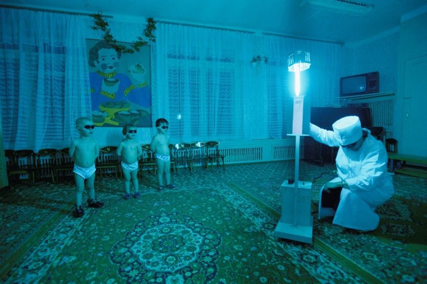
Sunlight can also be used to synthesize (from cholecalciferol, cholesterol in the skin) an essential
nutrient, Vitamin D (λmax = 300 nm, [34]). Simulated UV-exposure has been used in sunlight-deprivedInt. J. Mol. Sci. 2020, 21, 8020 4 of 22
Int. J. Mol.of
regions Sci.the
2020, 21, x FOR
world PEER REVIEW
(Figure 4 of 22
1). Vitamin D itself is the precursor to the steroid hormone calcitriol.
Calcitriol regulates a number of cellular pathways known to be central to cancer progression and
progression
risk and D
[35]. Vitamin risk
was[35]. Vitamin
originally D was
added originally
to milk in orderadded to rickets
to reduce milk in(weakened
order to bone
reduce rickets
especially
(weakened bone especially obvious with weight-bearing bones like the femur, which
obvious with weight-bearing bones like the femur, which caused kids to become bow-legged) that caused kids to
become bow-legged) that occurred especially in Black children in inner cities,
occurred especially in Black children in inner cities, where buildings block the sun [36]. where buildings block
the sun [36].
Figure
Figure 1. In sunless
1. In sunless Lovozero,
Lovozero, Russia,
Russia, children
children bathe
bathe in ultraviolet light
in ultraviolet light to produce vitamin
to produce vitamin D
D for
for their
their
bones. Photo © Joe McNally.
bones. Photo © Joe McNally.
Each of these systemic effects has its own specific wavelength dependency. For example, cell
Each of these systemic effects has its own specific wavelength dependency. For example, cell
culture studies have shown that maximum cell proliferation of fibroblasts occurs with light from
culture studies have shown that maximum cell proliferation of fibroblasts occurs with light from 665
665 to 675 nm, whereas slightly longer wavelengths (810 nm) inhibit cell division [37]. The field of
to 675 nm, whereas slightly longer wavelengths (810 nm) inhibit cell division [37]. The field of
biophotonics uses specific wavelengths of light to achieve imaging down to the cellular level [38]. It is
biophotonics uses specific wavelengths of light to achieve imaging down to the cellular level [38]. It
sometimes the case that even very small changes in wavelength can create large changes in biological
is sometimes the case that even very small changes in wavelength can create large changes in
response at the cellular level. Moon et al. exposed retinal pigment epithelial cells loaded with A2E to
biological response at the cellular level. Moon et al. exposed retinal pigment epithelial cells loaded
449, 458, and 470 nm light at varying time intervals [39]. Despite the equal energy of the exposure,
with A2E to 449, 458, and 470 nm light at varying time intervals [39]. Despite the equal energy of the
small changes in wavelength resulted in large changes in cell viability. For example, after 48 h of
exposure, small changes in wavelength resulted in large changes in cell viability. For example, after
exposure, the production of reactive oxygen species was three times higher for cells exposed to 449
48 h of exposure, the production of reactive oxygen species was three times higher for cells exposed
vs 470 nm light. Most recently, long wave light (670 nm) has been used to stimulate mitochondrial
to 449 vs 470 nm light. Most recently, long wave light (670 nm) has been used to stimulate
function in the retina as a means of retarding age-related decline in photoreceptor function [40].
mitochondrial function in the retina as a means of retarding age-related decline in photoreceptor
function
4. [40].
Phototoxicity, Dosimetry, and Action Spectrum of the Visual System
Light is the primary
4. Phototoxicity, Dosimetry,stimulus used by
and Action the visual
Spectrum system.
of the VisualWhat is less obvious is that different
System
wavelengths of light influence literally every aspect of the visual process. The early philosophers,
from Light is the to
the Greeks primary stimulus
the Chinese, usedmostly
focused by the on visual
howsystem?
wavelengthWhatrelated
is lesstoobvious is that different
the perception of color.
wavelengths of light influence literally every aspect of the visual process.
The first systematic/empirical study of wavelength and color has been attributed to Isaac The early philosophers,
Newton
from the Greeks
(Opticks, to the
1704), but, Chinese,
as art focused
and science mostly on
flourished how
hand inwavelength
hand through related to the perception
the Renaissance, so didof color.
interest
The
in first
the systematic/empirical
underlying study
optics of color. of wavelength
Prominent and color
intellectuals likehas been published
Goethe attributed treatises
to Isaac Newton
such as
(Opticks, 1704), but, as art and science flourished hand in hand through the
Zur Farbenlehre (‘Theory of Color,’ 1810). It was not until 1916 [41], however, that human spectral Renaissance, so did
interest in the
sensitivity underlying
curves optics ofand
were published color.
thenProminent
codified by intellectuals
the CIE [42]. like Goethe
What published
followed, and treatises
is beyondsuch
the
as Zur Farbenlehre (‘Theory of Color,’ 1810). It was not until 1916 [41], however,
scope of this review, were extensive studies of how the optics of the eye are affected by wavelength that human spectral
[43],
sensitivity
as curves were published
well as characteristics as diverse and
as then codified
spatial [44] andby temporal
the CIE [42].visionWhat followed,
[45]. and this
Along with is beyond
work
the scope of this review, were extensive studies of how the optics of the eye
came the realization that different wavelengths of light are more or less damaging to the visual system, are affected by
wavelength [43], as well as characteristics as diverse as spatial [44] and temporal vision [45]. Along
with this work came the realization that different wavelengths of light are more or less damaging to
the visual system, and other surface tissues such as skin. Van Norren and Vos published a
comprehensive review of the history of research on actinic action spectra [46]. This empirical workInt. J. Mol. Sci. 2020, 21, 8020 5 of 22
and other surface tissues such as skin. Van Norren and Vos published a comprehensive review of
the history of research on actinic action spectra [46]. This empirical work mostly began with animal
models (the work of Noell and Ham in the 1960s) but the damaging effects of light were also being
actively considered in the ophthalmic literature [17].
Light, in this case, with its potential to harm, refers mostly to the higher energy portions of the EM
spectrum, although significant exposure to lower energy light (say infrared) can also predispose an
individual to damage. For example, thermal lensing can defocus NIR at high intensities and increase
susceptibility [47]. Historically, the primary source for high-energy light came from the sun. Even now,
although many common devices (mobile phones, computers, etc.) emit light in the short-wave portions
of the spectrum, the amount is likely not significant enough to initiate photo-oxidative damage [48].
Light that damages the eye is termed phototoxic. Phototoxicity can take different forms depending
on the wavelength and intensity of the approaching EM radiation. Ocular phototoxicity is subdivided
into photothermal (photocoagulation), photomechanical (photoacoustic), or photochemical damage.
Photothermal damage typically occurs with the longer visible and NIR wavelengths of 600–1400 nm,
where photons have enough energy to cause molecular vibration without splitting electrons from
atoms within the molecule. This is inferred as heat and is particularly damaging to the crystalline lens
(e.g., “glassblowers” cataract) [49] and intentionally applied to the retina for laser photocoagulation of
certain disease states (e.g., diabetic retinopathy, retinal edema, etc.) [26]. Photomechanical damage
generally refers to a very short pulse (e.g., nanoseconds) of very high energy (e.g., megawatts/cm2 )
that results in a thermoelastic pressure wave [50]. This shock wave results in microcavitation bubbles
and permanent tissue change. A common example is the neodymium-doped yttrium aluminum
garnet (Nd:Yag) laser used to punch a hole in the peripheral iris (iridotomy) to help control glaucoma.
The Nd:Yag laser is often set to 532 nm, 700–900 mW, for 0.1 s [51]. Photochemical damage is thought to
occur when photons are absorbed by the molecules of a tissue, exciting the electrons from the ground to
excited state (i.e., the formation of free radicals or reactive oxygen species). This is typically associated
with long exposure times and higher energy light [26]. Photochemical ocular damage is associated
with the pathogenesis of diseases such as macular degeneration and choroidal neovascularization.
Photochemical exposures are additive according to the Bunsen-Roscoe law of photochemistry which
basically states that damage can occur from an intense radiation source for a short time, or a less intense
source for a longer period of time [52]. Paradoxically, the damaging photochemical process is also
utilized in photodynamic therapy (PDT) where unwanted anomalies are treated with a photosensitizing
drug then exposed to EM radiation (e.g., 689 nm) to induce the free radical process and reduce the
anomaly [53].
There are many factors to consider when calculating the dose of when a particular wavelength can
cause harm to a tissue. Apart from the numerous environmental factors [54,55], radiation that reaches
the ocular surface is also dependent upon anatomical factors that might limit its entry into the eye [56,57]
such as the size and shape of the nose, skin reflectance, protruding eye (exophthalmos), sunken eye
(enophthalmos), eyelid droop (ptosis), eyelashes, eyebrow, and prominence of the supraorbital ridge.
Once the radiation reaches the ocular surface, then factors such as wavelength (action spectra), aversion
responses (looking away, pupil constriction), tissue repair, size of the light source, exposure intensity,
exposure area, and exposure duration all play a role [52]. Nonetheless, general exposure guidelines are
provided in Tables 1–5 where appropriate.
Several recent reviews are available that discuss the impact that electromagnetic radiation has
on the eye [46,58], skin [59,60], and other biological functions [61]. In brief, as noted, UV damage
is photochemical, not photothermal. “Actinic ultraviolet” is most significant below 320 nm (UVC
and UVB) but affects deeper structures of the eye based on penetration (e.g., UVA penetrates deeper
and may be more significant for retina). More recent reports suggest that UVB [62] and near UVA
(360–400 nm) [63] are important for myopia control. Furthermore, blue (464 nm) and red (634 nm) may
be important for emmetropization when coupled with a multifocal lens [64].Int. J. Mol. Sci. 2020, 21, 8020 6 of 22
Although, significant exposure to lower energy light (say infrared) can also predispose an
individual to damage. For example, thermal lensing can defocus NIR at high intensities and increase
susceptibility [47].
4.1. The Eyelids
Table 1 summarizes the effects of high-energy light on the eyelids. As shown in the table, most
of the effects of light on the eyelids can be predicted by the general effects of light on skin, with the
exception that the skin of eyelids is thin (as light filters go, they have an average optical density of
around 2.1 ± 0.3 in the visible range, [65]). They are also somewhat protected by the supraorbital bone
and eyebrows [58]. Lids, ridges, and brows combine to also provide some protection to the cornea, the
other major ocular surface tissue. The action spectra for the cornea is summarized in Table 2.
Table 1. Electromagnetic radiation action spectra and dosimetry thresholds for the lids.
EMR Phototoxicity and Dosimetry of the Lids
Wavelength
Effect Reference
Average erythemal UV dose of Americans is about 25,000 J/m2 /year. [66]
3X more carcinogenic than UVA (e.g., squamous cell carcinoma, SCC). [66]
Squamous cell carcinomas account for about 8%, and melanomas about 2%, of all-cause cancer
[67]
in Croatia.
UVB Basal cell carcinomas (BCC) account for 80–90% of all malignant tumors of the eyelid. [68]
280 < 315 nm Excess exposure increases risk of erythema, melanogeneisis, DNA damage, immune
[66]
suppression, photo-aging.
UVB is major cause of sunburn, which is leading risk factor for melanoma and non-melanoma
[66]
skin cancers.
BCC may be due to strong UVB exposure at a young age, whereas SCC appears due to
[10]
chronic/cumulative exposure.
UVA
Photoaging: main effect on the lids is sagging skin. [66]
315 < 400 nm
Visible Intense visible (e.g., 532 nm laser pointer) has been shown to cause ecchymosis of upper and
[69]
380 < 760 nm lower eyelids.
Photodynamic therapy (at 634 nm) is an effective treatment for basal cell carcinoma. [70]
Infrared Thermal radiation can cause burns ranging from mild to third degree and eventually skin death. [71]
Exposure Limits UVR
(see text 3 mJ/cm2 for 8 h daily to avoid redness (erythemal), and this is one-third to one-quarter of the [72]
discussion) minimal erythemal dose.
Table 2. Electromagnetic radiation action spectra and dosimetry thresholds for the cornea.
EMR Phototoxicity and Dosimetry of the Cornea
Wavelength
Effect Reference
Momentary exposure to UVC can cause photokeratitis such as welders flash. [73]
UVC
100 < 280 nm Susceptibility to photokeratitis may peak at 270 nm with exposure thresholds as
[74]
low as 3 mJ/cm2 .
10 h of UVA or 23 min of UVB can cause photokeratitis. [75]
UVB absorption is 1.8 times higher in the anterior 100 nm of the human cornea
than in the posterior layers. The absorption coefficients of the epithelium and
[76]
Bowman’s membrane are higher than the stroma, but the stroma absorbs more
UVB due to thickness. Tryptophan and ascorbic acid absorb UVB.
280 < 315 nm 300 nm causes apoptosis in all three layers of the cornea and induces keratitis.
[77]
Apoptosis in all layers of the cornea occurs 5 h after exposure.
UVB light can accelerate the physiological loss of corneal epithelium be two
[10]
mechanisms, shedding and apoptosis.
The biological damage potential at 295 nm is 375 times more than the biological
[78]
damage potential at 320 nm.Int. J. Mol. Sci. 2020, 21, 8020 7 of 22
Table 2. Cont.
EMR Phototoxicity and Dosimetry of the Cornea
Wavelength
Effect Reference
Climatic droplet keratopathy—chronic UVA and UVB. [10]
Corneal crosslinking: primary treatment for corneal ectatic disease, involves
[79]
application of vitamin B2 (riboflavin) + 370 nm to stiffen the cornea.
Epithelium: pseudo-keratinization, polyhedral intermediate cells, necrosis,
UVA [80]
lymphatic infiltration.
315 < 400 nm
Bowman’s membrane: detachment from epithelium, thickened, micro-bleedings. [80]
Stroma: swelling and collagen disorganization, inflammatory cells, angiogenesis
[80]
blood vessels.
Endothelial detachment. [80]
Punctate keratitis caused by 532 nm laser pointer. [69]
Visible
380 < 760 nm Climatic droplet keratopathy with higher blue (400–500 nm) light exposures, in
[81]
addition to UVA and UVB.
The cornea transmits 96% of incident infrared in the 700–1400 nm range, limiting
sensitivity to IR harm, especially in the 750–990 nm range. Significant exposure
NIR results in causing protein coagulation which can cause irreversible damage
[71]
760 < 1400 nm especially on endothelium layer. High-dose IR damage to the cornea causes
immediate pain and vascularization, with potential of loss of transparency and
opacification in response to burns that causes ulcers.
315–400 nm:
1 J/cm2 for exposure time < 1000 s
1 mW/cm2 for time ≥ 1000 s
Exposure Limits 180–400 nm:
(see text 3 mJ/cm2 pulsed hazard [52]
discussion) 770–3000 nm
1.8t−0.75 W/cm2 for time < 20 s
0.1 W/cm2 for time > 20 s
1.8t0.25 J/cm2 for time < 45 s
4.2. The Cornea
As shown in Table 2, many of the significant effects to the cornea are highly wavelength-dependent
and in the low UV range. As ocular structures go, the cornea represents the main interface between the
eye and environment, and hence absorbs the largest amount of radiation [80]. This, perhaps, saves
deeper structures to a large degree but creates great susceptibility of the cornea to very low wavelengths.
This is a risk both indoors and out. With respect to the former, UV light is used in entertainment such as
black lights at nightclubs, and mass presentations of photokeratitis have been reported [82]. UV used
as a disinfectant has been reported in 41 cases of photokeratitis in poultry workers [83]. UV promotes
inflammation (keratitis) though a number of interleukins, cytokines, and matrix metalloproteinases
that, ultimately, mediate cell damage [80]. It should be noted that corneal crosslinking is a procedure
used to stiffen an ectatic cornea by the combination of a photoinducer (vitamin B2) and 370 nm light [84].
The light used to conduct the corneal crosslinking procedure (365 or 370 nm) can also be used to
effectively treat some cases of infectious keratitis [85].
4.3. The Conjunctiva
The other ocular surface tissue that is also an eye-environment interface is the membranous
conjunctiva. Effects of EM radiation on the conjunctiva are outlined in Table 3. The most obvious clinical
manifestations of light damage to the conjunctiva are pterygium and pinguecula (noncancerous growths,
often nasal, visible on the sclera). Since the conjunctiva interfaces with the external environment, many
factors can induce general inflammation of the conjunctiva (conjunctivitis). This has made recent
news, for example, as a relatively unique symptom of COVID-19. Early in the natural and recent
history of the novel coronavirus pandemic was the observation that the condition can cause changesInt. J. Mol. Sci. 2020, 21, 8020 8 of 22
(often irritation) to the conjunctiva (indeed, the first doctor who identified the new condition was an
ophthalmologist, Li Wenliang). These changes manifest as dry eye, blurred vision, and mild follicular
conjunctivitis [86]. Ocular changes, like conjunctivitis, do not typically accompany many other systemic
infections (like influenza or even other coronaviruses like SARS or MERS). Almost 80% of conjunctivitis
cases are specific to the adenovirus [87] and occur early in life (0–4 years of age). Reaction of the eye
to systemic infection is relatively rare. This implies that CoV may be relatively unique in its ability
to influence surface tissues of the eye. This may be why this strain of coronavirus (HCoV-NL63)
caused alarm when identified initially in an infant displaying conjunctivitis. A subsequent study of
28 cases of children with confirmed HCoV-NL63 infections showed that 17% had conjunctivitis [88].
Although the disease could come in contact with the conjunctiva as an aerosol, the presence of CoV
in the conjunctival sac [89] suggested it might also arise from within the patient and be expressing
through lacrimal fluid. Viruses often absorb in the UVC range [90] which is why UV light is sometimes
used as a germicidal (e.g., hospital rooms sometimes use upper room germicidal irradiation) and is
being actively considered to help retard the spread of CoV, and UVC appears most effective [91].
Table 3. Electromagnetic radiation action spectra and dosimetry thresholds for the conjunctiva.
EMR Phototoxicity and Dosimetry of the Conjunctiva
Wavelength
Effect Reference
UVC Erythema: Although only 1% of 254 nm may penetrate the stratum corneum,
[74]
200–280 nm mild erythema still results.
UVB Ocular surface squamous neoplasia (OSSN) declines by 49% for each 10
[92]
280–315 nm degree increase in latitude.
Pterygium: hyperplasia of the bulbar conjunctiva that grows over the cornea. [10]
Pterygium: Associated with long-term exposure to UVA and UVB. [10]
Pterygium: High prevalence between latitudes ± 37 degrees. [93]
Pterygium may initiate by UV-induced changes in corneal epithelial stem
[94]
cells.
Pinguecula: fibro-fatty degenerative change in bulbar conjunctiva. [10]
UVB, UVA and Pinguecula: Weak association with long-term UVA and UVB. [10]
Visible
Pinguecula: May be a histological link with sun-induced skin changes. [95]
448 nm at 0.8 mW/cm for 6 h resulted in lysosomal membrane
[96]
permeabilization of the conjunctiva.
Conjunctival UV autofluorescence can be used to accurately determine the
[97]
time spent outdoors.
Damage of conjunctiva caused by 532 nm laser pointer. [69]
Exposure Limits 270 nm:
(see text 3 mJ/cm2 within minutes can cause conjunctival injection, chemosis, [98]
discussion) damaged epithelial cells, and the presence of inflammatory cells.
Almost 80% of conjunctivitis cases are specific to the adenovirus [87] and occur early in life (0–4
years of age). The reaction of the eye to systemic infection is relatively rare. This implies that CoV may
be relatively unique in its ability to influence surface tissues of the eye.
4.4. The Iris and Crystalline Lens
Light passing through the cornea/conjunctiva (which now has removed much of the UV) passes
through the pupil but is also incident on the pigmented iris. The iris is contiguous with the retinal
pigment epithelium (to wit, a darker iris reflects a darker posterior fundus) and it seems clear that the
degree of pigmentation (unlike skin, iris pigmentation stays relatively constant) evolved to screen the
further penetration of high-energy light [99]. This is why light iris color (reduced filtering) is thought to
be a significant risk for uveal melanoma [100]. In contrast, darker irises (absorbing more light energy),Int. J. Mol. Sci. 2020, 21, 8020 9 of 22
are linked to a higher risk of cataract. Cumming et al. posited that the absorbance of thermal energy
by a darker iris could raise the energy state of the crystalline lens and predispose it to other forms of
photo-oxidative damage [101]. Action spectra for the crystalline lens is provided in Table 4.
Table 4. Electromagnetic radiation action spectra and dosimetry thresholds for the crystalline lens.
EMR Phototoxicity and Dosimetry of the Crystalline Lens
Wavelength
Phototoxic Effect Reference
295–325 nm associated with crystalline lens damage and cataract formation.
Cortical and posterior subcapsular cataracts associated with intense 295–325 [72,102]
nm exposure delivered over days.
315 nm contributes significantly to cataract formation [102]
Risk for cortical cataracts increased 1.6-fold when the cumulative UVB
exposure doubled. No association of nuclear cataracts and UVB, or between [103]
UVB cataracts and UVA—Chesapeake Bay Study.
280 < 315 nm
Chronic UVB exposure linked to cortical cataract. [72]
Variations in individual behavior can be a reason for up to a 18-fold
[104]
difference in UVB exposure.
280–315 nm is the most biologically active EM radiation band. [74]
Men with higher levels of average annual UVB were 1.36x more likely to have
[105]
more severe cortical opacities than men with lower levels.
If the radiant energy exposure was continuous and if no repair processes
occurred, it would take 10 h of UVA to damage the cornea but 26 h to damage
[106]
the lens. Lens damage due to UVB exposure would take 245 h, but the cornea
would be damaged in 23 min.
UVA
315 < 400 nm A generalization of the findings indicates that lens damage thresholds for
[106]
UVA are in the J/cm2 range, and for UVB in the mJ/cm2 range.
315–400 nm accelerates crystalline lens aging. [102]
325 nm at 260 J/cm2 created cataracts in rhesus monkeys. [107]
Cataracts induced in rats with 1090 nm, 197 W/cm2 , multiple exposure times. [108]
Infrared Infrared causes lens changes in molecular weight and protein backbone
[109,110]
structure, age-related types of cataract.
315–400 nm:
1 J/cm2 for exposure time < 1000 s
1 mW/cm2 for time ≥ 1000 s
Exposure Limits 180–400 nm:
(see text 3 mJ/cm2 pulsed hazard [52]
discussion) 770–3000 nm
1.8t−0.75 W/cm2 for time < 20 s
0.1 W/cm2 for time > 20 s
1.8t0.25 J/cm2 for time < 45 s
4.5. The Aqueous Humor and Vitreous Humor
UVB radiation is absorbed by the cornea, aqueous humor, and crystalline lens, withInt. J. Mol. Sci. 2020, 21, 8020 10 of 22
in eyes with UV-absorbing contact lenses [111]. Lledo et al. recently reported that yellow filters can
significantly increase the amount of melatonin in the anterior chamber of the eye. This, in turn, reduces
the production of aqueous humor, presumably by inhibition of the non-pigmented epithelium of
the ciliary body, and reduced intraocular pressure approximately 40% in rabbits [116]. Light effects
on vitreous are also apparent. An increase in vitreous catalase activity by 33% was shown in calf
eyes irradiated with UVA for 3 h [117]. As with most ocular structures, effects on vitreous are
wavelength-dependent. X-rays appear to have no effect on the vitreous [118] but IRA (ca 780–1400 nm)
can cause a blanched retinal lesion or edema proceeding to vitreous hemorrhage [71].
4.6. The Retina
Table 5 provides a summary of the action spectra for the retina. The retina is unique from the other
tissue types in that it contains cells that do not undergo mitosis (hence, damage is often irreversible).
It is also the highest metabolically active tissue in the body. This metabolic activity results in excessively
high oxygen tension, combined with high lipid content, and photosynthesizers (those that are always
there, like rhodopsin, and others that increase with age, like A2E) makes for an exceptionally vulnerable
tissue (retinal degeneration is the most common form of blindness in developed countries).
Table 5. Electromagnetic radiation action spectra and dosimetry thresholds for the retina.
EMR Phototoxicity and Dosimetry of the Retina
Wavelength
Effect Reference
UVC
UVC causes time-dependent apoptosis of RPE cells. [119]
200 < 280 nm
UVB
UVB energy from 0.2 to 0.4 J/cm2 induces decreased phagocytic activity of RPE cells. [120]
280 < 315 nm
338 nm: Lipofuscin, a conglomerate of modified lipids and bisretinoids, is susceptible to
[121]
photochemical changes leading to irreparable cellular damage.
A2E, a lipofuscin fluorophore, has two peak absorbance rates, including one in the UVA
[121]
range at 338 nm.
325 nm can produce retinal lesions. [122]
UVA 315–400 nm: AMD linked to strong UVA exposure, mitochondrial DNA and RPE cells are
315 < 400 nm particularly susceptible to 390–400 nm radiation within UVA, 380 nm was noted to cause [123]
damage to rat photoreceptor cells—particularly rods.
Patients with macular degeneration have a higher rate of poor tanning ability and glare
[124]
sensitivity.
The retina was exquisitely susceptible to damage by UVA light, requiring irradiances
50–80 times lower to cause permanent photoreceptor cell damage with this wave-band [125]
compared to green light.
Re-analysis of the Chesapeake Bay watermen study demonstrated an association between
blue light exposure and AMD. Individuals with more sunlight exposure are at a [81,126]
significantly increased risk of AMD.
400–480 induces photoreceptor cell death via apoptosis, consequently killing RPE cells. [123]
400–550 solar retinitis. Violet to blue light can cause temporary conditions such as ‘red
[72,102]
vision’ (erythropsia) and reduction in night vision—especially in aphakic patients.
Visible 400–550 are toxic to the aging retina as it loses antioxidant protection contributing to AMD. [127]
380 < 760 nm 415–555 provided maximum loss of RPE. [128]
390–550 can irradiate lipofuscin, compromising lysosomal integrity and impairing
activities of catalase, superoxide dismutase, and cathepsin while inducing lipid [123]
peroxidation—ultimately damaging mitochondrial DNA and RPE cells.
Removal of the “blue” component of light significantly decreases retinal damage after high
[129]
intensity exposure.
Toxicity of “blue” led light and A2E is associated to mitochondrial dynamics impairment
[130]
in ARPE-19 cells: implications for age-related macular degeneration.Int. J. Mol. Sci. 2020, 21, 8020 11 of 22
Table 5. Cont.
EMR Phototoxicity and Dosimetry of the Retina
Wavelength
Effect Reference
Short-wave light triggers cellular stress responses that may be involved in RPE disease
[131]
development, which has implications for pathogenesis of AMD.
Short-wave light increases production of reactive oxygen species (ROS), inducing oxidative
[132]
stress and triggering photoreceptor cell and RPE death, which are risk factors for AMD.
403 nm: regenerates rhodopsin. [123]
404 nm: cytochrome oxidase and RPE inhibited. [123]
430 nm: Maximum A2R excitation degrades RPE cells. [123]
432 nm: Damage to RPE cells via absorbance of all-trans-retinal. [121]
439 nm: Disrupt retina blood barrier in rats. [123]
447 nm: Peak A2E excitation rate harms RPE cells. [121]
448 nm: RPE disruption. [96]
460 nm: greater retinal damage by blue (460 nm) compared to green (530 nm) and red (620
[133]
nm) LEDs.
470 nm: Damage to photoreceptor rods and RPE. [123]
488 nm: RPE disruption above ANSI’s photochemical MPE. [121]
Thermal damage causing enzymes to denature which contributes to permanent damage to
Infrared [71]
the photoreceptor and RPE. Deep retinal coagulation, may involve choroid.
305–700 nm
2 mW/(cm2 sr) for time > 10,000 s (phakic eye)
Exposure Limit
White light source
(see text [52]
0.22 mW/cm2 for time > 10,000 s
discussion)
380–1400 nm
0.7 W/cm2 for time > 10 s and retinal image diameter > 1.7 mm
Although, as recently noted by Hammond et al., there is large (over a factor of 30) individual
variation in the quantity of UVB reaching the retina prior to the age of about 30 years [19].
Atmospheric oxygen, in its ground-state or triplet form (3 O2), is a diradical, meaning that it
possesses two unpaired electrons that spin in parallel orbits. This coordination of electron orbits tends
to stabilize the triplet form of oxygen. If triplet oxygen, however, absorbs enough light energy to
reverse the spin of one of its unpaired electron orbits, it converts to a more reactive singlet form (1 O2).
The singlet form of oxygen stays reactive for a relatively long time period since conversion back to the
triplet form is spin-forbidden. Receptoral outer segments contain high quantities of polyunsaturated
fatty acids (PUFA). Reactive oxygen species can abstract hydrogen atoms from PUFA-rich receptoral
membranes, ultimately leading to peroxidation of the lipid. The withdrawal of hydrogen will convert
the PUFA into an organic radical that may then react in a similar manner with adjacent PUFA molecules.
This chain sequence, left unquenched, slowly begins the formation of the lipid-based oxidation products
that populate the older retinal pigment epithelium (the RPE does not have the enzymes to break down
oxidized lipids, so they simply accumulate). This basic model [134] is a common one used to describe
the natural history of acquired retinal degeneration, often combined with other diatheses such as
genetic susceptibility, impairment in choroidal circulation, and thickening of Bruch’s membrane.
4.7. Other Ophthalmic-Related Effects
Finally, it is clear that wavelength can differentially influence the visual system as a whole. Table 6
is a sample of such conditions, largely deleterious, where wavelength differences are known to have a
meaningful effect. For example, Revell showed that exposure to 420 nm light had a significant effect on
increasing the alertness of young male subjects, whereas the same exposure to 600 nm had the opposite
effect [135].Int. J. Mol. Sci. 2020, 21, 8020 12 of 22
Table 6. The relation of wavelength to other ophthalmic-related issues.
Wavelength Ophthalmic-Related Effect Reference
Blurred vision: Secondary to photokeratitis. [136]
UVA
315–400 nm Myopia development: 360–400 nm might be important for both preventing
[63]
myopia progression and the onset of myopia.
Migraine sensitivity: 447, 590, and 627 nm aggravates migraines, while 530
[137]
nm lessens severity.
Glare via fluorescence: Near UV or short visible wavelengths induces a blue
green fluorescence, which can be a source of intraocular veiling glare.
Exposure to near UV/blue wavelength sources can influence a glare intense
[138]
enough to reduce visual performance. Exposure to wavelengths longer than
365 nm induce weaker but progresses more towards red color, with 365 nm a
lens absorption peak rate.
Filtering 465–480 nm light may lead to a decrease in intraocular pressure by
Visible [116]
increasing melatonin levels in the anterior chamber.
380 < 760 nm
Chromatic aberration: 2.50 D difference within the eye between violet (360
[139]
nm) and red wavelengths (760 nm): may impair high-frequency contrast.
Intrinsically photosensitive ganglion cells peak sensitivity at 480 nm. [140]
Blue haze /visual range: veiling due to short-wave dominant atmospheric
[141]
haze limits how far an individual can see.
High-intensity (blue or xenon) headlights: induce glare discomfort/disability
[142]
particularly in older drivers.
Photophobia/glare discomfort: shorter wavelengths are monotonically
[143]
related to higher photophobic (greater squint) and aversion responses.
Infrared Infrared may decrease discomfort. [102]
5. Photobiomodulation Effects on Human Health
In 2015, Juanita Anders (President of the American Society for Laser Medicine and Surgery)
and colleagues argued for the acceptance of the specific term “photobiomodulation,” as opposed
to say, low-level light/laser therapy, for the use of non-ionizing EM radiation (mostly visible and
infrared) as adjuvant clinical therapy (e.g., wound healing, anti-inflammatory) [144]. Recently, the term
“photobiomics” has been introduced to recognize the effects of light therapy on the microbiome [145].
The ideas behind this type of photomedicine (using light for clinical benefit), however, were introduced
far earlier. In the late 1960s, for example, Endre Mester, who was testing the effects of lasers as
carcinogens, discovered that infrared lasers, instead, stimulated hair growth in mice [146]. There are
now about 50 laser-based devices on the market for the purpose of stimulating hair growth in
humans—reviewed by Fayne et al. [147].
Applications of photobiomodulation (PBM), however, extend throughout medicine. For example,
NIR treatment of the heart is being considered for patients with acute myocardial infarction and ischemia
to promote revascularization, so-called cardio-light [148]. Transcranial NIR is being investigated as a
treatment for a number of psychiatric conditions such as anxiety disorders [149]. It has been known for
some time that a fraction of long-wave light delivered through the skull can reach neurons (hence,
the success of techniques such as optical neuroimaging or optogenetics) and moderate their activity.
Indeed, although in inchoate stages, there is hope that transcranial PBM may be an effective treatment
for Alzheimer’s disease and other dementias [150] and degenerative movement disorders such as
Parkinson’s [151]. A “photoceutical” approach has been used in wound care for years [152] with
clear evidence of its efficacy [153]. Longer-wave light tends to penetrate tissues and is absorbed by
enzymes like cytochrome c oxidase which can activate mitochondria. Markowitz et al. recently used
PBM therapy to treat patients with the dry (more common and difficult to treat) form of macular
degeneration. They found that application of multiple wavelengths (590, 660, 850 nm) over a year,
compared to a sham treatment, significantly reduced the volume of central drusen and improved
visual function [154].Int. J. Mol. Sci. 2020, 21, 8020 13 of 22
Although most of the work in this area has focused on long-wave and near-infrared light, short-and
mid-wave light may have therapeutic potential as well (e.g., modulating opsin signaling [155]). This has
long been obvious for issues such as jaundice in babies (e.g., blue light therapy breaks up bilirubin, [156])
or for more utilitarian purposes such as curing of dental compound [157]. In recent years, however,
the applications have expanded. For example, Magni et al. has shown that short-wave light
(ca. 420 nm) reduces the metabolism (and hence proliferation) of fibroblasts which could reduce keloid
scarring [158]. Mid-wave (green) light, introduced with implantable fiber optics, may be optimal for
bone regeneration [159]. Light in the range of 350–450 nm is known to photoreactivate the harmful
effects of far UV (200–300 nm) in some organisms. Interestingly, photoreactivation and circadian
photoentrainment may have both evolved from the same short-wave photoreceptors as a means of
DNA repair [160].
As noted throughout this review, different wavelengths prompt different effects. For example,
“red” and NIR light increase activity within the TGF-β signaling pathway (regulates cell
growth/differentiation), whereas “blue” light inhibits that same pathway [155]. Like alternating
cold and hot compresses for edema, alternating wavelength to achieve specific biological effects may
ultimately prove the most efficacious PBM approach for many conditions [161]. In addition, evidence
is mounting that red and NIR (600–1070 nm) are helpful in stopping the neurodegeneration of some
disease states such as Alzheimer’s and Parkinson’s [162].
6. Conclusions
Absorption spectroscopy is a technique that is used to measure wavelength absorption across the
EM spectrum. It is often used to determine the identity of specific chemical components. The what,
however, raises questions of the why, which can also often be inferred from the unique spectral
characteristics of a material. Studying spectra provide insights into function. This is obvious in fields
such as astronomy (e.g., cosmological redshift shows the universe is expanding not contracting). It is
true also, however, of biology. Mitochondria and chloroplasts, both utilizing similar cytochromic
enzymes, likely descended from the same ancient endosymbiosed bacteria [163]. Chlorophyll A
and B have absorption peaks in the short-wave and long-wave visible spectra (each peak effecting
electron excitation states differently). To photobiologists, the visible spectrum is therefore referred to
as Photosynthetically Active Radiation (PAR) [163]. Mitochrondria are also differentially influenced by
short- and long-wave light, albeit in opposition: For example, the absorption of short-wave light by
the mitochondria of retinal ganglion cells increases ischemic cell death but it is decreased by exposure
to long-wave visible light [164].
A specific, and often elegant interaction between wavelength and mechanism, tends to typify
visual biology. Humans can discriminate millions of colors based on relatively subtle variations in
retinal opsins (three amino acid substitutions account for the 30 nm difference between the absorption
peak of mid- and long-wave cones [165]). Even passive filtering appears to be carefully tuned.
The central cone photoreceptors can afford light loss and hence utilize pigments that (sometimes
strongly) filter the bottom third of the visible spectrum (lutein and zeaxanthin in the inner retinal
layers). In contrast, rod photoreceptors that are peripheral and adapted for high light sensitivity, take
advantage of molecules like alpha-tocopherol that are transparent to visible light. Each receptor type is
highly vulnerable to lipid peroxidation, but a different antioxidant system is used in each case.
The entire retina is screened by the natural crystalline lens which absorbs most of the UV [19] and
strongly in the visible range up to around 440 nm. Marie et al. used A2E-loaded RPE cells with 10 nm
stepped exposures across the visible spectrum to measure a precise action spectrum for this model of
retinal damage [166]. The authors concluded that the waveband between 415 and 455 nm generated
the largest magnitude of damage (reactive oxygen species and mitochondrial dysfunction).
If it is the case that human biology in general, and the visual system in specific, is very finely
adapted to the EM spectrum, some questions have taken on recent urgency. What is the consequence of
how modern technology is changing the landscape of our light exposure? What is the action spectrumInt. J. Mol. Sci. 2020, 21, 8020 14 of 22
of now common conditions like glare, ocular fatigue, and visual strain? Is the lack of natural light
during development fueling a pandemic of myopia? Can light be used therapeutically in conditions
ranging from mental state to wound healing?
Astrophysicists often define the habitability of exoplanets (the Goldilocks zone) based on EM
radiation (e.g., too much UV prevents the formation of nucleic acids). Human biology, like all life on
earth, evolved extreme sensitivity to even minor variation in wavelength exposure. Defining the exact
nature of these effects across a range of biological effects is an important goal.
Author Contributions: J.B. and B.H. were involved in all aspects of the writing and preparation of this manuscript.
All authors have read and agreed to the published version of the manuscript.
Funding: The authors received no funding for this article.
Conflicts of Interest: The authors declare no conflict of interest.
Abbreviations
A2E N-Retinylidene-N-retinylethanolamine
AMD Age-related macular degeneration
ANSI American National Standards Institute
ATP Adenosine triphosphate
BCC Basal cell carcinoma
CIE Commission Internationale de l’Eclairage
COVID Coronavirus disease
DNA Deoxyribonucleic acid
EM Electromagnetic
EMR Electromagnetic radiation
HEV High-energy visible (light)
IR Infrared
ISO International Organization for Standardization
MERS Middle East Respiratory Syndrome
MPE Maximum permissible exposure
OSSN Ocular surface squamous neoplasia
PAR Photosynthetically active radiation
PBM Photobiomodulation
PDT Photodynamic therapy
POS Reactive oxygen species
PUFA Polyunsaturated fatty acids
RPE Retinal pigment epithelium
SARS Severe acute respiratory syndrome
SCC Squamous cell carcinoma
UV Ultraviolet
References
1. Sengupta, D.L.; Sarkar, T.K. Maxwell, Hertz, the Maxwellians, and the early history of electromagnetic waves.
IEEE Antennas Propag. Mag. 2003, 45, 13–19. [CrossRef]
2. Lindsey, R. Climate and earth’s energy budget. Nasa Earth Obs. 2009, 9, 2017.
3. Gibson, J. UVB Radiation: Definition and Characteristics. USDA/CSU Website. 2003. Available online:
http://uvb.nrel.colostate.edu (accessed on 2 April 2020).
4. IARC. Non-ionizing radiation, Part 2: Radiofrequency electromagnetic fields. IARC Monogr. Eval. Carcinog.
Hum. 2013, 102 Pt 2, 1.
5. Smith-Roe, S.L.; Wyde, M.E.; Stout, M.D.; Winters, J.W.; Hobbs, C.A.; Shepard, K.G.; Green, A.S.; Kissling, G.E.;
Shockley, K.R.; Tice, R.R. Evaluation of the genotoxicity of cell phone radiofrequency radiation in male and
female rats and mice following subchronic exposure. Environ. Mol. Mutagenesis 2020, 61, 276–290. [CrossRef]
[PubMed]Int. J. Mol. Sci. 2020, 21, 8020 15 of 22
6. Chiaraviglio, L.; Fiore, M.; Rossi, E. 5G Technology: Which Risks from the Health Perspective. In The 5G Italy
Book 2019: A Multiperspective View of 5G; CNIT: Parma, Italy, 2019.
7. Zmyslony, M.; Bienkowski, P.; Bortkiewicz, A.; Karpowicz, J.; Kieliszek, J.; Politanski, P.; Rydzynski, K.
Protection of the population health from electronic challenges resulting from the implementation of the 5G
network planned in Poland (Ochrona zdrowia ludnosci przed zagrozeniami wynikajace z planowanego w
Polsce wdrozenia systemu radiokomunikacji standardu 5G). Med. Pr. 2020, 105–114. [CrossRef]
8. Barker, F.M.; Snodderly, D.M.; Johnson, E.J.; Schalch, W.; Koepcke, W.; Gerss, J.; Neuringer, M. Nutritional
manipulation of primate retinas, V: Effects of lutein, zeaxanthin, and n–3 fatty acids on retinal sensitivity to
blue-light–induced damage. Investig. Ophthalmol. Vis. Sci. 2011, 52, 3934–3942. [CrossRef] [PubMed]
9. McCarty, C.; Taylor, H. Light and risk for age-related eye diseases. Nutr. Environ. Influ. Vis. 1999, 135–150.
10. Yam, J.C.; Kwok, A.K. Ultraviolet light and ocular diseases. Int. Ophthalmol. 2014, 34, 383–400. [CrossRef]
[PubMed]
11. Altun, G.; Deniz, Ö.G.; Yurt, K.K.; Davis, D.; Kaplan, S. Effects of mobile phone exposure on metabolomics in
the male and female reproductive systems. Environ Res. 2018, 167, 700–707. [CrossRef] [PubMed]
12. Werner, J.S.; Steele, V.G.; Pfoff, D.S. Loss of human photoreceptor sensitivity associated with chronic exposure
to ultraviolet radiation. Ophthalmology 1989, 96, 1552–1558. [CrossRef]
13. Sunness, J.S.; Massof, R.W.; Bressler, N.M.; Bressler, S.B. S cone pathway sensitivity in eyes with high risk
and low risk drusen characteristics. Appl. Opt. 1989, 28, 1158–1164. [CrossRef] [PubMed]
14. Coohill, T.P. Action spectra again? Photochem. Photobiol. 1991, 54, 859–870. [CrossRef] [PubMed]
15. Ilv, C. International Lighting Vocabulary; CIE: Vienna, Austria, 2011.
16. Daubeny, C.G.B., XIII. On the action of light upon plants, and of plants upon the atmosphere. In Philosophical
Transactions of the Royal Society of London; The Royal Society: London, UK, 1836; pp. 149–175.
17. Bachem, A. Ophthalmic ultraviolet action spectra. Am. J. Ophthalmol. 1956, 41, 969–975. [CrossRef]
18. Sliney, D.H. Radiometric quantities and units used in photobiology and photochemistry: Recommendations
of the Commission Internationale de l’Eclairage (International Commission on Illumination). Photochem.
Photobiol. 2007, 83, 425–432. [CrossRef]
19. Hammond, B.R., Jr.; Renzi-Hammond, L. Individual variation in the transmission of UVB radiation in the
young adult eye. PLoS ONE 2018, 13, e0199940. [CrossRef] [PubMed]
20. Palczewska, G.; Vinberg, F.; Stremplewski, P.; Bircher, M.P.; Salom, D.; Komar, K.; Zhang, J.; Cascella, M.;
Wojtkowski, M.; Kefalov, V.J. Human infrared vision is triggered by two-photon chromophore isomerization.
Proc. Natl. Acad. Sci. USA 2014, 111, E5445–E5454. [CrossRef]
21. Ghetti, F.; Checcucci, G.; Bornman, J.F. Environmental UV Radiation: Impact on Ecosystems and Human
Health and Predictive Models. In Proceedings of the NATO Advanced Study Institute on Environmental UV
Radiation: Impact on Ecosystems and Human Health and Predictive Models, Pisa, Italy, June 2001; Springer
Science & Business: Media, Italy, 2006; Volume 57. printed in The Netherlands.
22. Barth, J.; Cadet, J.; Césarini, J.; Fitzpatrick, T.; McKinlay, A.; Mutzhas, M.; Pathak, M.; Peak, M.; Sliney, D.;
Urbach, F. CIE-134 Collection in Photobiology and Photochemistry; TC6-26 Report: Standardization of the Terms
UVA1, UVA2 and UVB; CIE: Vienna, Austria, 1999.
23. Gallas, J.; Eisner, M. Eye protection from sunlight damage. In Comprehensive Series in Photosciences; Elsevier:
Amsterdam, The Netherlands, 2001; Volume 3, pp. 437–455.
24. Kim, H.; Kim, H.-S.; Jung, C.-H.; Yoon, I. Monitor Calibration Approach to Reducing High-Energy Visible
Light from White Light-Emitting Diode. Adv. Sci. Technol. Lett. 2015, 87, 151–154.
25. Rai, V.; Dayan, N.; Michniak-Kohn, B. A comparative evaluation of photo-toxic effect of fractionated melanin
and chlorpromazine hydrochloride on human (dermal fibroblasts and epidermal keratinocytes) and mouse
cell line/s (fibroblast Balb/c 3T3). Toxicol. Vitr. 2011, 25, 538–544. [CrossRef]
26. Youssef, P.; Sheibani, N.; Albert, D. Retinal light toxicity. Eye 2011, 25, 1–14. [CrossRef]
27. Brainard, G.C.; Hanifin, J.P.; Greeson, J.M.; Byrne, B.; Glickman, G.; Gerner, E.; Rollag, M.D. Action spectrum
for melatonin regulation in humans: Evidence for a novel circadian photoreceptor. J. Neurosci. 2001, 21,
6405–6412. [CrossRef]You can also read