Electroanalytical Overview: Electrochemical Sensing Platforms for Food and Drink Safety
←
→
Page content transcription
If your browser does not render page correctly, please read the page content below
biosensors
Review
Electroanalytical Overview: Electrochemical Sensing Platforms
for Food and Drink Safety
Alejandro Garcia-Miranda Ferrari                   , Robert D. Crapnell        and Craig E. Banks *

                                          Faculty of Science and Engineering, Manchester Metropolitan University, Manchester M1 5GD, UK;
                                          A.Garcia-Miranda.Ferrari@mmu.ac.uk (A.G.-M.F.); R.Crapnell@mmu.ac.uk (R.D.C.)
                                          * Correspondence: c.banks@mmu.ac.uk; Tel.: +44-(0)16-1247-1196

                                          Abstract: Robust, reliable, and affordable analytical techniques are essential for screening and
                                          monitoring food and water safety from contaminants, pathogens, and allergens that might be
                                          harmful upon consumption. Recent advances in decentralised, miniaturised, and rapid tests for
                                          health and environmental monitoring can provide an alternative solution to the classic laboratory-
                                          based analytical techniques currently utilised. Electrochemical biosensors offer a promising option as
                                          portable sensing platforms to expedite the transition from laboratory benchtop to on-site analysis.
                                          A plethora of electroanalytical sensor platforms have been produced for the detection of small
                                          molecules, proteins, and microorganisms vital to ensuring food and drink safety. These utilise
                                          various recognition systems, from direct electrochemical redox processes to biological recognition
                                          elements such as antibodies, enzymes, and aptamers; however, further exploration needs to be
                                          carried out, with many systems requiring validation against standard benchtop laboratory-based
                                          techniques to offer increased confidence in the sensing platforms. This short review demonstrates
                                          that electroanalytical biosensors already offer a sensitive, fast, and low-cost sensor platform for
                                          food and drink safety monitoring. With continued research into the development of these sensors,
                                          increased confidence in the safety of food and drink products for manufacturers, policy makers, and

                                   end users will result.

Citation: Ferrari, A.G.-M.; Crapnell,
                                          Keywords: electroanalytical sensors; drug detection; bacteria detection; virus detection; allergen
R.D.; Banks, C.E. Electroanalytical
                                          detection; food safety; drink safety; food and drink
Overview: Electrochemical Sensing
Platforms for Food and Drink Safety.
Biosensors 2021, 11, 291. https://
doi.org/10.3390/bios11080291
                                          1. Introduction
Received: 27 July 2021                         The World Health Organisation (WHO) states that sufficient access to safe and nutri-
Accepted: 20 August 2021                  tious food is crucial to sustaining life and promoting good health. Foodborne diseases are
Published: 23 August 2021                 usually caused by bacteria, viruses, parasites, or chemical compounds that enter the body
                                          from polluted food or water, causing harm to the human body. An estimated 10% of the
Publisher’s Note: MDPI stays neutral      world’s population fall ill after ingesting contaminated food, with around 420,000 people
with regard to jurisdictional claims in   dying from it every year [1]. Foodborne diseases hinder socioeconomic development by
published maps and institutional affil-
                                          increasing the burden on health care systems and harming tourism, trade, and economies,
iations.
                                          and can cause a vicious cycle of malnutrition and disease particularly among children, the
                                          elderly, and those more vulnerable [1].
                                               Currently, food and water products are transported beyond national borders; therefore,
                                          well-established standards and effective governmental, producer, and consumer collabora-
Copyright: © 2021 by the authors.         tion help ensure food safety standards are maintained. It is for that reason that the Codex
Licensee MDPI, Basel, Switzerland.        Alimentarius (or “Food Code”) was produced. It is a collection of standards, guidelines,
This article is an open access article    and codes of good practice proposed and adopted by the 188 members (i.e., countries)
distributed under the terms and
                                          of the Codex Alimentarius Commission (CAC), which is a central part of the joint Food
conditions of the Creative Commons
                                          and Agriculture Organisation by the United Nations and the WHO (FAO-WHO) Food
Attribution (CC BY) license (https://
                                          Standards Programme, which was established in 1963 to promote and protect fair practices
creativecommons.org/licenses/by/
                                          and consumer health in the food trade [2]. To find out which Codex standards have been
4.0/).

Biosensors 2021, 11, 291. https://doi.org/10.3390/bios11080291                                          https://www.mdpi.com/journal/biosensors
Biosensors 2021, 11, 291 2 of 24
adopted or how can they be used, the latest copy of the List of Standards in reference [3]
should be sought. Pathogens can enter the body from water or undercooked/contaminated
food; therefore, it is of the highest priority to differentiate the presence of those pathogens
before they enter the body [4,5].
Multiple analytical methods, such as gas chromatography (GC) [6], high-performance liq-
uid chromatography (HPLC) [7], enzyme-linked immunosorbent assay (ELISA) [8], and lateral
flow immunoassay [9] have been used for foodborne disease detection [10]. Traditional analyt-
ical methods include desktop-based equipment and culture-based, immunological-, nucleic-
and biosensor-based detection methods which, although selective, often require complex
sample and equipment preparation added to labour-intensive and time-consuming meth-
ods in some cases [11] and, therefore, not ideal for the large-scale manufacture of sensors
towards on-site, decentralised, and affordable food safety analysis. Spectroscopic methods
such as Raman, surface-enhanced Raman spectroscopy (SERS), infrared (IR), or ultraviolet
(UV) spectroscopy also offer portable, rapid, sensitive, and non-destructive food safety
monitoring sensors. Among these, Raman and SERS exhibit higher spectral resolution
and narrower bandwidths, which translates into multi-elemental and multiplex detec-
tion [12–14]. Strong efforts are being made in order to couple SERS-based sensors with
microfluidic modules and control interfaces together in handheld devices; however, they
have not achieved yet such small sizes as electrochemical potentiostats and commercialised
sensors. Some recent trends to enhance the capabilities of Raman and SERS sensors is the
use of chemometric analysis to build databases and calibration curves/plots in order to
distinguish small differences from a variety of complex sample matrices. Although the
cost of these on-site spectroscopic sensors has decreased dramatically in recent years, more
efforts are needed to offer truly affordable alternatives to electrochemical on-site sensors.
Interested readers can find further information on Raman, SERS, and smartphone-based
optical assays for food safety monitoring, within the following recent reviews [12–14].
Indeed, modern electroanalytical methods offer a rapid, robust, and powerful an-
alytical solution with miniaturised, bulk-manufactured, and affordable sensors, with
little or no sample preparation whilst being a suitable solution for environmental [15–17],
forensics [18–20] biomedical [21,22] and food [23,24] control applications, to name just a
few [25]; Electrochemical methods achieve high selectivity and sensitivity when careful
attention is paid to tailor the electrode material, recognition elements, electrochemical
technique, potentials, etc. to the target analyte [26,27]. Multiple examples of successful
food safety biosensors are presented in the literature, see Table 1; however, many of them
utilise time-consuming electrode preparation that would be difficult to scale up when
commercialisation occurs; herein, therefore, we want to highlight the importance of bulk
modification and manufacture of biosensors when developing these type of on-site analysis
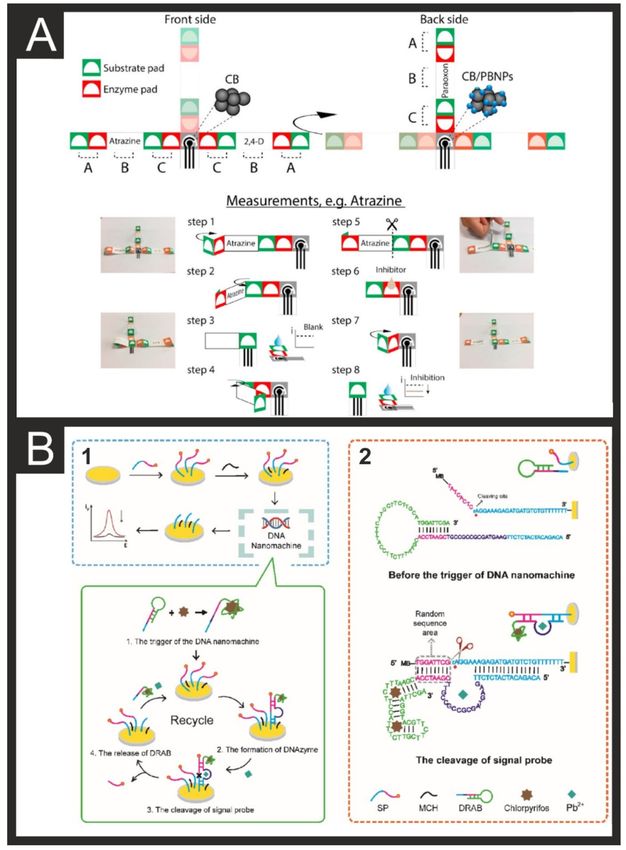
devices. Biosensors can be classified depending on their recognition element and/or their
transduction signalling. Figure 1 shows a biosensor classification (A) and schematic of the
importance of electroanalysis in food and drink quality control, including a variety of target
analytes that can be detected (B). As shown in Figure 1A, one of the main components of
biosensors is their bioreceptors, which can be enzymatic (most common), immunosensors
(highly specific and sensitive), aptamers or nucleic acid-based, or microbial or whole-cell
biosensors [28]. A classification based on the transducer and sensors can be also performed,
dividing the sensors as follows: electrochemical, electronic, thermal, optical, or gravimetric
sensors. Other categorisations can be performed such as analyte–bioreceptor combination,
detection systems, and type of applied technologies [28].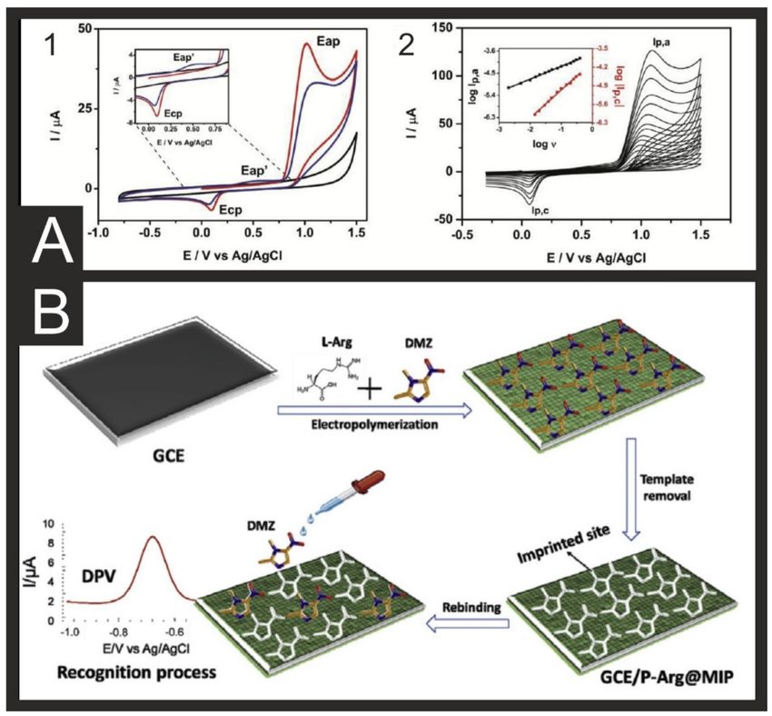
Biosensors 2021, 11, 291 3 of 24
Table 1. An overview comparing the different electrodes and their modifications for detection of different analytes relevant
to food safety alongside the limits of detection and linear ranges achieved and the composition of samples that were tested.
Electrode Electrode Target Detection Limit of Linear Sample
Reference
Materials Modification Analyte Method Detection Range Composition
Cu@Au labelled
ITO/GCE E. coli ASV 30 CFU/mL 50–50,000 CFU/mL Surface water [29]
Abs/Nafion and Hg
SPE Abs E. coli CA 103 CFU/mL 103 –107 CFU/mL Water [30]
Graphite Teflon/tyrosinase E. coli CA 10 CFU/mL 10–107 CFU/mL Drinking water [31]
6.4 × 10⁴ –3.3 × 10⁹
Ni disk NiOOH/Ni(OH)2 E. coli CA 104 CFU/mL Water [32]
CFU/mL
3 5 7 Drinking/Tap
ITO CNT E. coli CC 2 × 10 CFU/mL 10 –10 CFU/mL [33]
water
rGO- 8 Tap water, milk,
GCE E. coli DPV 9.34 CFU/mL 9.2–9.2 × 10 CFU/mL [34]
PVA/AuNP/Aptamer meat
SPE AuNP/Abs E. coli EIS 15 CFU/mL 101 –106 CFU/mL Water [35]
Au SAM/FcD/Peptide E. coli EIS 103 CFU/mL 103– 107 CFU/mL Water [36]
dsDNA/CeO2 /
GCE C. perfringens EIS 1.95 fM 10 fM–100 nM Dairy products [37]
CHIT
SPE N/A E. coli CV 10 ng/mL 10–1000 ng/mL Wastewater [38]
Nasal, mouth
Au DNA-TH/Abs S. pneumoniae SWV 0.093 CFU/mL 5–100 CFU/mL and axilla [39]
samples
ExtrAvidin® /
SPE V. cholerae CA 0.95 ng/µL 0.49–15.6 nM Water [40]
VHMR
GCE PDA/EPD/Abs S. aureus DPV 28.55 CFU/mL 104 –1010 CFU/mL Milk [41]
Au DNA walker/RP S. aureus DPV 9 CFU/mL 60–6 × 10⁷ CFU/mL Water, honey [42]
GCE CNF/Abs V. cholerae EIS 1.2 × 10⁻¹³ g/mL 10−13 –10−5 g/mL Water samples [43]
Ph- Orange juice,
GCE BoNT/E LSV 5 pg/mL 0.01–10 ng/mL [44]
PhNH2 /GNS/Abs milk
SPE AuNPs/Peptide BoNT/A&C SWV 10 pM 0.01–1 nM Orange juice [45]
SPE SWCNT ZEA DPASV 5 nM 0.0025–1 µM Cornflakes [46]
GCE GS/CHIT Microcystin-LR DPV 0.016 µg/L 0.05–15 µg/L Water [47]
SPE CB/ovalbumin DA/OA DPV 1.9/0.18 ng/mL 4–34/0.35–3.9 ng/mL Mussel extract [48]
Au-SPE DNA-capture probe A. minutum CA 25 pM 0.12–1 nM Ocean sample [49]
SPE CNF/Abs gliadin CA 0.005 mg/kg 0–80 µg/kg Flour samples [50]
Eggs, flour,
SPE MBs/Abs ovomucoid CA 0.1 ng/mL 0.3–25 ng/mL [51]
bread
GO/MBs/Abs/
SPE ovalbumin CA 0.2 fg/mL 0.01–10 pg/mL Wine [52]
HRP
Pharmaceutical
CPE - oxyclozanide SWASV 17.42 µg/L 0.058–4 mg/L [53]
formulation
Zn/Ni-ZIF-8
GCE monensin DPV 0.25–100 ng/mL 0.11 ng/mL Milk [54]
800/G/AuNp/Abs
Au MBs tetracycline EIS 1.2 pg/mL 0.1–1000 pg/mL Honey [55]
Pharmaceutical
GCE - xylazine DPV 120 nM 0.5–256 µM formula- [56]
tion/urine
GCE GNP xylazine ASV 0.1 mg/L 0.4–6 mg/L Beverages [57]
PGM MBs/Aptamer ampicillin - 0.25 nM 0.25–100 nM Milk [58]
Pigeon meat,
GCE Se-Co3 O4 /GO dimetridazole DPV 3.4 nM 0.02–83.72 µM [59]
eggs
GCE P-Arg-MIP dimetridazole DPV 0.1 nM 0.1 nM–10 µM Egg, milk, honey [60]
Carbofuran 0.6 nM 1.1–23 nM
SPE CB/acetylcholinesterase CA Olive oil [61]
chlorpyrifos 0.4 nM 0.7–14 nM
Carbofuran 0.048 µM
Isoprocarb 0.049 µM Wheat and
SPE CB DPV 0.1–100 µM [62]
Carbaryl 0.079 µM maize
fenobucarb 0.80 µM
100 nM–100 µM Tomato, beetroot,
SPE GONRs Metyl parathion CA 0.5 nM [63]
100–2500 µM broccoli
Paraoxen
2,4- 2–20 ppb 2 ppb
SPE CB/PB/Enzyme dichlorophenoxyacetic CA 100–600 ppb 50 ppb River water [64]
acid 10-100 ppb -
atrazine
Soil, fruit,
SPE CB/PB/BChE paraoxon CA 1.3 ng/mL 0.0013–3 µg/mL [65]
vegetables
1.82 × 10⁻³ –3.29 × 10⁴
SPE AuNP/PB/Abs OPs DPV 0.003 ng/mL Cabbage [66]
ng/mL
ITO MnNS OPs DPV 0.025 ng/mL 0.1–20 ng/mL Pakchoi [67]
ITO MB/ZIF-8/AChE paraoxon DPV 1.7 ng/mL 20–4000 ng/mL Apple, aubergine [68]
Profenofos 0.003 nM 0.01–100 nM
rGO- Phorate 0.3 nM 1–1000 nM Spinach,
SPE DPV [69]
CuNPs/Aptamer Isocarbophos 0.03 nM 0.1–1000 nM rapeseed
omethoate 0.3 nM 1–500 nM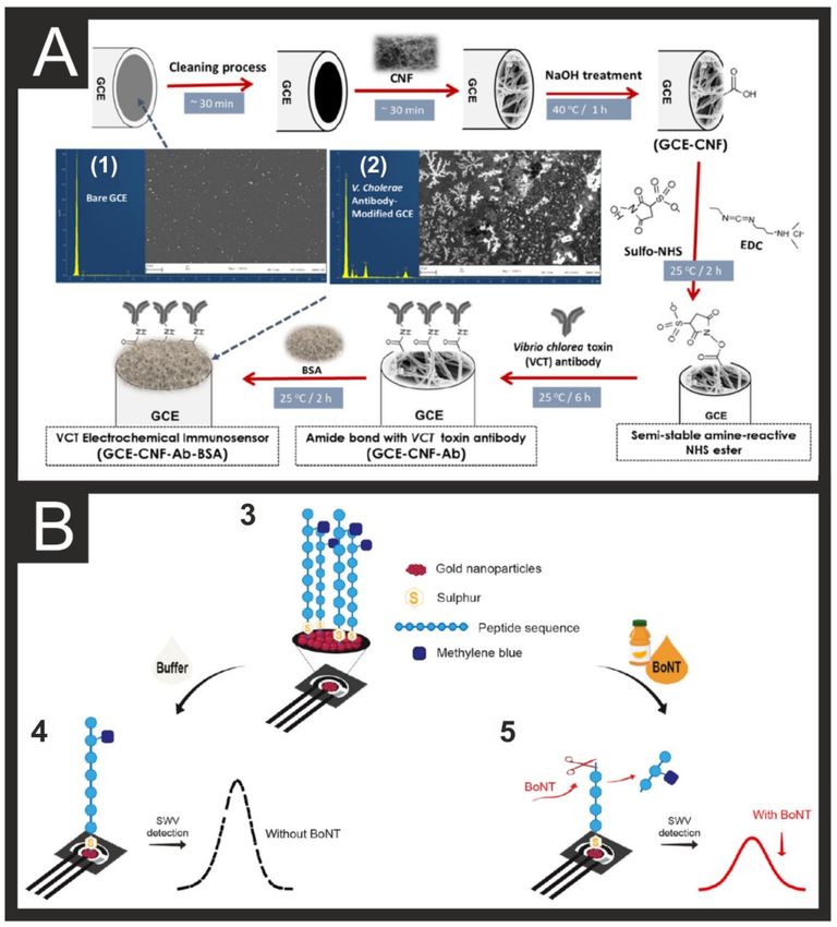
Biosensors 2021, 11, 291                                                                                                                  4 of 24

                                                                Table 1. Cont.

  Electrode          Electrode              Target         Detection      Limit of                Linear              Sample
                                                                                                                                     Reference
  Materials         Modification           Analyte         Method         Detection               Range             Composition
     GCE            PdNPs/BN            Paraoxon ethyl       LSV             3 nM              0.01–610.5 µM         River water        [70]
                                         Chlorpyrifos                      0.178 nM              0.5–500 nM         Apple, orange,
     Au                DRAB                                  DPV                                                                        [71]
                                             Pb                            0.034 nM              0.1–500 nM           cabbage
      ITO: indium-doped tin oxide; ASV: anodic stripping voltammetry; Abs: antibodies; GCE: glassy carbon electrode; SPE: screen-printed
      electrode; CA: chronoamperometry; CC: chronocoulometry; CNT: carbon nanotube; rGO: reduced graphene oxide; PVA: poly(vinyl alcohol);
      AuNP: gold nanoparticles; DPV: differential pulse voltammetry; EIS: electrochemical impedance spectroscopy; SAM: self-assembled
      monolayer; FcD: ferrocene derivative; dsDNA: double-stranded DNA; CHIT: chitosan; CV: cyclic voltammetry; DNA-TH: DNA tetrahedron;
      SWV: square-wave voltammetry; VHMR: target PCR amplicon; PDA: polydopamine; EPD: ε-poly-L-lysine-3,4-dihydroxy benzaldehyde;
      RP: RCA reaction primer; CNF: carbon nanofibers; BoNT/E: botulinum neurotoxin-E; LSV: linear sweep voltammetry; GNS: graphene
      nanosheets; ZEA: zearalenone; DPASV: differential pulse adsorptive stripping voltammetry; SWCNT: single-walled carbon nanotubes; GS:
      graphene sheets; DA: domoic acid; OA: okadaic acid; MBs: magnetic beads; HRP: horseradish peroxidase; GO: graphene oxide; CPE: carbon
      paste electrode; SWASV: square-wave adsorptive stripping voltammetry; G:graphene; Zn/Ni-ZIF-8 800: Zinc/Nickel-zeolitic imidazolate
      framework-8; GNP: graphene nanoplatelets; PGM: personal glucose meter; MIP: molecularly imprinted polymer; P-Arg: polyargenine; CB:
      carbon black; GONRs: graphene oxide nanoribbons; PB: Prussian blue; BChE: butyrylcholinesterase; Ops: organophosphorus pesticides;
      MnNS: manganese dioxide nanosheets; MB: methylene blue; AChE: acetylcholinesterase; BN: boron nitride; DRAB: dual-recognition
      aptazyme beacon.

      Figure 1. Classification of biosensors based on their bioreceptors, transducers, technology or detection system (A). Schematic
      representation of some of the multiple applications of tailored electrochemical sensing platforms towards food safety (B).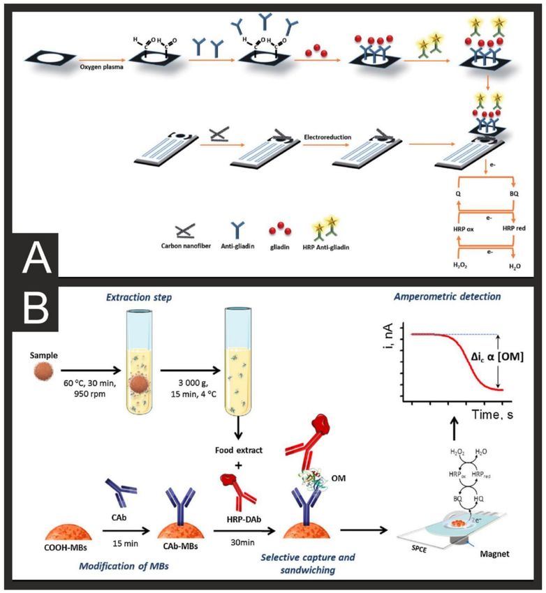
Biosensors 2021, 11, 291                                                                                         5 of 24

                           2. Electrochemical Sensors towards Food Safety
                                 The presence of undeclared substances in food, especially when processed, is a com-
                           plicated challenge for monitoring agencies due to the complex manufacturing, processing,
                           handling, etc., especially when heating and fermentation steps are applied [72]. Recent
                           scientific discoveries and analytical methods have allowed us to increase our understand-
                           ing of living organisms and their/our metabolism, which has helped identify a variety
                           of bioreceptors from biological organisms [73]. The discovery of these bioreceptors has
                           helped introduce a new generation of electrochemical biosensors that can provide an
                           alternative to classical analytical methods for the trace detection of food safety analytes.
                           The most common determination methods for food safety can be categorised between
                           immunological and DNA-based assays, or between direct and indirect methods (depend-
                           ing on whether the analyte in question is the harmful molecule itself or a characteristic
                           biomarker of its presence) [74]. Although these methods provide excellent results and are
                           well understood by researchers in the field, complete validation against the mentioned
                           lab-based methodologies is required to garner end-user confidence from people not within
                           the field.
                                 Immunological methods use the specific recognition involved in the antigen-antibody
                           coupling; apta- and genosensors use DNA/RNA to bind to the particular analyte or to
                           detect the encoding genes of the particular analyte via hybridisation with complementary
                           DNA sequences [73]. Imprinted polymers (IPs), or plastic antibodies, are also used as a
                           recognition scaffold to create exclusive non-covalent binding sites to the target analyte,
                           be it a small molecule [75], protein [76,77], or microorganism [78–80]. Affinity assay
                           methods, similar to ELISA tests, either sandwich or competitive, are also applied towards
                           food safety. Label-free non-competitive methods are also applied by immobilising the
                           bioreceptor directly on the transducer’s surface. In terms of electrochemical methods,
                           these are often using the following procedures: amperometry, potentiometry, impedance,
                           and voltammetry. Although progress is still needed in order to expand and have mass-
                           acceptance for the use of electrochemical biosensors in food safety monitoring, in this
                           review, we compile and explain some elegant and recent literature examples of novel
                           electrochemical biosensors towards food safety. We divide these into the different target
                           molecules—namely, pathogens, toxins, allergens, viruses, veterinary drugs, forensic drugs,
                           and pesticides. For an in-depth look into the recent advances in the portable sensing of
                           heavy metals, we refer the reader to a recent review paper [17]. Due to the importance of
                           these areas and the plethora of work undertaken in these fields, we will predominantly
                           focus on work completed in the last 5 years. We will establish the importance of the
                           individual areas, summarise key reports in the different fields, and offer our insight into
                           how the research community can seek to address and overcome future challenges. For
                           multiple foodborne biosensor examples described in this manuscript, Table 1 provides an
                           overview of many examples named herein, comparing their electrode material, electrode
                           modification, target analyte, detection method, limit of detection (LOD), linear range, and
                           sample composition.

                           2.1. Pathogen Detection
                                Pathogens are infectious agents capable of causing illness and include fungi, proto-
                           zoans, bacteria, viruses, and prions [81]. Pathogens include a plethora of microorganisms
                           and molecular-scale infectious agents whose virulence, transmission mode, reproduction
                           mode, etc. vary widely [82,83]. It is because of these complex differences that pathogen
                           sample matrices are intricate in nature, including aerosols, body fluids, and surfaces, and
                           this provides challenges in the optimisation of sample preparation for electrochemical
                           biosensors [84]. While these pathogenic bacteria are too small to be observed by the naked
                           eye, upon culture growth on an agar plate, they can form visible patches/grown patterns.
                           This remains the “gold standard” for bacteria detection, although this diagnostic scheme
                           takes a minimum of 24 h [85,86]. There are in excess of 1400 human pathogens; however,
                           the majority of healthcare-associated diseases are caused by a limited amount [87]. In this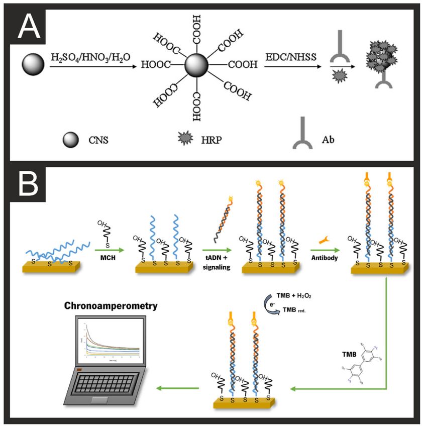
Biosensors 2021, 11, 291                                                                                            6 of 24

                           pathogenic electroanalytical section, we describe some recent reports towards the electroan-
                           alytical detection of some of the most common foodborne pathogens such as Escherichia coli,
                           Clostridium perfringens, Vibrio cholerae, Staphylococcus aureus, Listeria monocytogenes, etc.
                                Escherichia coli is the most common microorganism that affects warm-blooded intesti-
                           nal organs, and its presence is mainly associated with faecal contamination [29,30,88]. E. coli
                           causes diarrhoea, urinary infections, and peritonitis, predominantly in vulnerable people,
                           and it is often used as a microbiological marker for water quality [31]. E. coli biosensors are
                           one of the most commonly found in the literature, with various methods and electrodes
                           used such as rotating disk electrodes [32], indium-doped tin oxide [33], glassy carbon elec-
                           trodes (GCE) [34], and screen-printed electrodes (SPEs) [35]. For example, Kraazt et al. [36]
                           reported a label-free ferrocene-antimicrobial peptide (magainin I) modified gold electrode
                           biosensor, showing its preferential selectivity towards pathogenic strains (O157:H7) with a
                           LOD of 103 cfu/mL. This biosensor solution is based on rapid electrochemical impedance
                           spectroscopy (EIS) biosensor that uses antibacterial peptides as recognition elements for
                           a wide range of Gram+/−pathogens [36]. This biosensor, as shown within Figure 2A,
                           was produced through the deposition of an N-hydroxysuccinimide-based self-assembled
                           monolayer (SAM), followed by coupling to the ferrocene based probe, coupling to the
                           antimicrobial peptide probe, and finally bare surface blocking.

                           Figure 2. (A) Schematic diagram for the construction of a ferrocene-peptide-modified biosensor
                           using a gold macro-electrode. Reproduced with permission from [36]. Copyright Elsevier 2014.
                           (B) Schematic representation of the DNA Walker and DNA Nanoflower biosensor for S. aureus.
                           Reproduced with permission from [42]. Copyright American Chemical Society 2021.

                               Clostridium perfringens is a rod-shaped, Gram-positive, and spore-forming bacteria
                           widely found in meat, dairy, and water that is capable of causing diarrhoea and enteritis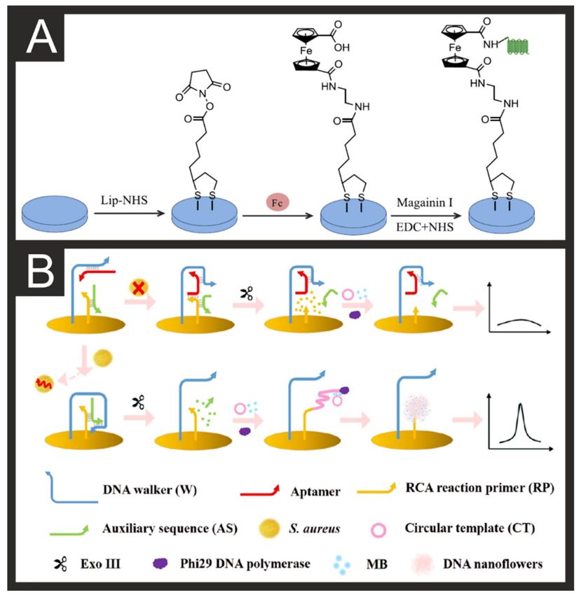
Biosensors 2021, 11, 291                                                                                            7 of 24

                           necroticans [89,90]. Qian et al. reported the use of ceria (CeO2 ) nanorods as a sensing mate-
                           rial towards C.perfringens due to its strong adsorption ability towards DNA and low toxicity,
                           compared to CeO2 nanoparticles used as control. They immobilised CeO2 nanorods onto
                           chitosan (CeO2 -CHIT), which was then modified upon a glassy carbon electrode (GCE) for
                           the electrochemical detection of C.perfringens, which was shown to be possible in pure milk
                           and milk powder samples, achieving a LOD of 7.06 and 1.95 × 10⁻¹⁵ mol/L when EIS and
                           differential pulse voltammetry (DPV), respectively. Their label-free biosensors exhibited an
                           easy-to-operate process, highly sensitive, and affordable, with RSD lower than 5% demon-
                           strated within dairy products [37]. Rochelet et al. [38] reported the first amperometric
                           detection of β-D glucuronidase using disposable carbon electrodes as a rapid wastewater
                           analysis method. The authors reported the indirect amperometric quantification of β-D
                           glucuronidase activity and correlated it to the presence of E. coli. β-D glucuronidase acts
                           as the electrochemical substrate for GLUase measurement, and the p-aminophenol (PAP)
                           released during the enzymatic hydrolysis was monitored by cyclic voltammetry with dis-
                           posable carbon SPEs achieving a LOD of 10 ng/mL. The amperometric assay was applied
                           to faecal wastewater contamination in raw and treated waters which was applied to turbid
                           sample quantification providing a reliable and decentralised method [38].
                                 A commonly used alternative bio-recognition element to the use of enzymes is anti-
                           bodies, specific for a certain protein present on the surface of bacteria [91]. Wang et al. [39]
                           reported an antibody-based platform for the pneumococcal surface protein A on
                           Streptococcus pneumonia. This bacteria accounted for 95% of the pneumonia cases reported
                           in the pre-antibiotic era and remains a pathogen of concern [92]. The sensor platform
                           utilised DNA tetrahedron nanostructures conjugated onto gold electrode surfaces as their
                           base, with the antibody conjugated on top of that. Through square wave voltammetry, they
                           were able to obtain a LOD of 0.218 ng/mL and applied it to detection in human samples.
                                 Vibrio cholerae is the pathogenic agent of cholera, which is a type of life-threatening
                           diarrhoea that can lead to extreme dehydration and death if not rapidly treated, being one
                           of the most rapidly fatal illnesses and therefore of worldwide concern [93–96]. The use of
                           horseradish peroxidase (HRP) enzyme-based carbon SPE genosensor towards V. cholerae
                           was reported by Low et al. [40]. Their strategy uses a double hybridisation strategy
                           (sandwich type) to target V. cholerae’s ssDNA to enhance the selectivity and specificity of
                           the detection, without the need for previous heat denaturation or purification, achieving
                           a 100% specificity and LOD of 0.85 ng/µL of V. cholerae genomic DNA when coupled to
                           asymmetric PCR amplification.
                                 Staphylococcus aureus is one of the most infectious agents in foodborne illness [97,98],
                           with a virulence that includes infective endocarditis, toxic shock syndrome, or osteomyeli-
                           tis [99]. Wu et al. recently reported the first study of an immunosensor with dual detection
                           and elimination of S. aureus in drinks, along with good selectivity, reproducibility, and
                           stability [41]. Their approach is a mussel-inspired scaffold of ε-poly-L-lysine-3,4-dihydroxy
                           benzaldehyde (EPD) that binds to polydopamine (PDA) on a pre-grafted gold macro-
                           electrode. Here, EPD acts as a biomimetic polymer to enhance the immunosensor’s
                           performance with robust binding of the antibody on the electrode’s surface, with pH-
                           responsive properties that allow on-demand ε-poly-l-lysine (ε-PL) delivery to eliminate
                           S. aureus. Recently, Cai et al. [42] have reported a DPV-based biosensor for the detection
                           of S. aureus with a working range of 60–6 × 10⁷ CFU/mL and LOD of 9 CFU/mL. This
                           platform uses DNA walkers, DNA nanoflowers, and aptamer-based recognition, as shown
                           in Figure 2B, to recognise the presence of the bacteria. DNA walkers can perform repeated
                           movements along a DNA orbit composed of part or all nucleic acids to produce signal
                           amplification [100]. DNA nanoflowers can assemble from localised high concentrations of
                           DNA and do not need full complementary base pairing and have excellent stability [101].
                           The combination of these two nano-DNA-based systems allows for enhanced signal am-
                           plification when the aptamer present is bound to the target bacteria and therefore not
                           interfering with the DNA walker. Using this system, they managed to accurately detect
                           levels of S. aureus in spiked lake water, tap water, and honey solutions.
Biosensors 2021, 11, 291 8 of 24
Listeriosis is a bacterial infection caused by Listeria monocytogenes that can cause severe
illness including sepsis, meningitis, or encephalitis [102]. Listeriosis is mainly a problem
from unpasteurised milk (or its derivatives such as cheese and ice cream), vegetables, meats,
and fish. Ruan et al. reported the automatic detection of L. monocytogenes in milk samples
by monitoring the relationship between oxygen consumption and the concentration of the
analyte by cyclic voltammetry, where the oxygen reduction decreases/disappears when
L. monocytogenes proliferates when using a gold disk macro-electrode (GDE) in a Listeria
enrichment broth (LEB) [103].
The presence of bacteria inside food and drink products is of vital concern, but these
microorganisms can also release toxins into these substances, which is where our focus
now turns.
2.2. Toxins/Mycotoxins
Toxins are harmful compounds produced by living cells or organisms such as bacteria,
fungi, or algae, whose composition varies from small molecules to large biomolecules such
as peptides or proteins [104,105]. Toxins are usually divided between exo- and endotoxins
depending on whether they are excreted by an organism or are a structural part of the
cell. In nature, toxins can have two primary functions—predation and defence—to kill
a potential meal or to discourage the action of a third party (predator). Another method
of classification can be in accordance with the location of the body that they affect the
most, such as hemotoxin (blood), phototoxin (light related), necrotoxin (damages tissue),
or neurotoxin (nervous system). For example, the botulinum toxin is a spore (endotoxin)
produced by Clostridium botulinum that can block neurotransmitter metabolic pathways
that cause muscle and lung paralysis, double vision, and muscle weakness in humans,
named botulism [106,107]. Botulinum toxin can be produced from foods with pH > 4.5 and
sufficient moisture, such as homemade tinned and fermented foods with long shelf life.
Another example is Staphylococcal enterotoxins (SEs) secreted by Staphylococcus aureus
(S. aureus), S. hyicus, and S. intermedius that are a major cause of food poisoning, with
nausea, violent vomiting, and diarrhoea as main symptoms and typically occur after
ingestion of dairy or processed meat that has been improperly handled or stored at elevated
temperatures [108]. Many other common bacterial foodborne toxins are cholera toxin (Ctx)
from Vibrio cholerae, Shiga Toxin from Shigella dysenteriae, and E. coli O157:H7, and CPE
enterotoxin from Clostridium perfringens [109].
A recent example of an electrochemical immunosensor for Ctx in water samples from
Ozoemena et al. utilises electrospun carbon nanofibers (CNFs) on a GCE, with the α-Ctx
antibody covalently immobilised using carbodiimide chemistry for amide bond forma-
tion (Figure 3A) [43]. Their electrochemical response is based on the suppression of the
electrical current, followed by EIS or square wave voltammetry (SWV), where the use of
the ferri-/ferro-redox probe enhances the sensitivity of the immunosensor to detect Ctx.
This immunosensor exhibited excellent sensitivity, selectivity, and regenerability; LOD of
ca. 1.2 × 10⁻¹³ g/mL and limit of quantification (LOQ) of ca. 1.3 × 10⁻¹³ g/mL [43]. An-
other example of a novel electrochemical sensor for the detection of botulinum neurotoxin
(BoNT/E) in milk and orange juice was reported by relying on graphene nanosheet-s-
aryldiazonium modified GC as a sensing platform, and enzyme-induced silver nanoparti-
cles (AgNPs) deposited on gold nanoparticles (AuNPs) as a signal amplifier. The authors
demonstrated a highly sensitive and specific electrochemical immunosensor for the detec-
tion of BoNT/E based on an enzyme-AuNPs accelerated silver deposition with ordered
graphene nanosheets to amplify the signal, increase the surface area and provide beneficial
antifouling properties [44]. Another system from Caratelli et al. [45] for the detection of
both BoNT/A and BoNT/C with LODs of 10 pM. A peptide labelled with methylene blue
is immobilised onto a paper-based electrode surface modified with AuNPs, as depicted in
Figure 3B. The BoNTs, when present, cleave the synthetic peptide, removing the methy-
lene blue and causing significant decreases in the measured square-wave signal. Last, a
zearalenone (ZEA; mycotoxin in cereals) sensor was developed using carbon nanotubesBiosensors 2021, 11, 291 9 of 24
(SWCNTs) on a screen-printed electrode (SWCNT-SPE) for the voltammetric (differential
pulse adsorptive stripping; DPASV) determination, with a LOD of 5.0 × 10⁻⁹ M in cornflake
samples. The use of SWCNTs here enhances the sensitivity attained by DPV, lowers the
LOD over a wide range of concentration, and shows antifouling properties [46]. Another
issue for the safety of especially drinks, in addition to food that originates from water-based
environments, is the presence of algae or algal toxins, which we will discuss next.
Figure 3. (A) Experimental protocol (and time needed) for the fabrication and sensing mechanism of an electrochemical
immunosensor for VCT. Inset (1) shows typical SEM and EDX of a bare GCE, while (2) shows SEM and EDX of V.
cholerae antibody-modified GCE. Reproduced with permission from [43]. Copyright American Chemical Society 2020.
(B) Experimental protocol for the BoNT detection using the signal-off method, including a representation of the peptide-
modified paper-based sensor (3), signal obtained before analyte presence (4), and signal obtained after analyte presence (5).
Reproduced with permission from [45]. Copyright Elsevier 2021. Algae/Algal toxins.Biosensors 2021, 11, 291 10 of 24
Different species of cyanobacteria or dinoflagellates are capable of producing a num-
ber of toxins and often cause episodes of harmful algal blooms (HABs) in fresh or marine
water bodies from eutrophication occurrence arising from human activities [110–113].
These anthropogenic activities usually include agricultural and urban waste, industrial
manufacture, and global warming [114,115]. HABs can create a change in colour, odour,
and taste due to the production of algae toxins, some of which have harmful effects on
humans. Examples of harmful algae toxins that can occur in drinking and recreational
water are microcystins, and cylindrospermopsin (hepatotoxin), anatoxins, and brevetox-
ins (neurotoxins), and certain lipopolysaccharides and lyngbyatoxin (dermatoxins) [116].
Algae toxins are often secondary metabolites that belong to a variety of class compounds
such as cyclic peptides, alkaloids, and lipopolysaccharides [113]. A recent example of
a cyanotoxin drinking water outbreak was the famous case of Lake Erie in Toledo (OH,
USA), where tap water was not suitable for human consumption during the summer of
2013–2014 due to the high presence of cyanotoxins in their supply [117,118]. Antibod-
ies, aptamers, carbohydrates, and antimicrobial peptides have been used as biorecogni-
tion elements for electrochemical biosensors [119]. Due to the small size of algae toxins
(MW < 1 KDa), competitive immunoassays are often employed for sensing purposes, in
which the working electrode surface would be linked to algal toxins and incubated with
antibodies, followed by the addition of the algal unknown samples. The unknown algal
toxins would then compete or displace the ones immobilised at the electrode’s surface,
offering enhanced electrochemical performance, compared to those based on antibodies
as biorecognition elements. An example of these types of competitive solutions is the
one reported by Quan et al. towards the detection of Microcystin-LR (MC-LR) in wa-
ter samples [47]. They reported a graphene sheet-chitosan (GS-CS) nanocomposite on a
glassy carbon electrode that acts as a scaffold for MC-LR detection. As signal reporters,
horseradish peroxidase (HRP), carbon nanospheres (CNS), and antibody conjugates were
used, Figure 4A. Their clever design increased the active area and sensitivity by using CNS,
which offers a 3D structure and good biocompatibility while offering enhanced electron
transfer properties that achieved a LOD of 0.016 µg/L and variation coefficients around 1%
for their proposed immunosensor [47].
Aptamers are single-stranded oligonucleotides capable of binding to a specific target
molecule that belong to a group of single-stranded (ss) DNA/RNA molecules [120]. Ap-
tamers are of small size and offer higher affinity, stability, and specificity to target molecules
than some antibodies and can be tailored to contain certain terminal moieties to bind to the
surface of the electrode (thiol, amino, disulphide, etc.) [121–123], although their stability
and shelf life is still a challenge for their large-scale commercialisation and use. It has
also been reported that the use of Mg2+ and pH changes can enhance the binding affinity
to algal toxins by controlling the secondary conformation of the aptamer and increase
their stability [124,125]. A recent example of detecting important biotoxins domoic acid
(DA) and okadaic acid (OA) in mussels was reported by Nelis et al. [48]. They function-
alised carbon-black-modified carbon SPEs (drop-casted CB-SPE) with protein conjugates
specific for DA and OA and utilised them in indirect competitive DPV immunosensors.
Through this approach, the authors achieved LODs of 1.9 ng/mL and 0.18 ng/mL in
mussel extract samples.
The dinoflagellates from the Alexandium genus can include some of the most toxic
species [126]. Morais et al. [49] have reported a disposable electrochemical genosensor
for the detection of Alexandrium minutum through the immobilisation of a DNA-capture
probe onto a screen-printed gold electrode (SPGE) targeting a specific coding sequence
(Figure 4B). The chronoamperometric detection achieved a linear range of 0.12–1.0 nM and
a LOD of 24.78 pM, with RSD < 5.2 % shown to be possible.Biosensors 2021, 11, 291 11 of 24
Figure 4. (A) Schematic illustration of the carboxylation procedures of CNS and the subsequent HRP-CNSs-Ab conjugation.
Reproduced with permission from [47]. Copyright Elsevier 2013. (B) Schematic for the development of the electrochemical
genosensor for A. minutum. Reproduced with permission from [49]. Copyright Elsevier 2021.
2.3. Allergen Examples
Allergens are a type of antigen that trigger an abnormal immune response against an
otherwise harmless substance in the body; these types of reactions are commonly known as
allergies and are believed to affect around 3% of adults and 10% of children in industrialised
countries [127]. These allergies can be divided into toxic or nontoxic, depending on the
individual sensitivity, and can be further subdivided into immunological (food allergies)
or non-immunological (food intolerances) [128,129]. Overall, 14 ingredients have been
included in the European Union list of allergenic food ingredients—eggs, milk, peanuts,
nuts, gluten-containing cereals, lupin, soybeans, celery, mustard, sesame, fish, crustaceans,Biosensors 2021, 11, 291 12 of 24
molluscs, and sulphites [130–132]. Although detailed food labelling is required in most
countries, cross-contamination, adulteration, or fraud of undeclared allergens often occurs.
Pereira et al. [50] reported a disposable and low-cost biosensor strategy towards gliadin
in common flour samples. Gliadin is a protein component of gluten which provides the
rising ability during baking and is present in wheat and many other cereals from the
grass genus Triticum. Their elegant solution uses a carbon SPE modified with carbon
nanofibers, which are coupled to a paper immunoaffinity platform for the gliadin sensing
in flour samples (Figure 5A). The choice of carbon nanofibers herein allows an increase in
electron transfer efficiency and in the electroactive area, enabling the determination of low
levels of analyte. This paper platform uses a covalently functionalised micro-zone, with
specific anti-gliadin antibodies placed for the voltammetric detection of gliadin, reporting
a LOD of 0.005 mg/kg and a 4.11% variation coefficient for a 20 µg/kg gliadin sample.
Their activated cellulose paper offers hydroxyl groups for bioconjugation purposes, which
allows the covalent bonding of the amino groups from the antibodies. The use of an
HRP saturation measurement to quantify the available sites not previously occupied by
antibodies will catalyse the catechol–benzoquinone reaction in the presence of H2 O2 ,
generating a current response that is inversely proportional to the amount of immobilised
antibody [50]. Another recent example of an allergen electrochemical biosensor is the one
reported by Pingarron et al. [51] for ovomucoid allergen (OM; egg white allergen) by using
magnetic bioconjugates captured on a carbon SPE surface to perform an amperometric
detection in the presence of hydroquinone and H2 O2 . Their immunoplatform involves
the selective capture of sandwich antibody-target analyte-HRP labelled detector antibody
sandwich onto a carboxylic acid-functionalised magnetic bead (HOOC-MBs), as shown in
Figure 5B, exhibiting a LOD of 0.1 nm/mL for the determination of OM in unprocessed
eggs and flour and baked bread [51]. Another example targeting egg allergen (ovalbumin,
OVA), this time in wine samples, has been reported by Baldo et al. [52]. In the wine industry,
egg white can be utilised as a fining agent to assist in the removal of tannins. The authors
report a very similar sandwich-based immunoassay to that described above using OVA-
specific antibodies and HRP decorated onto magnetic beads (Figure 5). An array of 8 SPEs
modified with poly(diallyldimethylammonium chloride) and graphene oxide, which aided
with the biomodification, was modified with specific antibodies through carbodiimide
coupling. The modified MBs were mixed with the sample and then incubated onto the
electrode. If the target was present, they would bind to the surface Abs and provide an
enhanced electrochemical signal through the HRP.Biosensors 2021, 11, 291 13 of 24
Figure 5. (A) Schematic representation of the electrode modification and gliadin determination procedures. Reproduced
with permission from [50]. Copyright Royal Society of Chemistry 2019. (B) Schematic display of the fundamentals involved
in the immunosensing platform developed for OM determination. Reproduced with permission from [51]. Copyright
Elsevier 2018.Biosensors 2021, 11, 291 14 of 24
2.4. Veterinary Drugs
Antimicrobial, antiparasitic, and growth promoters are normally included in the
veterinary drugs category due to their work in treating and promoting animal growth and
feed efficiency and preventing diseases [133]. Their quantification is of public concern
due to the possible presence of these drugs in animal-derived foods, which might cause
harmful effects to the final food product if their quantities are higher than the maximum
residue limits (MRL) defined on the basis of food safety [134]. Common antibiotics used in
veterinary practice often include sulphonamides, lincosamides, nitrofurans, etc. [135–137].
From these compounds, trace residues present in the final food (milk, meat, egg, honey,
etc.) product can cause serious harm to humans [138]. An added risk recently discovered is
that the presence of these antibiotics in food can also trigger antimicrobial resistance later in
the food chain if inefficient antibiotic therapies emerge [139,140]. There are many examples
of various electrochemical sensing platforms for veterinary drugs found throughout the
literature, such as oxyclozanide [53], monensin [54], and tetracycline [55].
As an example of a non-approved substance for human use in food, xylazine is a
clonidine derivative that is an analgesic and sedative for animals [140,141]. In humans,
xylazine causes diarrhoea and drowsiness, acting on the central nervous system, and if
long exposure occurs, it can develop a drug dependence that might lead to depression,
sleepiness, and lowered respiratory rates [142]. Xylazine can be detected electrochemically
through its irreversible oxidation peak at approximately +0.9 V (Figure 6A) [56]. Saisahas
et al. recently reported a simple portable electrochemical sensor for xylazine based on drop-
casted graphene nanoplatelets (GNPs) on carbon SPEs for the voltammetric determination
in spiked alcoholic and non-alcoholic drinks. Their choice of GBPs allowed enhanced
adsorption and a unique electrochemical profile, which is based on adsorptive stripping
voltammetry (AdSV) [57]. Zhang et al. reported an ingenious use of commercial personal
glucose meters (PGM) coupled with a novel sensitive method for ampicillin detection in
milk. This method uses magnetic beads (MBs), combining an ampicillin aptamer as a recog-
nition element and streptavidin as a linking agent, for the indirect relationship between
ampicillin and the sucrose to glucose hydrolysis, achieving a LOD of 2.5 × 10⁻¹⁰ mol/L in
milk [58].
Dimetridazole (DMZ) is used to treat protozoal and bacterial infections and is com-
monly added to poultry feed. Residues of this, however, can produce carcinogenicity,
genotoxicity, and mutagenicity in humans [143]. Recently, Umesh et al. [59] reported a
biosensing platform for the detection of DMZ in milk, pigeon meat, and eggs based on Se
nanorods capped with Co3 O4 nanoflowers decorated onto graphene oxide. This system
combined these materials together to produce enhanced electron transfer and a higher
conductivity and are deposited onto the surface of the GCD through simple drop-casting.
They achieved a wide linear range of 0.02–83.72 µM, a LOD of 3.4 Nm, and excellent
recoveries in real samples. Another example for the detection of DMZ was reported by Ali
et al. [60], who used a poly-arginine (PAG) based MIP on a GCE (Figure 6B). The PAG MIP
was formed directly onto the surface of the GCE through electrodeposition in the presence
of DMZ using cyclic voltammetry. Upon rebinding of the DMZ with the MIP, increases in
the DPV signal were obtained leading to a linear range of 0.1 nM–100 µM and a LOD of
0.1 nM.Biosensors 2021, 11, 291 15 of 24
Figure 6. (A) (1) Cyclic voltammograms (scan rate: 50 mVs−1 ) recorded using a GCE in a BR buffer (0.1 M, pH = 7.0)
in the absence (black line) and presence of 0.4 mM xylazine (red line—first scan, blue line—second scan). Shown in the
insert is the zoom plot of the anodic process on the second scan. (2) Scan rate study (2–400 mVs−1 ) of the above solution.
Inserted is the plot of the log of the peak current versus the log of the scan rate. Reproduced with permission from [56].
Copyright Elsevier 2019. (B) Schematic for the formation of the PAG MIP sensor for the detection of DMZ. Reproduced
with permission from [60]. Copyright Elsevier 2020.
2.5. Pesticides
Herbicides, insecticides, and fungicides are commonly used within food production to
control their pests and increase production [144]. These pesticides, although toxic for their
targets, can also be harmful to other animals and humans, causing carcinogenicity, infertility,
neurological diseases, respiratory problems, etc. [145]. As an example, an organophos-
phorus and carbamate pesticides biosensor was recently reported for olive oil samples,
comparing the different protocols for AChE deposition on CB drop-casted on carbon SPEs,
using laser-induced forward transfer (LIFT) technique to modify the electrodes [61]. The
use of LIFT minimises the amount of deposited material, reduces the cost, and produces
high spatial resolution allowing the biofunctionalisation of small-area surfaces [146,147].Biosensors 2021, 11, 291 16 of 24
Their olive oil pesticide sensors exhibited LODs of 0.6 and 0.4 × 10⁻⁹ mol/L for carbofuran
and chlorpyrifos, respectively [61]. Della Pelle et al. also reported a CB-SPE voltammetric
sensor for carbofuran, isoprocarb, carbaryl, and fenobucarb in grain samples with compara-
ble results to that of UPLC-MS/MS [62]. Validation against these standard laboratory-based
techniques is vital in providing confidence in the electrochemical platforms for potential
end users and legislation creators. Govindasamy et al. also reported the use of graphene
oxide nanoribbon (GONRs) modified SPEs for a disposable real-time detection of methyl
parathion in broccoli, beetroot, tomato, and Ugli fruits [63]. GONR-SPEs exhibited im-
proved electrocatalytic ability towards methyl parathion in comparison with the CNT
counterparts, due to the rich edge chemistry and abundant functional groups, higher
area-normalised edge-plane structures, and chemical active sites [63].
An interesting approach to the detection of pesticides is the origami-paper-based
sensors presented by Arduini et al. [64]. In this work, they utilised office-paper-based
carbon SPEs alongside multiple filter paper pads for the loading of enzymes and enzymatic
substrates. A hydrophobic wax before printing, creating hydrophilic zones, defines the elec-
trode areas. Thereafter, the working electrode was modified with a carbon black/Prussian
blue nanoparticle dispersion. The pads on either side of the SPE are loaded with the appro-
priate enzymes and substrates. When samples testing is required, the appropriate outer
pads are folded onto the SPE surface before the droplet is added and analysis can occur
(Figure 7A). This was applied to the detection of paraoxon, 2,4-dichlorophenoxyacetic acid,
and atrazine, whereby the presence of these analytes inhibits the immobilised enzymes
and substrates. This was monitored using chronoamperometry and used to evaluate the
analytes at the ppb level. Similar paper-based devices have been recently applied to the
detection of organophosphorus pesticides in soil with a LOD of 1.3 ng/mL, with this sys-
tem validated against LC-MS lab-based techniques [65]. Organophosphorus pesticides are
some of the most commonly used throughout the agriculture industry to control pests and
diseases and increase crop production. As such, there have been various electrochemical
biosensors designed for their detection utilising different recognition elements such as
antibodies [66], nanozymes [67], enzymes [68], and aptamers [69]. The aptamer-based
system reported by Fu et al. utilised reduced graphene oxide (rGO) as a base for the
immobilisation of copper nanoparticles (CuNPs) and the aptamer element. The use of
micro and nanomaterials in biosensor development has been regularly used to increase the
surface area, conductivity, and analytical performance [148], such as graphene oxide [149],
reduced graphene oxide [150], and carbon nanotubes [151]. A recent example utilising
a mix of materials was reported by Renganathan et al. [70], where they used palladium
nanoparticles adorned with boron nitride for the electrochemical detection of paraoxon
ethyl (PXL). Boron nitride provides a high specific surface area and an effective pathway for
mass transport, with the nanoparticles improving the electrochemical performance. This
allowed for the direct electrochemical detection of PXL, through linear sweep voltammetry,
with a linear range of 0.01–210 µM and a LOD of 3 nM. Finally, a novel methodology for
dual detection of the pesticide chlorpyrifos and heavy metal ions. Wang et al. [71] utilised
a dual-recognition aptazyme beacon (DRAB), which contains both the aptamer specific
for chlorpyrifos and an enzyme strand of Pb2+ DNAzyme. The aptamer is bound to the 30
terminus of the enzyme strand preventing it from forming the activeated DNAzyme. In
the presence of the analyte chlorpyrifos, the DNA nanomachine is activated and DRAB
unfolded (Figure 7B). Upon the binding of Pb2+ , the DRAB is catalysed and the signal
probe is cleaved from the surface, resulting in a decrease in the DPV signal. This was
applied to the detection of the analytes in fresh fruit and vegetable samples that had been
turned into a puree. This is a good example of analysis in complex real samples, which
remains a challenge in this field. As seen throughout this section, the majority of pesticide
detection has been performed on water samples, typically looking for contamination in
open water sources.Biosensors 2021, 11, 291 17 of 24
Figure 7. (A) Schematic representation and photographs of the configuration of the paper-based platform and measurement
procedure. (B) The principle of DRAB-based electrochemical biosensors (1) Modification and detection procedure of the
proposed sensor. (2) The secondary structure of the DRAB.Biosensors 2021, 11, 291 18 of 24
3. Future Trends
Currently, and due to the 2019 SARS-CoV/COVID pandemic, the general public has
seen the importance of rapid, reliable, and robust diagnostic kits for the early detection of
diseases by using rapid serological tests. However, the need for rapid on-site monitoring
does not stop at the management of pandemics, but it is applicable to healthcare and
forensic biomarkers, environmental monitoring, and food safety. The benefit of obtaining
fast results in decentralised environments makes these tests part of an on-growing trend
for their rapid turnarounds and ability to mitigate consequences and harmful exposure to
humans and the environment.
The fundamental concept of point-of-care (PoC) and/or on-site testing devices is
to carry out the test in the most appropriate, comfortable, and immediate way without
extensive sample preparation, expensive kits, or specialised experimentalists. Based on this,
electrochemical biosensors offer most of the ideal requirements for their miniaturisation
for PoC applications. One of the main challenges that experimentalists encounter when
developing a biosensor for food safety is the application to real samples. One can easily find
reports in the literature showcasing contaminants, pathogens, and allergens biosensors;
however, their application to real-world samples is often limited. As summarised in
Table 1, most of the real-world applications of these food safety biosensors are in water or
drink samples. Therefore, there is a need for improving the performance of these devices,
particularly in complex matrices, in order to transition from laboratory-based methods to
PoC solutions.
Lastly, with the recent increase in computing power and the ever-decreasing cost of
computers, researchers are moving towards a trend of design and development of experi-
ments (DoW), artificial intelligence (AI), cloud-based and Internet-of-Things (IoT) solutions
that, when coupled with extensive databases and complex algorithms and chemometric
studies, will boost the sensing capabilities of PoC devices which eventually will also lead to
new scientific discoveries in terms of epidemiology, environmental monitoring and remedi-
ation, water sanitation, early diagnostics, etc. In summary, we encourage electrochemists to
explore and invest in developing a new generation of intelligent databases and algorithms
capable of resolve matrix- and real-sample problems by applying computer modelling
and AI.
4. Conclusions
Food and drink safety is of critical importance to the health and well-being of the
human population. The rapid, in situ detection of possible contamination is becoming
increasingly important as the importing and consumption of products from all areas of
the globe continue to grow. Electrochemical sensing platforms offer an alternative route to
solving this challenge due to their rapid testing, portability, and low cost. This has been
demonstrated through the plethora of literature articles published on the detection of a wide
variety of possible targets. There are numerous different strategies employed by researchers
with examples of direct detection of redox-active targets, use of enzyme catalysis, antibody
sandwich assays, etc. Although these sensing platforms have shown excellent performance
and promise towards their application, more research must be conducted to overcome
future challenges such as sample preparation and interferent substances for detection in
the more complex matrices. We also recommend that authors consistently validate their
results against the commonly used laboratory standards to raise confidence in the results.
This will help to gain trust in the results from food and drink producers, policymakers, and
consumers, leading to enhanced chances of commercialisation.
Author Contributions: Conceptualization, A.G.-M.F., R.D.C. and C.E.B.; writing—original draft
preparation, A.G.-M.F., R.D.C. and C.E.B.; writing—review and editing, A.G.-M.F., R.D.C. and C.E.B.;
visualization, A.G.-M.F., R.D.C. and C.E.B.; supervision, C.E.B.; funding acquisition, C.E.B. All
authors have read and agreed to the published version of the manuscript.
Funding: Innovate UK: Knowledge Transfer Partnership (KTP Reference: 11606).Biosensors 2021, 11, 291 19 of 24
Institutional Review Board Statement: Not applicable.
Informed Consent Statement: Not applicable.
Data Availability Statement: Not applicable.
Acknowledgments: A.G.-M.F. would like to acknowledge Innovate UK for funding his Knowledge
Transfer Partnership (KTP Reference: 11606).
Conflicts of Interest: The authors declare no conflict of interest.
References
1. (WHO), W.H.O. Food Safety. Available online: https://www.who.int/news-room/fact-sheets/detail/food-safety (accessed on
15 July 2021).
2. FAO/WHO. Codex Alimentarius. Available online: http://www.fao.org/fao-who-codexalimentarius/en (accessed on 15
July 2021).
3. FAO-WHO. Codex Alimentarius Text. Available online: http://www.fao.org/fao-who-codexalimentarius/codex-texts/en
(accessed on 15 July 2021).
4. Yáñez, L.; Ortiz, D.; Calderón, J.; Batres, L.; Carrizales, L.; Mejía, J.; Martínez, L.; García-Nieto, E.; Díaz-Barriga, F. Overview
of human health and chemical mixtures: Problems facing developing countries. Environ. Health Perspect. 2002, 110, 901–909.
[CrossRef]
5. (CDC), C.f.D.C.a.P. Available online: http://www.cdc.gov/foodborneburden/2011-foodborne-estimates.html (accessed on 15
July 2021).
6. Nolvachai, Y.; Kulsing, C.; Marriott, P.J. Multidimensional gas chromatography in food analysis. TrAC Trends Anal. Chem. 2017,
96, 124–137. [CrossRef]
7. Armutcu, C.; Uzun, L.; Denizli, A. Determination of Ochratoxin A traces in foodstuffs: Comparison of an automated on-line two-
dimensional high-performance liquid chromatography and off-line immunoaffinity-high-performance liquid chromatography
system. J. Chromatogr. A 2018, 1569, 139–148. [CrossRef] [PubMed]
8. Ma, L.; Nilghaz, A.; Choi, J.R.; Liu, X.; Lu, X. Rapid detection of clenbuterol in milk using microfluidic paper-based ELISA.
Food Chem. 2018, 246, 437–441. [CrossRef] [PubMed]
9. Raeisossadati, M.J.; Danesh, N.M.; Borna, F.; Gholamzad, M.; Ramezani, M.; Abnous, K.; Taghdisi, S.M. Lateral flow based
immunobiosensors for detection of food contaminants. Biosens. Bioelectron. 2016, 86, 235–246. [CrossRef]
10. Zeng, L.; Peng, L.; Wu, D.; Yang, B. Electrochemical sensors for food safety. In Nutrition in Health and Disease: Our Challenges Now
and Forthcoming Time; IntechOpen: London, UK, 2018.
11. Yadav, N.; Mishra, A.; Narang, J. 31—Electrochemical sensor method for food quality evaluation. In Evaluation Technologies for
Food Quality; Zhong, J., Wang, X., Eds.; Woodhead Publishing: Cambridge, UK, 2019; pp. 793–815. [CrossRef]
12. Petersen, M.; Yu, Z.; Lu, X. Application of Raman Spectroscopic Methods in Food Safety: A Review. Biosensors 2021, 11, 187.
[CrossRef]
13. Tang, H.; Zhu, C.; Meng, G.; Wu, N. Review—Surface-Enhanced Raman Scattering Sensors for Food Safety and Environmental
Monitoring. J. Electrochem. Soc. 2018, 165, B3098–B3118. [CrossRef]
14. Nelis, J.L.D.; Tsagkaris, A.S.; Dillon, M.J.; Hajslova, J.; Elliott, C.T. Smartphone-based optical assays in the food safety field. TrAC
Trends Anal. Chem. 2020, 129, 115934. [CrossRef]
15. Kadara, R.O.; Jenkinson, N.; Banks, C.E. Disposable Bismuth Oxide Screen Printed Electrodes for the High Throughput Screening
of Heavy Metals. Electroanalysis 2009, 21, 2410–2414. [CrossRef]
16. Foster, C.W.; de Souza, A.P.; Metters, J.P.; Bertotti, M.; Banks, C.E. Metallic modified (bismuth, antimony, tin and combinations
thereof) film carbon electrodes. Analyst 2015, 140, 7598–7612. [CrossRef]
17. García-Miranda Ferrari, A.; Carrington, P.; Rowley-Neale, S.J.; Banks, C.E. Recent advances in portable heavy metal electrochemi-
cal sensing platforms. Environ. Sci. Water Res. Technol. 2020. [CrossRef]
18. Elbardisy, H.M.; Ferrari, A.G.M.; Foster, C.W.; Sutcliffe, O.B.; Brownson, D.A.C.; Belal, T.S.; Talaat, W.; Daabees, H.G.; Banks, C.E.
Forensic Electrochemistry: The Electroanalytical Sensing of Mephedrone Metabolites. ACS Omega 2019, 4, 1947–1954. [CrossRef]
19. Smith, J.P.; Randviir, E.P.; Banks, C.E. An Introduction to Forensic Electrochemistry. In Forensic Science; John Wiley & Sons:
Hoboken, NJ, USA, 2016; pp. 89–102.
20. Zuway, K.Y.; Smith, J.P.; Foster, C.W.; Kapur, N.; Banks, C.E.; Sutcliffe, O.B. Detection and quantification of new psychoactive
substances (NPSs) within the evolved “legal high” product, NRG-2, using high performance liquid chromatography-amperometric
detection (HPLC-AD). Analyst 2015, 140, 6283–6294. [CrossRef] [PubMed]
21. Hernández-Ibáñez, N.; García-Cruz, L.; Montiel, V.; Foster, C.W.; Banks, C.E.; Iniesta, J. Electrochemical lactate biosensor based
upon chitosan/carbon nanotubes modified screen-printed graphite electrodes for the determination of lactate in embryonic cell
cultures. Biosens. Bioelectron. 2016, 77, 1168–1174. [CrossRef]
22. Hernández-Ibáñez, N.; Sanjuán, I.; Montiel, M.Á.; Foster, C.W.; Banks, C.E.; Iniesta, J. l-Cysteine determination in embryo cell
culture media using Co (II)-phthalocyanine modified disposable screen-printed electrodes. J. Electroanal. Chem. 2016, 780, 303–310.
[CrossRef]You can also read