The role of Nkx3.2 and Gdf5 during zebrafish skeletal development - Diva Portal
←
→
Page content transcription
If your browser does not render page correctly, please read the page content below
Digital Comprehensive Summaries of Uppsala Dissertations
                from the Faculty of Science and Technology 2002

                The role of Nkx3.2 and Gdf5
                during zebrafish skeletal
                development

                LAURA WALDMANN

    ACTA
UNIVERSITATIS
 UPSALIENSIS    ISSN 1651-6214
                ISBN 978-91-513-1110-4
  UPPSALA       urn:nbn:se:uu:diva-430399
    2021
Dissertation presented at Uppsala University to be publicly examined in Ekmansalen,
Evolutionsbiologiskt centrum, Norbyvägen 16, Uppsala, Friday, 26 February 2021 at 14:15
for the degree of Doctor of Philosophy. The examination will be conducted in English.
Faculty examiner: Professor Abigail Tucker (King´s College London ).

Abstract
Waldmann, L. 2021. The role of Nkx3.2 and Gdf5 during zebrafish skeletal development.
Digital Comprehensive Summaries of Uppsala Dissertations from the Faculty of Science and
Technology 2002. 53 pp. Uppsala: Acta Universitatis Upsaliensis. ISBN 978-91-513-1110-4.

The vertebrate skeleton is composed of bony and cartilaginous structures that are developed
under the control of numerous genetic networks. The transcription factor Nkx3.2 and the
signaling molecule Gdf5 play a fundamental role during joint development and chondrogenesis,
a process whereby mesenchyme cells form precartilaginous condensations followed by
chondrocyte differentiation. Mutations in these genes can lead to some rare human skeletal
diseases and are furthermore thought to play a role during osteoarthritis, whereby the articular
cartilage in synovial joints degrades. Both genes are fairly well studied in amniotes, but their
full function and regulation are not completely understood. This thesis focuses on further
characterization of Nkx3.2 and Gdf5 function, by using the zebrafish Danio rerio, a small
vertebrate, as a model organism.
   We generated a CRISPR/Cas9 nkx3.2 mutant zebrafish line and detected broad phenotypes
in the axial skeleton. Nkx3.2 deficiency in knockout zebrafish confirms previously reported jaw
joint loss, but also revealed new phenotypes in the occipital region, the Weberian apparatus, the
vertebrae and some fins.
   By identifying a cis-regulatory element of nkx3.2 in zebrafish, we were able to generate
a transgenic zebrafish line labelling the developing jaw joint and jaw joint progenitor cells.
This line enables detailed documentation of jaw joint development and paves the way for
a better understanding of joint development. Knockout of this nkx3.2 enhancer sequence in
zebrafish did not result in any phenotypic differences, indicating a redundant function. Besides
the identification of a nkx3.2 enhancer in the zebrafish genome, we identified homologous
nkx3.2 enhancer sequences in the genomes of multiple gnathostome species and found that they
display a high degree of functional conservation.
   To study the role of Gdf5, we generated a CRISPR/Cas9 gdf5 mutant line. gdf5 mutant
zebrafish displayed abnormalities in endoskeletal elements of all median and the pectoral fins
showing truncation of median fin endoskeletal elements and partial absence of pectoral fin
radials.
   Finally, we developed an optical projection tomography (OPT) based automated workflow
to generate 3D reconstructions of in situ and skeletal-stained zebrafish embryos and larvae. The
acquired imaging data of skeletal-stained larval zebrafish was subsequently used to quantify
phenotypic differences between mutant and wild-type zebrafish groups. This technique allows
for the identification of even subtle phenotypic differences at early stages of development.
   To conclude, the work presented in this thesis provides further understanding of the role
of Nkx3.2 and Gdf5 during skeletogenesis in zebrafish and contributes to the development of
zebrafish imaging techniques.

Keywords: Nkx3.2, Gdf5, zebrafish, jaw joint, joints, axial skeleton, appendicular skeleton,
fin, enhancer conservation, CRISPR/Cas9, OPT

Laura Waldmann, Department of Organismal Biology, Norbyv 18 A, Uppsala University,
SE-75236 Uppsala, Sweden.

© Laura Waldmann 2021

ISSN 1651-6214
ISBN 978-91-513-1110-4
urn:nbn:se:uu:diva-430399 (http://urn.kb.se/resolve?urn=urn:nbn:se:uu:diva-430399)
To my family

The cover shows a 30 day old skeletal-stained zebrafish (lateral view).
Alcian blue stains cartilage in blue, and Alizarin red stains bone in red.
List of Papers

This thesis is based on the following papers, which are referred to in the text
by their Roman numerals.

      I      Waldmann, L.*, Leyhr, J.*, Zhang, H., Öhman-Mägi, C., Al-
             lalou, A., Haitina, T. (2020). The broad role of Nkx3.2 in the
             development of the zebrafish axial skeleton.
             BioRxiv. doi: 10.1101/2020.12.30.424496
             Submitted manuscript
             * These authors contributed equally to the work

      II     Waldmann, L., Leyhr, J., Filipek-Górniok, B., Zhang, H., Al-
             lalou, A., Haitina, T. (2021). An evolutionarily conserved cis-
             regulatory element of nkx3.2 drives jaw joint-specific expres-
             sion in zebrafish.
             Manuscript

      III    Waldmann, L.*, Leyhr, J.*, Zhang, H., Allalou, A., Öhman-
             Mägi, C., Haitina, T. (2021). The role of Gdf5 in the develop-
             ment of the zebrafish fin endoskeleton.
             Submitted manuscript
             * These authors contributed equally to the work

      IV     Zhang, H., Waldmann, L., Manuel, R., Boije, H., Haitina, T.,
             Allalou, A. (2020). zOPT: an open source optical projection to-
             mography system and methods for rapid 3D zebrafish imaging.
             Biomedical Optics Express, 11, 4290-4305.

Reprints were made with permission from the respective publishers.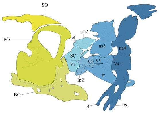
Contents

Introduction ...............................................................................................11
Background ...............................................................................................12
  Zebrafish anatomy and development ...................................................12
  Chondrogenesis and chondrocyte maturation ......................................15
  Patterning of the pharyngeal arches .....................................................16
  The transcription factor Nkx3.2 ...........................................................16
     The role of Nkx3.2 during skeletogenesis .......................................17
     Nkx3.2 related human diseases .......................................................18
  Growth and differentiation factor 5 ......................................................18
     The role of Gdf5 during skeletogenesis ...........................................19
     Gdf5 related human diseases ...........................................................19
  The transition of the primary jaw joint into the middle ear .................20
  Characteristics and formation of a synovial joint.................................21
  Transcriptional enhancers ....................................................................23
  Zebrafish as a model organism.............................................................24
  Methods ................................................................................................25
     Transgenesis in zebrafish using the Tol2 system ............................25
     CRISPR/Cas9 genome editing.........................................................27
     Histology .........................................................................................31
     Skeletal staining ...............................................................................31
     Imaging with confocal microscopy and Optical Projection
     Tomography.....................................................................................31
Aims ..........................................................................................................32
Results and Discussion .............................................................................33
  nkx3.2 knockout in zebrafish (Paper I).................................................33
  Conservation of the cis-regulatory nkx3.2 element in gnathostomes
  (Paper II) ..............................................................................................34
  gdf5 knockout in zebrafish (Paper III) .................................................34
  Development of automated OPT workflow to quantify phenotypic
  differences in zebrafish mutants (Paper IV) .........................................35
Conclusions and future perspectives .........................................................36
  Nkx3.2 deficiency in zebrafish - similar to what is seen in mammals .36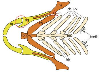
nkx3.2:mCherry transgenic zebrafish line labels jaw joint progenitor
    cells.......................................................................................................36
    Conservation of the nkx3.2 enhancer in gnathostomes ........................37
    Gdf5 deficiency in zebrafish affects the development of the fin
    endoskeleton .........................................................................................37
    Do zebrafish have true synovial joints? ...............................................38
    zOPT automated workflow for analyzing skeletal phenotypes in
    zebrafish ...............................................................................................38
Svensk Sammanfattning............................................................................39
Acknowledgements ...................................................................................41
References .................................................................................................44
Abbreviations

bp               base pair
Cas              CRISPR associated
CRISPR           Clustered interspaced short palindromic repeats
crRNA            CRISPR RNA
dpf              days post fertilization
DNA              Deoxyribonucleic acid
DSB              Double-strand break
GFP              Green fluorescent protein
H&E              Hematoxylin-Eosin
hpf              hours post fertilization
micro CT/µCT     Micro computed tomography
MO               Morpholino
mRNA             messenger ribonucleic acid
NCCs             Neural crest cells
NHEJ             Non-homologous end joining
OPT              Optical Projection Tomography
PAM              Protospacer adjacent motif
RNA              Ribonucleic acid
sgRNA            Single guide ribonucleic acid
SMMD             Spondylo-megaepiphyseal-metaphyseal dysplasia
tracrRNA         trans-activating crRNA
WT               wild-type
gene symbols are not listed
Introduction
The vertebrate skeleton consists of numerous bones that provide support for
the body and organ protection, but it also serves as the location for blood cell
production and mineral storage. The development of the skeleton is complex
and requires an interaction of several factors that ensure correct shaping and
positioning of all skeletal elements. Bony tissue can be formed by intramem-
branous ossification, endochondral ossification, and perichondral ossification.
Skeletal elements developing via intramembranous ossification originate from
mesenchymal cell condensations, which undergo ossification without any in-
termediate steps. Endochondrally ossifying skeletal elements also originate
from mesenchymal cell condensations, but the actual bone is formed from a
cartilage template. In this case, mesenchymal cells undergo chondrogenesis
and chondrocyte maturation to establish the cartilage template, which subse-
quently gets replaced by bony tissue. In order to provide skeletal mobility,
bones require articulation in the form of joints. Joint development involves a
complex interaction of both signaling molecules and transcription factors.
This work mainly focuses on two genes, one coding for the transcription
factor Nkx3.2 and the other coding for the signaling molecule Gdf5. They are
both involved in chondrogenesis (by repressing respectively enhancing carti-
lage maturation during endochondral ossification) as well as joint develop-
ment during skeletogenesis. All studies presented here were carried out in the
zebrafish (Danio rerio) at different developmental stages.
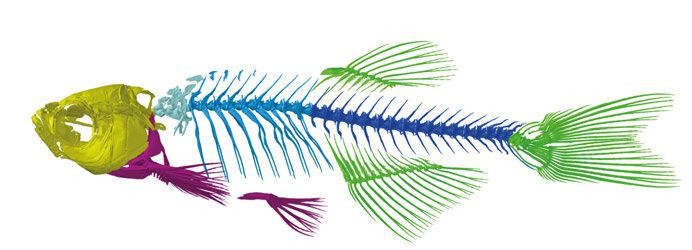
11Background Zebrafish anatomy and development The zebrafish skeleton consists of both dermal and endochondral (or chondral) bones that comprise both exoskeletal and endoskeletal structures. Skeletal structures in the zebrafish include the skull, paired and median fins, and the vertebral column (Fig. 1). Figure 1. Zebrafish (Danio rerio) skeleton. The zebrafish skeleton comprises the skull (yellow), paired fins (magenta), median fins (green), and the vertebral column (different shades of blue). The first four vertebrae are referred to as the Weberian apparatus (light blue), followed by the rib-bearing abdominal region (medium blue) and the caudal region (dark blue). The zebrafish skull can be divided into two main regions: the neurocranium and the viscerocranium. The viscerocranium embryonic cartilage elements originate from neural crest cells (NCCs) and form among others the pharyn- geal arches, which give raise to lower and upper jaws as well as the gill bearing structures (Schilling and Kimmel, 1994). In the adult zebrafish skull, these cartilage elements are replaced by bone. Meckel´s cartilage and the pala- toquadrate are derived from the first pharyngeal arch, also referred to as man- dibular arch (Fig. 2). The first arch elements serve as a feeding apparatus where the U-shaped, ventrally-located Meckel´s cartilages function as the lower jaw and the dorsally-located palatoquadrate cartilages as the upper jaw. Flexibility and movement of the jaws are enabled by the articulation between Meckel´s cartilage and the palatoquadrate, which is referred to as the primary jaw joint. In the adult zebrafish, the first pharyngeal arch elements are partly replaced by the anguloarticular and quadrate bones. Another joint (hyoid) is located in the second pharyngeal arch or hyoid arch, linking the jaw support- ing hyosymplectic and ceratohyal cartilages via a small element called the interhyal cartilage. The paired second arch ceratohyals and the paired cerato- branchial cartilages of the third to seventh arches connect to a ventral midline 12
consisting of the basihyal and posterior basibranchial (Fig. 2). Arches three to
six are attached to the basibranchial via small cartilage elements called hypo-
branchials. In contrast, the paired ceratobranchial cartilages of arch seven do
not connect to the branchial midline and are also the only elements containing
dermal teeth in zebrafish and other Cypriniformes (Kimmel et al., 1995).

Figure 2. Pharyngeal arches of the zebrafish larvae. Schematic of craniofacial car-
tilage elements (ventral view). First pharyngeal arch in yellow, second pharyngeal
arch in orange, arch 3-7 in beige.The circle marks the jaw joint, the triangle marks the
hyoid joint. bb: basibranchial; bh: basihyal; cb: ceratobranchial cartilages; ch: cerato-
hyal; hb: hypobranchial; hs: hyosymplectic; ih: interhyal cartilage; m: Meckel´s car-
tilage; pq: palatoquadrate.

   Skeletal structures in the neurocranium support the brain and sensory or-
gans and derive from both NCCs and mesoderm (Kague et al., 2012). For my
work, the occipital region is of special interest. The occipital region is the most
posterior region of the skull and connects the skull to the vertebral column.
The bones of the occipital series are the basioccipital, exoccipital, and su-
praoccipital (Fig. 3).
   The segmented vertebral column in zebrafish can be subdivided into the
Weberian, abdominal (precaudal), caudal, and caudal fin regions. The number
of vertebrae can slightly vary between individual fish, with an average of 32
elements (Morin-Kensicki et al., 2002). The vertebral bodies (or centra) do
not form from cartilage templates but develop from notochord secreted bone
matrix (Fleming et al., 2004). The first four vertebrae, which bear modified
ribs and neural arches are referred to as the Weberian ossicles/apparatus (Fig.
3). These structures connect the gas bladder to the inner ear and functions in
sound transmission (von Frisch, 1938; Rosen and Greenwood, 1970).

                                                                                      13
Figure 3. Schematic drawing of the occipital bones and the Weberian apparatus in zebrafish (lateral view). Occipital region is displayed in different shades of yel- low. BO: basioccipital; EO: exoccipital; SO: supraoccipital. Weberian apparatus structures in different shades of blue. cl: claustrum ; lp2: lateral process 2 , na: neural arch; os: os suspensorium ; r4: rib 4; SC: scaphium; sn2: supraneural 2; tr: tripus; v: vertebrae. The fins can be divided into paired and median fins. The pectoral and pelvic fins are paired fins whereas the dorsal, anal, and caudal fin are classified as median fins (Fig. 4). Here the focus will be on the pectoral and all median fins. The dorsal and anal fin are anatomically similar as they consist of proximal and distal radials connected to fin rays (lepidotrichia). The radials serve as internal support and originate from mesoderm. They initially develop from a single mesenchyme condensation and the distal radials segment away from the proximal radials later in development (Bird and Mabee, 2003). The skeletal elements of the caudal fin that are most relevant for this thesis include the parhypural and hypurals 1-5, which all develop from individual mesoderm-derived mesenchyme condensations (Fig. 4) (Bird and Mabee, 2003). The pectoral fin endoskeleton is composed of four proximal and six to eight distal radials (Fig. 4) (Grandel and Schulte-Merker, 1998). The proximal ra- dials of the pectoral fin develop from a single cartilaginous disk which is sub- divided by a zone of cartilage dedifferentiation that gives rise to two cartilage disk halves that then divide once more forming four proximal radials aligned along the anteroposterior axis (Dewit et al., 2011). Distal radials do not seg- ment away from the proximal radials as in the dorsal and anal fin, but instead form independently (Grandel and Schulte-Merker, 1998). The pectoral fin en- doskeleton belongs to the appendicular skeleton, homologous to the tetrapod 14
forelimb endoskeleton (Gehrke et al., 2015; Grandel and Schulte-Merker,
1998).

Figure 4. Skeletal structures of zebrafish fins. Schematic drawing of median (cau-
dal, dorsal and anal) fins and pectoral fin (lateral view, anterior to the left). ep: epural;
hsp: hemal spine; hy: hypural; nsp: neural spine; phy: parhypural.

Chondrogenesis and chondrocyte maturation
The first stage of the development of endochondral bones is chondrogenesis
and chondrocyte maturation, whereby the cartilage templates are established.
Chondrogenesis begins with the formation of mesenchyme condensations
which then differentiate into chondrocytes. These chondrocytes then mature
into pre-hypertrophic and later hypertrophic chondrocytes. Hypertrophic
chondrocytes then enter terminal maturation and get gradually replaced by en-
dochondral bone (reviwed by Kozhemyakina et al., 2015). There are a number
of genes expressed at different stages during this developmental process. Mes-
enchyme cells expressing Sox9 are committed to a chondrogenic cell lineage,
develop into chondroprogenitor cells and produce a high number of extracel-
lular matrix proteins (Bi et al., 1999; Bi et al., 2001). Factors promoting chon-
drocyte maturation are, among others, Runx2, Runx3, and Wnt5a (Choi et al.,

                                                                                         15
2012; Yoshida et al., 2004). Indian Hedgehog (Ihh) is essential for chondro-
cyte hypertrophy and bone formation and positively regulates Runx2 and
Wnt5a (Choi et al., 2012; Yoshida et al., 2004). In amniotes, it has been shown
that Runx2 upregulates Col10a1 in hypertrophic chondrocytes, which later
undergo apoptosis or osteoblast differentiation (Fisher et al., 2003; Zheng et
al., 2003).
Patterning of the pharyngeal arches
The craniofacial skeleton of vertebrates originates from NCCs, a cell popula-
tion that delaminates from the dorsal part of the developing neural tube and
migrates in three streams to populate the seven pharyngeal arches (Schilling
and Kimmel, 1994). Interacting signaling pathways guiding migrating NCCs
are essential since the pharyngeal arches consist of complex elements located
along the dorsoventral axis. The signals coordinating the migrated cells derive
from endodermal and ectodermal epithelia and divide NCCs into dorsal, in-
termediate, and ventral subdomains.
The dorsal subdomain forms both the dorsal part of the palatoquadrate and
the hyomandibula, and is controlled by Jagged-Notch signaling. Jagged-Notch
is restricted to the dorsal domain by Endothelin 1 (Edn1), which is active in
the ventral/intermediate domain (Zuniga et al., 2010; Barske et al. 2016). El-
ements of the intermediate domain include the posterior Meckel´s cartilage,
the anterior palatoquadrate, the symplectic, the interhyal cartilage, and the
posterior part of the ceratohyal. Distal-less (Dlx) genes in the intermediate
domain are positively regulated by Edn1 and repressed by dorsal Jagged-
Notch (Talbot et al., 2010; Zuniga et al., 2010). Bone morphogenetic protein
(Bmp) signaling is active in the ventral domain, specifying the anterior
Meckel´s cartilage and the anterior ceratohyal (Alexander et al., 2011). Bmp
signaling suppresses Jagged-Notch and activates both Edn1 and Hand2, which
restricts Dlx gene expression to the intermediate domain (Alexander et al.
2011; Miller et al. 2003; Zuniga et al. 2011). Both nkx3.2 and gdf5 are ex-
pressed within the jaw joint forming region in the intermediate domain
(Bruneau et al., 1997; Miller et al., 2003).
The transcription factor Nkx3.2
NK3 Homeobox 2 (Nkx3.2) is an evolutionary conserved homeobox-contain-
ing gene encoding for a transcription factor acting as a transcriptional re-
pressor. The gene was first discovered in Drosophila (bagpipe, bap) where it
plays a major role in the formation of the midgut musculature by subdividing
mesoderm (Azpiazu and Frasch, 1993). In vertebrates, Nkx3.2 (or Bapx1) is
mainly expressed within the first mandibular arch and is necessary for primary
16jaw joint development in non-mammalian vertebrates (Lukas and Olsson,
2018a; Miller et al., 2003; Square et al., 2015). Knock-down experiments in
both Xenopus and zebrafish result in loss of the jaw joint and as a consequence
fusion of Meckel´s cartilage and palatoquadrate (Lukas and Olsson, 2018a;
Miller et al., 2003). Zebrafish embryos injected with nkx3.2-MO furthermore
display loss of the retroarticular process (RAP), a cartilaginous process at the
dorsoventral tip of Meckel´s cartilage (Miller et al., 2003). Upregulation of
bapx1 expression induces ectopic joint development in the first pharyngeal
arch of Xenopus (Lukas and Olsson, 2018b). In zebrafish, nkx3.2 is positively
regulated by Edn1 signaling (Miller et al., 2003). Loss of Edn1 or its receptor
Endothelin type-A receptor (EdnrA) causes transformations in ventral and in-
termediate arch domains including jaw joint loss (Miller et al., 2003; Nair et
al., 2007). Ventrally expressed hand2 furthermore restricts nkx3.2 to the jaw
joint forming region in the intermediate domain (Miller et al., 2003). By taking
together those findings it can be concluded that Nkx3.2 displays joint promot-
ing abilities and is important for the primary jaw joint development. Further
nkx3.2 expression in zebrafish has been described to be present in the occipital
region, the vertebrae, and the median fins (Crotwell and Mabee, 2007).
The pharyngeal skeleton of extant jawless vertebrates is cartilaginous and
derived, as in all vertebrates, from NCCs (McCauley and Bronner-Fraser,
2003). nkx3.2 in lamprey, a jawless vertebrate, is expressed in ectomesen-
chyme surrounding the pharyngeal arches (Kuraku et al., 2010; Miyashita,
2018). A hypothesis proposed by Cerny et al. (2010) suggests that incorpora-
tion of nkx3.2 into the intermediate domain of the first arch during evolution
led to the emergence of the jaw.
In mice, Nkx3.2 is expressed within the middle ear associated bones tym-
panic ring and gonium as well as in the incudomalleolar joint. Mice embryos
deficient in Nkx3.2 do not show any skeletal defects in the middle ear ossicles,
but the tympanic ring is hypoplastic and the gonium absent (Tucker et al.
2004). More drastic effects of Nkx3.2 loss in mice are prominent in the axial
skeleton including the skull. The two cranial bones basioccipital and ba-
sisphenoid are reduced in size and several bones of the vertebral column are
either lost or deformed in response to Nkx3.2 deficiency (Lettice et al., 1999;
Tribioli and Lufkin, 1999).
The role of Nkx3.2 during skeletogenesis
The transcription factor Nkx3.2 has been shown to act as chondrocyte matu-
ration inhibitor (Kim et al., 2015; Provot et al., 2006). In endochondrally os-
sifying chicken and mouse long bones, Nkx3.2 expression is restricted to pro-
liferative immature chondrocytes and inhibits chondrocyte maturation via re-
pression of the chondrocyte maturation factor Runx2 (Lengner et al., 2005;
Provot et al., 2006). Overexpression of Nkx3.2 in mice resulted in cartilage
17hypertrophy delay and dwarfism of endochondrally ossifying skeletal ele- ments (Jeong et al., 2017). Nkx3.2 is directly upregulated by Sox9, a tran- scription factor that promotes early stages of chondrogenesis but represses hy- pertrophic differentiation (Akiyama et al., 2004; Yamashita et al., 2009). In- hibition of Nkx3.2 is promoted by Phosphatidylinositol-3-kinase (PI3K) (Kim et al., 2015). Inhibition of PI3K in pharmacologically treated cell cultures leads to increased Nkx3.2 expression leading to suppression of chondrocyte maturation (Kim et al., 2015). Nkx3.2 expression in the mandibular arch in chicken is restricted by oral epithelium expressed fibroblast growth factor 8 (Fgf8) and distal mandibular arch expressed bone and morphogenic protein 4 (Bmp4) (Wilson and Tucker, 2004). Degradation of Nkx3.2 can be induced by Indian Hedgehog (Ihh) via a Wnt5a dependent pathway promoting carti- lage development (Choi et al., 2012). Nkx3.2 related human diseases Nkx3.2 is linked to the rare human disease spondylo-megaepiphyseal-metaph- yseal dysplasia (SMMD) (Hellemans et al., 2009; Silverman and Reiley, 1985; Simsek-Kiper et al., 2019). The disease is caused by a homozygous mutation in the first or second exon of nkx3.2 resulting in a frameshift and a premature stop codon (Hellemans et al., 2009; Simsek-Kiper et al., 2019). Patients suf- fering from SMMD display the following phenotypic differences: short stature and trunk, long limbs, enlarged head, reduced neck and back mobility, lack of vertebral body ossification, large epiphyseal ossification centres, wide carti- lage zones between epiphyseal ossification and metaphyses, truncated ribs and pseudoepiphyses in hands and feet (Hellemans et al., 2009). Nkx3.2 might furthermore play a role during osteoarthritis, a chronic joint disease, caused by gradual degradation of articular cartilage in synovial joints. It is suggested that Nkx3.2 might be essential to prevent articular cartilage cells from differ- entiation into hypertrophic chondrocytes (Caron et al., 2015). Growth and differentiation factor 5 Growth and differentiation factor 5 (Gdf5) belongs to the TGF-beta (trans- forming growth factor-beta) superfamily and is also previously known as Con- tact, BMP14, or CDMP1. Gdf5 binds to BMP Receptor 1B and 1A, and to- gether with other BMPs in the TGF-beta superfamily is involved in skele- togenesis (Nishitoh et al., 1996). The expression pattern of gdf5 in zebrafish embryos and larvae has been extensively studied and is present in the primary jaw joint, the midline of the future basihyal cartilage element, the pectoral fins and all median fins at the onset and during chondrogenesis (Bruneau et al., 1997; Crotwell et al., 2001; Miller et al., 2003; Schwend and Ahlgren, 2009). 18
nxk3.2-MO injection in zebrafish results in the loss of craniofacial gdf5 ex-
pression, indicating that gdf5 expression is regulated by Nkx3.2 (Miller et al.,
2003). However, functional studies of gdf5 in zebrafish are lacking. In am-
phibians, gdf5 is expressed in the intramandibular joint as well as in the limb
buds of the appendicular skeleton (Satoh et al., 2005; Square et al., 2015b). In
amniotes, gdf5 expression is restricted to the limbs and prominent in develop-
ing autopod mesenchyme, cartilage, and joints (Merino et al., 1999; Storm and
Kingsley, 1996). Gdf5 loss in mice and the resulting phenotype, referred to as
brachypodism, has been widely studied (Gruneberg and Lee, 1973; Storm and
Kingsley, 1996; Storm et al., 1994). Brachypodism mice display shortened
limb bones and severe deformations in the digits in the form of shortened pha-
langes, lost digit elements and occasional loss of digit joints (Gruneberg and
Lee, 1973; Storm and Kingsley, 1996; Storm et al., 1994).
The role of Gdf5 during skeletogenesis
The role and function of Gdf5 during skeletogenesis have been mainly studied
in mice, chicken, and cell cultures. Overexpression and ectopic supply of Gdf5
enhances both maturation and differentiation of chondrogenic cells in a stage-
dependent manner (Buxton et al., 2001; Coleman and Tuan, 2003; Merino et
al., 1999; Storm and Kingsley, 1999). It has furthermore been shown that Gdf5
is able to stimulate mesenchyme cells to differentiate into cartilage in both
mesenchymal cell culture and in mouse digits, at certain developmental stages
(Merino et al., 1999). These results are consistent with observations in Gdf5
deficient mice, where hypertrophic cartilage zones are reduced in size and
consequently lead to a delay of ossification (Storm and Kingsley, 1999).
Sox11, a factor expressed in cartilage condensations during early stages of
chondrogenesis and in the joint interzone at later stages, has been suggested
to positively regulate Gdf5 expression (Kan et al., 2013). Sox11 binding sites
have been identified in the 5´UTR region of the Gdf5 gene and overexpression
of Sox11 both in vivo and in vitro increases levels of Gdf5 mRNA expression
(Kan et al., 2013). Wnt14, an important factor for synovial joint development
is able to activate and increase Gdf5 expression during elbow joint develop-
ment and in digit regions in mouse (Guo et al., 2004). It has furthermore been
suggested that Gdf5 binding to the TGF-beta superfamily antagonist Noggin
might play an important role during joint development and cartilage shape
regulation (Brunet et al., 1998; Guo et al., 2004; Merino et al., 1999).
Gdf5 related human diseases
The two rare human chondroplasias, Grebe type (CGT) and Hunter-Thomson
type (CHTT) are caused by mutations within the gdf5 gene sequence
(Martinez-Garcia et al., 2016; Thomas et al., 1996). Patients that possess these
gdf5 mutations display a similar phenotype to what is seen in Gdf5 deficient
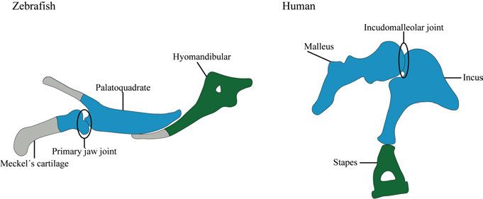
19mice. Both CGT and CHTT patients display phenotypes restricted to the limbs whereby long bones and digit bones (especially the phalanges) are reduced in size. In both cases, proximal elements are more affected than distally located elements (Martinez-Garcia et al., 2016; Thomas et al., 1996). Several studies furthermore indicate that functional single-nucleotide pol- ymorphisms in the Gdf5 gene are a risk factor for the development of osteoar- thritis (Chapman et al., 2008; Daans et al., 2011; Miyamoto et al., 2007). The transition of the primary jaw joint into the middle ear A common feature for all gnathostomes is, as the name already indicates (gna- thos=jaw, stoma=mouth), a jawed mouth. Elements forming the jaw within gnathostomes however differ significantly between mammalian and non- mammalian vertebrates. The jaw in non-mammalian vertebrates is formed by the articular and quad- rate bones that articulate via the primary jaw joint. By taking a closer look into mammalian evolution it becomes clear what leads to the mammalian condition where the dentary and squamosal, connected via the secondary jaw joint, func- tion as jaw elements. The fossil record of mammalian ancestors from the Per- mian and Triassic display a gradual expansion of the dentary and incorpora- tion of the primary jaw joint elements into the middle ear (reviewed by Takechi and Kuratani 2010). This transformation was accompanied by the de- velopment of the secondary jaw joint stepwise taking over the role of the pri- mary jaw joint, allowing its integration and development into the middle ear. Fossils of some extinct synapsids such as Diarthrognathus (200 Mya) display a double-joint state whereby the secondary dermally ossified jaw joint was applied for feeding, enabling the medial movement of the primary endochon- dral jaw joint into the middle ear (Crompton, 1963). A similar transition is observable in living marsupials (Maier, 1987). During early developmental stages in marsupials, elements forming the middle ear bones incus and malleus support the feeding apparatus and enable suckling action. A few weeks after birth, the secondary jaw joint comprised of the dentary and squamosal arises leading to the state of a double jaw joint, enabling the incus and malleus to gradually move into the middle ear (Clark and Smith, 1993; Filan, 1991). Reichert (1837) was the first to launch the theory about the homology be- tween mammalian middle ear ossicles and non-mammalian first and second pharyngeal arch elements based on anatomy studies. Further studies con- firmed homology between non-mammalian first pharyngeal arch elements Meckel´s cartilage and palatoquadrate and mammalian middle ear ossicles malleus and incus, respectively, as well as second arch element hyosymplectic 20
and middle ear ossicle stapes (Gaupp, 1912). As Meckel´s cartilage and pala-
toquadrate derive from a single mesenchymal condensation, which, later in
development, is divided by the primary jaw articulation (Wilson and Tucker,
2004), the question arose whether malleus and incus also develop from a sin-
gle mesenchymal condensation. Experiments in mice revealed that malleus
and incus are united at the onset of cartilage development before they separate
during joint development (Amin and Tucker, 2006) (Fig. 5).
Figure 5. Schematic representation of homology between the first and second
pharyngeal arch in zebrafish to the middle ear in humans. The posterior part of
Meckel´s cartilage and the palatoquadrate are homologous to the mammalian middle
ear ossicles malleus and incus (highlighted in blue). The second pharyngeal arch ele-
ment hyomandibular of non-mammalian vertebrates (including zebrafish) is homolo-
gous to the middle ear ossicle stapes (highlighted in green).
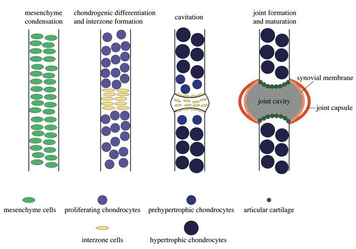
Characteristics and formation of a synovial joint
The evolution of jaw joints was a major event providing jaw flexibility and
enabling active feeding. The fossil record verifies that articulated jaws did not
evolve as originally suggested in Osteichthyes, but were already present in
Silurian placoderms (Zhu et al., 2013). The most sophisticated joints in verte-
brates are referred to as synovial joints. They enable a high degree of flexibil-
ity and enable movement of the skeleton. Synovial joints are present in the
knee and elbow joints, among others, of tetrapods. They are characterized by
a cavity filled with synovial fluid, allowing frictionless movement of the ar-
ticulating bones. The fluid mainly consists of macromolecules like hyaluronic
acid and lubricin (Tanaka et al., 2008) and is produced by synovial cells form-
ing the capsule around the cavity, and by articular cartilage lining the surface
of the articulating bones. The jaw joint of zebrafish has been shown to display
synovial-like characteristics as the expression and function of the lubricin en-
coding gene prg4b has been described in joint-lining cells (Askary et al.,
2016). However, whether the zebrafish jaw joints display additional synovial
21joint characteristics such as synoviocytes remains unclear. At the onset of long bone formation mesenchyme cells condense before undergoing chondrogene- sis. However, at the site of joint development mesenchyme cells do not un- dergo chondrogenesis but flatten and form an area referred to as interzone (Archer et al., 2003; Craig et al., 1987). Cells from the interzone partly give rise to joint-specific cells and structures (Archer et al., 1994; Mitrovic, 1977). A more recent study suggests that an additional continuous influx of cells into the interzone is necessary for the formation of joint components (Shwartz et al., 2016). Interzone formation is followed by joint cavitation, a process, which is suggested to be primarily facilitated by increasing extracellular ma- trix synthesis in order to separate the two articulating cartilage elements (Edwards et al., 1994). After cavitation, synovial-specific structures are formed, including the fluid filled cavity (Fig. 6). Figure 6. Schematic drawing of synovial joint development. Mesenchyme conden- sation giving rise to bony elements. Mesenchyme cells committed to the chondrogenic line undergo chondrogenesis and differentiate into chondrocytes which mature into prehypertrophic and hypertrophic chondrocytes. The interzone is formed by non- chondrogenic interzone cells which determine the position of the developing joint. Cavitation takes place, which separates the adjacent skeletal elements from each other. The mature synovial joint consists of a fluid-filled joint cavity, which is surrounded by a synovial membrane and the joint capsule. Articular cartilage is present on the cavity facing surfaces of the articulating elements. 22
Transcriptional enhancers
Cis- and trans-regulatory elements are DNA sequences that control and regu-
late gene expression in eukaryotes. Studies have shown that these elements
have contributed significantly to creating phenotypic differences and variety
during evolution (Indjeian et al., 2016; Shapiro et al., 2004; Sucena and Stern,
2000). Enhancers are categorized as cis-regulatory elements and are non-cod-
ing sequences that can activate gene transcription. The cis-regulatory element
can be in close proximity to the gene it is regulating but can even be located
further away (Kleinjan et al., 2001; Lettice et al., 2002). Enhancers contain a
variety of binding sites, also called motifs, that interact with transcription fac-
tors and other regulatory molecules (Fig. 7). For regulating transcription, the
enhancer has to be in close proximity to the promoter of the gene it is regulat-
ing, which is achieved by cohesin and mediator complexes that create a DNA
loop (Amano et al., 2009; Kagey et al., 2010; Schmidt et al., 2010). Transcrip-
tion factors and mediator proteins interact with the enhancer sequence and
recruit RNA polymerase to initiate gene transcription (Fig. 7) (Malik and
Roeder, 2010).

Figure 7. Schematic drawing of enhancer function. Enhancer sequences can be up-
or downstream of the gene they are regulating and contain transcription factor binding
sites (motifs). Transcription factor binding accompanied by cohesin mediated DNA
looping and mediator complex binding brings the enhancer into close proximity to the
regulating gene. The complex recruits RNA polymerase, which subsequently activates
or enhances gene transcription.

                                                                                   23
Zebrafish as a model organism
The zebrafish (Danio rerio) is a small freshwater tropical teleost fish of the
family Cyprinidae. Their natural habitats are rivers and small streams in south
Asia (reviewed by Engeszer et al., 2007). Starting in the 1970s when Georg
Streisinger first used the zebrafish to study the nervous system, it became
more and more popular and established as a model organism. The early devel-
opment of the zebrafish has been extensively studied and described by Kim-
mel et al. (1995). They have described the development from fertilization until
early larval stages at an incubation temperature of 28.5°C, which was set as
the standard temperature for experimental use (Kimmel et al., 1995).
In contrast to other model organisms such as mice and rats, zebrafish are
comparatively inexpensive to keep and easy to breed and handle. Other ad-
vantages include a high reproduction rate, external fertilization and develop-
ment, rapid development, and transparency at early embryonic stages. The
transparency of embryos is particularly convenient for in vivo functional stud-
ies in transgenic zebrafish lines. The high reproduction rate is especially use-
ful to perform large-scale forward genetic screens. For studying craniofacial
development in zebrafish, forward genetics techniques have been applied to
identify multiple mutants displaying defects in the formation of the pharyn-
geal arches and cartilage differentiation (Piotrowski et al., 1996; Schilling et
al., 1996).
Subsequently, to study the function and role of specific genes, reverse ge-
netic approaches were developed and applied. One method which can be used
to for generating gene knock-downs are microinjection of morpholino oligo-
nucleotides (MOs). These are synthetic antisense oligonucleotides targeted at
specific mRNAs and can inhibit both splicing and translation, resulting in a
gene knock-down (reviewed by Summerton and Weller, 1997). Several nega-
tive side effects caused by MO-injections such as high off-target rates and
non-specific toxicity, resulted in increased use of knock-out techniques
(Nasevicius and Ekker, 2000). To generate more efficient and reliable
zebrafish null mutants, new targeted mutagenesis tools such as TALENs
(Christian et al., 2010), zinc-finger nucleases (Foley et al., 2009) and CRISPR-
Cas9 (Cong et al., 2013) have been developed.
24Methods
Transgenesis in zebrafish using the Tol2 system
Transgenic zebrafish lines are a great tool to label and study specific cells and
tissues in the living organism. The principle of transgenesis is rather straight-
forward and first requires the identification of gene-specific enhancer or pro-
moter, which is incorporated into the genome in combination with a fluores-
cent protein (reporter). After successful genome integration, only cells with
the capability of enhancer/promoter activation are going to express the fluo-
rescent protein, which can be visualized by using in vivo fluorescent micros-
copy. A very common method for effective genome integration is the Tol2
transposon system (Kawakami, 2007). Transposable elements are naturally
occurring DNA sequences with the ability to change their location in the ge-
nome by excision and reintegration. Tol2 is an autonomous transposon which
has been identified in the medaka fish Oryzias latipes (Koga et al., 1996). The
Tol2 transposase recognizes inverted terminal repeats (ITR) and excises and
reinserts DNA elements flanked by ITRs.
For generating zebrafish transgenic lines, a plasmid containing the desired
enhancer or promoter sequence and a fluorescent protein is generated, flanked
by Tol2 recognition sites (Fisher et al., 2006). The plasmid is injected into
zebrafish embryos at the one-cell stage, together with transposase mRNA,
which facilitates the excision of the Tol2 flanked DNA construct from the
plasmid and integration into the zebrafish genome (Fig. 8). Since the construct
is not evenly distributed after injection and subsequent cell division, the in-
jected embryos (F0 generation) display mosaic fluorescent expression. For
generating stable transgenic lines, the construct needs to be integrated into the
germline of F0 fish. Germline transmission can be detected by outcrossing
founder F0 with wild-type fish and analyzing F1 generation embryos (Fig. 8).
For vector assembly we used multisite Gateway-based cloning system for
Tol2 transgenesis constructed by Kwan et al. (2007) in combination with a
commercially available Gateway system from Invitrogen.
25Figure 8. Overview of the Tol2 mediated transgenesis workflow in zebrafish. A construct containing the cis-regulatory element of interest and a fluorescent reporter gene flanked by Tol2 recognition sites is co-injected together with Tol2 transposase mRNA into a fertilized one-cell stage zebrafish embryo. After injection, Tol2 trans- posase mRNA is translated into Tol2 transposase which recognizes Tol2 sites on the vector and leads to the excision of the Tol2 site flanked construct and random integra- tion into the zebrafish genome. Injected embryos display mosaic expression and are outcrossed in wild-type fish to confirm germline transmission in the F1 embryos, lead- ing to the generation of a stable transgenic line. 26
CRISPR/Cas9 genome editing
From defense in prokaryotes to genome editing
The CRISPR/Cas system is a naturally occurring adaptive immunity response
in bacteria and archaea to protect them against phages and viruses (Barrangou
et al., 2007). It all started with the discovery of repeated structures separated
by variable spacer sequences in the Escherichia coli and the halophilic Ar-
chaea Haloferax mediterranei genome (Ishino et al., 1987; Mojica et al.,
1993). These structures, identified in more prokaryote species and named for
Clustered Regulatory Interspaced Short Palindromic Repeats (CRISPR)
(Jansen et al., 2002; Mojica et al., 2000) have been shown to be of viral and
bacteriophage origin (Bolotin et al., 2005; Mojica et al., 2005; Pourcel et al.,
2005). Prokaryotes containing spacers specific for certain viruses or phages
display resistance against the invader, leading to the conclusion that CRISPRs
act as adaptive immunity system (Barrangou et al., 2007; Bolotin et al., 2005;
Mojica et al., 2005). Cas (CRISPR associated sequence) genes that have been
detected in close proximity to CRISPRs helped to further investigate on how
CRISPR is working (Haft et al., 2005; Jansen et al., 2002; Makarova et al.,
2006). CRISPRs are transcribed and processed into crRNAs (CRISPR-
RNAs), which are short RNA sequences (Mojica et al., 2000; Tang et al.,
2002). crRNAs form a complex with Cas proteins and detect invading proto-
spacer sequences which are subsequently degraded by the Cas-proteins
(Brouns et al., 2008; Garneau et al., 2010; Marraffini and Sontheimer, 2008).
Additionally, Cas proteins have been shown to be involved in integrating new
spacer sequences into the CRISPR loci and in generating crRNAs (Bhaya et
al., 2011; Terns and Terns, 2011). Another crucial element necessary for in-
vader DNA recognition and detection are protospacer adjacent motifs
(PAMs). PAMs are flanking protospacer sequences and are essential for Cas9
proteins to detect invading sequences. PAMs are not present in CRISPR loci
spacers which protects the prokaryote from degrading their own CRISPR
spacers (Bolotin et al., 2005; Horvath et al., 2008; Mojica et al., 2009).
The idea to use the CRISPR/Cas system as a genome editing tool emerged
after the discovery of the CRISPR/Cas9 system. In 2020, two researchers;
Emmanuelle Charpentier and Jennifer A. Doudna were awarded the Nobel
Prize in Chemistry for their research leading to the use of the CRISPR/Cas9
system for genome editing. Together, they discovered the tracrRNA (trans-
activating crRNA) and its function, another important puzzle piece to under-
stand CRISPR/Cas9 system in more detail (Deltcheva et al., 2011; Jinek et al.,
2012). The tracrRNA sequence is partly complementary to the repeated re-
gions of the CRISPR locus and is involved in the maturation process of the
crRNA (Deltcheva et al., 2011). tracrRNA is furthermore essential for the
Cas9 endonuclease to introduce DNA double-strand breaks into the targeted
protospacer sequences (Deltcheva et al., 2011; Jinek et al., 2012).
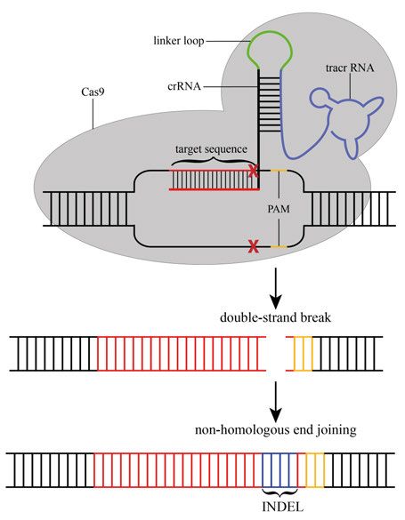
27The genome editing technique using CRISPR/Cas9 is based on a synthe- tized chimeric sgRNA (single guide RNA) molecule consisting of the target DNA sequence (around 23 nucleotides) and the crRNA:tracrRNA. After co- injection of sgRNA and Cas9 protein or mRNA, the sgRNA forms a complex with the Cas9 protein and guides it to the target sequence. The Cas9 endonu- clease subsequently introduces a double-strand break about 3 bp upstream of the PAM site (Fig. 9). Double strand breaks are repaired by the natural repair mechanism of the cell by either non-homologous end joining (NHEJ) or ho- mology-directed repair (HDR). The DNA repair machinery however is not error-proof and often leads to the introduction of insertions and deletions (IN- DELS), which in most cases cause a frameshift and consequently the occur- rence of a premature stop codon (Fig. 9). Figure 9. Schematic drawing of CRISPR/Cas9 mediated genome-editing mecha- nism. A single guide RNA, consisting of tracrRNA which is connected via a linker loop to the crRNA that recognizes the genomic region of interest. The sgRNA is de- signed to recognize sequences which are in close proximity to a PAM sequence (5'- NGG-3') and able to guide/recruit the Cas9 endonuclease which introduces a double- strand break (DSB). The DSB is repaired by non-homologous end joining (NHEJ), an error-prone pathway that often results base-pair insertions or deletions (INDELS). This often leads to a frameshift and the occurrence of a premature stop codon. 28
CRISPR/Cas9 in zebrafish
A number of optimization steps are required for successful targeted mutagen-
esis with CRISPR/Cas9 in zebrafish. In order to achieve high sgRNA activity,
the target sequence should be designed with a high GC-content and a length
of around 18 bp (Gagnon et al., 2014; Moreno-Mateos et al., 2015). A
zebrafish codon-optimized version of the Cas9 mRNA sequence has been gen-
erated and shown to increase nuclear targeting which leads to higher mutagen-
esis frequencies (Jao et al., 2013).
   In our experimental set up we applied a cloning-free sgRNA synthesis pro-
tocol developed by the Burgess Lab (Varshney et al., 2015). After co-injection
of one to two sgRNAs and Cas9 mRNA into one-cell stage embryos, we fol-
lowed a high-throughput workflow protocol for CRISPR/Cas9 in zebrafish,
allowing somatic mutation efficiency verification in the F0 injected embryos
(Varshney et al., 2016). Once sufficient sgRNA-Cas9 activity was detected,
F0 fish were raised to adulthood and outcrossed with wild-type fish (Fig. 10).
Mutation screens were performed in F1 generation to identify heterozygous
mutant fish. To obtain uniform mutant fish, we performed additional outcross
steps of the F1 heterozygote mutant founders. The homozygote mutant phe-
notypes were analyzed in the offspring of the F2 fish carrying heterozygous
uniform alleles (Fig. 10).

                                                                           29
Figure 10. Overview of the CRISPR/Cas9 workflow in zebrafish. One to two sgR- NAs targeting the same gene are co-injected together with Cas9 mRNA into a ferti- lized one-cell stage zebrafish embryo, resulting in chimeric embryos which are raised to adulthood. F0 mosaic founder fish are outcrossed with wild-type fish and the F1 generation is screened for germline-transmitted mutations. In order to obtain a suffi- cient number of heterozygous fish carrying the uniform allele of interest (here red mark in the chromosome), additional crosses with wild-type are necessary. F2 heter- ozygous fish carrying the uniform mutant allele are incrossed to obtain homozygote mutant fish in the F3 generation. 30
Histology
For histological examination of zebrafish tissues I performed Hematoxylin-
Eosin (H&E) and Nuclear Fast Red staining on deparaffinized thin sections.
H&E stain is a standard stain that marks cells and tissues in blue and different
shades of pink. Hematoxylin is a positively charged basic dye and stains nu-
cleic acids blue (Chan, 2014; Fischer et al., 2008). Eosin is a negatively
charged acid dye and stains cytoplasm and extracellular matrix in different
shades of pink/red (Chan, 2014; Fischer et al., 2008).
   In contrast to H&E staining, Nuclear Fast Red staining is faster to perform,
but the obtained staining is less complex as it only stains nucleic acids in pink,
making it more challenging to distinguish different tissues from one another.

Skeletal staining
In order to analyze skeletal phenotypes in mutant zebrafish and compare these
to wild-type fish, whole mount cartilage and bone staining, which is a fast and
inexpensive method, can be easily performed. I performed double staining us-
ing Alcian blue to stain cartilage and Alizarin red for bone staining. Alcian
blue is taken up by proteoglycans present in cartilage tissue whereas Alizarin
red stain binds to calcium present in mineralized tissue such as bones and teeth
(Puchtler et al., 1969; Scott, 1996). For double staining, I applied an acid-free
staining protocol modified from the original developed by Walker and
Kimmel (2007) whereby demineralization of bone by acid is avoided.

Imaging with confocal microscopy and Optical Projection
Tomography
Imaging of zebrafish embryos or larvae can often be quite challenging due to
their small size. For 3D imaging, confocal laser scanning microscopy is fre-
quently used. Hereby, the sample is fixed in agarose on a glass-bottomed plate
and placed on the microscope stage. 2D images, that are taken by sectioning
in depth acquiring Z stacks, are used for subsequent 3D reconstruction or max-
imum intensity projections. In this thesis, confocal microscopy was used to
perform live imaging of transgenic zebrafish up to 14 dpf.
   Another alternative to generate brightfield high-resolution 3D imaging is
Optical Projection Tomography (OPT) (Sharpe et al., 2002). The data ac-
quired by OPT is, in contrast to confocal imaging, obtained by rotating the
sample during the acquisition, resulting in isotropic high-resolution images.
OPT uses the same principle as X-ray computed tomography but utilizing vis-
ible radiation instead of X-rays. 2D images are acquired at multiple angles and
by using tomographic reconstruction, a 3D image can be reconstructed. OPT
was used to image and analyze cartilage and bone stained wild-type and mu-
tant zebrafish at 5 dpf and 9 dpf.

                                                                               31
Aims
The overall aim of this thesis was to achieve a better understanding of genes
involved in chondrogenesis and joint development in gnathostomes by using
the zebrafish Danio rerio as a model. We furthermore aimed to generate a
transgenic line labeling the jaw joint in zebrafish and to improve imaging and
analysis techniques for zebrafish embryos and larvae.
Paper I
• To generate a CRISPR/Cas9 induced nkx3.2 knockout line in
zebrafish and analyze the phenotype at different developmental
stages
Paper II
• To identify the regulatory elements of nkx3.2 in gnathostome ge-
nomes and investigate whether they are evolutionary conserved
• To generate a stable transgenic zebrafish line using the nkx3.2 en-
hancer element to study jaw joint development
• To generate a nkx3.2 enhancer knockout in zebrafish by using
CRISPR/Cas9 to study the resulting phenotype
Paper III
• To generate a CRISPR/Cas9 induced gdf5 knockout line in
zebrafish and analyze the phenotype at different developmental
stages
Paper IV
• Using OPT to generate an automated workflow for imaging and
analysis of in situ whole mount and skeletal stained zebrafish
32Results and Discussion
nkx3.2 knockout in zebrafish (Paper I)
To study the role of nkx3.2 during zebrafish development, we generated
CRISPR/Cas9-induced nkx3.2 mutant zebrafish and analyzed the resulting
phenotype at different developmental stages. The generated mutant allele has
a 7 bp deletion in the first exon leading to a premature stop codon after 95
amino acids. The most obvious mutant phenotype, detectable by a simple vis-
ual inspection, was the "open-mouth" phenotype caused by jaw joint loss. De-
spite this severe phenotype homozygote mutant fish were able to feed and
reach adulthood. OPT combined with 3D reconstruction and statistical com-
ponent analysis performed on cartilage- and bone-stained larval wild-type and
mutant fish revealed additional mutant phenotypes in the skull around the otic
capsule. We applied cartilage and bone staining as well as µCT analysis on
adult nkx3.2 mutant fish for further investigations on the posterior skull phe-
notype. µCT analysis revealed severe fusions and losses of skeletal elements
in the occipital region and the Weberian apparatus in nkx3.2 mutant adult fish.
The exoccipital and basioccipital were partially or completely fused in Nkx3.2
deficient fish causing partial or complete loss of the cavum sinus impar. De-
fects in the Weberian apparatus in response to nkx3.2 knockout included loss
of the first cervical vertebrae including the associated scaphium and claus-
trum. In some analyzed homozygote mutant fish, we could detect loss of cer-
vical vertebra two and reduction of its lateral process. The third cervical ver-
tebra displayed deformation and absence of the anterior ramus and the articu-
lating process. The parapophyses, small bones articulating ribs 5-11 with the
vertebral centra, were completely absent in nkx3.2 mutant fish causing direct
attachment of the ribs to the vertebrae. Skeletal abnormalities were detected
in dorsal and anal fins, showing increased proximal radial length in the mutant
line when compared to wild-type. Our results confirm the key role for nkx3.2
during jaw joint development but more importantly reveal novel phenotypes
in response to Nkx3.2 loss in zebrafish, which are reminiscent of phenotypes
reported in Nkx3.2 deficient amniotes.
33You can also read