Biological properties and therapeutic effects of plant-derived nanovesicles
←
→
Page content transcription
If your browser does not render page correctly, please read the page content below
Open Medicine 2020; 15: 1096–1122
Review
Sante Di Gioia, Md Niamat Hossain, Massimo Conese*
Biological properties and therapeutic effects of
plant-derived nanovesicles
https://doi.org/10.1515/med-2020-0160
received April 23, 2020; accepted September 23, 2020
1 Introduction
Abstract: Exosomes-like nanoparticles can be released by Natural products and their derivatives have been widely
a variety of plants and vegetables. The relevance of plant- used throughout the human history, and today, they con-
derived nanovesicles (PDNVs) in interspecies communi- stitutes a large part of the pharmaceutical market [1,2].
cation is derived from their content in biomolecules (li- For years, pharmaceuticals companies have not paid due
pids, proteins, and miRNAs), absence of toxicity, easy attention to these classes of compounds for many rea-
internalization by mammalian cells, as well as for their sons, such as the wrong idea that natural products are
anti-inflammatory, immunomodulatory, and regenerative only useful as a restricted class of drugs, e.g., antibiotics:
properties. Due to these interesting features, we review natural products had huge success in the post-World War
here their potential application in the treatment of inflam- II era as antibiotics, and the two terms have become sy-
matory bowel disease (IBD), liver diseases, and cancer as nonymous [1]. Generally, large pharmaceutical compa-
well as their potentiality as drug carriers. Current evi- nies have focused their attention on screening synthetic
dence indicate that PDNVs can improve the disease state compound libraries for drug discovery, whereas small
at the level of intestine in IBD mouse models by affecting companies have started to explore the role of natural
inflammation and promoting prohealing effects. While compounds against diseases such as cancer, microbial
few reports suggest that anticancer effects can be derived infection, and inflammatory processes [3,4]. When a po-
from antiproliferative and immunomodulatory properties tential therapeutic application is considered, one of the
of PDNVs, other studies have shown that PDNVs can be biggest issues that make the use of natural compounds
used as effective delivery systems for small molecule
quite challenging is their low bioavailability [4]. For in-
agents and nucleic acids with therapeutic effects (siRNAs,
stance, it has been calculated that the administration of
miRNAs, and DNAs). Finally, since PDNVs are character-
curcumin orally requires doses of 3.6 g/day to reach
ized by a proven stability in the gastrointestinal tract, they
serum levels of 11.1 nmol/L, and subjects who take lower
have been considered as promising delivery systems for
doses of curcumin did not have detectable plasma levels
natural products contained therein and drugs (including
[5,6]. Similar results were observed with other natural
nucleic acids) via the oral route.
compounds such as polyphenols and flavonoids [7,8].
Keywords: exosome-like nanoparticles, antitumoral, In this context, the use of nanoparticles can enhance
miRNAs, drug delivery, inflammatory bowel disease the efficacy of natural products in disease treatment by
increasing their bioavailability. The benefits of nanopar-
ticles are not just a “size-matter” but something more
complicated. Indeed, it is well recognized that at a na-
noscale level, particles can acquire unique properties.
Basically, nanodelivery systems might be useful to over-
come the limitations of the traditional natural com-
pounds administration because of the following reasons:
1. Nanoparticles improve the solubility of natural com-
* Corresponding author: Massimo Conese, Laboratory of pounds [9].
Experimental and Regenerative Medicine, Department of Medical 2. Nanoparticles could target the natural products to spe-
and Surgical Sciences, University of Foggia, Foggia, Italy,
cific organ, which improves the selectivity, drug de-
e-mail: massimo.conese@unifg.it
Sante Di Gioia, Md Niamat Hossain: Department of Medical and livery, efficacy, and safety and thereby reduces dose
Surgical Sciences, University of Foggia, 71122 Foggia, Italy and increases the patient compliance.
Open Access. © 2020 Sante Di Gioia et al., published by De Gruyter. This work is licensed under the Creative Commons Attribution 4.0
International License.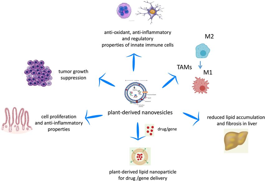
Biological properties and therapeutic effects of PDNVs 1097
3. They appear to be able to deliver high concentrations cells undergoing apoptosis (see Vesiclepedia, www.micro-
of drugs to disease sites because of their unique size vesicles.org and ref. [13]). Recently, ISEV has reviewed many
and high loading capacities. features of EVs, for examples, subtypes should be defined by
4. Delivering the drug in small particle size enhances the physical and biochemical characteristics and/or conditions/
entire surface area of the drugs, therefore allocating sources rather than on conventional nomenclature [14].
quicker dissolution in the blood. However, since these recommendations are still to be ac-
5. Nanoparticles show enhanced permeation and reten- cepted universally, we refer to terms referred earlier.
tion (EPR) effect, i.e., enhanced permeation through For many years, researchers had been guided by the
the barriers because of the small size and retention outdated idea that exosomes are waste products obtained
due to poor lymphatic drainage such as in tumor [10]. from the shedding of plasma membranes, whereas exo-
6. Nanoparticles exhibit passive targeting to the disease some vesicles cargo is composed of proteins, lipids, and
site of action without the addition of any particular nucleic acids. All of these “cargo” biomolecules accumu-
ligand moiety. late inside exosomes and are wrapped by the phospho-
7. Due to the previous properties, nanoparticle applica- lipid bilayer. This structure allows them to take part
tion decreases the side effects. in processes such as intercellular communication, ex-
change of materials with other cells, elimination of un-
There are different types of synthetic nanoparticles wanted products from cells, and immune surveillance
used for drug delivery, such as polymer nanoparticles, [15,16]. Growing evidence show EVs (MVs along with exo-
solid lipid nanoparticles (SLNs), crystal nanoparticles, somes) participate in various cell signaling process and
liposomes, micelles, and dendrimers. Each of these are likely involved in pathophysiological processes such
nanoparticles has its own advantages and disadvan- as cardioprotection [17] and cancer [18]. Currently, there is
tages as a drug delivery vehicle. For example, polymer evidence that PDNVs may be involved not only in plant–cell
nanoparticles between 10 and 1,000 nm in diameter communication but also in interspecies communication be-
can have the characteristics desired for an efficient de- tween plants and animals [19]. For example, a plant-derived
livery of molecules [4,11]. Unfortunately, these sys- miRNA such as miR-168 has been reported to enter the cir-
tems are “artificial” as they are obtained by chemical culation of rice-fed mice enclosed in vesicles and to modu-
synthesis, and this poses a strong limitation for their late the expression of target genes [20]. Besides, exosomes
application in vivo because synthetic nanoparticles found in cell culture medium and biological fluids such as
have two major limitations: (1) each of their constitu- urine, saliva, breast milk, plasma, and cerebrospinal fluid
ents must be evaluated for potential in vivo toxicity [21], as well as matrix-bound nanovesicles (MBVs), em-
before clinical application and (2) the production scale bedded within biological scaffold composed of extracellular
is limited. matrix (ECM), have been identified [22,23]. Since they are
To overcome these limitations, many research groups localized to collagen fibrils, likely anchoring via adhesion
focused their attention in developing green, sustainable molecules, MBVs have been isolated by enzymatic digestion
and biocompatible materials for the delivery of bioactive of ECM bioscaffolds obtained from urinary bladder matrix,
compounds within pharmaceutical and medical industries. small intestinal submucosa, and dermis [22,23]. Although
Basically, plant-derived nanovesicles (PDNVs) would be MBVs share features in common with exosomes and micro-
excellent candidates for the delivery of therapeutic agents vesicles, such as their size (10–1,000 nm) and the presence
(e.g., anticancerous drugs, small interfering RNAs (siRNAs), of miRNA cargoes, the lack of identifiable surface markers
microRNAs (miRNAs)), or poorly soluble natural compounds (such as CD63, CD81, CD9, and Hsp70) and unique nucleic
(e.g., curcumin) [12]. Among the various PDNVs, exosomes acid and protein cargoes suggest that they represent a
have gained attention as a potential nanodelivery system as different population of signaling vesicles [22–24]. MBVs
they are characterized by various desirable properties such and their miRNA cargoes can recapitulate many of their
as small size, biocompatibility, and high stability. According parent ECM’s effects, including the promotion of a regula-
to the International Society for Extracellular Vesicles (ISEV; tory macrophage phenotype, thus facilitating tissue repair
www.isev.org), there are three main extracellular vesicles [22]. In addition, MBVs have been shown to positively reg-
(EVs): exosomes, ranging from 30 to 120 nm and that are ulate primary neuron survival and growth [25] as well as to
produced through the endocytic pathway; microvesicles reduce proinflammatory, neurotoxic glial signaling enhan-
(MVs), 100–1,000 nm vesicles, that are released from the cing healing responses in the retina and the optic nerve [26].
plasma membrane through outward protrusion or budding; Although PDNVs were initially conceived as more similar in
and apoptotic bodies, 1–6 µm in diameter, derived from structure and function to mammalian exosomes than MBVs1098 Sante Di Gioia et al.
[27], they are quite heterogeneous in size (see below) and improving inflammatory bowel disease prevention and
may have distinct features from mammalian exosomes and treatment by blocking damaging factors and promoting
MBVs. However, as outlined briefly earlier, the field of EVs is healing factors [29,30]. Moreover, the positive action of
continuously evolving and we may have to deal with new exosome-like EVs was demonstrated in the liver when
concepts about them in the close future [14]. According to injured by alcohol [31]. Also, PDNVs are not cytotoxic
the recent literature, PDNVs, the focus of this review, display and can be considered as novel drug carrier systems
many biological properties, which are illustrated in the fol- used in combination with nucleic acids (siRNAs and
lowing sections (Figure 1). miRNAs) as well as with anticancer therapeutics [28,32].
PDNVs display many properties that make them sui- Altogether, today, PDNVs may be considered a real
table for clinical applications, including a relatively alternative to synthetic nanoparticles for their complex
high internalization rate, low immunogenicity, stability biological properties, therapeutic applications, and drug
in the gastrointestinal (GI) tract, and the ability to over- delivery systems. The aim of this review is to provide up-
come the blood–brain barrier but not the placental bar- to-date consideration to PDNVs, highlighting their phy-
rier [28]. Since the GI tract is the first tissue receiving EVs sicochemical properties, their biological effects, as well
from edible fruits and plants, many researchers focused as their anti-inflammatory, anti-oxidant, and proregen-
their attention onto EVs biological functions on the in- erative properties. Finally, we will give an insight in the
testinal barrier. These studies have shown that these treatment of gastrointestinal/liver diseases and cancer
exosome-like plant-derived EVs can be used for through PDNV-mediated drug delivery.
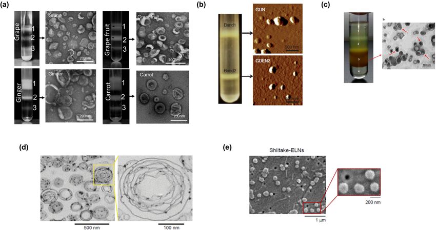
Figure 1: Biological properties of PDNVs. PDNVs can regulate in vitro and in vivo the function of macrophages and dendritic cells by inducing
anti-inflammatory and regulatory functions, as well as shifting tumor-associated macrophages (TAMs) from a M2 to a M1 phenotype. PDNVs
have been demonstrated to participate in intestinal tissue homeostasis in in vivo animal models and have validated functions against
inflammation-related diseases and cancers. Finally, the efficacy of PDNVs for gene or drug delivery has been shown.Biological properties and therapeutic effects of PDNVs 1099
2 Isolation and purification of the case of liposomes that are the more approximate drug
vehicle to exosomes and PDNVs. The high costs strongly
PDNVs limit the large-scale production of artificial nanovesicles
and mammalian cell-secreted exosomes to be employed
The isolation of PDNVs is mainly based on differential cen-
in humans as commercial products [28,43]. Although
trifugation plus density gradient centrifugation. Plants are
PDNVs can be produced economically [44] and have a
ground to juice in a mixer, and low-speed centrifugations
significant potential for large-scale production [19,45], due
are used to remove large plant debris and aggregates,
to the high yield as outlined earlier, the field lacks scalable
whereas ultracentrifugation is used to pellet PDNVs. A “stan-
methods to efficiently isolate and assemble PDNVs of uni-
dard protocol” to isolate PDNVs utilizes multiple centrifuga-
form size [28].
tion steps (low, medium, and high speeds). Generally, the
amount of a raw material (plant or fruit) used to isolate
PDNVs is variable, ranging from 2 to 10 g [33,34] to 250 g
[31]. Initially, intact cells are removed by low-speed centri-
fugation (e.g., 1,000 × g). The supernatant is then subjected 3 Physicochemical and biological
to centrifugal forces in the range of 10,000–20,000 × g to properties of PDNVs
remove large debris and intact organelles. This supernatant
is then subjected to centrifugation at high speed (100,000– PDNVs from edible plants and fruit have been characterized
150,000 × g). While the stated methodology is relatively for their physical parameters (size and surface charge),
straightforward, the type, quantity, and quality of the PDNVs biomolecules content (lipids, protein, and miRNAs), and
isolated by ultracentrifugation is highly sensitive to multiple biological properties. Here, we provide an overview of
parameters, including the g force, the rotor type (fixed angle PDNV characteristics (Table 1).
or swinging bucket), the angle of rotor sedimentation, radius
of the centrifugal force, pelleting efficiency (rotor and tube
k-factors), and solution viscosity [35–37]. Moreover, the
highest speed used (100,000 × g or greater) also sediments 3.1 Chemical properties
other vesicles, proteins, and/or protein/RNA aggregates.
Thus, a subsequent sucrose density gradient step is used Lipids are key components of the lipid bilayer structures
to separate the PDNVs from contaminants of different den- of PDNVs, which have distinct composition from mam-
sities. Gradient ultracentrifugation requires an extended malian cell-derived exosomes and artificially synthesized
centrifugation time (1–5 h), but provides a more purified liposomes [46–48]. Lipid profiling has ascertained that
edible plant nanoparticle isolate than ultracentrifugation two major classes of lipids in PDNVs are phospholipids and
alone [38]. Other methods such as ultrafiltration and immu- glycerol lipids, but they lack cholesterol [28]. Ju et al. [49]
noisolation have been implemented for animal-derived exo- identified exosome-like nanoparticles from grapes (GELNs)
somes to obtain purer preparations [38,39]; however, they using electron microscopy examination and found they con-
have some drawbacks, including higher costs and have not sist of 98% phospholipids, among which approximately 50%
been extensively used for PDNVs purification. has been identified as phosphatidic acids (PA). Differential
The yield is variable depending on the plant source centrifugation and sucrose gradient ultracentrifugation were
and the method used for quantification. Zhang et al. [40] used to isolate GELNs and a triple quadrupole mass spectro-
reported a high yield of exosome-like vesicles of 48.5 ± meter was used to determine their lipid composition. The
4.8 mg per 1 kg of ginger. In another study, the produc- presence of an extremely small amount (∼2%) of usual plant
tion yields of PDNVs are very similar across the fruits and lipids (e.g., galactolipids, such as digalactosyldiacylglycerol
root-derived edible plants, with 100 g of edible plant ma- (DGDG) and monogalactosyldiacylglycerol (MGDG)) have
terial producing about 350–450 mg of nanoparticles [41]. also been reported [49]. PA is a cell-signaling lipid with
In alternative to weighing, the yield of PDNVs can be many biological activities, including the activation of the
measured by a zeta-sizer. For instance, among the mush- mammalian target of rapamycin (mTOR) and mitogen-acti-
rooms tested, oyster mushroom-derived exosome-like vated protein kinase (MAPK) pathways which could explain
nanoparticles had the lowest yield of 2.3 ± 1.5 × 10¹¹/g, PDNV action on cell growth and proliferation [50,51]. PA is
whereas white button mushroom-derived PDNVs had the also involved in vesicular trafficking, secretion, and endocy-
highest yield of 8.1 ± 1.6 × 10¹¹/g [42]. tosis, likely by affecting the cytoskeletal organization [50,51],
Scalability in the production of nanoscale materials suggesting its role in the uptake of PDNVs by mammalian
is an inherent problem for nanomedicine, for instance, in cells. More recently, it has been shown that PDNVs (obtainedTable 1: Physicochemical and biological properties of plant-derived nanovesicles
1100
Source of plant- Chemical properties Physical properties Biological properties Ref.
derived
Lipids Proteins RNAs Size and surface Structure Toxicity Cell uptake Natural Stability
nanoparticles
charge (zeta targeting
(PDNVs)
potential) properties
Grape exosome- 98% 28 proteins 96 miRNAs Size Referred as Uptaken by [49]
like phospholipids 380.5 ± exosome like mouse
nanoparticles (50% Proteins 37.47 nm intestinal
(GELNs) phosphatidic regulating the epithelial cell
Sante Di Gioia et al.
acids), 2% carbohydrate/ Charge line CT26
galactolipids lipid metabolism −26.3 ± 8.14 mV
Grapefruit- 29% 137 proteins Size Nontoxic to Uptaken by Intestinal Very stable at [53]
derived phosphatidylcho- 210.8 ± mouse mouse macrophages physiologic
nanovesicles line, 46% Proteins 48.62 nm macrophage intestinal of mouse temperature
(GFDNs) phosphatidyle- regulating the cell line macrophages model (37 °C)
thanolamine carbohydrate/ Charge
lipid metabolism −49.2 to
−1.52 mV
Naringin and
naringenin
Grapefruit- 24% Size Multilayer Nontoxic to A549 Uptaken by Splenic and Very stable at [32]
derived phosphatidyle- 180 to 200 nm flower-like and CT26 cells GL26, A549, liver cells of 4 °C for more
nanovesicles thanolamine, structures (nonhemato- SW620, CT26, mouse model than one
(GFDNs) 23% poietic cells) and 4T1 cells month and a
phosphatidylcho- (tumor cell loaded cargo
line, 13% lines) (curcumin)
phosphatidylino-
sitol, 10%
diacylglycerol
Ginger-derived 37.03 and Size Uptaken by Hepatocytes Very stable in [31]
nanovesicles 40.41% ∼386.6 nm for primary are the stomach-like
(GDNs) phosphatidic band 1, hepatocytes primary (pH 2.0) and
acid for band 1 ∼294.1 nm from targeted cells small
and band 2 band 2 intestine-like
respectively, Charge solutions
39.93 and −24.6 mV to (pH 6.5)
32.88% −29.7 mVTable 1: Continued
Source of plant- Chemical properties Physical properties Biological properties Ref.
derived
Lipids Proteins RNAs Size and surface Structure Toxicity Cell uptake Natural Stability
nanoparticles
charge (zeta targeting
(PDNVs)
potential) properties
digalactosyldi-
acylglycerol for
band 1 and band
2 respectively,
16.92 and 19.65%
monogalactosyl-
diacylglycerol for
band 1 and band
2, respectively
Ginger-derived ∼25–40% actin and 125 different Size Nontoxic to Colon of Extremely [44]
nanovesicles phosphatidic proteolysis miRNAs ∼292.5 nm form Colon-26 mouse model stable at room
(GDNs) acid, ∼25–40% enzymes, (15–27 nt) band 1, 232 nm epithelial-like cell temperature
digalactosyldia- membrane from band 2, lines, RAW 264.7 over 7 days
cylglycerol, channel/ 220 nm from macrophage-like (band 1 and 2),
∼20–30% transporters band 3. cell lines tolerate
monogalactosyl- Charge freeze/thaw
diacyglycerol −12 mV at pH 6 cycles
for band 1 and 2,
−2.1 mV for
band 3
Ginger-derived Size Exosome-like Low cytotoxicity [68]
nanovesicles Cushion method: on somatic cell
(GDNs) 123.5 nm (HEK293), cancer
Pellet method: cell (KB)
124.5 nm
Ginger-derived Size Uptaken by [33]
nanovesicles DLS: ∼130 nm BMDM (bone
(GDNs) SEM: marrow-
120–150 nm derived
macrophages)
Biological properties and therapeutic effects of PDNVs
Citrus limon L. 580 proteins Size ranged Exosome-like Uptaken by Liver, spleen, [55]
derived between 50 A549 (human and partially
nanovesicles and 70 nm lung carcinoma by kidneys of
(CDNs) cell line), mouse model
LAMA84
1101Table 1: Continued
1102
Source of plant- Chemical properties Physical properties Biological properties Ref.
derived
Lipids Proteins RNAs Size and surface Structure Toxicity Cell uptake Natural Stability
nanoparticles
charge (zeta targeting
(PDNVs)
potential) properties
(chronic
myeloid
leukemia
Sante Di Gioia et al.
cell line)
Citrus limon L. 23 nucleotide Size Round or Nontoxic to MSCs Uptaken [66]
derived small RNAs 30–100 nm cup-shaped (mesenchymal by MSCs
nanovesicles objects stromal cells)
(CDNs)
Citrus 1,018 proteins, Size [56]
clementina fruit 162 proteins 75–345 nm
juice-derived associated with
nanovesicles transport
(CFDNs) Gene
Ontology (GO):
71
transmembrane
transporters, 53
vesicle-mediated
transporters and
50 intracellular
transporters
Edible plant- Lipid profile in 418 conserved Size Round or oval [61]
derived grape- and miRNAs, Ginger 100–1,000 nm
exosome-like grapefruit- (n = 32),
nanoparticles (11 derived NVs Soybean
fruits and different from (n = 127)
vegetables) that in ginger and
carrot
Edible plant- Total RNAs Intestinal The size of NVs [41]
derived extracted from macrophages was altered in
exosome-like grape and and stem stomach-like
nanoparticles grapefruit much cells and intestinal-
(grape, less abundant like conditions
grapefruit, than in ginger in a pH-
ginger, carrot) and carrot.Table 1: Continued
Source of plant- Chemical properties Physical properties Biological properties Ref.
derived
Lipids Proteins RNAs Size and surface Structure Toxicity Cell uptake Natural Stability
nanoparticles
charge (zeta targeting
(PDNVs)
potential) properties
Grape NVs dependent
contain miRNAs manner.
that are
enriched for
miR169 family.
Exosome-like 36 miRNAs Size [34]
nanoparticles coconut water
from coconut 59.72 nm, milk
water sample
100.40 nm
Broccoli-derived Lipids Size [64]
nanoparticles (sulforaphane) ∼18 and 118 nm
(BDNs) Charge
−39 to −2.6 mV
Broccoli Size Uptaken by Excellent [65]
phytochem- hydrodynamic breast (triple stability in
icals–coated size as 90 ± 5 nm negative) biological
gold Charge cancer cell fluids (0.5%
nanoparticles −29.0 mV lines MDA-MB- cysteine, 0.2 M
(B-AuNPs) 231 and histidine, 0.5%
prostate human serum
cancer cell albumin (HSA),
lines PC-3 0.5% bovine
serum albumin
(BSA), and 1%
NaCl solutions)
at
physiological
pH (pH 7 and 9)
Wheat-derived Size Exosome-like Nontoxic to HDF [69]
Biological properties and therapeutic effects of PDNVs
nanovesicles Between 40 and (primary human
(WDNPs) 100 nm dermal fibroblast
cell line), HUVEC
(human umbilical
vein endothelial
1103Table 1: Continued
Source of plant- Chemical properties Physical properties Biological properties Ref.
1104
derived
Lipids Proteins RNAs Size and surface Structure Toxicity Cell uptake Natural Stability
nanoparticles
charge (zeta targeting
(PDNVs)
potential) properties
cells), and HaCaT
cells (human
keratinocyte
Sante Di Gioia et al.
cell line)
Ginseng-derived 59.4% 3,129 proteins Size Similar to Nontoxic to Uptaken by Liver and [54]
nanovesicles digalactosyl mammalian- BMDMs (bone BMDMs (bone spleen of
(GSNVs) monoacylgly- derived marrow-derived marrow- mouse model
cerol, 16.8% extracellular macrophages) derived mouse
phosphatidyl ∼344.8 nm for vesicles B16F10 (mouse macrophages)
ethanolamine, band 3 melanoma
13.8% ceramide cell line)
Charge 4T1 (mouse
−25.4 mV mammary
carcinoma line),
HEK293T (human
embtyonic kidney
cell line)
Apple-derived RNAs ranging in Size Uptaken by Disappear [67]
nanoparticles size from 20 to 100–400 nm by Caco.2 cells when boiled or
(APNPs) 30 nt and from nanosizer (intestinal sonicated
50 to 70 nt 100–200 nm by epithelium)
eectron
microscopyBiological properties and therapeutic effects of PDNVs 1105
from ginger) are preferentially taken up by Lactobacillus (GeLC-MS/MS) and liquid chromatography (LC)-MS/MS
rhamnosus, a process mediated by PA, and this alters the system. Approximately 57% of these proteins overlapped
composition of gut microbiota. Mass spectrometry (MS) ana- with those found in mammalian cell-derived exosomes,
lysis was done to assess the comparative lipid profiles, irrespective of the cell origin. Another similar study iden-
which showed that PDNVs from ginger are enriched with tified 1,018 proteins from Citrus clementina juice-derived
PA (35.2%) [52]. nanovesicles (CFDNs) using MS-based organelle proteo-
Another study reported that grapefruit-derived nano- mics [56]. In a study on GSDNs, Cao et al. [54] identified
vesicles (GFDNs) encompassed higher levels of phospha- 3,129 proteins by analyzing through MS, which were clas-
tidylcholine (PC, 29%) and phosphatidylethanolamine sified using the gene ontology (GO) analysis into three
(PE, 46%) [53]. A triple quadrupole tandem mass spectro- categories: biological process, cellular compartment, and
meter was used to analyze the lipid composition and the molecular function.
data were represented as percentage of total signal of the The PDNV content of mRNAs, miRNAs, and noncoding
molecular species determined after normalization of the RNAs is similar with that of mammalian cell-derived exo-
signals to internal standards of the same lipid class [53]. somes [44,57]. miRNAs are a group of molecules that are
Similar study with GFDNs revealed the composition as single-stranded RNAs and small in size containing only
24% PE, 23% PC, 13% phosphatidylinositol (PI), and 18–24 nucleotides [58]. They play important roles in post-
only 10% diacylglycerol (DG) [32]. Like the other scien- transcriptional regulation in animals and plants [59,60].
tific groups, tandem mass spectrometer similar to the PDNVs carried a significant number of miRNAs, as in the
previous study was used to determine the lipid composi- case of 125 for GDNs as estimated by deep sequencing [44].
tion. On the other hand, the scenario is different in case of Among them, 124 miRNAs could potentially target and reg-
the study of ginger-derived nanoparticles (GDNs) which ulate the expression of human genes by binding to their
reported that the lipid composition analyzed by triple quad- 3′-untranslated regions (3′-UTRs). Ninety-six miRNAs have
rupole mass spectrometer encompassed 42% PA, 27% DGDG, been reported from GELNs as obtained by the MS analysis
and 19% MGDG. In this case the data represented as mol% of [49]. Recently, 418 conserved miRNAs were identified from
the total lipid analyzed [44]. Study with ginseng-derived na- 11 edible fruit and vegetables (from 32 for ginger EVs to 127
noparticles (GSDNs) by MS revealed that they are comprised for soybean EVs), which were sequenced by Illumina Hi-
of DGMG (59.4%), PE (16.8%) and ceramide (13.8%). Among Seq 2,500 platform and identified in miRBase21.0 against
them DGMG and ceramide are not familiar lipids with other known plant mature miRNAs [61]. Bioinformatics ana-
plant derived nanoparticles [54]. As we shall see in the lyses were performed to predict functional relationships
Section 3.4 (“PDNVs as drug carrier”), lipids from PDNVs between plant-derived miRNAs and their potential
can be applied as suitable agents for transporting different target genes in the mammalian genome. Interestingly,
therapeutic agents. it was found that these mammalian genes were involved
According to Yang et al. [28], there are only few re- in immune response and cancer-related pathways, the
ports available considering the proteins of PDNVs and two main functions associated with plant-derived
also the results are not consistent enough. It is reported exosomes.
that PDNVs have a relatively low protein content in com- The comparison among different methods of detec-
parison to mammalian cell-derived exosomes and that tion of lipids and proteins may explain the differences in
the protein compositions were different from those of detected proteins and lipid composition and also biolo-
mammalian exosomes [44]. As mentioned by Ju et al. gical properties shown by the various PDNVs.
[49], GELNs represented 28 detected proteins by analysis
using MS. Conversely, around 137 proteins have been
isolated from GFDNs through the MS analysis [53]. Zhang
et al. [44] reported that GDNs mostly consist of cytosolic 3.2 Physical properties
proteins (mainly actins and proteolytic enzymes). A few
low-abundance membrane proteins such as membrane The normal size distribution of PDNVs ranges from 30 to
channels/transporters (e.g., aquaporin and chloride chan- 1,000 nm. Structures smaller than 30 nm are excluded
nels) were also identified and quantified by ultra-performance from consideration because of the difficulty of packing
liquid chromatography tandem mass-spectrometry lipids inside a strongly curved geometry. Their drug-
(UPLC-MS/MS) [44]. Raimondo et al. [55] identified 580 loading capacity is also correspondingly low [62].
proteins from Citrus limon L. juice-derived nanovesicles Generally, PDNVs display negative zeta potential value
(CDNs) which were characterized by gel-based approach ranging from −100 to around 0 mV, illustrating their1106 Sante Di Gioia et al. Figure 2: Physical characteristics of PDNVs obtained from different edible plants. (a) Three bands were formed after sucrose gradient ultracentrifugation. NVs from grape, grapefruit, ginger, and carrot from the 30%/45% interface were visualized by the electron microscopy. Reprinted from ref. [41] with permission from John Wiley and Sons. (b) Two bands from sucrose-banded ginger rhizome root-derived samples were formed after gradient ultracentrifugation (left). GDN and GDN2 particles were visualized by AFM (right). From ref. [31]. (c) Ginseng root juice was purified by sucrose gradient ultracentrifugation, and the band from the interface of 45% (band 3) was harvested (left panel) and characterized by TEM (right panel). Adapted from ref. [54]. (d) Grapefruit-derived lipids were analyzed by electron microscopy. Original magnification was 50,000×. A multilayer flower-like structure is observable. Reprinted from ref. [32] with permission from Springer Nature. (e) Shiitake mushroom–derived exosome-like NVs under SEM. Main figure: magnification 20,000×, inset: magnifi- cation 50,000× (from ref. [42]). mutual repulsion and lacking aggregation tendency [41]. studies revealed that the particles have mean diameters In one of the first reports, PDNVs from grape, grapefruit, of 60 and 13 nm, respectively [34]. Interestingly, those de- ginger, and carrot were characterized as exosomes based rived from coconut milk were greater, i.e., around 100 nm. on the electron microscopic estimation of a sucrose gra- The size distribution of broccoli-derived nanovesicles dient purified band 2 (Figure 2a), and other determina- (BDNs) was evaluated by a nanosizer and affirmed by tions [41]. GDNs were identified as band 1 and band 2 by electron microscopy and ranged from ∼18 to 118 nm. The sucrose gradient ultracentrifugation. The average size zeta potential value was also measured, which showed a distribution was ∼292.5 nm for band 1 and 232 nm for negative zeta potential value approximately from −39 to band 2. The zeta potential value detected at pH 6 (the −2.6 mV [64]. Another study with broccoli phytochemical– pH of the duodenum–jejunum) was −12 mV for both coated gold nanoparticles (B-AuNPs) reported the hydro- band 1 and band 2 [44]. Atomic force microscopy (AFM) dynamic size as 90 ± 5 nm by DLS. The zeta potential value revealed that GDNs are spherical nanoparticles (Figure 2b). was −29.0 mV, which provided the necessary repulsive Chen et al. [33] found that GDNs were approximately forces for the particles to remain stable in solution [65]. 130 nm in diameter by dynamic light scattering (DLS) GFDN-derived lipids resembled multilayer flower-like stru- and precisely 120–150 by scanning electron microscopy ctures by electron microscopy, with a size distribution by (SEM). Conversely, GDNs had a size distribution between the DLS analysis ranging from 180 to 200 nm in diameter ∼294 and 386 nm by the sucrose density gradient system [32] (Figure 2d). In the previous study, Cao et al. [54] and a zeta potential value of −25 mV [63]. The size dis- obtained four bands (named 1, 2, 3, and 4) from GSDNs by tribution of exosome-like nanoparticles from coconut sucrose gradient ultracentrifugation. Among them, band 3 water (CCDNs) was evaluated by DLS and SEM. These (spherical in shape, Figure 2c) was identified as prominent
Biological properties and therapeutic effects of PDNVs 1107
with an average diameter (as determined by DLS) of cytokines (TNF-α, IL-6, or IL-1β, as assessed at the
∼344.8 nm [8]. For the band 3, the zeta potential analysis mRNA and protein levels) dosed by gavage for 7 days
revealed the value of −25.4 mV. Baldini et al. [66] isolated [44]. The colonic tissues of these mice observed no
CDNs by differential centrifugation and found the round- changes in hematoxylin and eosin (H&E) staining, intest-
or cup-shaped objects by transmission electron micro- inal epithelial cell (IEC) proliferation, or IEC apoptosis.
scopy (TEM). The size distribution range was from 30 to Histological examination of H&E-stained heart, liver,
100 nm in diameter. Seven edible mushrooms, including spleen, kidney, and lung found unaffected tissues in
white bottom, Swiss brown, king oyster, shiitake, white terms of morphological or pathological changes in the
beech, brown beech, and oyster, were characterized for GDNs gavage groups compared with controls [44]. An-
the presence of exosome-like nanoparticles that were other MTT assay reported low cytotoxicity (more than
100–140 nm in range by a NanoSight NS300 instrument 80% cell proliferation) on somatic cell (HEK293) and
and presented a sphere-shaped morphology by SEM [42] cancer cell (KB) by GDNs band 2 (20 µg/mL) after 24 h
(Figure 2e). Finally, apple-derived NPs (APNPs) were incubation [68]. In an in vitro study, it was revealed
shown to have a size ranging from 100 to 400 nm by that ATPlite assays and PI/Annexin V staining with
measurement with a qNano using a NP200 nanopore at GFDNs treatment (up to 200 nmol lipid/mL) had no sig-
a 47 mm stretch [67]. nificant effect on cell proliferation or death rates of A549
Most PDNVs have a simple lipid bilayer structure, (human type II pneumocytes) and CT26 cells (mouse fi-
which is similar to that of the eukaryotic cellular mem- broblasts derived from a colon carcinoma), compared to
brane and are spherical in nature [53]. PDNVs can be cationic 1,2-dioleoyl-3-trimethylammoniumpropane/dio-
fabricated into various multilayer substructures after ex- leoylphosphatidylethanolamine (DOTAP/DOPE) lipo-
traction and reassembly of the lipid. Wang et al. [32] some-treated cells after 24 h [32]. In the same report,
reported that GFDNs had a unique multilayer flower- the histological analysis of tissues from GFDN-treated
like structure that could be used to deliver chemothera- mice (up to 200 nmol lipid/mL) showed no pathological
peutic agents, siRNAs, and proteins to diverse cell types. change in the lung, kidney, liver, or spleen when com-
pared with untreated mice when injected intravenously
(i.v.) [32]. Another in vitro study (WST-1 cell proliferation
assay) considering wheat-derived nanovesicles (WDNs)
3.3 Biological properties showed that there was no lethal effect of WDN treatment
with concentrations up to 200 µg protein/mL on HDF (pri-
Generally, PDNVs derived from edible plants are nontoxic mary human dermal fibroblast cell line), human umbi-
and nonimmunogenic as shown in many supporting stu- lical vein endothelial cells (HUVECs), and HaCaT cells
dies. For example, a MTT (3-(4,5-dimethylthiazol-2-yl)- (human keratinocyte cell line) within 3 days of the incu-
2,5-diphenyltetrazolium bromide) study on Colon-26 bation and spectrophotometric measurement at 540 nm.
(mouse epithelial cell line derived from colon carcinoma) Alongside, the cell proliferation was significantly in-
and RAW 264.7 cells (mouse macrophage-like cells) re- creased in a dose-dependent manner (concentrations of
vealed that treatment with GDNs (up to 100 µg/mL) for 30–200 µg protein/mL up to 3 days in all cell types) [69].
24 h had no effect on the viability of these cells after Besides, the number of apoptotic cells was substantially
spectrophotometric measurement at 570 nm [44]. In the reduced when cells were treated with WDNs, whereas
same study, electric cell substrate impedance sensing there was no change in the cell cycle phase distribution,
(ECIS) assay was used to prove that the integrity of the indicating that WDNs exert their proliferative effects due
barrier function of Caco2-BBE monolayers was unaffected in part by their anti-apoptotic properties. An in vitro
irrespective of the treatment with GDNs and that experi- study revealed that GSDN treatment (from the interface of
ments with propidium iodide (PI)/Annexin V staining 45% sucrose gradient (band 3) at concentrations up to
represented that the treatment with the same concentra- 30 µg/mL) exhibited no cytotoxicity on cells B16F10 cells
tions for 24 h had no effects on the percentage of apop- (mouse melanoma), 4T1 cells (mouse mammary carci-
totic Colon-26 or RAW 264.7 cells [44]. These findings noma), and nonmalignant HEK293 cells (human em-
showed that GDNs seem to be nontoxic in vitro. In vivo bryonic kidney) for 72 h. In vivo studies with mice re-
toxicity evaluation considering healthy mice showed that vealed that intraperitoneal (i.p.)-injected GSDNs did not
with the administration of GDNs (0.3 mg protein/mouse) lead to any changes in blood cells, hemoglobin, and pla-
there was no significant change in colonic myeloperoxi- telets. No statistically significant differences were also
dase (MPO) activity or induction of proinflammatory detected by evaluating liver enzymes, kidney function,1108 Sante Di Gioia et al.
and hematologic toxicity. H&E staining experiment re- that food-derived NPs could be used to deliver large
vealed that no apparent organ or tissue damages in the molecules to treat diseases of the GI tract.
brain, heart, kidney, liver, lungs, or spleen were observed The main focus of the targeted therapy is to custo-
in GSDN-administrated mice, compared with those in the mize nanoparticles in such a way that they accumulate at
control group [54]. Collectively, these studies suggest that the target site instead of at the disease-unrelated periph-
PDNVs are very safe in vitro and in vivo. eral tissues. Hence, tissue distribution studies are an im-
For the use of PDNVs as a vehicle for intracellular portant step in designing nanoparticles and determining
drug delivery, it is very important to ensure the efficient their potential targets [47]. Studies showed that DiR-la-
uptake of nanoparticles by target cells [70]. Wang et al. beled GFDNs administered by intramuscular injection
[32] evaluated the GFDN uptake efficiency by different were mostly localized in the muscle, and on the other
cell types. The cells were treated with PKH26-labeled hand, those applied via the intranasal (i.n.) route were
GFDNs and examined by confocal microscopy or fluores- located in the lung and the brain after 72 h, which was
cence-activated cell sorting (FACS) to analyze quantita- evaluated by Kodak Image Station 4000MM Pro system or
tively. The results indicated that the majority of GL26 the Odyssey imaging system [32]. On the other hand, the
(mouse glioma cell line), A549, SW620 (human colon car- analysis by fluorescence-activated cell sorting (FACS) in-
cinoma cell lines), CT26, and 4T1 cells internalized the dicated that i.v.-injected DiR-labeled GFDNs were taken
PKH26-GFDNs. Another study [66] revealed that PKH26-la- up by splenic liver cells and remained in the liver, spleen,
beled CDNs were uptaken by mesenchymal stromal cells and lung for 20 days. The particles were cleared from the
(MSCs), as examined by the fluorescence microscopy. The kidney and the brain by days 1 and 5, accordingly [32].
intracellular signals from uptaken PKH26-labeled CDNs Another in vivo study [31] was carried out to determine the
were found after 24 h, whereas no fluorescent signal was tissue distribution of GDNs (both band 1 and band 2) by
detected in the negative control. An indirect viability assay oral administration of (50 mg proteins) DiR-labeled nano-
was also carried to demonstrate the nontoxic effect of vesicles. DiR fluorescent signals were predominantly de-
PKH26-labeled CDNs on MSCs. Another study [65] was car- tected in the liver and in mesenteric lymph nodes (MLNs)
ried out to demonstrate the uptake of B-AuNPs by breast after 12 h of oral administration and evaluated by Kodak
(triple negative) cancer cell lines (MDA-MB-231) and pros- Image Station 4,000 MM Pro system. The fluorescent sig-
tate cancer cell lines (PC-3). The experiments were evalu- nals were not detected in the lung, spleen, or other organs.
ated considering two concentrations (25 and 50 µg/mL) by Confocal immune-staining for albumin was also used to
dark field optical microscopy and also through the TEM further confirm the presence of DiR-labeled GDNs in the
image analysis at two different time points. The results liver, which suggested that hepatocytes are the primary
showed the confirmation of internalized into both prostate cells targeted by the nanoparticles. In another in vivo study
and breast cancer cells and that the identity of individual [54], i.p. and i.v. administrated DiR-labeled GSDNs were
nanoparticles remained intact inside the cells. APNPs up- detected in the liver and spleen of an experimental mouse
take was observed in the human epithelial colorectal ade- model after 72 h. No signal was detected in the lung, heart,
nocarcinoma (Caco-2 cells) within 6 h by confocal micro- kidney, and brain of an experimental mouse model.
scopy [67]. Stability of particles, including chemical stability as
Another important issue in the field of PDNVs is to well as colloidal stability can change depending on the
see if they may alter the transport properties of intestinal incubation time. The chemical stability is susceptible to
epithelial cells. APNPs were considered in this context degradation and dissolution of the particles, whereas col-
[67] and were found to decrease the expression of many loidal stability is influenced by pH, ions, and macromo-
colonic epithelial transports, among which organic- lecules in the biological fluid [44]. There are many recent
anion-transporting polypeptide (OATP) 2B1 is pharma- studies that have challenged the common belief that
cologically important for humans due to the transport of PDNVs are not stable. Wang et al. [32] reported that
fexofenadine, an antihistaminic drug [71]. APNPs de- GFDNs are more stable than cationic liposomes DOTAP/
creased OATP2B1 as both mRNA and protein and also DOPE at 37°C in the presence of 10% bovine serum. In the
the uptake of [3H] Estrone-3-sulfate. Further studies de- same study, it was mentioned that the GFDNs were very
termined that APNP-derived miRNAs were internalized stable at 4°C for more than 1 month, and a loaded cargo
by Caco-2 cells and that inhibited OATP2B1 expression (curcumin) maintained its biological activity during this
by binding to the 3′-UTR of its gene [67]. These results, period [32]. Zhang et al. [44] discovered that GDNs were
beyond considering that PDNVs can alter the transport very stable in stomach- and intestine-like solutions and
function of GI tract epithelial cells, offer the possibility tolerant of freeze/thaw cycles. The stability of B-AuNPsBiological properties and therapeutic effects of PDNVs 1109
was evaluated by monitoring the plasmon (λmax) signal in nucleotide-binding domain and leucine-rich repeat con-
various biological fluids (0.5% cysteine, 0.2 M histidine, taining family, pyrin domain containing 3 (NLRP3) in-
0.5% human serum albumin, 0.5% bovine serum al- flammasome [33]. Grapefruit, rhizomes of ginger and
bumin, and 1% NaCl solution) at different time points turmeric, garlic cloves, leaves of cilantro, aloe vera, dan-
(1, 4, 24, 48 h, and 1 week). Stability at phosphate buffer delion, lavender, and cactus stem were processed, and
solutions at pH 7 and 9 were also observed. The results the derived exosome-like NVs were assessed for their effects
had confirmed that the B-AuNPs demonstrated excellent on NLRP3 activation in bone marrow-derived macrophages
stability in biological fluids at the physiological pH [65]. (BMDMs). While the most of the NVs tested had mild inhi-
bitory or stimulatory effects on NLRP3 activation (Caspase 1
cleavage) and downstream effects (IL-1β release), GDNs (up
to 3 × 10¹⁰ mL) strongly suppressed both Caspase 1 cleavage
3.4 Anti-oxidant, anti-inflammatory, and and IL-1β release with a 16 h-incubation followed by a
regenerative activities NLRP3 activation step. GDNs were easily taken up by
BMDM (after a 16 h incubation time), which were inhibited
Exosome-like NVs from edible plants and fruit have been in the secretion of IL-18, another cytokine dependent on
demonstrated to modulate the inflammatory and immune inflammasome activation, and in the pyroptotic response
responses [33,41,42,49,53,64]. Table 2 summarizes the re- as assessed by the release of lactate dehydrogenase (LDH).
sults of these studies. In particular, ginger, grapefruit, Lipids, rather than proteins and RNAs, were the active bio-
and broccoli NVs are able to increase anti-oxidant and molecules responsible for the inhibition of NLRP3 inflamma-
anti-inflammatory mediators in macrophages, while lim- some. Since GDNs blocked the assembly and activation of
iting the production of proinflammatory cytokines such NLRP3 inflammasome, these NVs represent a new promising
as TNF-α and IL-1β [41,53,64]. However, the molecular class of NLRP3 inflammasome inhibitors. Exosome-like na-
mechanisms underlying these effects are not known. Re- noparticles from six mushrooms presented various effects on
cently, nine fruit and vegetables were tested for the the NLRP3 inflammasome in BMDM [42]. Interestingly, NVs
activity of their exosome-like NVs in respect with the from Shiitake mushrooms (SMNs), at a concentration range
Table 2: Anti-oxidant, anti-inflammatory, and regenerative properties of plant-derived nanovesicles
Vegetable/fruit Properties Ref.
Anti-oxidant and anti-inflammatory properties
Grapefruit GFDNs enhance nuclear translocation of Nrf2 in RAW 264.7 macrophages [41]
Carrot NVs enhance expression of IL-10; enhance nuclear translocation of Nrf2 in RAW 264.7 macrophages [41]
Ginger GDNs (band 2) enhance expression of HO-1 and IL-10; lower expression of IL-6; enhance nuclear [41]
translocation of Nrf2 in RAW 264.7 macrophages
GDNs block NLRP3 activation in BMDM as judged by inhibition of Caspase 1 cleavage and IL-1β release [33]
GDNs (band 1 and band 2) increase Nrf2 nuclear translocation and reduction of ROS in mouse hepatocytes [31]
Grapefruit GFDNs upregulate the expression of HO-1 and inhibit of the production of TNF-α and IL-1β in intestinal [53]
macrophages
Broccoli BDN-derived lipids impair the ability of BMDCs to respond to LPS; they induce an anti-inflammatory [64]
response in BMDC-T cell co-cultures
Shiitake mushroom SMNs block NLRP3 activation in BMDM, as judged by inhibition of Caspase 1 cleavage and IL-1β release, [42]
upon different inflammasome activators
Regenerative properties
Grape GELNs promote ex vivo intestinal stem cell proliferation and organoid structure formation [49]
Ginger GDNs dampen inflammation and epithelial erosion in the DSS-induced ulcerative colitis in mouse; reduce [44]
anti-inflammatory cytokines (TNF-α, IL-1β, and IL-6) and induce anti-inflammatory and pro-healing
cytokines (IL-10, IL-22); accelerate wound healing in Caco2-BBE monolayers
Wheat grass WDNs promote proliferation and exert anti-apoptotic effects on HDF (primary human dermal fibroblasts), [69]
HUVEC (human endothelial vascular endothelial cells), and HaCaT (human keratinocytes), induce tube
branching in HUVEC, and increase collagen type I expression in HaCaT as both protein and mRNA
BMDCs: bone marrow-derived dendritic cells; BMDM: bone marrow-derived macrophages; HO-1: heme oxygenase-1; IL: interleukin; Nrf2:
nuclear factor (erythroid-derived 2)-like 2; NVs: nanovesicles; LPS: lipopolysaccharide; ROS: reactive oxygen species; TNF: tumor necrosis
factor.1110 Sante Di Gioia et al.
of 1–9 × 10¹⁰ mL, remarkably inhibited both IL-1β secretion branches in HUVEC seeded into matrix resembling base-
and Casp1 activation with a 16 h incubation time followed by ment membrane compared with negative controls. Both
the NLRP3 inflammasome activation step. Moreover, SMNs immunocytochemistry and RT-PCR analysis showed that
inhibited IL-18 secretion and suppressed LDH release. Inter- WDNs also induced an increase in collagen type 1 (Col1)
estingly, these NVs were able to inhibit the inflammasome protein and mRNA expression. Although Triticum aes-
activation when preincubated with BMDM before the sti- tivum Linn. (wheat grass) has been shown to possess
mulus, as lipopolysaccharide (LPS) plus sodium palmi- anticancer, anti-ulcer, and anti-arthritic activities [82],
tate, or by three other activators, such as alum, nigericin, due to its high chlorophyll content, essential vitamins,
or ATP. minerals, vital enzymes, amino acids, and dietary fibers,
The nuclear translocation of nuclear factor (erythroid- as well as anti-oxidant properties for the presence of bio-
derived 2)-like 2 (Nrf2) leads to the activation of a pleio- flavonoids such as apigenin, quercetin, and luteolin,
tropic cytoprotective defense processes that includes anti- further studies are needed to understand which compo-
oxidant and protects against inflammatory diseases by in- nent of WDNs from T. aestivum is involved in these
hibiting oxidative stress-mediated tissue injuries [72–75]. regenerative and reparative responses.
Grapefruit and ginger-derived NVs (band 2 was analyzed Would healing properties of GDNs have been studies
for ginger) can increase Nrf2 translocation to the nucleus in vitro and in vivo in the context of colon inflammation
of macrophages (RAW 264.7 cells) after a 24 h incubation [44]. The dextran sulfate sodium (DSS) model in mice
time, where this transcription factor exert its cytoprotec- was used to induce colitis with ulceration, a well-estab-
tive effects [41]. Primary hepatocytes treated with band 1 lished model for the study of human ulcerative colitis.
and band 2 GDNs (100 µg/mL for 4 h) have also a signifi- Immunocytochemistry studies revealed that, in mice sa-
cant increased nuclear translocation of Nrf2 and reduced crificed 7 days after the start of treatment, DSS had in-
production of reactive oxygen species (ROS) [31]. These duced robust signs of inflammation, with epithelial ero-
effects were mediated by shogaol, confirming the previous sion, and intestinal edema. In experiments evaluating the
results on the regulation of Nrf2 by shogaol-rich ginger effects of GDNs, mice in both DSS and GDN treatment
extracts [76]. groups were first subjected to 7 days of DSS. Then, both
Wound healing is a multistep process that includes groups were changed to regular water for an additional 7
hemostasis, inflammation, angiogenesis, fibroblast pro- days wound healing period; mice in the treatment group
liferation, collagen deposition, and tissue remodeling were orally administered GDNs (0.3 mg protein/mouse)
[77]. In particular, during skin wound healing, re- every day at the same time. Interestingly, treatment
epithelization involves keratinocyte proliferation and mi- with GDNs band 2, but not with GDNs band 1, prevented
gration [78]. It is well known that plant-derived extracts these signs of intestinal inflammation. Notably, the ex-
and their natural compounds have demonstrated high pression of E-cadherin, which contributes to maintain
activity in the management of wounds for different epithelial integrity and tissue architecture, was increased
targets, such as suppressing the production of pro- in GDN band 2-treated mice. It was also shown that orally
inflammatory cytokines, reducing oxidative factors and administered GDNs dramatically decreased the levels
enhancing anti-oxidative enzymes, and promoting neo- of pro-inflammatory cytokines (TNF-α, IL-1β, and IL-6),
vascularization and angiogenic pathways [79]. Moreover, while increasing the levels of anti-inflammatory and pro-
a number of preclinical studies have revealed that pro- healing cytokines (IL-10, IL-22). To assess the effects of
ducts extracted from plants can be successfully applied GDNs on intestinal tissue homeostasis and the ability to
in modulating proliferation and differentiation of me- trigger repair mechanisms after injury, the apoptosis and
senchymal stem cells [80]. Furthermore, in the field of proliferation of IEC were studied in vivo using TUNEL
tissue engineering, plant-derived compounds or plant (terminal deoxynucleotidyl transferase dUTP nick-end
extracts can be incorporated into biomaterials to labeling) assays and by staining for the proliferation
achieve their controlled release or used as biomaterials marker Ki67, respectively. They found that the GDN ad-
for cell transplantation [80,81]. ministration reduced IEC apoptosis while increasing IEC
A scratch assay revealed that while controls slightly proliferation. By using ECIS technology, it was further
closed the wound after 48 h, WDN-treated HDF, HUVEC, demonstrated that Caco2-BBE monolayers, subjected to
and HaCaT cells were found to migrate faster during the a 30 second pulse with a frequency of 40 kHz and ampli-
24 h incubation period, indicating that wheat exosomes tude of 4.5 V, healed faster when cultured in the presence
have an important role in cell migration. WDNs also pro- of GDNs (500 µL of a 0.1 mg/mL solution) compared with
moted tube-like structures by increasing the number of PBS controls (500 µL). These results were confirmed inBiological properties and therapeutic effects of PDNVs 1111
vivo when mice were subjected to DSS-induced colitis for by a remarkable increase in Lgr5+ Ki67+ double-positive
7 days (wounding phase) and then administered with stem cells as well as activation of the Wnt/β-catenin
either GDNs in water or water alone for an additional 7 pathway, indicating that GELNs accelerated mucosal
days (healing phase). The assessment of body weight, epithelium regeneration and induced a rapid restoring
histological analysis, and mRNA for various cytokines of the intestinal architecture under DSS-induced colitis.
demonstrated that GDNs accelerated healing of intestinal Further studies showed that PDNVs from ginger (band
mucosal injuries. 2), carrot, grape, and grapefruit are taken up by F4/80+
Ju et al. [49] were the first to demonstrate that GELNs, intestinal macrophages and by Lgr5+ intestinal stem cells
when given by gavage (1 mg per mouse in 200 µL PBS) 6 h after oral administration to mice (1 mg per mouse in
and imaged after 6 h, crossed the mouse intestinal mucus 200 µL PBS) [41]. However, in vitro studies in RAW 264.7
barrier and were taken up by Lgr5+ crypt stem cells by a macrophages revealed that only ginger-derived exosomes
macropinocytotic mechanism. Importantly, GELNs were (at 1 µg/mL and after 24 h of incubation) significantly en-
resistant to degradation by the stomach acidity and the hanced heme oxygenase-1 (HO-1) and IL-10 expression,
proteolytic enzymes residing along the intestinal tract. two genes involved in the control of oxidative stress and
Furthermore, gavage administration of GELNs (2 mg per inflammation. The nuclear translocation of Nfr2, a key
mouse in 200 µL PBS) every day for 7 days was shown to regulator of the HO-1 gene, was induced at higher levels
induce proliferation of intestinal epithelium and in par- by ginger exosomes, followed by carrot and grapefruit.
ticular of Lgr5+ stem cells that determined the increase in Ginger-derived exosomes (from band 2) were also able to
the stem cell-derived organoid growth ex vivo. This pro- induce the pro-inflammatory cytokine IL-6, indicating
liferation was due in part to the activation by GELNs of that ginger may have a role in maintaining intestinal
homeostasis in terms of production of anti-inflammatory
the Wnt/β-catenin/Tcf4 pathway as assessed by the in-
and pro-inflammatory cytokines. All four of the plant-de-
crease in downstream genes such as AXIN-2, Cyclin D1, c-
rived exosomes were able to increase the number of Wnt/
Myc, and EGFR and by nuclear migration of β-catenin. Tcf4-positive intestinal cells when orally administrated to
mice (gavage administration twice a day for 3 days with
2 mg of PDNVs per mouse in 200 µL PBS). Overall, it seems
that GDNs show a higher beneficial effect for maintaining gut
4 Therapeutic effects of PDNVs homeostasis compared to other plants; however, further stu-
dies have to be carried out to comprehend if eating different
plants may have additive or even synergic actions.
4.1 Therapeutic effects on inflammatory The same group produced another investigation on
bowel diseases GFDNs and their effects on intestinal macrophages [53].
First, their physicochemical features did not change across
IBD is composed of chronic, relapsing inflammatory dis- a wide range of pH. These GFDNs were not toxic at both
orders of the gastrointestinal tract, including Crohn’s local and systemic levels (mice were daily given 10 mg
disease and ulcerative colitis [83]. PDNVs can influence protein/kg GFDNs for 7 days), as assessed by immune
intestinal regeneration positively, show immunomodula- cell population and serum cytokine levels, and pretreat-
tory properties, and protect the gut from inflammatory ment with GFDNs protected mice from DSS-induced co-
diseases [41,49,53]. As mentioned earlier, GELN gavage litis, as it was previously observed for GELNs [49]. The
administration induced the proliferation of intestinal inflammatory profile was changed by the GFDN treatment,
stem cells [49], which are crucial in epithelial cell differ- as demonstrated by the reduction in the expression of IL-6
entiation and required intestinal tissue homeostasis and and Il-1β, two pro-inflammatory cytokines, as well as of
repair [84,85]. Due to these premises, the effect of orally MCP-1, CXCL-9, and CXCL-10, chemokines involved in
administrated GELNs on DSS-induced colitis injury was the recruitment of inflammatory monocytes and T cells.
studied. GELN treatment (2 mg/mouse/day) reduced the GFDNs were internalized by intestinal macrophages that
mortality of mice treated by DSS, i.e., within 13 days, were demonstrated ex vivo to express enhanced HO-1 and
there was 100% mortality of the control group, whereas IL-10 and to produce less IL-1β and TNF-α when isolated
it took 25 days for 100% mortality of the GELN-fed mice. from mice prefed with GFDNs. To improve methotrexate
The treatment prevented the progression of the disease as (MTX) pharmacodynamics, MTX was incorporated into
evidenced by the little reduction in the intestine length GFDNs (GMTX) and administered to mice in the DSS-in-
and the villus height at day 7 of administration, compar- duced colitis model. On day 3, 5, and 6 of DSS-induced
able to those of naive mice. These results were paralleled colitis, mice were treated with free MTX (5 mg/kg bodyYou can also read