Provided for non-commercial research and educational use only. Not for reproduction, distribution or commercial use.
←
→
Page content transcription
If your browser does not render page correctly, please read the page content below

Provided for non-commercial research and educational use only.
                 Not for reproduction, distribution or commercial use.

 This chapter was originally published in the book Heart Development and Regeneration, published by
 Elsevier, and the attached copy is provided by Elsevier for the author’s benefit and for the benefit of the
author’s institution, for non-commercial research and educational use including without limitation use in
 instruction at your institution, sending it to specific colleagues who know you, and providing a copy to
                                       your institution’s administrator.

All other uses, reproduction and distribution, including without limitation commercial reprints, selling
or licensing copies or access, or posting on open internet sites, your personal or institution’s website or
 repository, are prohibited. For exceptions, permission may be sought for such use through Elsevier’s
                                           permissions site at:
                         http://www.elsevier.com/locate/permissionusematerial

  From Brian L. Black, Myocyte Enhancer Factor 2 Transcription Factors in Heart Development and
  Disease. In: Nadia Rosenthal and Richard P. Harvey, editors, Heart Development and Regeneration.
                            Oxford: Academic Press, 2010, pp. 673-699.
                                     ISBN: 978-0-12-381332-9
                                  © Copyright 2010 Elsevier Inc.
                                          Academic Press.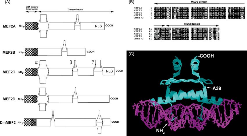
Author’s personal copy

                                                                                                                      Chapter 9.5

Myocyte Enhancer Factor 2 Transcription
Factors in Heart Development and
Disease
Brian L. Black1 and Richard M. Cripps2
1
 Cardiovascular Research Institute and Department of Biochemistry and Biophysics, University of California, San Francisco, CA, USA
2
 Department of Biology, University of New Mexico, Albuquerque, NM, USA

I. Introduction                                                                   are highly-regulated by signaling cascades that control
                                                                                  MEF2’s function as a transcriptional switch. We highlight
Members of the myocyte enhancer factor 2 (MEF2) fam-                              the many signaling pathways and kinases that regulate
ily of transcription factors are important regulators of                          MEF2-HDAC interactions, MEF2 post-translational modi-
gene expression in numerous tissues, including the heart,                         fication, and how these pathways influence MEF2 activ-
where MEF2 plays important roles in development and in                            ity. We also discuss the genetic function of Mef2 genes in
postnatal adaptation to a wide array of physiological and                         flies, fish and mice. These studies demonstrate the essen-
pathological signals. MEF2 functions as a transcriptional                         tial function for MEF2 proteins in heart development, as
switch, by potently activating or repressing transcription                        well as the development of numerous other lineages, and
through interaction with a variety of co-factors which                            reflect the general conservation of MEF2 function through-
serve as positive and negative regulators of transcription.                       out much of metazoan evolution. Many genes have been
The interaction of MEF2 with its co-factors is controlled                         identified as direct transcriptional targets of MEF2 through
by a multitude of signaling pathways that result in post-                         direct MEF2 binding to their promoter and enhancer ele-
translational modification of MEF2, and in the subsequent                         ments, and we summarize the known direct transcriptional
MEF2-dependent repression or activation of target gene                            targets of MEF2 in the heart.
transcription. This allows MEF2 to link the extracellular                              The Mef2 genes themselves are regulated at the tran-
environment to distinct and highly-regulated transcrip-                           scriptional level by the activity of multiple, independent
tional outputs through intracellular signaling cascades                           modular enhancers. These discrete enhancer modules con-
and co-factor interactions. In the heart, MEF2 is essential                       trol Mef2 expression in a restricted subset of the gene’s
for development and plays fundamental roles in myocyte                            complete expression pattern. We review the transcriptional
differentiation and gene activation. MEF2 is also crucial                         regulation of the single Mef2 gene in Drosophila and the
in the postnatal heart for integrating the transcriptional                        mouse Mef2c gene, which has been the best-characterized
response to numerous environmental cues, and regulating                           vertebrate Mef2 gene in terms of transcriptional regula-
normal physiological and pathological growth and adapta-                          tion. These studies have uncovered many new roles for
tion of the heart.                                                                Mef2 genes in the heart and other tissues by identifying
    In this chapter, we review what is known about the                            unexpected expression patterns and regulatory interactions
general regulation and function of MEF2 transcription fac-                        upstream of MEF2. Finally, in this chapter, we highlight
tors, with a focus on their role in the heart. We discuss the                     several important areas for future investigation regarding
many co-factors of MEF2 with a particular attention to                            the role of MEF2 transcription factors, how they are regu-
the interaction of MEF2 with class II histone deacetylases                        lated and function in the developing and postnatal heart, and
(HDACs) (see Chapter 10.2). MEF2-HDAC interactions                                their possible involvement in human cardiovascular disease.

Heart Development and Regeneration
Copyright © 2010 Elsevier Inc. All rights of reproduction in any form reserved.                                                            673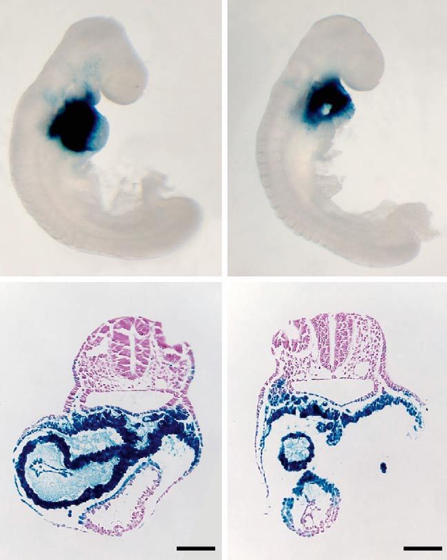
Author’s personal copy
674                                                 PART | 9 Transcriptional Circuits in Cardiac Development and Disease

 II. The mef2 family of transcription                            embryo (Martin et al., 1993; Edmondson et al., 1994).
 factors                                                         It is now well-established that most vertebrate genomes
                                                                 contain at least four MEF2-encoding genes, whereas
 II.A. Discovery of MEF2 Transcription                           simpler animals such as Drosophila melanogaster,
 Factors                                                         Caenorhabditis elegans and Ciona intestinalis con-
                                                                 tain only a single Mef2 gene each (Olson et al., 1995;
 In the late-1980s and early-1990s, numerous transcrip-          Davidson, 2007).
 tion factors involved in skeletal muscle development were
 identified. These studies were highlighted by the seminal
 observations of Davis et al. who showed that the myogenic       II.B. The MEF2 Family in the Context of the
 basic helix-loop-helix (bHLH) transcription factor MyoD         MADS Domain Superfamily
 alone could initiate skeletal myogenesis in a broad range
 of cell types in culture (Davis et al., 1987). MEF2 proteins    MEF2 proteins share, with several other factors, an N-
 were also identified during this time as essential regulators   terminal 57-amino acid sequence termed the MADS
 of skeletal muscle transcription, and as partners for MyoD      domain, which is responsible for protein dimeriza-
 and other myogenic bHLH proteins. In addition, it soon          tion and sequence-specific DNA-binding (Shore and
 became apparent that MEF2 was a critical regulator of           Sharrocks, 1995). The MADS domain is an acronym for
 muscle development in all muscle lineages, including the        the earliest-described members of the protein family:
 heart (Black and Olson, 1998).                                  the yeast mating type regulator MCM1; the plant floral
     MEF2 transcription factors were discovered independ-        determinants Agamous and Deficiens/Apetala3; and the
 ently using two different approaches. On the one hand,          animal protein Serum response factor (SRF) (Black and
 several groups had determined that muscle cells contained       Olson, 1998).
 different DNA-binding activities that could interact with           Phylogenetic analyses have concluded that the MADS
 muscle structural gene promoter sequences in vitro and          protein domain is ancient and originated in a common
 in vivo. MEF2-binding activity interacted strongly and          ancestor of prokaryotes and eukaryotes, based on simi-
 specifically with an AT-rich sequence that was found in         larities in primary structure between eukaryotic MADS
 the promoters of numerous muscle-specific genes, and            proteins and the Escherichia coli universal stress protein
 the integrity of these sites was required for full muscle-      UspA (Mushegian and Koonin, 1996). Current evolution-
 specific gene activation (Gossett et al., 1989; Horlick and     ary models propose that an ancestral MADS-box gene was
 Benfield, 1989; Mueller and Wold, 1989). Meanwhile,             duplicated prior to the divergence of the plant and animal
 Treisman and colleagues were studying the function of           kingdoms, and these duplicates formed the founders of the
 the serum response factor (SRF) protein. Serum response         two major classes of eukaryotic MADS domain proteins
 factor had been shown to be a potent activator of genes         found today: type I and type II MADS domain proteins
 important to proliferation, as well as to differentiation of    (Alvarez-Buylla et al., 2000). In higher animals, type I pro-
 vascular muscle lineages, interacting with DNA via a con-       teins are represented by serum response factor, which con-
 served binding domain (Treisman, 1990). By screening for        tains a conserved SAM domain immediately C-terminal to
 cDNAs similar to that of serum response factor, Pollock         the MADS domain. The SAM domain functions in homo-
 and Treisman identified factors named Related to SRF4           and heterodimerization (Ling et al., 1998). Interestingly,
 (RSRF4, now called MEF2D) and RSRFR2 (MEF2B)                    despite the ancient evolution of serum response factor pro-
 (Pollock and Treisman, 1991). These investigators went on       teins, sequenced animal genomes contain only one serum
 to demonstrate that RSRF proteins probably corresponded         response factor gene member per haploid genome. By con-
 to the muscle-specific binding activities previously            trast, type II MADS domain proteins have diverged signifi-
 defined as MEF2. Using a similar approach, Chambers             cantly to generate additional family members, particularly
 et al. (1992) identified Xenopus SL-1 (MEF2D) and SL-2          in plants (Theissen et al., 1996; Becker and Theissen,
 (MEF2A).                                                        2003). In addition to the MADS domain, plant type II
     A more direct connection between MEF2 binding               proteins also contain a conserved K domain for protein–
 activity and the RSRF proteins was achieved by Yu et al.        protein interaction (Yang and Jack, 2004), whereas animal
 (1992), who used a concatamerized MEF2 binding site             type II proteins acquired a 29-amino acid sequence which
 from the muscle creatine kinase (MCK) gene to screen            was termed the MEF2 domain, based on its inclusion in
 a cDNA expression library for factors that bound to             animal MEF2 proteins (Theissen et al., 1996; Black and
 the MEF2 sequence. This resulted in the identifica-             Olson, 1998).
 tion of human MEF2A. The final MEF2 family mem-                     The diversity of organisms encoding type I and type
 ber, MEF2C, was cloned based on its similarity to               II MADS domain proteins indicates that MEF2 proteins
 existing MEF2 factors, and was the first to show sig-           arose early in the evolution of life on Earth, and have been
 nificant enrichment in muscle tissues in the developing         retained in the genomes of organisms since that time to
Author’s personal copy
Chapter | 9.5 Myocyte Enhancer Factor 2 Transcription Factors in Heart Development and Disease 675
fulfill functions essential to the development and survival As might be expected from the high sequence conser-
of the organism. Indeed, it appears that all eukaryotic vation within N-termini of MEF2 proteins, critical func-
genomes contain at least one member of each of the type I tions are imparted by the MADS and MEF2 domains.
and type II MADS domain families. Deletion analyses confirmed that the MADS domain was
required, although not sufficient, for DNA-binding. Full
DNA-binding by MEF2 also required the presence of the
II.C. Structure of MEF2 Proteins MEF2 domain (Huang et al., 2000). Furthermore, MEF2
A striking feature of the MEF2 family is the retention of factors also dimerize via the MADS and MEF2 domains
the MADS domain and the adjacent MEF2 domain at the (Pollock and Treisman, 1991).
N-terminus of all known MEF2 proteins. This is clearly To define the residues within MEF2 that contribute
apparent in comparisons of the overall domain structures to dimerization and DNA-binding, extensive mutagen-
of mammalian MEF2 proteins with that of Drosophila esis studies were conducted by Molkentin and colleagues.
(Fig. 1A). In serum response factor (see Chapter 9.3), by These authors generated a series of 22 point mutants of
contrast, the MADS domain begins at amino acid 141. MEF2C, containing alterations in the sequence of the
Structural studies comparing MEF2 to serum response fac- MADS and MEF2 domains, and assayed each mutant pro-
tor indicate that the presence of the MADS domain at the tein for dimerization, DNA-binding and transcriptional
N-terminus affects DNA-binding and likely accounts for activation potential (Molkentin et al., 1996a). These studies
the differential binding sites preferred by MEF2 compared identified three critical regions within the MADS domain
to serum response factor (West et al., 1997; Santelli and and one region in the MEF2 domain that were required
Richmond, 2000). The MADS and MEF2 domains have for DNA binding: amino acids 3–5; 23–24; 30–31; and
been deeply conserved throughout evolution. For exam- 68–72 (Molkentin et al., 1996a). In addition, residues
ple, within the combined MADS and MEF2 domains, 35–50 of the MADS domain were critical for dimerization.
Drosophila MEF2 differs from mouse MEF2D at only 9 of Consistent with the in vitro mutagenesis studies, randomly-
86 amino acid residues (Fig. 1B). induced point mutants of the Drosophila Mef2 gene have
Figure 1 Structure of MEF2 factors. (A) Domain structure of the four mammalian MEF2 factors MEF2A–D, and Drosophila MEF2. Note that each
protein comprises N-terminally located MADS and MEF2 domains (shaded boxes) which function in dimerization and DNA-binding. The C-terminal
regions (open boxes) are highly variable. In mammals, variability usually centers around three main regions termed , and (indicated on MEF2C).
(B) Sequence conservation among the MADS and MEF2 domains of murine and Drosophila MEF2 proteins. (C) Structure of a human MEF2A dimer
complexed with DNA. Amino acids 1–85 are shown. DNA strands are shown in two shades of purple, and the two MEF2A polypeptides are shown in
two shades of blue. Amino acid A39 is shown for orientation purposes. Image was created using Protein Explorer and the structure coordinates con-
tained in Accession #1C7U. Huang et al. (2000).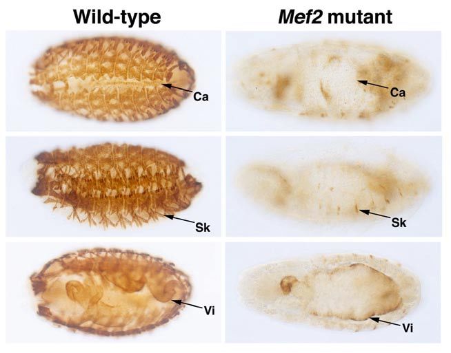
Author’s personal copy
676 PART | 9 Transcriptional Circuits in Cardiac Development and Disease
also established the importance of the MADS domain for are consistent with the large number of co-factors that
MEF2 function in vivo (Nguyen et al., 2002). Three point interact with MEF2 through the N-terminal domains, as
mutants of Drosophila MEF2 that affect conserved MADS well as the observation that the phosphorylation of a ser-
domain residues ablated MEF2 DNA-binding (Nguyen ine at the junction of the MADS and MEF2 domains is
et al., 2002). Notably, one of the residues affected in an important post-translational mechanism for MEF2
these mutants, R24, mimics the biochemical defects of regulation (Molkentin et al., 1996a; Cox et al., 2003).
the orthologous mutant mammalian protein (Molkentin Phosphorylation and other post-translational modifications
et al., 1996a; Nguyen et al., 2002). of MEF2 will be discussed in detail in Section III of this
More recently, a rigorous evaluation of the MEF2C chapter.
mutagenesis data has been possible, with the determina- In contrast to the MADS and MEF2 domains, the C-
tion by two separate groups of the crystal structure of the terminal regions of MEF2 proteins are highly-divergent
N-terminus of MEF2A bound to DNA (Fig. 1C) (Huang and also highly-variable within a single gene, as a result
et al., 2000; Santelli and Richmond, 2000). The two struc- of regulated RNA splicing. MEF2 primary transcripts are
tures are in close agreement with each other, and also subjected to alternative splicing, skip splicing and cryp-
with the published mutagenesis studies from Molkentin tic splice site selection, which generates a large number
and colleagues. The structural analyses show that the N- of potential MEF2 isoforms (Black and Olson, 1998; Zhu
terminus of MEF2A (amino acids 1–10) forms an exten- and Gulick, 2004; Zhu et al., 2005). For Mef2d, the alter-
sion that contacts the minor groove of DNA, presumably native splicing of the exon immediately C-terminal to the
to stabilize protein–DNA interactions, while amino acids MEF2 domain is regulated in a tissue-specific manner to
13–36 form an -helix that contacts DNA at several sites give rise to a muscle-specific isoform (Fig. 1A) (Breitbart
in the major groove (Huang et al., 2000; Santelli and et al., 1993; Martin et al., 1994), and a similar pattern of
Richmond, 2000). Critical interacting residues are dis- alternative splicing of an equivalent domain has been
persed in the region encompassing amino acids 13–36, but observed for Mef2a and Mef2c transcripts (Martin et al.,
also include amino acids 23–24 and 30–31, which were 1993; McDermott et al., 1993; Zhu and Gulick, 2004). The
defined by functional studies. Following the -helical Drosophila Mef2 primary transcript is also subject to regu-
region, the MADS domain forms two antiparallel -sheets, lated splicing (Taylor et al., 1995).
comprising approximately amino acids 40–60. In the crys- Recent studies from Gulick and colleagues have cat-
tal structure, these motifs are critical contact points for egorized the different protein domains resulting from regu-
dimerization of MEF2 (Huang et al., 2000; Santelli and lated splicing of mammalian Mef2c transcripts as , and
Richmond, 2000). These observations are also in agree- (Fig. 1A) (Zhu and Gulick, 2004). Mef2a and Mef2d
ment with the mutagenesis studies, which showed that transcripts also show alternative splicing of the and
residues 35–50 were critical for protein–protein interac- regions, and constitutively include sequences encoding the
tions within the dimer (Molkentin et al., 1996a). Finally, domain (Zhu et al., 2005). The domain enhances tran-
the MEF2 domain forms a short -helical region (amino scriptional activation by the parent MEF2 molecule, and
acids 63–73), which also appears to function in dimer is also preferentially-included in brain and muscle tran-
ization, since the location of the helix is remote from the scripts. In contrast, the domain is alternatively spliced
DNA, and the structure predicts a contact point between in Mef2c, and acts as a phosphorylation-dependent tran-
dimerized MEF2A polypeptides. Here, the structural data scriptional repressor (Zhu et al., 2005). These studies indi-
diverge slightly from those predicted by mutagenesis stud- cate that MEF2 function is modulated by alternative RNA
ies, which suggested that the MEF2 domain mutations splicing. Given the recent observation that alternate splic-
affected DNA-binding but not dimerization (Molkentin ing of Mef2b transcripts is altered in Mef2c-null hearts
et al., 1996a). These differences might be explained if (Vong et al., 2006), pathological conditions might signifi-
the function of the MEF2 domain -helix residues was to cantly affect the patterns of Mef2 transcript splicing, and
stabilize the dimer after it had formed. In vitro dimeriza- thus alter MEF2 function.
tion studies might not be sensitive enough to detect a mild In addition to transcriptional activation functions, the
destabilization, but this could be reflected in attenuated C-termini of MEF2A, MEF2C and MEF2D each contain
DNA-binding. nuclear localization signals (Fig. 1A), which are criti-
There is also compelling evidence that the MADS and cal to in vivo function (Yu, 1996; Borghi et al., 2001).
MEF2 domains function critically in the activation of tar- Furthermore, a Drosophila Mef2 mutant allele encodes a
get gene expression. Again, the first evidence in support of C-terminally truncated isoform that does not localize to the
this came from the mutagenesis studies, which showed that nucleus, suggesting that the location of nuclear localization
individual mutation of several different residues scattered signals is generally conserved across MEF2 proteins
throughout the first 86 amino acids did not affect DNA- (Ranganayakulu et al., 1995). Furthermore, and as dis-
binding, but had severe effects on transcriptional activa- cussed in the next section, the C-terminal regions of MEF2
tion ability (Molkentin et al., 1996a). These observations proteins contain potent transcriptional activation domains,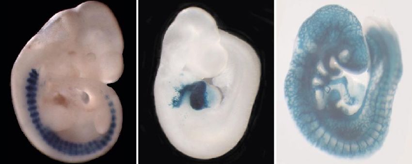
Author’s personal copy
Chapter | 9.5 Myocyte Enhancer Factor 2 Transcription Factors in Heart Development and Disease 677
and are also important targets for phosphorylation and 2004). The homeodomain protein Pitx2 also interacts with
other post-translational modifications that regulate MEF2 MEF2A to activate the Nppa promoter synergistically (Toro
function. et al., 2004). This interaction requires MEF2-binding to the
promoter, suggesting that MEF2 may serve as a platform
for Pitx2-binding, and that direct interaction of Pitx2 with
III. Regulation of mef2 activity by a cis-acting element in the promoter may not be required
(Toro et al., 2004).
post-translational modification
The majority of MEF2-interacting transcription fac-
III.A. MEF2 Functions as a Transcriptional tors discussed above contact MEF2 through the MADS and
MEF2 domain. The transcription factor TEF-1 is an exam-
Co-Factor ple of a MEF2 co-factor that does not bind to the MADS
MEF2 transcription factors interact with a diverse array of domain. Rather, TEF-1 interacts with motifs present near the
co-factors that modulate MEF2 activity. MEF2 can func- C-terminus of MEF2C (Maeda et al., 2002). TEF-1 usually
tion either as an activator or as a repressor, depending on binds to MCAT elements, which are present in the promot-
co-factor interactions, and several MEF2 co-factors facili- ers and enhancers of numerous skeletal and cardiac muscle
tate the ability of MEF2 to respond to intracellular signal- genes (Mar and Ordahl, 1990). Many of these gene promot-
ing. MEF2 function as a transcriptional co-factor was first ers also contain conserved MEF2 sites, which suggest the
described from studies in skeletal muscle, where MEF2 possibility that MEF2C and TEF-1 may co-regulate multi-
proteins were shown to function as essential co-factors ple genes involved in cardiac development and differentia-
for the myogenic bHLH proteins, including MyoD and tion. In addition, it has been observed that TEF-1 can bind
myogenin (Molkentin et al., 1995; Ornatsky et al., 1997; directly to MEF2 sites or to MEF2-like AT-rich elements,
Black et al., 1998). Myogenic bHLH proteins have the suggesting a further interplay between MEF2 and TEF-1
remarkable ability to convert nonmuscle cells to muscle during muscle development, possibly through competition
cells in culture, and it was observed that this activity was for shared binding elements (Karasseva et al., 2003).
dependent on interaction with MEF2 (Molkentin et al., Thyroid hormone receptor (TR) is another MEF2
1995; Ornatsky et al., 1997; Black et al., 1998). MEF2 co-factor that interacts with MEF2 in the heart, and this
and MyoD physically associate, and their binding sites interaction is facilitated by p300/CBP, which is thought
are frequently coordinately positioned in the enhancers to bridge the two factors and promote transcriptional acti-
and promoters of muscle-specific genes (Molkentin et al., vation (De Luca et al., 2003). TR and MEF2 interaction
1995; Fickett, 1996; Black et al., 1998). The interaction of has been shown to be important for the activation of the
MyoD and MEF2 occurs through the DNA-binding motifs -MHC gene via closely-positioned binding sites for the
of each factor, the bHLH domain on MyoD and the MADS two factors in the proximal promoter region (Lee et al.,
and MEF2 domains of MEF2, raising the possibility that 1997). The interaction of MEF2 and MyoD in skeletal
MEF2 factors may interact with a wide array of bHLH muscle is also facilitated by p300 (Sartorelli et al., 1997).
proteins. In addition to acetylation of histones and subsequent chro-
Indeed, MEF2 proteins interact with several other matin relaxation, p300 also directly acetylates MEF2,
bHLH proteins in diverse contexts. MEF2 forms a com- which promotes MEF2 transactivation, probably through
plex and potently activates transcription of target genes negative regulation of sumoylation (Ma et al., 2005; Zhao
in cooperation with the neural bHLH protein mammalian et al., 2005a), which will be discussed later in this chapter.
achaete-scute homolog 1 (MASH1), and this interaction is The cooperative transcriptional activation of the -MHC
also dependent on the MADS and MEF2 domains (Black promoter by TR and MEF2 is attenuated by the action of
et al., 1996; Mao and Nadal-Ginard, 1996). In the heart, Jumonji/Jarid2, which is also a direct MEF2 partner (Kim
MEF2C cooperatively activates transcription of the Nppa et al., 2005). Jumonji, a histone demethylase, directly
gene with the bHLH proteins HAND1 and HAND2, which interacts with the MADS domain of MEF2A to negatively
like MEF2, are essential regulators of cardiac development regulate MEF2-dependent transcription (Kim et al., 2005).
(Srivastava et al., 1995; Zang et al., 2004; Morin et al., Jumonji interaction with MEF2A probably blocks TR
2005). interaction with the MADS domain, and represses tran-
GATA4 is another cardiac-enriched transcription fac- scription via a direct effect on histones.
tor that interacts with MEF2 to activate the Nppa promoter In addition to MEF2 co-factors that interact with DNA
(Morin et al., 2000) (see Chapter 9.2). Given the broad and MEF2, there are a number of tissue-specific and ubi
overlap in the expression of GATA and MEF2 transcrip- quitous proteins that modulate MEF2 activity solely
tion factors and the prevalence of GATA and MEF2 sites through protein–protein interactions. One such group of
in cardiac promoters, these members of these two families MEF2-interacting factors includes the SAP domain pro-
of transcription factors may participate in the co-activation teins myocardin and MASTR. Myocardin was first iden-
of numerous other genes in the heart (Vanpoucke et al., tified as a potent transcriptional activation partner forAuthor’s personal copy
678 PART | 9 Transcriptional Circuits in Cardiac Development and Disease
serum response factor (see Chapter 9.3), a MADS domain and serum response factor bound to their respective cog-
protein that is closely related to MEF2, in cardiac and nate binding sites, but it seems likely that this mechanism
smooth muscle (Wang et al., 2003b; Yoshida et al., 2003). will function in the heart and other tissues. If this notion is
Myocardin itself does not bind DNA, but uses DNA-bound correct, it would provide an additional mode for MEF2 to
serum response factor as a platform to interact with chro- serve as a transcriptional switch through the assembly of a
matin through protein–protein interactions. Once bound multi-protein complex.
to serum response factor, myocardin very potently acti-
vates transcription (Wang et al., 2003b). One molecule
of myocardin interacts with each serum response factor III.B. Chromatin Remodeling by MEF2
dimer bound to a CArG box, and myocardin itself prob- through Interaction with Histone
ably dimerizes to bridge two CArG elements (Wang et al.,
Deacetylases
2003b; Yoshida et al., 2003). A longer form of myocardin,
generated from a distinct splicing event can interact with It is now becoming increasingly appreciated that binding
either serum response factor or MEF2 (Creemers et al., sites for regulatory factors must be accessible in the con-
2006). The MEF2 interaction domain in the myocardin text of the overall chromatin structure of the cell in order
long-form comprises a short amino acid sequence at the to be recognized, and for gene expression to be control-
N-terminus that is distinct from the N-terminus of the led. Along these lines, it now appears that a major func-
short-form of myocardin, which facilitates serum response tion of MEF2 is to control the balance between chromatin
factor interaction (Creemers et al., 2006). The unique acetylation and deacetylation, and thereby regulate the
N-terminus of the myocardin long-form directly interacts relative accessibility of promoters and enhancers to the
with MEF2. Interestingly, however, the myocardin long- transcriptional machinery (Fig. 2). Accordingly, MEF2
form still retains the sequences necessary to facilitate factors interact with multiple histone acetylases and
interaction with serum response factor, such that this iso- deacetylases. Most notably, MEF2 forms a complex with
form of myocardin can interact with either serum response class II histone deacetylases (HDACs), which include
factor or MEF2 (Creemers et al., 2006). HDACs 4, 5, 6, 7 and 9 (McKinsey et al., 2001a) (see
Olson and colleagues used the unique N-terminal Chapter 10.2). Interaction with class II HDACs occurs
sequence of the myocardin long-form to identify another through the MADS domain at the N-terminus of MEF2
SAP domain-containing protein that also contains the (Lu et al., 2000b; Dressel et al., 2001; Zhang et al., 2001a).
MEF2 interacting sequence (Creemers et al., 2006). This Similarly, a conserved N-terminal domain in HDAC dic-
myocardin homolog was named MASTR (MEF2-associated tates interaction with MEF2 (Wang et al., 1999; Lemercier
SAP domain transcriptional regulator). Unlike the myocar- et al., 2000; Dressel et al., 2001). MEF2-HDAC complexes
din long-form, MASTR only contains sequences sufficient repress transcription by deacetylating histones, resulting
to interact with MEF2 and not with serum response fac- in chromatin condensation and reduced accessibility of
tor (Creemers et al., 2006). Thus, the myocardin family core transcriptional machinery to promoter and enhancer
includes: the myocardin short-form, which only interacts regions of MEF2 target genes (Lu et al., 2000b; Kao et al.,
with serum response factor; MASTR, which only inter- 2001; McKinsey et al., 2001a). MEF2 also interacts with
acts with MEF2; and the myocardin long-form, which several histone acetyltransferases, including p300/CBP and
can interact with either MEF2 or serum response factor. SIRT1, which likely serve to balance the repressive effects
The myocardin long- and short-forms contain sequences of HDAC on MEF2 and allow MEF2 to function as a tran-
that should allow dimerization between the two isoforms, scriptional switch (Sartorelli et al., 1999; Ma et al., 2005;
which raises the intriguing possibility that myocardin het- Zhao et al., 2005a; Stankovic-Valentin et al., 2007).
erodimers, containing long- and short-forms, might bridge Class II HDACs are important regulators of transcrip-
serum response factor and MEF2 binding sites. Since tion in the developing and postnatal heart that help to reg-
numerous promoters contain binding elements for each ulate the hypertrophic response (Zhang et al., 2002; Olson
of these classes of MADS domain transcription factors, et al., 2006). Normal growth of the myocardium requires
myocardin members may bridge the sites and allow syn- large amounts of structural and other regulatory proteins
ergistic activation mediated by MEF2 and serum response to be synthesized as cells enlarge, but excessive enlarge-
factor. This possible relationship between serum response ment of the heart can result in pathologic hypertrophy,
factor and MEF2 through myocardin may be further facili- which ultimately can lead to heart failure (Olson et al.,
tated by the presence of binding sites that can be bound 2006). Thus, HDACs serve as a kind of regulated braking
by either MEF2 or serum response factor (L’Honore et al., mechanism, keeping the MEF2-dependent transcriptional
2007). These composite SRF/MEF2 cis-elements provide response in check until signals that stimulate myocardial
additional targets for myocardin and MASTR regulation of growth are received. Hypertrophic induction results in sig-
cardiac genes. To date, no specific genes have been identi- nal-dependent export of HDACs from the nucleus, which
fied as myocardin targets through the bridging of MEF2 results in chromatin relaxation due to increased histoneAuthor’s personal copy
Chapter | 9.5 Myocyte Enhancer Factor 2 Transcription Factors in Heart Development and Disease 679
Normal and Pathologic Stimulation
catecholamine stimulation electrical activity exercise
P
P
CamK 14-3-3
HDAC
MAPK
HDAC P300 GRIP
MEF2 X MEF2
Initial activation signals received: MEF2 Phosphorylation of HDAC results in
and HDAC are phosphorylated by MAPK association with 14-3-3 proteins and nuclear
and CamK signaling export. MEF2 associates with HATs (P300)
and potent coactivator
Figure 2 MEF2 functions as a signal-dependent transcriptional switch. MEF2 functions as a repressor by recruiting class II HDACs to promoter
and enhancer regions of target genes. In response to a variety of developmental and pathological signals, CamK and MAPK signaling pathways are
activated. These signals result in the phosphorylation of MEF2 and HDACs. HDAC phosphorylation by CamK results in exposure of a nuclear export
signal at the C-terminus, interaction with 14-3-3 proteins, and export from the nucleus. MEF2 phosphorylation and HDAC dissociation result in recruit-
ment of HATs and co-activator molecules, such as GRIP-1, and the conversion of MEF2 to an activator complex.
acetylation and subsequent MEF2-dependent transcription post-translational modifications have mutually-exclusive
(McKinsey et al., 2000a; Kao et al., 2001; McKinsey et al., and opposing functions in promoting MEF2-dependent
2001a,b; Olson et al., 2006). activation and repression, respectively (Zhao et al., 2005a;
Intriguingly, the deacetylase activity of class II HDACs Gregoire et al., 2006; Shalizi et al., 2006; Stankovic-
is not required for interaction with MEF2, nor is this activ- Valentin et al., 2007). In addition to its role in deacetyl
ity required for transcriptional repression by MEF2-HDAC ation of histones, HDAC4 also inhibits MEF2-dependent
(Lemercier et al., 2000; Zhang et al., 2001b; Chan et al., transcription by promoting sumoylation of MEF2 (Zhao
2003). The dispensability of deacetylase activity is con- et al., 2005a; Gregoire et al., 2006; Stankovic-Valentin
sistent with the observations that MITR (MEF2-interacting et al., 2007).
transcriptional repressor), an HDAC homolog lacking the As noted above, it was recently discovered that MEF2
deacetylase domain, also interacts with MEF2 and facili- proteins are modified by sumoylation (Gregoire et al.,
tates strong transcriptional repression, as its name implies 2006; Kang et al., 2006; Riquelme et al., 2006; Shalizi
(Youn et al., 2000; Zhang et al., 2001b). This may occur et al., 2006). Sumoylation is the process by which a pro-
because class II HDACs and MITR have the ability to tein moiety, SUMO, is covalently added to proteins by
recruit the potent co-repressor protein CtBP (Dressel the activity of SUMO-conjugating enzymes (Gill, 2005).
et al., 2001; Zhang et al., 2001a). CtBP co-repressors Addition of SUMO-1 to the C-terminus of MEF2 modi-
repress transcription via recruitment of HDACs (Bertos fies MEF2 to a repressor form, which has been demon-
et al., 2001). CtBP physically associates with the N-terminus strated for MEF2A, MEF2C and MEF2D (Gregoire and
of HDAC4, HDAC5 and the HDAC homolog MITR, Yang, 2005; Zhao et al., 2005a; Kang et al., 2006; Shalizi
which interact with MEF2 to repress its activity (Zhang et al., 2006). Interestingly, SUMO addition to MEF2
et al., 2001a). The deacetylase activity-independent repres- occurs at a lysine residue in the C-terminal activation
sion might also result from the observation that HDACs domain and is controlled by MAPK phosphorylation at a
can multimerize, allowing deactylase-defective HDACs nearby serine residue for MEF2A, MEF2C and MEF2D
to recruit other HDACs that possess full enzymatic activ- (Fig. 3) (Gregoire et al., 2006; Kang et al., 2006; Shalizi
ity, although this may not explain the strong transcrip- et al., 2006). In each case, dephosphorylation of MEF2
tional repression conferred to MEF2 by MITR (Youn promotes a switch from acetylation to sumoylation at the
et al., 2000; Zhang et al., 2001b). Alternatively, a crucial neighboring lysine and the conversion of MEF2 from an
function of class II HDACs may be to help control the bal- activator to a repressor (Gregoire et al., 2006; Kang et al.,
ance of acetylation and sumoylation of MEF2. These two 2006; Shalizi et al., 2006). The dephosphorylation event isAuthor’s personal copy
680 PART | 9 Transcriptional Circuits in Cardiac Development and Disease
P38
BMK1
ERK5 BMK1
CKII P38 P38 cdk5 CKII P38 ERK5 P38 CnA P38
?
S
U
P P P P P PP P M Ac P P
O
S59 S98 S192 S255 S289 T312 S355 K403 S408 S479
MEF2A T319
MADS MEF2
Figure 3 MEF2 proteins are extensively modified post-translationally. A schematic of human MEF2A shows sites of phosphorylation (red ovals),
sumoylation (pink oval) and acetylation (yellow oval). Signaling pathways that are known or thought to modify MEF2 are noted above and the amino
acid residues are noted below. The MADS and MEF2 domains are depicted. Differential modification of lysine 403 by acetylation and sumoylation is
controlled by phosphorylation and dephosphorylation by p38 and calcineurin at the neighboring serine 408. Note that similar (but not identical) post-
translational modifications depicted here for hMEF2A also occur on other MEF2 isoforms.
controlled by calcineurin, which serves as a link between of these two broad classes of HDACs with MEF2 result in
calcium-mediated hypertrophic signaling and post- different biological outputs.
translational modification of MEF2 by sumoylation (Flavell
et al., 2006; Shalizi et al., 2006).
In contrast to sumoylation, direct acetylation of III.C. MEF2 Functions as a Signal-Dependent
MEF2C at the C-terminus by CBP promotes MEF2 activ-
Transcriptional Switch
ity, as noted earlier in this chapter (Sartorelli et al., 1997).
The balance between sumoylation and acetylation of The interaction of MEF2 with HDACs underscores the
MEF2 is also controlled directly by HDACs themselves. function of MEF2 proteins as both positive and negative
HDAC4 has been shown to promote SUMO addition to regulators of transcription. Prior to those groundbreaking
MEF2, providing a dual mechanism for repression of observations, MEF2 proteins were generally thought of
MEF2 activity by class II HDACs through deacetylation only as transcriptional activators that functioned through
of chromatin, and further through addition of SUMO to protein–protein interactions with other transcription fac-
MEF2 (Zhao et al., 2005a; Gregoire et al., 2006). This tors containing more potent activation domains (Molkentin
may partially explain why the enzymatic activity of and Olson, 1996; Black and Olson, 1998). The notion
HDACs is not required for repression of MEF2, since the that MEF2 functions as both a repressor and an activator,
catalytic domain of HDAC4 is not required for sumoyl depending on the gene, cell type and cellular differen-
ation of MEF2 (Zhao et al., 2005a). Interestingly, HDAC4 tiation state led to the idea that MEF2 serves as a switch
is also regulated by sumoylation. However, sumoylation capable of interpreting distinct signals into opposing tran-
of HDAC4 inhibits its ability to promote SUMO addition scriptional outputs. Therefore, it has been important to
to MEF2, which provides a post-translational negative- define what dictates whether MEF2 functions as an acti-
feedback mechanism for control of MEF2 sumoylation vator or a repressor. Over the last decade, numerous intra
and repression (Zhao et al., 2005a; Gregoire et al., 2006). cellular signaling pathways have been identified to interact
It was previously believed that only class II HDACs with MEF2 and class II HDACs. Not surprisingly, MEF2
interacted with MEF2, but several reports have now dem- and HDAC proteins are each regulated by their phosphor-
onstrated that class I HDACs also interact with MEF2, and ylation state (McKinsey et al., 2001a, 2002).
that these interactions play an important role in the heart Class II HDACs are phosphorylated in response to
(Montgomery et al., 2007; Trivedi et al., 2007). The class a variety of extracellular signals, including electrical
I HDAC, HDAC3, was shown recently to interact with and activity, pressure, adrenergic signaling and other normal
supress the transcriptional activity of MEF2 (Gregoire developmental and postnatal cues. These signals result
et al., 2007). The nature of the interaction between class I in an increase in the concentration of Ca2 in the cyto-
HDACs and MEF2 is not as well-characterized as the inter- plasm, which activates the phosphatase calcineurin and
action with class II HDACs, so it remains unclear which stimulates the activity of calcium/calmodulin-dependent
residues within the MEF2 MADS domain make direct kinases (CaMK) I, II and VI (Lu et al., 2000a; Kao
contact with the class I HDAC (Gregoire et al., 2007). et al., 2001; McKinsey et al., 2001b; Little et al., 2007).
Structural studies should resolve how the interactions Phosphorylation of class II HDACs by CaMK occurs onAuthor’s personal copy
Chapter | 9.5 Myocyte Enhancer Factor 2 Transcription Factors in Heart Development and Disease 681
two residues near the N-terminus of the HDAC protein serine 289 in response to p38 MAPK signaling (Fig. 3)
(McKinsey et al., 2000a,b; Kao et al., 2001). CaMK phos- (Cox et al., 2003).
phorylation of HDAC facilitates interaction with 14-3-3 An interesting study of MEF2 function in dominant
proteins, which activates a nuclear export sequence at the induction of skeletal myogenesis in culture showed that
C-terminus (McKinsey et al., 2000b; Choi et al., 2001; RAF kinase inhibits MyoD-induced conversion of fibro
McKinsey et al., 2001b; Wang and Yang, 2001). HDAC blasts into muscle cells by blocking MEF2 nuclear local
nuclear export, in turn, results in MEF2 activation of gene ization (Winter and Arnold, 2000). These studies were the
expression in the heart and other MEF2-dependent tissues first to suggest a regulated nuclear localization of MEF2
(Fig. 2). In addition, SIK1 kinase phosphorylates class II proteins themselves, which may have important implica-
HDACs, which affects MEF2-dependent transcription tions if MEF2 is shuttled between the nucleus and cyto-
through dissociation of MEF2 from HDAC (Berdeaux plasm during the development of the heart and other
et al., 2007). tissues.
MEF2 factors themselves are extensively phosphor- Cyclins and cyclin-dependent kinase pathways have
ylated in response to a host of intracellular and extracel- also been shown to regulate MEF2 activity and to provide
lular cues (Fig. 3). The p38, BMK1/ERK5 and ERK1 a link between the cell-cycle and MEF2-dependent tran-
mitogen-activated protein kinases (MAPK) each play scription. For example, cdk5 phosphorylates MEF2C in
a role in MEF2 regulation through phosphorylation. neurons, supporting the notion that a cell-cycle-dependent
BMK1/ERK5 signaling results in the phosphorylation of signaling event functions via post-translational modifica-
MEF2C at serine-387 in the C-terminal activation domain tion of MEF2C in neurons (Gong et al., 2003; Tang et al.,
in response to a variety of extracellular signals, including 2005; Smith et al., 2006). The role of cdk phosphorylation
adrenergic signaling and pressure overload in the myocar- of MEF2 in the heart has not been examined, but it is likely
dium (Kato et al., 1997; Yang et al., 1998; Yan et al., 1999; that MEF2 plays a role in cell-cycle control along with
Nadruz et al., 2003). Each of these cues stimulates the GATA4, which is known to regulate cardiac cell-cycle, and
nuclear localization of BMK1/ERK5, which leads to phos- to interact with MEF2 and other partners downstream of
phorylation and activation of MEF2 (Kato et al., 2000; Yan these signaling pathways (Morin et al., 2000; Vanpoucke
et al., 2001). Although these studies investigated BMK1/ et al., 2004; Zeisberg et al., 2005; Xin et al., 2006).
ERK5 signaling in the postnatal heart, it is also quite likely MEF2 factors themselves have only weak inherent tran-
that BMK1/ERK5 signaling regulates MEF2 phosphoryla- scriptional activation potential (Molkentin et al., 1996a,b).
tion during cardiac development, since all of these signals This is also the case for several of the many MEF2 co-
lead to MEF2-dependent c-jun transcription, which func- factors that have been described to date, such as GATA4.
tions in numerous developmental contexts (Kato et al., However, MEF2 proteins and their co-factors are sufficient
1997; Marinissen et al., 1999; Nadruz et al., 2003). to direct extremely robust activation of many genes and
The p38 MAPK signaling pathway also plays a funda- reporter genes, both in vivo and in reporter assays in cell
mentally important role in the post-translational modifi- culture. A likely mechanism for how MEF2 is able to drive
cation of MEF2 in myocytes (Han and Molkentin, 2000). strong transcriptional activation is through interaction with
Signaling by p38 results in phosphorylation of MEF2A potent transcriptional co-activators, such as GRIP-1, which
and MEF2C, but not MEF2B or MEF2D (Han et al., 1997; belongs to the p160 steroid receptor co-activator (SRC)
Ornatsky et al., 1999; Zhao et al., 1999; Chang et al., family of transcriptional co-activator proteins (Chen et al.,
2002). p38 phosphorylation promotes the role of MEF2 as 2000; Leo and Chen, 2000; Lazaro et al., 2002; Xu and Li,
a transcriptional activator in response to normal develop- 2003; Liu et al., 2004). Signaling downstream of D cyclins
mental and postnatal hypertrophic growth of the heart, as and cdk4 activity blocks muscle-differentiation by disrupt-
well as to pathologic hypertrophic cues (Han et al., 1997; ing the interaction between MEF2C and GRIP-1 (Lazaro
Kolodziejczyk et al., 1999; Ornatsky et al., 1999; Zhao et al., 2002).
et al., 1999; Han and Molkentin, 2000; Cox et al., 2003). Interactions with transcriptional co-activators and co-
Other studies have shown that retinoic acid (RA) signal- repressors provide another mechanism for MEF2 to serve
ing during myocardial development results in the phosphor as a transcriptional switch, repressing transcription in
ylation of MEF2 via p38 MAPK (Ren et al., 2007), which some contexts, while activating it in others. In this regard,
potentially links normal growth and development of the the interaction between MEF2 and GRIP-1 is also targeted
myocardium via RA signaling to MEF2 (Tran and Sucov, by the TGF signaling pathway (Liu et al., 2004). TGF
1998; Lavine et al., 2005). Casein kinase II (CKII) results signaling is transmitted intracellularly by Smads transcrip-
in phosphorylation of a conserved serine (S59) found at tion factors, and MEF2 interacts with several different
the junction of the MADS and MEF2 domains in MEF2A members of the Smads family, including Smads 2, 3 and 4
and MEF2C (Molkentin et al., 1996c; Cox et al., 2003). (Quinn et al., 2001; Derynck and Zhang, 2003; Liu et al.,
Work from McDermott and colleagues showed that CKII 2004). Smads 2 and 4 interact with MEF2 to influence
also results in the direct phosphorylation of MEF2A at transcription positively in skeletal muscle by promotingAuthor’s personal copy
682 PART | 9 Transcriptional Circuits in Cardiac Development and Disease
interaction between MEF2 and MyoD (Quinn et al., 2001). transcriptional co-factors, such as SRC family co-activators,
Presumably, TGF or BMP signaling influences MEF2 myocardin proteins and histone acetyltransferases, or
activity in the heart through interactions with Smads as co-factors that repress transcription, including class II
well, although this has not been demonstrated. In contrast, HDACs, MAML, CABIN1 and Jumonji proteins (Czubryt
Smad3 is an inhibitory Smad and functions as a negative and Olson, 2004; Backs and Olson, 2006; Liu and Olson,
regulator of transcription (Liu et al., 2004). Smad3 asso- 2006). In addition, MEF2 interacts with multiple other
ciates with MEF2 and disrupts MEF2 association with transcription factors, which provides additional combina-
GRIP-1, thereby repressing transcription by targeting the torial complexity and allows for more precise co-factor
interaction of MEF2 and one of its co-activators (Liu et al., recruitment. It is likely that additional MEF2 co-factors
2004). will continue to be identified, but the general role for
MAML1 (mastermind like 1) is also a potent transcrip- MEF2 as a nodal point for balancing growth and differen-
tional co-activator that interacts with MEF2 (Shen et al., tiation signals through post-translational modification and
2006). MAML factors belong to a family of co-activator co-factor interaction is now well-established.
proteins that are required for Notch signaling. Notch sig
naling inhibits skeletal muscle differentiation and the mus-
cle-inducing activity of myogenic bHLH proteins (Kopan IV. MEF2 gene function in the heart
et al., 1994; Kuroda et al., 1999; Wilson-Rawls et al., 1999; and other tissues
Shen et al., 2006). Activated Notch interacts directly with
MEF2C via a specific splice isoform of MEF2C found in IV.A. MEF2 Proteins are Expressed in
muscle lineages (Wilson-Rawls et al., 1999). Thus, it is Multiple Lineages During Development and
clear that this pathway interacts with MEF2 via Notch itself
and via the Notch co-activator MAML1 to inhibit skeletal
in Adulthood
myogenesis (Wilson-Rawls et al., 1999; Shen et al., 2006). Despite the initial identification of MEF2 proteins as fac-
The presence of the muscle-specific exon has not been tors that are bound to skeletal muscle gene promoters,
examined in cardiac muscle, so it is not known whether early studies suggested that MEF2-binding activity was
Notch interacts directly with MEF2C in the heart. present in a broad tissue distribution (Olson et al., 1995).
In addition to direct interactions with co-activator mol- Indeed, Mef2d transcripts are detected in many adult tis-
ecules, MEF2 also interacts with a number of co-repressor sues, although the muscle-specific splice variant of Mef2d
proteins. For example, CABIN1/CAIN is a potent tran- is expressed only in adult muscles (Breitbart et al., 1993;
scriptional co-repressor protein that binds to calcineurin Martin et al., 1994). Furthermore, Xenopus Mef2 genes
and interacts directly with MEF2 (Esau et al., 2001; Han are broadly-expressed in the adult frog (Chambers et al.,
et al., 2003). Interaction of CABIN1 with MEF2 blocks 1992). By contrast, in situ hybridization studies performed
the activation of MEF2 by BMK1/ERK5 MAP kinase on vertebrate embryos indicate that expression of Mef2
signaling (Kasler et al., 2000). CABIN1 further represses genes is indeed predominantly muscle-specific at earlier
MEF2 by recruitment of mSIN3 and associated HDACs, stages of development (Edmondson et al., 1994). Mef2
and CABIN1 also recruits the histone methyltransferase gene expression in frogs and fish is restricted to muscle
SUV39H1 to the complex (Youn and Liu, 2000; Jang lineages in the embryo (Chambers et al., 1992; Ticho et al.,
et al., 2007). Chen and colleagues solved the crystal struc- 1996), and in the mouse embryo, Mef2a, Mef2c and Mef2d
ture of the MADS domain of MEF2B in a complex with expression is largely restricted to skeletal and cardiac
CABIN1 (Han et al., 2003). The structure showed that muscle early in development (Edmondson et al., 1994).
CABIN1 contacts the MADS domain in a similar, but not Similarly, mouse Mef2b is a marker of early myogenic lin-
identical, fashion to class II HDACs, raising the possibility eages (Molkentin et al., 1996b).
that the binding of HDAC and CABIN1 to MEF2 might be Nevertheless, expression of Mef2 genes was also
mutually exclusive (Han et al., 2003, 2005). The discrete detected in the developing nervous system of mice, sug-
contacts of each of these MEF2 co-regulators suggest that gesting that it might also play a role in neuronal devel-
they likely affect MEF2 conformation differently, but this opment (Leifer et al., 1994; Ikeshima et al., 1995; Lyons
remains to be determined. et al., 1995; Lin et al., 1996b). In addition, specific func-
In general, it is clear that the major function of MEF2 tions for MEF2 have been described more recently in spe-
is to respond to intracellular signaling pathways and to cialized cells of the blood and other tissues of mesodermal
respond to those signaling cues through co-factor interac- and ectodermal origin. MEF2 is an activator of cell death
tions (Figs 2; 3). In response to growth and differentiation in T-lymphocytes via direct transcriptional activation of
signals, MEF2 proteins are modified post-translationally, the steroid receptor gene Nur77 (Youn et al., 1999; Youn
which is primarily via phosphorylation but also includes and Liu, 2000; Liu et al., 2001a). Similarly, immunoglobu-
acetylation and sumoylation (Fig. 3). These modifica- lin gene expression in B-cells is regulated by MEF2 func-
tions influence whether MEF2 interacts with positive tion (Rao et al., 1998; Satyaraj and Storb, 1998; WallinAuthor’s personal copy
Chapter | 9.5 Myocyte Enhancer Factor 2 Transcription Factors in Heart Development and Disease 683
et al., 1999). In addition, MEF2C is an essential regulator expression of several of these genes have been described
of B-cell proliferation in response to B-cell receptor sig (Bour et al., 1995; Lilly et al., 1995; Ranganayakulu et al.,
naling, which results in direct phosphorylation of MEF2C 1995; Kelly et al., 2002; Marin et al., 2004). Interestingly,
via p38 MAPK (Khiem et al., 2008; Wilker et al., 2008). expression of Tropomyosin 2 in Mef2 mutants was reduced
Furthermore, Mef2c and Mef2d are expressed in chondro- in skeletal and visceral muscles, but was relatively nor-
cytes during bone formation in the embryo (Arnold et al., mal in the heart (Lin et al., 1996a). Similarly, expression
2007), and Mef2c is highly-expressed in developing neu- of the -tubulin gene Tub60D was strongly reduced in
ral crest cells in the mouse and fish (Miller et al., 2007; the skeletal muscles of Mef2 mutants, but was expressed
Verzi et al., 2007). Based on these studies, it is clear normally in the cardiac tube (Damm et al., 1998). These
that although MEF2 is not restricted to muscle lineages, two examples emphasize that transcriptional programs can
muscle cells in the embryo are a major location of Mef2 differ significantly between different muscle types in the
gene expression. Later in vertebrate development, how- embryo. Nevertheless, the Drosophila studies represent
ever, transcription of Mef2 genes is detected in a number the first example of whole-organism inactivation of MEF2
of additional tissues and organs, although in many cases function and its developmental consequences. These stud-
the precise cells that express MEF2 within these organs ies remain the clearest examples of MEF2 function and its
have yet to be defined. In Drosophila, embryonic expres- requirement for the differentiation of muscle lineages.
sion of the single Mef2 gene is restricted to the develop- To define the function of vertebrate Mef2 genes in
ing mesodermal tissues, including the cardiac tube (Lilly cardiac development, a variety of approaches have been
et al., 1994; Nguyen et al., 1994). Later in development, employed. In mice, global knockouts of Mef2c and Mef2a
there is accumulation of MEF2 protein in portions of the have been performed and have shown that mice lacking
Drosophila central nervous system (Schulz et al., 1996). these genes have severe defects in cardiac development
In summary, although they were initially identified as and function (Lin et al., 1997; Naya et al., 2002). By con-
muscle-enriched binding factors, it is now clear that MEF2 trast, a recent publication indicated that homozygotes for
proteins function in many tissues during development and a mutation in Mef2d are phenotypically normal (Arnold
in adulthood, including all muscle lineages, neuronal cells, et al., 2007). Targeted inactivation of Mef2c in mice results
blood cells, neural crest, bone, chondrocytes and vascular in death by embryonic day (E) 10.5, and mutant mice
endothelium. Direct transcriptional targets of MEF2 have exhibit severe defects in cardiac development (Lin et al.,
been identified in each of these tissues and, as discussed 1997). Affected embryos display pericardial edema, a
below, gene knockout strategies and targeted ablation of
MEF2 function have begun to define the roles of Mef2 in
many of these cell types more precisely.
IV.B. Genetic Analyses of Mef2 Gene
Function
To determine the function of MEF2 in tissues where it is
expressed, numerous genetic studies have been performed
in flies, mice and fish. The fact that vertebrate genomes
have four distinct Mef2 genes makes defining the precise
function of MEF2 complicated in higher animals, due
to issues of redundancy and overlapping expression. As
noted earlier in this chapter, Drosophila has only a single
Mef2 gene, which has made genetic analyses of Mef2 func-
tion highly tractable in that organism. Inactivation of the
Drosophila Mef2 gene results in a severe failure in muscle
Figure 4 Requirement for MEF2 function for cardiac development
differentiation for cardiac, skeletal and visceral mesoderm,
in Drosophila. Muscle tissue can be visualized in mature embryos via
even though precursors for each of these muscle lineages immunohistochemical staining for myosin heavy-chain (MHC) protein.
are normally specified in mutant embryos (Fig. 4) (Bour Left column: In wild-type embryos, cardiac muscle (Ca) forms at the dor-
et al., 1995; Lilly et al., 1995; Ranganayakulu et al., 1995). sal midline; skeletal muscles (Sk) are apparent in dorsal and lateral views;
In Mef2 mutants, the expression of a large number of mus- and visceral muscle (Vi) showing MHC accumulation can be observed in
a ventral view. Right column: in Mef2-null mutants, there is no detectable
cle structural genes was essentially absent. These potential
MHC accumulation in cardiac muscle, only a handful of skeletal muscle
target genes include the single Myosin heavy-chain gene, cells accumulate MHC, and the visceral muscle accumulates low levels
Actin57B, Myosin light chain-2, Myosin light chain-alkali of MHC. In addition, morphogenesis of the mutant gut is abnormal, with
and Troponin I, and MEF2 binding sites critical for the the gut predominantly comprising a single lumen.You can also read