Cytochrome P450 Metabolism of Polyunsaturated Fatty Acids and Neurodegeneration - MDPI
←
→
Page content transcription
If your browser does not render page correctly, please read the page content below
nutrients
Review
Cytochrome P450 Metabolism of Polyunsaturated
Fatty Acids and Neurodegeneration
Morteza Sarparast 1 , Devon Dattmore 2 , Jamie Alan 2, * and Kin Sing Stephen Lee 1,2, *
 1   Department of Chemistry, Michigan State University, East Lansing, MI 48824, USA; sarparas@msu.edu
 2   Department of Pharmacology and Toxicology, Michigan State University, East Lansing, MI 48824, USA;
     dattmore@msu.edu
 *   Correspondence: alanjami@msu.edu (J.A.); sing@msu.edu (K.S.S.L.)

 Received: 1 October 2020; Accepted: 10 November 2020; Published: 16 November 2020

 Abstract: Due to the aging population in the world, neurodegenerative diseases have become a serious
 public health issue that greatly impacts patients’ quality of life and adds a huge economic burden.
 Even after decades of research, there is no effective curative treatment for neurodegenerative diseases.
 Polyunsaturated fatty acids (PUFAs) have become an emerging dietary medical intervention for
 health maintenance and treatment of diseases, including neurodegenerative diseases. Recent research
 demonstrated that the oxidized metabolites, particularly the cytochrome P450 (CYP) metabolites,
 of PUFAs are beneficial to several neurodegenerative diseases, including Alzheimer’s disease and
 Parkinson’s disease; however, their mechanism(s) remains unclear. The endogenous levels of CYP
 metabolites are greatly affected by our diet, endogenous synthesis, and the downstream metabolism.
 While the activity of omega-3 (ω-3) CYP PUFA metabolites and omega-6 (ω-6) CYP PUFA metabolites
 largely overlap, the ω-3 CYP PUFA metabolites are more active in general. In this review, we will briefly
 summarize recent findings regarding the biosynthesis and metabolism of CYP PUFA metabolites.
 We will also discuss the potential mechanism(s) of CYP PUFA metabolites in neurodegeneration,
 which will ultimately improve our understanding of how PUFAs affect neurodegeneration and may
 identify potential drug targets for neurodegenerative diseases.

 Keywords: polyunsaturated fatty acid; cytochrome P450; epoxy-polyunsaturated fatty acid;
 neurodegeneration; neurodegenerative disease; dietary intervention

1. Introduction
     Neurodegenerative diseases (NDs) are affected by both genetic and environmental factors
suggesting that there are likely multiple etiologies for these diseases [1,2]. In addition, the prevalence
of NDs correlates well with age [3]. According to the United Nations, the population over the
age of 65 is expected to increase from approximately 9% (2019) to roughly 20% by 2050. With this
demographic change, a coinciding increased incidence of age-related NDs is expected in the near
future [4]. Despite decades of effort, no curative treatment has been developed for these diseases,
and almost all medication interventions are aimed at reducing the symptoms. Perhaps the primary
reason for the lack of treatments for NDs is that the underlying mechanism(s) for ND pathologies
has yet to be identified. Human genetic studies revealed several genes responsible for NDs, such as
apolipoprotein E (APOE), which has been extensively reviewed and is not the focus of this review [4,5].
On the other hand, numerous studies reveal that environmental factors or a complex interaction
between environmental and genetic factors result in slow and sustained dysfunctions in the nervous
system during aging and could be major causes of NDs. Among the environmental factors, exposure
to pesticides and trace metals, head injuries, lifestyle, and diet, are deemed to be most influential to
ND risk [6]. Interestingly, one pathway that appears to be involved in the aging process, but remains

Nutrients 2020, 12, 3523; doi:10.3390/nu12113523                             www.mdpi.com/journal/nutrients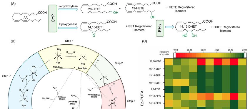
Nutrients 2020, 12, 3523                                                                          2 of 40

to be fully explored, is the metabolism of polyunsaturated fatty acids (PUFAs). In this review paper,
we focus primarily on dietary factors, specifically, the relationship between cytochrome P450 (CYP)
metabolism of polyunsaturated fatty acids (PUFAs) and age-associated NDs [1,2].
       In mammals, ω-3 PUFAs cannot be synthesized endogenously; therefore, they must be obtained
from dietary sources [7]. PUFAs are suspected to play significant roles in neural functions due to their
abundance in neural tissues. Arachidonic acid (AA) and docosahexaenoic acid (DHA) are the two
most abundant PUFAs within the nervous system and together comprise roughly 35% of the lipid
content in the brain tissue [8]. It has been demonstrated that dietary PUFA intake is beneficial for
neurodevelopment and attenuating neurodegeneration [9]. The Rotterdam study, which assessed a
cohort of 5289 subjects aged 55 and older, showed an association between PUFA intake and reduced
incidence of PD [10]. In rodents fed with a diet deficient in ω-3 PUFAs, a dramatic decrease in brain
PUFA content have been observed, which were also accompanied with a decrease in the numbers
of dopaminergic neurons in the substantia nigra [11]. Manipulating the PUFA composition of the
cell membrane has been shown to change the function and/or signaling of a variety of receptors,
including cholinergic, dopaminergic, and GABAergic receptors [12], while the detailed mechanism
remains largely unknown. The endogenous level of PUFAs greatly affected the composition of their
downstream metabolites in vivo. Therefore, it has been suggested that the downstream metabolites
may be partly responsible for the action of ω-3 and ω-6 PUFAs in neurons.
       PUFAs are metabolized by three main oxidative pathways, which include (i) lipoxygenase (LOX),
(ii) cyclooxygenase (COX), and (iii) cytochrome P450 (CYP) pathways to produce different oxidized lipid
mediators called oxylipins [13]. Generally, ω-6 PUFA oxylipins tend to be proinflammatory, while ω-3
PUFA oxylipins are considered anti-inflammatory and pro-resolving. Oxylipins are mediators in
physiological processes in various tissues including neurons [13]. AA oxylipins, such as lipoxin A4,
as well as prostaglandin D2 and E2, have shown neuroprotective properties [13]. The pro-resolving
mediators derived from DHA such as protectin D1, as well as resolvin D1 and D2, seem to inhibit
age-related memory decline and protect the brain from cell injury and death [13,14]. Furthermore,
the oxylipins generated by COX and LOX, such as prostaglandin and leukotrienes, respectively,
tend to be pro-inflammatory and exert excitatory effects on neurons. On the other hand, epoxy-PUFAs
(Ep-PUFAs), oxylipins generated by CYP enzymes, seem to produce the opposite effects, and are
neuroprotective, anti-hypertensive, and analgesic [13,15,16]. While the activity of ω-3 Ep-PUFAs and
ω-6 Ep-PUFAs largely overlap, the ω-3 Ep-PUFAs are generally more active than ω-6 Ep-PUFAs.
The beneficial properties of Ep-PUFAs appear to be diminished when Ep-PUFAs are converted
to their corresponding 1,2-diols by soluble epoxy hydrolase (sEH) [17], which will be discussed
later in this review. As more research has accumulated, CYP metabolites of PUFAs have been
suggested to be a key class of oxylipins that affect neurodegeneration. Inhibition of sEH, the enzyme
largely responsible for the degradation of CYP PUFA metabolites, has been shown to be beneficial in
Alzheimer’s disease (AD) and Parkinson’s disease (PD) [18]. Additionally, inhibition or the genetic
knockout of sEH can protect the dopaminergic neurons in the mouse brain against neurotoxins [19].
Even though there is a controversy, in randomized clinical trials studying the effects of ω-3 PUFAs
in AD, most observational studies have shown the beneficial effects of ω-3 intake on reducing
the incidence of AD [9,13,18]. Additionally, the oxylipin profiles of blood serum in AD subjects
show around 20% higher levels of dihydroxyeicosatrienoic acid (which is the product of the sEH
metabolism of the epoxy-metabolite of AA) as compared to the elderly individuals that were cognitively
healthy [20]. Therefore, sEH is an important enzyme in PUFA metabolism and a lucrative drug target
for NDs [21]. However, the mechanism of action of the CYP PUFA metabolites remains largely
unknown. The endogenous level of CYP PUFA metabolites is not only affected by their metabolism
but is also greatly affected by the endogenous level of their PUFA precursors as well as their uptake
by diet [22,23]. Therefore, in this review, we will: (1) describe the biosynthesis and metabolism of
PUFAs and downstream CYP metabolites and the role of biosynthesis and metabolism of PUFAs in
neurodegeneration; (2) highlight the key CYP and EH enzymes present in central nervous system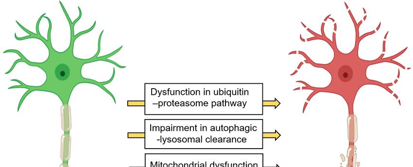
Nutrients 2020, 12, 3523 3 of 40
(CNS); and (3) elaborate on the potential mechanism(s) of action of CYP PUFA metabolites in
neuroinflammation and corresponding neurodegeneration.
Nutrients 2020, 12, x FOR PEER
2. Neurodegeneration: REVIEW
A Brief Overview 3 of 41
2.Neurons have an incredibly
Neurodegeneration: high respiratory rate due to the presence of highly active processes
A Brief Overview
such as vesicle trafficking, neurotransmitter synthesis, protein synthesis, molecular/ion transport,
Neurons have an incredibly high respiratory rate due to the presence of highly active processes
etc.,such
andasthus generate a great deal of oxidative stress [1,24]. This further necessitates a high
vesicle trafficking, neurotransmitter synthesis, protein synthesis, molecular/ion transport,
demand of energy
etc., and thus generate for the maintenance
a great of redox
deal of oxidative homeostasis
stress and, organelle
[1,24]. This further and
necessitates protein
a high quality
demand
control [1,24].
of energy forChronic and excessive
the maintenance perturbations
of redox in these
homeostasis and, pathways
organelle can cause
and protein neurodegeneration.
quality control [1,24].
Figure 1 displays
Chronic commonperturbations
and excessive neuronal pathways
in thesethat are disturbed
pathways in neurodegenerative
can cause neurodegeneration. diseases
Figure [1,25].
1
These pathways
displays common areneuronal
mostly interdependent.
pathways that are For instance,
disturbed chronic neuroinflammation
in neurodegenerative can
diseases [1,25]. lead to
These
protein accumulation,
pathways are mostlyendoplasmic
interdependent.reticulum (ER) chronic
For instance, stress, mitochondria dysfunction,
neuroinflammation uncontrolled
can lead to protein
accumulation,
oxidative stress, endoplasmic reticulum
axonal transport (ER) stress,and
impairment, mitochondria
apoptosis,dysfunction,
which areuncontrolled
detrimentaloxidative
to neurons,
stress,toaxonal
leading transportThe
NDs [26,27]. impairment,
major NDand apoptosis, include,
pathologies which arebut detrimental to neurons,
are not limited leadingmisfolding,
to protein to NDs
[26,27]. The major ND pathologies include, but are not limited to protein misfolding,
synaptic dystrophy, changes in neurotransmitter production, an increased oxidative stress response, synaptic
anddystrophy, changes
loss of neurons in neurotransmitter
[2,28–30]. In addition, production,
reductionan in increased oxidative
brain volume stress
is often response,(in
observed and lossgray
both
of neurons [2,28–30]. In addition, reduction in brain volume is often observed (in both gray and white
and white matter), along with increased lesions in white matter and dysfunction of the blood–brain
matter), along with increased lesions in white matter and dysfunction of the blood–brain barrier
barrier (BBB) [28]. While the mechanisms that are involved in the pathology of NDs are pertinent
(BBB) [28]. While the mechanisms that are involved in the pathology of NDs are pertinent to this
to this review, the details are not included here, as they have been extensively discussed in other
review, the details are not included here, as they have been extensively discussed in other reviews
reviews [1,24,25]. Our focus here is to review and discuss how CYP metabolism of PUFAs is potentially
[1,24,25]. Our focus here is to review and discuss how CYP metabolism of PUFAs is potentially
involved
involved in neurodegeneration.
in neurodegeneration.
Figure 1. 1.Common
Figure Common neuronal pathwaysthat
neuronal pathways thatareare changed
changed in different
in different neurodegenerative
neurodegenerative diseasesdiseases
(1)
(1) Dysfunction in the ubiquitin–proteasome pathway increases the intracellular misfolded,
Dysfunction in the ubiquitin–proteasome pathway increases the intracellular misfolded, damaged, or damaged,
or unneeded
unneeded protein. (2)Dysfunction
protein. (2) Dysfunctionininthetheautophagy–lysosomal
autophagy–lysosomal pathway
pathway triggers
triggers the the accumulation
accumulation
of pathogenic
of pathogenic protein
proteinaggregates
aggregatesandanddamaged
damaged mitochondria.
mitochondria. (3) (3)Mitochondria
Mitochondria dysfunction
dysfunction causes
causes
dysfunction
dysfunction andanduncontrolled
uncontrolledrelease
release of
of reactive
reactive oxygen
oxygen species.
species.(4)(4)ERER stress occurs
stress when
occurs whenthe the
homeostatic
homeostatic proteinfolding
protein folding and
andtrafficking
traffickingin in
thethe
cellcell
is overwhelmed
is overwhelmedor unbalanced, leading to
or unbalanced, UPR. to
leading
UPR.(5) (5)
Transcellular propagation
Transcellular and seeding
propagation of protein
and seeding aggregates
of protein cause disease
aggregates cause progression. (6) The
disease progression.
(6) aggregation of misfolded
The aggregation proteinsproteins
of misfolded contributes to toxicity.
contributes to(7) A chronic
toxicity. (7) neuroinflammatory state can
A chronic neuroinflammatory
lead to protein accumulation, ER stress, mitochondria dysfunction, uncontrolled oxidative
state can lead to protein accumulation, ER stress, mitochondria dysfunction, uncontrolled oxidative stress, and
axonal transport impairment. Abbreviations- ER: endoplasmic reticulum, UPR: unfolded protein
stress, and axonal transport impairment. Abbreviations- ER: endoplasmic reticulum, UPR: unfolded
response.
protein response.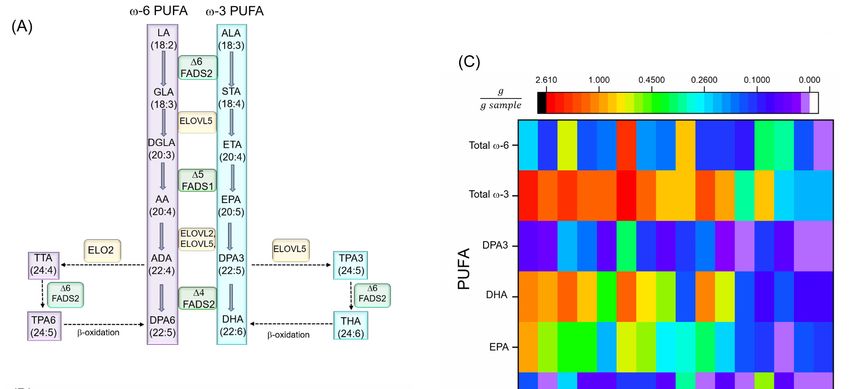
Nutrients 2020, 12, 3523                                                                            4 of 40

3. Overview of PUFAs
     PUFAs are long-chain fatty acids comprising of at least two carbon-carbon double bonds that play
crucial roles in a variety of physiological processes. The endogenous levels of ω-3 and ω-6 PUFAs
significantly modulate the in vivo levels of their downstream metabolites, particularly the CYP PUFA
metabolites [31,32]. In mammals, small quantities of PUFAs can be synthesized endogenously, and the
rest are obtained from the diet. An overview of the the biological levels of PUFAs, and thus their
downstream metabolism is presented in this section.

3.1. Biosynthesis of PUFAs and Neurodegenerative Diseases
     The major steps in PUFA biosynthesis in organisms are: (i) elongation, by elongase enzymes
(elongase of very long chain fatty acids, ELOVL); and (ii) desaturation by desaturase enzymes (fatty acid
desaturase, FADS) [7,33]. Some metazoans, such as Caenorhabditis elegans, are capable of endogenous
production of ω-6 and ω-3 PUFAs through the conversion of ω-9 monounsaturated oleic acid (OA) to
ω-6 linoleic acid (LA) via a ∆12 desaturase, and then further conversion of ω-6 PUFAs to ω-3 PUFAs
via ∆15 (ω-3) desaturase [34]. These animals do not require dietary sources of PUFAs. Vertebrates,
however, do not have genes encoding functional ∆12 and ω-3 desaturase enzymes, and thus cannot
endogenously synthesize PUFAs from OA [35]. Despite this, vertebrates do have the ability to use LA
and α-linoleic acid (ALA), which must be obtained from the diet, as precursors for the biosynthesis of the
other ω-6 and ω-3 PUFAs, respectively, through desaturation and elongation [7,33]. The conventional
enzymatic pathway that produces these PUFAs is depicted in Figure 2A.
     Although humans only synthesize a small amount of PUFAs, recent studies provide evidence
that FADS in humans play an important role in modulating endogenous levels of different PUFAs.
A genome-wide genotyping study conducted by Ameur et al. revealed two common haplotypes
of the FADS gene: (i) haplotype D that can drive more active conversion in PUFAs, and (ii) the
less active haplotype A [36]. People homozygous for the D type have higher levels of AA (43%)
and DHA (24%), as well as greater plasma lipid levels compared to those who are homozygous
for haplotype A [36]. Several studies have shown the association between breastfeeding and child
brain development, as a FADS gene variant can control the PUFA composition of breast milk [37–40].
Furthermore, from rare human studies, a very low level of ∆6 desaturase was found in patients
with Sjögren–Larsson syndrome, which is characterized by neurological, skin, and eye problems [41].
A single-nucleotide polymorphism (SNP) in FADS2 was also discovered to be associated with the
occurrence of attention-deficit/hyperactivity disorder (ADHD), which is suggested to be a result from
abnormal regulation of DA at the neural synapse [42]. Most of these rare studies can be considered as
preliminary data, and replication in a larger population, and consideration of other PUFA metabolic
genes such as ELOVL are necessary. In addition to human studies, mouse models with genetic defects
in Fads1 and Fads2 have been conducted [43,44]. The Fads1 knockout mice failed to thrive and died
before reaching 12 weeks of age. These mice also exhibited perturbed immune cell homeostasis and
severe inflammatory problems. Dietary supplementation with AA prolonged the lifespan of these
mice to levels comparable to wild-type mice [43]. Surprisingly, knockout of the Fads2 gene in mice
did not result in significant impairment in lifespan and viability, but rather produced sterility (in both
sexes) and increased bleeding time [44]. Brown and colleagues observed that a selective knockdown
of Fads1 in adult hyperlipidemic mice results in striking changes of both ω-3 and ω-6 PUFA levels
and their corresponding proinflammatory and proresolving lipid mediators, suggesting an important
regulatory effect of ∆-5 desaturase in inflammation initiation and resolution [45]. Thus, variability in
gene expression may be one of the key factors impacting PUFA downstream metabolism by controlling
the Fads/FADS (in mouse/human) genes. Considering this idea, researchers studying the effects of PUFA
supplementation should consider genetic effects in the specific population under study, which might
be the reason for the inconsistency found in the clinical and epidemiology studies regarding the PUFA
effects on human health and disease.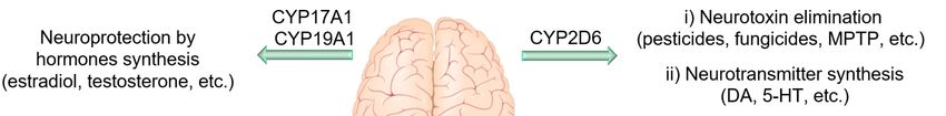
Nutrients
Nutrients2020,
2020,12,
12,x3523
FOR PEER REVIEW 55of
of41
40
3.2. Dietary PUFA
3.2. Dietary PUFA
Although humans make both desaturase and elongase enzymes, the conversion/biosynthesis of
PUFAs Although
is quite humans
limited in make both desaturase
humans. and elongase
Because of this, PUFA levels enzymes, the and
in blood conversion/biosynthesis
tissues are modulated of
PUFAs
by is quite
dietary intakelimited
[7,46].in humans.
Hence, thereBecause
is aofgreat
this, PUFA
interestlevels in blood and
surrounding the tissues are benefits
potential modulated of
by dietary intakeand
supplementation, [7,46]. Hence,are
ω-3 PUFAs there
one is
of athegreat
mostinterest
consumed surrounding the potential
dietary supplements [7].benefits
The main of
supplementation,
sources of fatty acids and are PUFAs are
ω-3 different one ofcountries,
among the most consumed
and mainly dietary supplements
controlled by food[7]. The main
availability,
sources ofand
economy, fattyculture
acids are different
[7,39]. among countries,
ω-3 PUFAs and mainly
primarily originate controlled
from by food
plant, algal, availability,
marine, economy,
and protozoan
and culture
sources. While [7,39].
plants PUFAs
ω-3such primarily
as nuts, someoriginate
seeds, and from plant, algal,
vegetable marine,
oil are and protozoan
the primary source of sources.
ALA
While plants
(Figure such as nuts,
2B), marine animalssome areseeds, and vegetable
the main source of oil eicosapentaenoic
are the primary sourceacid of ALA DHA,
(EPA), (Figureand
2B),
marine animals are the main source of eicosapentaenoic acid (EPA), DHA,
docosapentaenoic acid (ω-3) (DPA3) (Figure 2C) [47]. Dietary supplementation studies in randomized and docosapentaenoic
acid (ω-3)
clinical trials(DPA3)
show an (Figure 2C) [47].
inconclusive Dietary
results supplementation
regarding the effects ofstudies
PUFAsinonrandomized clinical trials
neurodegeneration, that
show be
could an due
inconclusive results
to the genetic regardingdiet
variability, theand
effects of PUFAs
different on neurodegeneration,
lifestyles that could be
and habits in the population due
under
to the [46].
study genetic variability,studies
Furthermore, diet and indifferent
humans lifestyles
demonstrate and ahabits
causal inrelationship
the population underthe
between study [46].
dietary
Furthermore,
lipid studies in humans
ratio supplementation of ω-3demonstrate
and ω-6 PUFAs a causal relationship
and NDs, such asbetween
AD and thePD,dietary
which is lipid ratio
beyond
supplementation
the scope of this review of ω-3[6,10,46,48,49].
and ω-6 PUFAs and NDs, such as AD and PD, which is beyond the scope of
this review [6,10,46,48,49].
Figure 2.
Figure (A) PUFA
2. (A) PUFA biosynthesis
biosynthesis pathways.
pathways. (B) (B)Common
Commonvegetable
vegetableoiloil or
or seeds.
seeds. The
The total
total ω-PUFA
ω-PUFA
content is almost exclusively ALA, with little to no EPA, DHA, and DPA3 [47]. (C)
content is almost exclusively ALA, with little to no EPA, DHA, and DPA3 [47]. (C) PUFA amounts in PUFA amounts in
different fish
different fish and
and seafood
seafood (all
(all are
are cooked
cooked or or baked
baked unless
unless otherwise
otherwise mentioned)
mentioned)[47].[47].Abbreviations-
Abbreviations-
AA: arachidonic
AA: arachidonic acid,acid, AD:
AD: adrenic
adrenic acid,
acid, ALA: α-linolenic ∆6: delta-4
α-linolenic Acid, Δ6: delta-4 desaturase,
desaturase, Δ5:∆5: delta-5
delta-5
desaturase,Δ6:
desaturase, ∆6:delta-6
delta-6 desaturase,
desaturase, DGLA:
DGLA: dihomo-γ-linolenic
dihomo-γ-linolenic acid,
acid, DPA3:
DPA3: docosapentaenoic
docosapentaenoic acidacid
(ω-
(ω-3), DPA6: docosapentaenoic acid (ω-6), DHA: docosahexaenoic acid, ELOVL:
3), DPA6: docosapentaenoic acid (ω-6), DHA: docosahexaenoic acid, ELOVL: elongase of very long elongase of very long
chain fatty
chain fatty acids,
acids, EPA:
EPA: eicosapentaenoic
eicosapentaenoic acid,
acid, ETA:
ETA:eicosatrienoic
eicosatrienoicacid,
acid,FADS
FADS1:1:fatty
fattyacid
aciddesaturase
desaturase1,
1,FADS2:
FADS2: fatty acid
fatty desaturase
acid desaturase2, GLA:
2, GLA:γ-linolenic
γ-linolenicacid, LA:LA:
acid, Linoleic acid,acid,
Linoleic PUFA: polyunsaturated
PUFA: fatty
polyunsaturated
acid, THA:
fatty acid, tetracosahexaenoic acid, TPA3:acid,
THA: tetracosahexaenoic tetracosapentenoic acid (ω-3), TPA6:
TPA3: tetracosapentenoic tetracosapentenoic
acid (ω-3), TPA6:
acid (ω-6), TTA: tetracosatetraenoic
tetracosapentenoic acid, STA: stearidonic
acid (ω-6), TTA: tetracosatetraenoic acid.
acid, STA: stearidonic acid.
4. CYP: A Key Monooxygenase Enzyme in PUFAs Metabolism
Nutrients 2020, 12, 3523                                                                            6 of 40

4. CYP: A Key Monooxygenase Enzyme in PUFAs Metabolism
       PUFAs are mainly metabolized through three oxidative (pathways: (i) lipoxygenases (LOX),
(ii) cyclooxygenases (COX), and (iii) CYPs) to produce lipid signaling molecules called oxylipins [50–52].
Most of these mono-oxygenated lipid metabolites are key lipid mediators in physiological processes
in mammals. Research regarding the effects of the lipoxygenase and cyclooxygenase pathways on
neurodegeneration has been extensively reviewed [52,53]; thus, these are not the focus of this review.
In this section, we will summarize the recent findings on a CYP pathway of PUFAs and the effects
of CYP selectivity on mammalian physiology. The CYP metabolic pathway of AA is depicted in
Figure 3A.

4.1. Characteristics of CYP
      CYPs are heme-thiolate proteins primarily involved in synthesis and metabolism of xenobiotics,
as well as endogenous biological molecules such as steroid hormones, fatty acids, cholesterol, drugs,
vitamin D, etc., via oxidation [54]. CYPs, in general, contain a signature residue sequence of
FXXGXbXXCXG, in which Xb is a basic residue and the cysteine is located at the axial position to the
heme, and a Soret peak at 450 nm when a carbon monoxide binds to the Fe(II) of the heme group [55,56].
Note that there are some other proteins with the same heme group, axial cysteine residue, and similar
Soret peak in the presence of carbon monoxide, as well as some related catalytic properties such as some
peroxidases and nitric oxide synthases, but they are not considered CYP enzymes. The 3D structures
of these proteins also differentiate them from CYP enzymes, which share the same folding [57,58].
      The human genome project has identified 57 genes expressing different CYP enzymes, which are
grouped into 18 families (43 sub-families) based on the amino acid sequence homology [59,60]. Thus,
each CYP enzyme is named by a number representing the family and a letter indicating the subfamily,
followed by a second number specific for an individual CYP enzyme (e.g., CYP2J2). In contrast to
prokaryotes that have soluble CYP enzymes, in mammals CYPs are primarily membrane-associated
proteins located either on ER or mitochondria membranes [61,62]. The catalytic domain of these
enzymes is partially immersed in the membrane and can move along the membrane surface. The active
site connection to both the cytosolic environment and the membrane through networks of access
channels allows them to interact with substrates in either compartment [62]. Of the 57 human CYP
enzymes, 50 are located on the ER and are usually involved in xenobiotic metabolism (i.e., drugs and
environmental pollutants), while the rest are located in the mitochondria membrane and are generally
engaged in the metabolism/biosynthesis of endogenous molecules [63]. Even though these enzymes
are mostly expressed in the liver, they can also be expressed in many other tissues including, but not
limited to, kidney, brain, intestinal mucosa, skin, and lung [64].

4.2. Catalytic Function and Mechanism of CYP
     Historically, the first CYP enzyme was described by Klingenberg and Garfinkel as an unknown
pigment that binds carbon monoxide in its reduced form and produces a Soret absorption peak at
450 nm [65,66]. This unknown pigment was then identified as a new cytochrome by Omura et al. [55].
About ten years later, in 1979, Benhamou and colleagues further confirmed this observation by
studying the inhibitory effect of AA administration on metabolizing the hepatic drug by CYP enzymes
in mice [67].
     CYPs catalyze a large variety of reactions including oxidation of heteroatoms, heteroatom
dealkylation, carbon-carbon bond cleavage, desaturation, ring formation, aryl ring couplings,
and rearrangements of oxygenated molecules [68–70]. The monooxygenase activity of CYPs has been
discussed thoroughly elsewhere [71–73], and, therefore, we will only provide a brief description here.
Figure 3B illustrates the epoxidation mechanism of CYPs, related to its epoxygenase activity, in seven
steps: (1) before binding of the substrate to the CYP protein, there is an equilibrium between the hexa-
and pentacoordinate Fe(III); substrate binding to the CYP enzymes shifts the equilibrium in favor of
Nutrients 2020, 12, 3523                                                                                7 of 40

pentacoordinate; (2) an electron will transfer to this complex either directly from NADPH or through a
redox protein partner, to reduce Fe (III) to Fe (II) (note that this step is critical for substrate oxidation,
as the diatom oxygen cannot bind to Fe (III)); (3) oxygen binds to the Fe(II); (4) the next electron is
either transferred directly from NAD(P)H or through a redox protein partner; (5) two subsequent
protonations occur; (6) the complex gets deprotonated by releasing a water molecule, which results
in an iron-oxo complex; (7) finally, the oxygen atom is transferred to the substrate and results in an
oxidized product [71–73].

4.3. Major CYP Responsible for PUFA Metabolism
     CYPs metabolize PUFAs to either Ep-PUFAs (epoxygenase activity) or hydroxy-PUFAs
(with hydroxylase activity). The final product depends on the specific CYP enzyme, as well as
the type of PUFA substrate. In general, the regio/stereoselectivity, catalytic properties, and structure of
each CYP enzyme might be significantly determined by a B-C loop in their structure, controlling the
type of reaction and prevalence of a specific regio/stereoisomer product [56,74,75]. In this section, we
discuss the major CYPs and their products when ω-3 and ω-6 PUFAs are substrates.

4.3.1. Regio- and Stereoselective Epoxidation by Major CYPs
      In the case of AA, the main CYPs that generate Ep-PUFAs are CYP2B, 2C, and 2J sub-families
(known as AA epoxygenase), while 1A, 4A, and 4F subfamilies produce the majority of ω and ω-1
hydroxylated AA (known as AA hydroxylase) (Figure 3A) [76]. Both CYPs families are regio- and
stereoselective [77,78]. For instance, CYP2C and 2J subfamilies can convert AA into four regioisomers
of epoxyeicosatrienoic acids (EETs) depending on which double bond is involved in oxygen insertion,
which results in 5,6-EET, 8,9-EET, 11,12-EET, and 14,15-EET. Each of the EET products can be either
the R,S- or the S,R stereoisomer [77,78]. This regio- and the stereoselectivity of the epoxidation of
AA to EET is CYP isoform-specific. For instance, while CYP2C8 in humans metabolizes AA with
high regio/stereoselectivity to 14,15- and 11,12-EET (with ratio of 1.3:1 and more than 80% optical
purity (OP) of R,S enantiomers), CYP2C9 shows very low regio- and stereoselectivity [79,80]. CYP2J2
is not regioselective as it produces all four regioisomers of EETs, in which 8,9- and 11,12-EET are
largely a racemic mixture, and 14,15 -EET is mainly in the form of 14(R),15(S)-EET (OP ≈ 76%) [81].
CYP2C23, which is the main CYP involved in AA epoxidation in the rat kidney, generates 8,9-, 11,12-,
and 14,15-EET in a ratio of 1:2:0.7 with high stereoselectivity (OP ≈ 95, 85, and 75%, respectively) [82].
Likewise, CYP2C44 in mice results in the same products [83]. The regioselectivity of different CYPs
and the relative amount of each regioisomer are depicted in Figure 3C.
      Even though the CYPs subfamilies involved in LA metabolism have not yet been meticulously
examined, several studies proposed that all CYP isoforms can metabolize LA with high efficiency.
For instance, CYP2C9 is known as the main LA monooxygenase in the human liver and generates
both 9,10- and 12,13-epoxyoctadecamonoenic acids (EpOMEs) [84]. Other AA metabolizing CYP
isoforms that can also accept LA as a substrate are CYP2C8 and -19; CYP2J2, -3, -5, and -9; CYP1A2;
and CYP3A4 [85–87]. It is worth mentioning that the same CYP isoform can have different preferences
as to whether they act as an epoxygenase or hydroxylase when the substrate is changed from AA to
LA. For example, while human CYP2E1 has primarily hydroxylase activity on AA, it is a major LA
monooxygenase [87].
      Like LA, EPA and DHA have been shown to be substrates for CYP isoforms in human, rat,
and mouse [22]. For example, the human isoforms CYP2C8, 9, 18, and 19, as well as CYP2J2 can
epoxidize both EPA and DHA [88,89]. Moreover, the catalytic activities of CYP2C isoforms for
EPA and DHA are almost the same as for AA, while CYP2J2 displays 9 and 2 times higher rates in
metabolizing EPA and DHA, respectively, compared to AA [88,90]. Furthermore, these enzymes have
different regioselectivity for EPA and DHA [88,90,91]. For instance, while CYP2C23 metabolizes AA to
8,9-, 11,12-, and 14,15-EET in a ratio of about 1:2:0.6, the epoxidation of EPA by CYP2C23 results in
17,18-, 14,15-, 11,12-, and 8,9- epoxy eicosatetraenoic acids (EEQs) in a ratio of about 6:1:1:1 [90,91].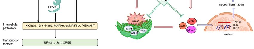
Nutrients 2020, 12, 3523 8 of 40
Furthermore, while human CYP2C8 produces mainly 11,12- and 14,15-EET with AA as a substrate,
it produces exclusively a terminal ω-3 PUFA epoxide with omega-3 PUFAs as a substrate, such as
17,18-EEQ with EPA, and 19,20- epoxy docosapentaenoic acid (19,20-EDPs) with DHA [88,90,91].
However, very low regioselectivity has been reported for other CYP2C isoforms toward EPA and
Nutrients 2020, 12, x FOR PEER REVIEW 8 of 41
DHA [22,89]. Furthermore, as previously mentioned, CYP2J2 has very low regioselectivity toward
AAregioselectivity
and high regioselectivity towards
towards ω-3 PUFAs ω-3 PUFAsgenerating
(preferentially (preferentially generating
terminal ω-3 PUFAterminal
epoxides)ω-3 PUFA
[88,90].
epoxides) [88,90]. Furthermore, it should be noted that CYP2J2 and all CYP2C isoforms
Furthermore, it should be noted that CYP2J2 and all CYP2C isoforms (except CYP2C8) exhibit high (except
CYP2C8) exhibit high
stereoselectivity, stereoselectivity,
favoring favoring
the production the production of 17,18-EEQ
of the R,S-enantiomers the R,S-enantiomers of 17,18-EEQ
and 19,20-EDP [91,92].
and 19,20-EDP [91,92].
Figure 3. 3.(A)
Figure (A)CYP
CYPhydroxylase
hydroxylase and andepoxygenase
epoxygenase activities
activities on on
AA,AA, as well
as well as EH asproducts
EH products of (B)
of EETs. EETs.
(B)CYP
CYPmechanism
mechanism of function
of function for monooxygenase
for monooxygenase activity.
activity. (C) Regioselectivity
(C) Regioselectivity in epoxygenase
in epoxygenase activity
of different
activity CYP adapted
of different from [88].
CYP adapted Note
from that
[88]. all EETs
Note and
that all HETEs
EETs and(except
HETEs20-HETE) can be exist
(except 20-HETE) as be
can
either
exist the R-the
as either or S-enantiomers. Abbreviations-
R- or S-enantiomers. AA: arachidonic
Abbreviations- acid, CYP:
AA: arachidonic cytochrome
acid, P450, DHET:
CYP: cytochrome P450,
dihydroxyeicosatrienoic
DHET: dihydroxyeicosatrienoicacid, EDP:
acid, epoxydocosapentaenoic
EDP: epoxydocosapentaenoic acid, EEQ:
acid,epoxyeicosatetraenoic acid,
EEQ: epoxyeicosatetraenoic
EET:
acid, epoxyeicosatrienoic
EET: epoxyeicosatrienoicacid,acid,
EH: EH:
epoxide hydrolase,
epoxide HETE:
hydrolase, hydroxyeicosatetraenoic
HETE: acid. acid.
hydroxyeicosatetraenoic
4.3.2. Regio-
4.3.2. and
Regio- andStereoselective
StereoselectiveHydroxylation
Hydroxylation of PUFAs by
by CYPs
CYPs
Like epoxidation
Like epoxidationactivity,
activity,CYPs
CYPsalso
also hydroxylate
hydroxylate in a regio-selective
regio-selectivemanner.
manner.CYP4ACYP4A andand4F4F
subfamilies
subfamilies hydroxylate
hydroxylate AA AA at the
at the terminal
terminal methyl
methyl group
group to produce
to produce 20-hydroxyeicosatetraenoic
20-hydroxyeicosatetraenoic acid
acid (20-HETE)
(20-HETE) as the product,
as the major major product, and 19-HETE
and 19-HETE as theasminor
the minor product.
product. Specifically,
Specifically, 20-and
20-and 19-
19-HETE
canHETE can be generated
be generated by humanby human
CYP4A11, CYP4A11, rat CYP4A1,
rat CYP4A1, and mouse
and mouse CYP4A12A,
CYP4A12A, in ain a ratio
ratio of 90:10,
of 90:10, 93:7,
and 87:13, respectively [93–95]. CYP4F isoforms such as CYP4F2, CYP4F3A, and CYP4F3B are are
93:7, and 87:13, respectively [93–95]. CYP4F isoforms such as CYP4F2, CYP4F3A, and CYP4F3B more
more regioselective
regioselective than in
than CYP4A CYP4A in ω-hydroxylation
ω-hydroxylation [96]. On[96].
the On thehand,
other otherthe
hand, the CYP1A1,
CYP1A1, CYP1A2, CYP1A2,
and CYP
and CYP 2E1 mainly have ω-1 hydroxylase activity on AA, yielding 19-HETE
2E1 mainly have ω-1 hydroxylase activity on AA, yielding 19-HETE as the predominant product, as the predominant
product, and 16-, 17-HETE (CYP1A1 and CYP1A2) and 18-HETE (CYP2E1) as minor products, while
and 16-, 17-HETE (CYP1A1 and CYP1A2) and 18-HETE (CYP2E1) as minor products, while 20-HETE
20-HETE is not produced [22,76]. Interestingly, CYP2J9, unlike other members in the CYP2J
is not produced [22,76]. Interestingly, CYP2J9, unlike other members in the CYP2J subfamily, generates
subfamily, generates the 19-HETE from AA almost exclusively, while the other members in the
the 19-HETE from AA almost exclusively, while the other members in the subfamily such as human
subfamily such as human CYP2J2 and rat CYP2J3 mainly display epoxygenase activity towards AA
CYP2J2 and rat CYP2J3 mainly display epoxygenase activity towards AA [81,96,97]. CYP4A1, which is
[81,96,97]. CYP4A1, which is the main PUFA hydroxylase in rats, can metabolize LA at the same rate
the main PUFA hydroxylase in rats, can metabolize LA at the same rate as AA and generates 18- and
as AA and generates 18- and 17-hydroxy octadecadienoic acids (HODE) in a ratio of 3:1 [94].
Likewise, human CYP4A11 has shown hydroxylase activity on LA [87,98].
EPA and DHA can also be effectively hydroxylated by CYP hydroxylases such as human
CYP4A11, CYP4F2, CYP4F3A, and CYP4F3B, as well as mouse CYP4A12, and rat CYP4A1
[95,96,99,100]. Again, when the substrate of these CYPs changes from AA to EPA or DHA, the
regioselectivity, as well as reactivity, is altered. For instance, while CYP4A1 hydroxylates AA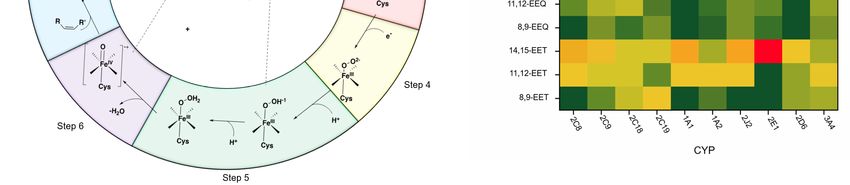
Nutrients 2020, 12, 3523                                                                         9 of 40

17-hydroxy octadecadienoic acids (HODE) in a ratio of 3:1 [94]. Likewise, human CYP4A11 has shown
hydroxylase activity on LA [87,98].
     EPA and DHA can also be effectively hydroxylated by CYP hydroxylases such as human CYP4A11,
CYP4F2, CYP4F3A, and CYP4F3B, as well as mouse CYP4A12, and rat CYP4A1 [95,96,99,100].
Again, when the substrate of these CYPs changes from AA to EPA or DHA, the regioselectivity,
as well as reactivity, is altered. For instance, while CYP4A1 hydroxylates AA (generating mainly
20- and 19-HETE), it epoxidizes and hydroxylates EPA to produce predominantly 17,18-EEQ (68%)
and 19-HEPE (31%) [94,100,101]. Furthermore, when DHA is a substrate, the CYP4A1 exclusively
produces the epoxidized product: 19,20-EDP [88,92]. Likewise, CYP4A12A can function exclusively
as ω/(ω-1)-hydroxylase with AA, producing 20- and 19-HETE in a ratio of 8:2, and can metabolize
EPA exclusively to 17,18-EEQ with a minor amount of 20- and 19-HEPE [95]. This trend has also been
observed for CYP2E1, as it acts as a hydroxylase to AA, and an epoxygenase to EPA and DHA [88,90].
Note that the change in catalytic preference from ω to (ω-1)-hydroxylase activity of CYPs has also
been observed. For instance, by changing the substrate from AA to EPA and DHA the ratio of ω to
(ω-1)-hydroxylase of CYP4A1 shifts from 4:1 to 1:3 and 1:2, respectively [96]. Considering CYP4F
subfamilies, CYP4F3A and CYP4F3B display higher catalytic activity for AA and DHA compared to
EPA, while CYP4F2 has a higher preference for hydroxylating DHA compared to AA and EPA [96].
CYP4F8 and CYP4F12 mainly function as (ω-n)-hydroxylases, with AA producing 18- and 19-HETE,
and producing 17,18-EEQ and 19,20-EDP as the main products of EPA and DHA, respectively [102].
CYP2U1 is another CYP isoform that is expressed in the brain, which mainly acts as a ω-hydroxylase
for ALA, AA, EPA, and DHA [103,104].
     The regio- and stereoselectivity of CYP enzymes ultimately affects the physiology because
numerous studies have suggested that the biological activity of Ep-PUFAs is controlled by the
regio/stereoselectivity of the target receptors, which will be discussed in the following paragraphs
(Section 4.4).

4.3.3. Physiological Functions of Regio-/Stereoisomers of CYP Products: Ep-PUFAs and Hydroxy-PUFAs
      The regio- and stereoselectivity of CYP enzymes ultimately could alter their effects on mammalian
physiology because numerous studies have suggested that the biological activity of Ep-PUFAs is
regio/stereoselective. These results will be briefly summarized in this section, whereas the specific
effects of CYP PUFA metabolites on neurodegeneration will be discussed in Section 7.
      One study regarding the regioselective effects of EET by Node et al., demonstrated that
11,12-EETs exert significant anti-inflammatory effects by inhibiting the TNF-α-induced vascular
cell adhesion molecule-1 (VCAM-1) expression, while no effects were observed for 14,15-EETs [105].
In addition, peroxisome proliferator-activated receptor-α (PPARα), which plays an important role
in inflammation, can only be activated by 8,9-EET, and 11,12-EET, but not 14,15-EET [106]. Besides,
the omega-3 Ep-PUFAs also affect the regioselectivity of physiological processes. For example,
EDPs alleviate nociception response in rodent inflammatory pain model with relative potencies of
13,14-EDPs > 16,17-EDPs > 19,20-EDPs [107]. The biological effects of Ep-PUFAs in mammals are
also stereospecific. For instance, Ding et al. found that a Gs -coupled receptor on the membrane
of endothelial cells responds to 11(R),12(S)-EET, but not 11(S),12(R)-EET, which mediates protein
kinase A (PKA)-dependent translocation and activation of transient receptor potential (TRP) C6
channels [108]. Over the last few decades, several studies have shown that the Ep-PUFAs likely act
stereospecifically [100,109–111]. In addition to Ep-PUFAs, the biological functions of hydroxy-PUFAs
are also stereo- and regioselective [22,112–115].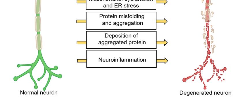
Nutrients 2020, 12, 3523                                                                          10 of 40

      Overall, these studies show the significance of regio- and stereochemistry on the physiological
effects of Ep- and hydroxy-PUFAs. One of the main challenges in investigating the regio-/stereoisomers
of PUFA metabolites is obtaining significant quantities of pure regioisomers and their antipodes.
Chemical epoxidation is neither chemoselective nor enantioselective, and the purification process of
positional isomers and enantiomers is a tedious process [116]. While enzymatic epoxidation seems to
be an effective way to generate single enantiomers, using a chemical inversion process is inevitable
to access the corresponding enantiomers. Besides, this method is non-diversifiable and cannot be
utilized in synthesize the corresponding analogs [116,117]. Progress in synthetic routes to achieve large
quantities of specific regio-/stereoisomers with high purity is the key to improve our understanding of
these isomers and the mechanisms of their functions.

4.4. CYP Enzymes in the Central Nervous System
      The first evaluation of CYP in the brain was conducted in 1977 by Sasame et al. [118]. They found
30 pmol/mg of CYP enzymes in the rat brain, which was approximately 3% of the corresponding levels
in the liver, with 30 times lower activity [118]. Since then, many efforts have been made to identify
different CYP enzymes, their activity, as well as expression patterns within the brain. To date, 41 of 57
CYP enzymes have been identified in various brain regions [119]. Studies on CYP enzyme expression
and function in the brain revealed some remarkable information. CYPs can be found in both glial
cells and neurons either in the cell bodies or throughout the cell processes. For instance, isoforms
such as CYP2E1, 1A1, 3A, and 2B, are mostly expressed in neurons, while others such as CYP2D6,
are predominately expressed in both glial and neuron cells [120,121]. CYP enzyme expression in the
brain is heterogeneous among different parts of the brain due to the presence of various cell types with
different needs and functionality [119]. Furthermore, some CYP enzyme levels in specific neurons
are even greater than their counterparts in hepatocytes. For example, CYP46A1, which regulates the
cholesterol homeostasis in the brain, and CYP2D6, which is involved in the biosynthesis of serotonin
and DA, are mainly expressed in the brain [122,123]. Even though CYP enzymes (especially 2C, 2J,
and 4A) can potentially be highly influential in brain function through their monooxygenase activity in
PUFAs metabolism (which will be discussed later), they are also key players in hormone, cholesterol,
endocannabinoids, and neurotransmitter metabolism [18,124]. Therefore, these enzymes can affect
neuronal activity and homeostasis through other mechanisms, independent of their PUFA metabolism
(Figure 4). For instance, CYP2D6 expressed in the brain is involved in the metabolism of endogenous
neural compounds such as catecholamines and can metabolize drugs and inactivate neurotoxins such
as 1-methyl-4-phenyl-1,2,3,6-tet-rahydropyridine (MPTP) and 1-methyl-4-phenylpyridinium (MPP+ ).
Thus, low activity of this enzyme can result in neuronal hypofunction, especially in dopaminergic
neurons. Mann et al. demonstrated that CYP2D6 levels increase with age; however, in PD patients,
this enzyme is expressed 40% less compared to in a healthy brain [125]. Lower levels of this enzyme
in PD patients may reduce their ability to inactivate PD-causing neurotoxins. Furthermore, CYP46
and CYP27 seem to be important in AD and Huntington disease, probably due to their role in the
brain cholesterol homeostasis and metabolism [126–128]. In addition, genetic variations in CYP19
and CYP2J2 have been associated with enhanced susceptibility AD [129,130]. The rs890293 variant of
CYP2J2, which results in a CYP2J2 enzyme with reduced function, has been shown to be associated with
late-onset AD in the Chinese Han population [131]. These data suggest a possible role of Ep-PUFAs,
dihydroxy-PUFAs, or hydroxy-PUFAs in neuroprotection and reduced risk of AD, as CYP2J2 is one of
the key isoforms of CYP responsible for Ep-PUFAs metabolism. There are extensive studies on the
CYP enzymes effects in neuronal function and homeostasis, which is beyond the scope of this paper,
and we are only focusing on the role they have in Ep-PUFAs or dihydroxy-PUFAs metabolism.
reduced function, has been shown to be associated with late-onset AD in the Chinese Han population
[131]. These data suggest a possible role of Ep-PUFAs, dihydroxy-PUFAs, or hydroxy-PUFAs in
neuroprotection and reduced risk of AD, as CYP2J2 is one of the key isoforms of CYP responsible for
Ep-PUFAs metabolism. There are extensive studies on the CYP enzymes effects in neuronal function
andNutrients 2020, 12,which
homeostasis, 3523 is beyond the scope of this paper, and we are only focusing on the role they11 of 40
have in Ep-PUFAs or dihydroxy-PUFAs metabolism.
Figure 4. The
Figure protective
4. The rolerole
protective of some cytochrome
of some P450s
cytochrome (CYPs)
P450s in neurodegenerative
(CYPs) diseases.
in neurodegenerative diseases.
Abbreviations- CYP:CYP:
Abbreviations- cytochrome P450,P450,
cytochrome DA:dopamine, 5-HT:5-HT:
DA:dopamine, 5-hydroxytryptamine (or serotonin),
5-hydroxytryptamine (or serotonin),
MPTP: 1-methyl-4-phenyl-1,2,3,6-tetrahydropyridine, ROS: reactive oxygen species.
MPTP: 1-methyl-4-phenyl-1,2,3,6-tetrahydropyridine, ROS: reactive oxygen species.
5. EH: Epoxy
5. EH: Hydrolase,
Epoxy a Critical
Hydrolase, Member
a Critical in Ep-PUFA
Member Metabolism
in Ep-PUFA Metabolism
TheThe
epoxide is a is
epoxide three-membered
a three-memberedheterocycle that that
heterocycle has unfavorable bondbond
has unfavorable angles and aand
angles polarized
a polarized
C-OC-O
bond leading
bond to significant
leading electrophilic
to significant activity
electrophilic [132].
activity Thus,
[132]. epoxides
Thus, could
epoxides covalently
could reactreact
covalently withwith a
a nucleophile and cause
nucleophile and cause a a wide range of biological and pathological effects. For instance, styrene
range of biological and pathological effects. For instance, styrene epoxide
epoxide derivatives
derivatives can becan be attacked
attacked by nucleophilic
by nucleophilic exocyclic
exocyclic aminoamino groups
groups of nucleotides
of nucleotides or N7ormoiety
N7 of
purines causing DNA adducts and mutations [133,134]. There is extensive evidence confirming the
potential of some epoxides, particularly Ep-PUFAs, to act as secondary messengers in the initiation of
different physiological pathways. Epoxide hydrolases (EHs) catalyze the hydrolysis of both endogenous
and exogenous epoxides, resulting in corresponding 1,2-diol compounds [135]. Therefore, EHs are
involved in detoxification and regulating signaling molecule metabolism by hydrolyzing epoxides and
modulating their endogenous levels.
The EHs can be detected in both prokaryotes and eukaryotes. There are seven different EHs
identified in mammals (i) microsomal epoxide hydrolase (mEH, encoded by EPHX1), (ii) soluble
epoxide hydrolase (sEH, encoded by EPHX2), (iii) epoxide hydrolase 3 (EH3, encoded by EPHX3),
(iv) epoxide hydrolase 4 (EH4, encoded by EPHX4), (v) hepoxilin hydrolase, (vi) leukotriene A4
(LTA4) hydrolase, and (vii) cholesterol epoxide hydrolase [135–140]. The mEH, sEH, EH3, and EH4
enzymes can be considered as EH subfamilies, which are members of α/β-fold hydrolases superfamily,
comprising of eight anti-parallel β-strands as the core domain connected together by α-helices
that are interrupted by an adjustable lid domain. The other three enzymes can be categorized in
different families due to their different catalytic mechanisms and substrate preferences that are well
reviewed elsewhere and will not be discussed further [136,137,139]. Note that paternally expressed
gene 1 (Peg1)/mesoderm-specific transcript (Mest) gene (Peg1/Mest) is another candidate that might be
considered an EH due to its considerable sequence similarity to α/β hydrolases. However, its enzymatic
function is currently unknown [141]. Thus, we are using EH to refer to mEH, sEH, EH3, and EH4
in this review, unless otherwise mentioned. In order to avoid confusion, when we are refering to
the genes for EHs, we will use the abbreviation EPHX (for humans; Ephx for rodents and other
non-domesticated animals).
5.1. Characteristics of the Main EH Enzymes
Among the EHs, mEH is the first identified mammalian EH, which is encoded by the EPHX1 gene
and has a primary structure of 455 amino acids [142,143]. This membrane-bound enzyme is attached to
the surface of the ER or the plasma membrane by its N-terminal membrane anchor [144,145]. The mEH
also has an N-terminal extension that wraps around the protein and holds the lid domain down to
the α/β hydrolase fold [146,147]. The localization of N-terminal in the membrane is a mechanism
by which the C-terminal region with epoxide hydrolase activity faces the cytosol on ER membranes,
and on the plasma membrane it is exposed to the extracellular medium. The mEH and CYP enzymes
are type I membrane-bound proteins, and there is evidence of close proximity of CYP and mEH in
the endoplasmic reticulum [148], suggesting a possible physical interaction. Interestingly, when mEHNutrients 2020, 12, 3523 12 of 40
dissociates from the membrane and is found in the blood, this is considered a preneoplastic antigen,
a marker for tissue damage and cancer [149]. sEH, on the other hand, can selectively hydrolyze
lipid epoxides with a high catalytic rate, while having an unknown role in xenobiotic metabolism
in the liver [140]. Human sEH, encoded by EPHX2, is a 62 kDa homodimeric enzyme located in
the intracellular environment (cytosol and peroxisomes) [140]. Each monomer has two regions:
a C-terminal region with epoxide hydroxylase activity, and an N-terminal region with phosphatase
activity, which are linked together by a proline-rich linker (Figure 5A) [150]. Both mEH and sEH are
widely found in different tissues with the highest levels in the liver, and their expression as well as
specific
Nutrientsactivity
2020, 12, can bePEER
x FOR altered by tissue, sex, and age [140,151].
REVIEW 12 of 41
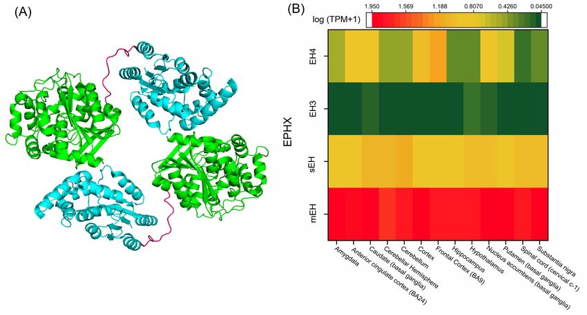
Figure 5. (A) Crystal structure of the sEH dimer (PDB accession code 1S8O) [150]. The sEH monomer is
Figure 5.of(A)
composed twoCrystal structure
globular regionsofrepresenting
the sEH dimer the(PDB accession
α/β tertiary code 1S8O)
structure, and [150]. The
a short sEH monomer
proline-rich linker
is composed
(pink) connectsofthe
two globular regions
C-terminal representing
of one monomer the N-terminal
to the α/β tertiaryregion
structure, and a short
of another. The proline-rich
catalytic site
linker
with (pink)hydrolase
epoxide connectsactivity
the C-terminal
is locatedofwithin
one monomer to theregion,
the C-terminal N-terminal
whereasregion of another.region
the N-terminal The
catalytic site with epoxide hydrolase activity is located within the C-terminal region,
possesses phosphatase activity. (B) EH distribution in the human brain based on mRNA expression. whereas the N-
terminal region possesses phosphatase activity. (B) EH distribution in the human brain
Expression values are shown as a median of TPM, calculated from a gene model with isoforms collapsed based on
tomRNA
a singleexpression.
gene with noExpression values are shown
other normalization [152]. as a median of TPM,
Abbreviations- calculated
EH: epoxide from a gene
hydrolase, EH3:model
epoxy
with isoforms
hydrolase 3, EH4:collapsed to a single4,gene
epoxy hydrolase EPHX:with no other
epoxide normalization
hydrolase [152]. microsomal
gene, mEH: Abbreviations- EH:
epoxide
epoxide hydrolase,
hydrolase, EH3:
PDB: Protein epoxy
Data hydrolase
Bank, 3, EH4:epoxide
sEH: soluble epoxy hydrolase
hydrolase, 4, TPM:
EPHX:transcripts
epoxide hydrolase gene,
per million.
mEH: microsomal epoxide hydrolase, PDB: Protein Data Bank, sEH: soluble epoxide hydrolase, TPM:
Both EH3 and
transcripts EH4 are predicted to be single-pass type II membrane proteins and possess
per million.
N-terminal membrane anchors based on their amino acid sequences, yet these properties need to be
Both EH3confirmed.
experimentally and EH4 areEH3predicted
and EH4 to have
be single-pass type IIinmembrane
45% homology proteins
their sequence andand possess
were N-
originally
terminal
named membrane
as α/β anchors
hydrolase domainbased on their protein
containing amino acid sequences,
9 (ABHD 9) andyetprotein
these properties
7 (ABHD 7), need
buttowere
be
experimentally
renamed confirmed.
after studies done byEH3 and EH4
Arand’s haveshowed
group 45% homology in their
their epoxide sequenceactivity
hydrolase and were originally
toward epoxy
named as α/β
octadecenoic hydrolase
acids (EpOMEs)domain containing
and EETs protein
[153,154]. 9 (ABHD
However, their9)specific
and protein
in vivo7 (ABHD
function7),
is but
still were
under
renamed after studies done by Arand’s group showed their epoxide hydrolase activity
investigation. EH3 is a 41 kDa (360 residues) protein encoded by the EPHX3 gene, and its microsomal toward epoxy
octadecenoic
properties haveacids
been(EpOMEs)
identifiedand
by aEETs [153,154].
related gene inHowever, their
insect cells specific
[154]. EH3in vivo function
expression is still
is generally
under investigation. EH3 is a 41 kDa (360 residues) protein encoded by the
low compared to mEH and sEH. Isolated mRNA from a representative set of mouse organs revealedEPHX3 gene, and its
microsomal properties have been identified by a related gene in insect cells [154]. EH3
the lowest expression in the liver and kidney, while the highest expression was observed in the skin, expression is
generallylung,
stomach, low compared
and tongueto mEH
[154].and sEH.
This Isolated mRNA
expression patternfrom a representative
suggests a potentialset of mouse
function oforgans
EH3 in
revealed the lowest expression in the liver and kidney, while the highest expression was observed in
barrier formation, which is supported by a recent study showing high turnover hydrolyzing activity
the skin, stomach, lung, and tongue [154]. This expression pattern suggests a potential function of
toward a skin-related epoxide involved in a key step of water permeability barrier formation in the
EH3 in barrier formation, which is supported by a recent study showing high turnover hydrolyzing
outer epidermis [155]. EH3 is also involved in leukotoxin metabolism to mediate acute respiratory
activity toward a skin-related epoxide involved in a key step of water permeability barrier formation
in the outer epidermis [155]. EH3 is also involved in leukotoxin metabolism to mediate acute
respiratory distress syndrome (ARDS) [156]. In addition, several studies identified the epigenetic
silencing of EH3 in different types of cancers. For instance, hypermethylation of EH3 is associated
with prostate cancer relapse [157], and has been observed in human colorectal carcinomas [158],Nutrients 2020, 12, 3523 13 of 40
distress syndrome (ARDS) [156]. In addition, several studies identified the epigenetic silencing of
EH3 in different types of cancers. For instance, hypermethylation of EH3 is associated with prostate
cancer relapse [157], and has been observed in human colorectal carcinomas [158], gastric cancers [159],
as well as malignant melanomas [160].
EH4 is a 42 kDa (362 residue) protein encoded by the EPHX4 gene located on chromosome 1p22.1.
As already mentioned, like mEH and sEH, both EH3 and EH4 possess an aspartate (Asp) nucleophile,
distinguishing them from other ABHD proteins that have either a serine (Ser) or Cys as nucleophile
residue in the catalytic triad, placing them in the EH family [153,154]. EH4 expression is much higher
in the brain compared to other tissues [141]. EH4 has been recognized as a target gene for the zinc
finger protein 217, an important oncogene that is amplified and overexpressed in a variety of human
tumors [161]. In addition, hypermethylated EH4 has been found in human colorectal cancer. Thus,
EH4 is associated with the pathogenesis of some cancers [162]. In general, very little is known about
the EH4 substrate spectrum and enzymatic function. However, the presence of a nucleophilic Asp in
the catalytic triad (that distinguishes EH3 and 4 from other ABHDs), and high homology with two
EHs in C. elegans [17], suggest the potential for EH4 to be an active EH with important physiological
activity in the brain [153].
5.2. Catalytic Function and Mechanism of EHs
The major function of EH is to hydrolyze xenobiotic epoxides and Ep-PUFAs (Figure 3A).
The catalytic activity of EHs can be described in three steps, with the contribution of four amino
acids, which are involved in transforming the epoxide to a diol [135,163]. In the first step, the epoxide
enters through the L-shaped hydrophobic tunnel with the nucleophilic aspartate in the center, and it is
stabilized there by van der Walls and hydrogen bonding with the hydrophobic pocket and two tyrosines,
respectively. The second step is the formation of an ester intermediate, in which the nucleophilic
aspartate and polarizing tyrosine are involved. In this step, the oxygen of the epoxide is polarized by
hydrogen bonds between the hydroxyl group of two tyrosine residues. At the same time, the carboxylate
of Asp (a nucleophilic amino acid), which is opposite of the tyrosine residues, gets orientated and
activated by other amino acids in the active site including histidine and an acidic residue (aspartate in
sEH and glutamate in mEH) adjacent to the nucleophilic aspartate. Then, the activated nucleophilic
aspartate attacks the electrophilic carbon of the epoxide resulting in a hydroxyl acylated-enzyme
intermediate. The third step is diol formation. When the intermediate is formed in the second step,
the His orients and then the charge relay system between the glutamate/aspartate and His activates the
His to facilitate hydrolysis of the acylated intermediate, forming the corresponding 1,2-diol product.
Table 1 shows the different residues in the catalytic triad of mEH, sEH, EH3, and EH4. Besides these
highly conserved regions, the amino acid sequences and the N-terminal region are largely different
among different EHs, probably due to their origin in evolutionary pathways.
Table 1. Different residues in the catalytic triad of mEH, sEH, EH3, and EH4 and their function.
Involved in Orientation and
EH Type Polarizing Residues Nucleophilic Residue
Activation of Nucleophilic Residues
Tyr299 His431
mEH Asp226
Tyr374 Glu404
Tyr383 His524
sEH Asp335
Tyr466 Asp496
Tyr220 His337
EH3 Asp173
Tyr281 Asp307
Tyr216 His336
Eh4 Asp169
Tyr281 Asp307
Abbreviations—Asp: Aspartic acid, EH: epoxide hydrolase, EH3: epoxy hydrolase 3, EH4: epoxy hydrolase 4, Glu:
Glutamic acid, His: histidine, mEH: microsomal epoxy hydrolase, sEH: soluble epoxy hydrolase, Tyr: Tyrosine.You can also read


