Hyaluronic Acid in the Third Millennium - Review - MDPI
←
→
Page content transcription
If your browser does not render page correctly, please read the page content below
polymers
Review
Hyaluronic Acid in the Third Millennium
Arianna Fallacara     ID
                           , Erika Baldini, Stefano Manfredini * and Silvia Vertuani
 Department of Life Sciences and Biotechnology, Master Course in Cosmetic Science and
 Technology (COSMAST), University of Ferrara, Via L. Borsari 46, 44121 Ferrara, Italy;
 arianna.fallacara@student.unife.it (A.F.); erika.baldini@student.unife.it (E.B.); vrs@unife.it (S.V.)
 * Correspondence: smanfred@unife.it; Tel.: +39-0532-455294; Fax: +39-0532-455378

 Received: 28 May 2018; Accepted: 20 June 2018; Published: 25 June 2018

 Abstract: Since its first isolation in 1934, hyaluronic acid (HA) has been studied across a variety
 of research areas. This unbranched glycosaminoglycan consisting of repeating disaccharide
 units of N-acetyl-D-glucosamine and D-glucuronic acid is almost ubiquitous in humans and
 in other vertebrates. HA is involved in many key processes, including cell signaling, wound
 reparation, tissue regeneration, morphogenesis, matrix organization and pathobiology, and has
 unique physico-chemical properties, such as biocompatibility, biodegradability, mucoadhesivity,
 hygroscopicity and viscoelasticity. For these reasons, exogenous HA has been investigated as a drug
 delivery system and treatment in cancer, ophthalmology, arthrology, pneumology, rhinology, urology,
 aesthetic medicine and cosmetics. To improve and customize its properties and applications, HA can
 be subjected to chemical modifications: conjugation and crosslinking. The present review gives an
 overview regarding HA, describing its history, physico-chemical, structural and hydrodynamic
 properties and biology (occurrence, biosynthesis (by hyaluronan synthases), degradation (by
 hyaluronidases and oxidative stress), roles, mechanisms of action and receptors). Furthermore,
 both conventional and recently emerging methods developed for the industrial production of HA
 and its chemical derivatization are presented. Finally, the medical, pharmaceutical and cosmetic
 applications of HA and its derivatives are reviewed, reporting examples of HA-based products that
 currently are on the market or are undergoing further investigations.

 Keywords: biological activity; crosslinking; drug delivery; cosmetic; food-supplement;
 functionalization; hyaluronan applications; hyaluronan derivatives; hyaluronan synthases;
 hyaluronic acid; hyaluronidases; physico-chemical properties

1. Introduction and Historical Background of HA
     Research on hyaluronic acid (HA) has expanded over more than one century.
     The first study that can be referred to regarding HA dates from 1880: the French scientist Portes
observed that mucin from vitreous body was different from other mucoids in cornea and cartilage
and called it “hyalomucine” [1]. Nevertheless, only in 1934, Meyer and Palmer isolated from bovine
vitreous humor a new polysaccharide containing an amino sugar and a uronic acid and named it HA,
from “hyaloid” (vitreous) and “uronic acid” [2]. During the 1930s and 1950s, HA was isolated also
from human umbilical cord, rooster comb and streptococci [3,4].
     The physico-chemical properties of HA were widely studied from the 1940s [5–9], and its chemical
structure was solved in 1954 by Meyer and Weissmann [10]. During the second half of the Twentieth
Century, the progressive understanding of HA’s biological roles [11–13] determined an increasing
interest in its production and development as a medical product for a number of clinical applications.
Hence, the extraction processes from animal tissues were progressively optimized, but still carried
several problems of purification from unwanted contaminants (i.e., microorganisms, proteins). The first

Polymers 2018, 10, 701; doi:10.3390/polym10070701                                   www.mdpi.com/journal/polymers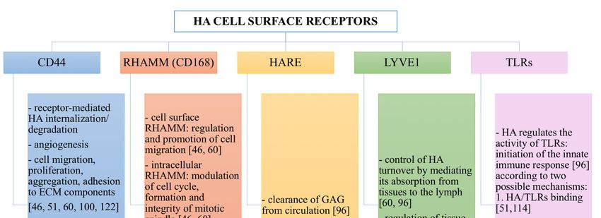
Polymers 2018, 10, 701                                                                               2 of 36

studies on HA production through bacterial fermentation and chemical synthesis were carried out
before the 1970s [1].
      The first pharmaceutical-grade HA was produced in 1979 by Balazs, who developed an efficient
method to extract and purify the polymer from rooster combs and human umbilical cords [14]. Balazs’
procedure set the basis for the industrial production of HA [14]. Since the early 1980s, HA has been
widely investigated as a raw material to develop intraocular lenses for implantation, becoming a
major product in ophthalmology for its safety and protective effect on corneal endothelium [15–22].
Additionally, HA was found to be beneficial also for the treatment of joint [23–27] and skin
diseases [28,29], for wound healing [30–33] and for soft tissue augmentation [34,35]. Since the late
1980s, HA has also been used to formulate drug delivery systems [36–41], and efforts continue still
to today to develop HA-based vehicles to improve therapeutic efficacy [42–45]. During the 1990s
and 2000s, particular attention was paid to identifying and characterizing the enzymes involved
in HA metabolism, as well as developing bacterial fermentation techniques to produce HA with
controlled size and polydispersity [1]. Nowadays, HA represents a key molecule in a variety of
medical, pharmaceutical, nutritional and cosmetic applications. For this reason, HA is still widely
studied to elucidate its biosynthetic pathways and molecular biology, to optimize its biotechnological
production, to synthesize derivatives with improved properties and to optimize and implement its
therapeutic and aesthetic uses [1,42–44,46–58].
      Considering the great interest in HA from different fields, the fast growing number of studies
and our interest in this topic, we decided to provide a comprehensive overview regarding HA and
its potentialities, giving a concise update on the latest progress. As an example, a search on the
most common public databases (i.e., Pubmed, Scopus, Isi Web of Science, ScienceDirect, Google
Scholar, ResearchGate and Patent Data Base Questel) with the keyword “hyaluron*”, gave a total of
161,863 hits: 142,575 papers and 19,288 patents. This huge amount of data are continuously growing.
Thus, with the aim to give a clearer picture about where researches and applications in the field are
going, the present work starts with an update of HA’s physico-chemical, structural and hydrodynamic
properties and proceeds with the discussion of HA biology: occurrence, biosynthesis (by hyaluronan
synthases), degradation (by hyaluronidases and oxidative stress), roles, mechanisms of action and
receptors. Furthermore, both conventional and recently-emerging methods developed for the industrial
production of HA and its chemical derivatization are described. Finally, the medical, pharmaceutical,
cosmetic and dietary applications of HA and its derivatives are reviewed, reporting examples of
HA-based products that currently are on the market or are undergoing further investigations.
      Literature search: we searched the Cochrane Controlled Trials Register (Central), Medline,
EMBase and Cinahl from inception to November 2006 using truncated variations of preparation
names including brand names combined with truncated variations of terms related to osteoarthritis,
all as text. No methodologic filter for controlled clinical trials was applied (the exact search strategy is
available from the authors). We entered relevant articles into the Science Citation Index to retrieve
reports that have cited these articles, manually searched conference proceedings and textbooks,
screened reference lists of all obtained articles and checked the proceedings of the U.S. Food and Drug
Administration advisory panel related to relevant approval applications. Finally, we asked authors
and content experts for relevant references and contacted manufacturers known to have conducted
trials on viscosupplementation.

2. Physico-Chemical, Structural and Hydrodynamic Properties of HA
     HA is a natural and unbranched polymer belonging to a group of heteropolysaccharides named
glycosaminoglycans (GAGs), which are diffused in the epithelial, connective and nervous tissues
of vertebrates [46,59,60]. All the GAGs (i.e., HA, chondroitin sulfate, dermatan sulfate, keratin
sulfate, heparin sulfate and heparin) are characterized by the same basic structure consisting of
disaccharide units of an amino sugar (N-acetyl-galactosamine or N-acetyl-glucosamine) and a uronic
sugar (glucuronic acid, iduronic acid or galactose). However, HA differs as it is not sulfated and it
Polymers 2018, 10, 701                                                                                                         3 of 36

is not synthesized by Golgi enzymes in association with proteins [46,59,60]. Indeed, HA is produced
at thePolymers
        inner2018,
                face10,ofx FOR
                           the PEER
                                plasmaREVIEW
                                           membrane without any covalent bond to a protein core. Additionally,             3 of 35

HA can reach a very high molecular weight (HMW, 108 Da), while the other GAGs are relatively
      and it is not synthesized by Golgi enzymes in association with proteins [46,59,60]. Indeed, HA is
smaller   in sizeat(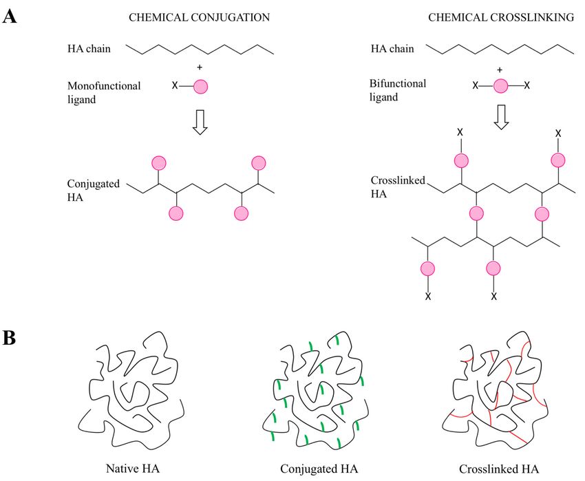
Polymers 2018, 10, x FOR PEER REVIEW 4 of 35
Polymers 2018, 10, 701 4 of 36
The establishment of this network depends on HA molecular weight (MW) and concentration;
for example, HMW native
The establishment of thisHA (>106depends
network Da) forms on HA an molecular
extended weight
network (MW) even andatconcentration;
a very low
concentration
for example, HMW of 1 native
µg/mL HA[64,69]. 6
(>10 Da) With formsincreasing MWnetwork
an extended and concentration,
even at a veryHA low networks
concentrationare
strengthened,
of 1 µg/mL [64,69].and consequently,
With increasing HA solutions display progressively
MW and concentration, HA networksincreased are viscosity
strengthened,and
viscoelasticity
and consequently, [70]. HASince hyaluronan
solutions is a progressively
display polyelectrolyteincreased
[71], its rheological
viscosity and properties in aqueous
viscoelasticity [70].
solutions are influenced
Since hyaluronan also by ionic
is a polyelectrolyte [71],strength, pH and
its rheological temperature
properties in aqueous[46,70,72]:
solutions as are
these factors
influenced
increase, HAstrength,
also by ionic viscositypH declines markedly, [46,70,72]:
and temperature suggestingasatheseweakening of the interactions
factors increase, HA viscosity among the
declines
polymer
markedly,chains [73]. In
suggesting particular, HA
a weakening of the is interactions
highly sensitiveamong to pH alterations:
the polymer in acidic
chains [73]. Inand alkaline
particular,
environments, a critical
HA is highly sensitive balance
to pH between
alterations: repulsive
in acidic and and attractive
alkaline forces occurs
environments, [74],
a critical and when
balance the
between
pH is lower
repulsive andthan four or forces
attractive higheroccurs
than 11, HAand
[74], is degraded
when theby pHhydrolysis
is lower than [75].four
In alkaline
or higher conditions,
than 11,
this
HA effect is morebypronounced,
is degraded due to
hydrolysis [75]. Inthe disruption
alkaline of H bonds,
conditions, which
this effect take part
is more in the structural
pronounced, due to
organization
the disruptionof of H HA chains
bonds, which [74,76,77].
take partTherefore, both organization
in the structural the structural properties
of HA and the
chains [74,76,77].
polyelectrolyte
Therefore, bothcharacter of HAproperties
the structural determineand its the
rheological profile [65,73,78,79].
polyelectrolyte character of HA HA determine
solutions are its
characterized by a [65,73,78,79].
rheological profile non-Newtonian, HAshear-thinning and viscoelastic
solutions are characterized by abehavior. The shear-thinning
non-Newtonian, shear-thinning (or
pseudoplastic)
and viscoelasticprofile
behavior. of HA is due to the breakdown
The shear-thinning of the inter-molecular
(or pseudoplastic) profile of HA is due hydrogen bonds and
to the breakdown
hydrophobic interactions
of the inter-molecular underbonds
hydrogen increasing shear rates:interactions
and hydrophobic HA chainsunder deform and align
increasing shearinrates:
the
streamlines of flow, and this results in a viscosity decrease [74,78] (Figure
HA chains deform and align in the streamlines of flow, and this results in a viscosity decrease [74,78] 2). Additionally, HA
solutions2).are
(Figure non-thixotropic:
Additionally, as the shear
HA solutions rate decreases and
are non-thixotropic: as theends, they
shear raterecover
decreases theirandoriginal
ends,
structure
they recoverandtheirviscosity
original proceeding
structure andthroughviscositythe same intermediate
proceeding through the same statesintermediate
of the breakdown states of
process [73] (Figure
the breakdown process2). Hence, the breakdown
[73] (Figure 2). Hence, the of the polymericofnetwork
breakdown is transient
the polymeric network andisreversible.
transient
This unique rheological
and reversible. This uniquebehavior
rheological is peculiar
behavior and extremely
is peculiar important,
and extremely as it determines
important, many
as it determines
physiological roles and pharmaceutical, medical, food and cosmetic applications
many physiological roles and pharmaceutical, medical, food and cosmetic applications of hyaluronan. of hyaluronan.
Figure 2. Shear-thinning
Figure 2. Shear-thinning andand non-thixotropic
non-thixotropic behavior
behavior ofof 0.5%
0.5% HA solution (2
HA solution MDa) analyzed
(2 MDa) analyzed using
using
the rotational rheometer AR2000 (TA instruments, New Castle, DE, USA), connected
the rotational rheometer AR2000 (TA instruments, New Castle, DE, USA), connected to the Rheology to the
Rheology Advantage
Advantage software
software (Version (Version
V7.20) V7.20) with
and equipped and anequipped
aluminumwith an aluminum
cone/plate geometrycone/plate
(diameter
geometry (diameter
◦ 40 mm, angle 2°, 64-µm truncation). The viscosity decreases
40 mm, angle 2 , 64-µm truncation). The viscosity decreases in response to gradual increases in response to
of the
gradual increases of the shear rate over time (upward ramp), and then, the viscosity increases
shear rate over time (upward ramp), and then, the viscosity increases in response to gradual decreases in
response to gradual
of the shear rate overdecreases of the shear
time (downward rate over
ramp). time (downward
The initial viscosity isramp). The through
recovered initial viscosity
the same is
recovered
intermediatethrough
states the same
of the intermediate
breakdown states
process: theofbreakdown
the breakdown
of the process:
polymeric thenetwork
breakdown of the
is transient
polymeric network is transient and reversible, and therefore,
and reversible, and therefore, the original structure of HA is recovered. the original structure of HA
is recovered.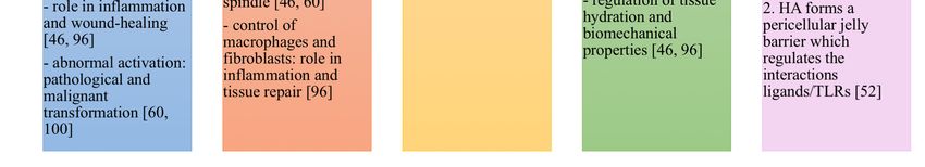
Polymers 2018, 10, 701                                                                              5 of 36

3. Biology of HA

3.1. HA Occurrence in Living Organism and Diffusion in the Human Body
     Hyaluronan is widely diffused in nature: it is present in humans, animals, such as, rabbits, bovines,
roosters, bacteria, such as Streptococcus equi, Streptococcus zooepidermicus, Streptococcus equisimilis,
Streptococcus pyogenes, Streptococcus uberis, Pasteurella multocida [49,80–82], algae, such as the green
algae Chlorella sp. infected by the Chlorovirus [49,83], yeasts, such as Cryptococcus neoformans [49],
and mollusks [84]. However, it is not found in fungi, plants and insects [85].
     In the human body, the total content of HA is about 15 g for a 70-kg adult [86]. HA is prevalently
distributed around cells, where it forms a pericellular coating, and in the extracellular matrix (ECM) of
connective tissues [61,82]. Approximately 50% of the total HA resides in the skin, both in the dermis and
the epidermis [82]. Synovial joint fluid and eye vitreous body, being mainly composed of ECM, contain
important amounts of hyaluronan: 3–4 mg/mL and 0.1 mg/mL (wet weight), respectively [61,82].
Moreover, HA is also abundant in the umbilical cord (4 mg/mL), where it represents the major
component of Wharton’s jelly together with chondroitin sulfate [87,88]. The turnover of HA is fast
(5 g/day) and is finely regulated through enzymatic synthesis and degradation [86].

3.2. HA Synthesis in the Human Body
     In the human body, HA is synthesized as a free linear polymer by three transmembrane
glycosyltransferase isoenzymes named hyaluronan synthases, HAS: HAS1, HAS2 and HAS3,
whose catalytic sites are located on the inner face of the plasma membrane. HA growing chains
are extruded onto the cell surface or into the ECM through the plasma membrane and HAS protein
complexes [89,90] (Figure 3). The three HAS isoforms share the 50–71% of their amino acid sequences
(55% HAS1/HAS2, 57% HAS1/HAS3, 71% HAS2/HAS3), and indeed, they are all characterized by
seven membrane-spanning regions and a central cytoplasmic domain [50,86,89]. However, HAS gene
sequences are located on different chromosomes (hCh19-HAS1, hCh8-HAS2 and hCh16-HAS3) [91,92],
and the expression and the activity of HAS isoforms are controlled by growth factors, cytokines and
other proteins such as kinases in different fashions, which appear cell and tissue specific [50,90,93,94].
Hence, the three HAS genes may respond differently to transcriptional signals: for example, in
human fibroblasts like synoviocytes, transforming growth factor ß upregulates HAS1 expression,
but downregulates HAS3 expression [95]. Moreover, HAS biochemical and synthetic properties are
different: HAS1 is the least active isoenzyme and produces HMW hyaluronan (from 2 × 10⁵ to
2 × 10⁶ Da). HAS2 is more active and synthesizes HA chains greater than 2 × 10⁶ Da. It represents the
main hyaluronan synthetic enzyme in normal adult cells, and its activity is finely regulated [96]. HAS2
also regulates the developmental and reparation processes of tissue growth, and it may be involved in
inflammation, cancer, pulmonary fibrosis and keloid scarring [55,86,97–99]. HAS3 is the most active
isoenzyme and produces HA molecules with MW lower than 3 × 10⁵ Da [60].
     Dysregulation and misregulation of HAS genes’ expression result in abnormal production of HA
and, therefore, in increased risk of pathological events, altered cell responses to injury and aberrant
biological processes such as malignant transformation and metastasis [47,48,50,100].
     Even if the exact regulation mechanisms and functions of each HAS isoenzyme have not been
fully elucidated yet [96], all the aforementioned studies suggest that HAS are critical mediators of
physiological and pathological processes, as they are involved in development, injury and disease.
Polymers 2018, 10, 701 6 of 36
Polymers 2018, 10, x FOR PEER REVIEW 6 of 35
Figure 3. Schematic diagram showing HA key steps from its synthesis to its degradation.
Figure 3. Schematic diagram showing HA key steps from its synthesis to its degradation.
3.3. HA Degradation in the Human Body
3.3. HA Degradation in the Human Body
HA degradation in the human body is accomplished by two different mechanisms: one is
HA degradation in the human body is accomplished by two different mechanisms: one is specific,
specific, mediated by enzymes (hyaluronidases (HYAL)), while the other is nonspecific, determined
mediated
by oxidativeby enzymes
damage(hyaluronidases
due to reactive(HYAL)),oxygen whilespeciesthe(ROS)
other (Figure
is nonspecific, determined
3). Together, HYAL by oxidative
and ROS
damage
locally degrade roughly 30% of the 15 g HA present in the human body. The remainingdegrade
due to reactive oxygen species (ROS) (Figure 3). Together, HYAL and ROS locally 70% is
roughly 30% of
catabolized the 15 g HA hyaluronan
systemically: present in theishuman mostlybody. The remaining
transported by the70% is catabolized
lymph to the lymph systemically:
nodes,
hyaluronan
where it isis internalized
mostly transported by the lymph
and catabolized by to thethe lymph nodes,
endothelial cellswhereof theit is internalized
lymphatic and
vessels.
catabolized
Additionally, by the a endothelial
small partcells of ofHA the islymphatic
carried vessels.
to the Additionally,
bloodstreama small and part of HA is
degraded bycarried
liver
toendothelial
the bloodstream and
cells [50]. degraded by liver endothelial cells [50].
HYAL
HYALhave haveaapivotal
pivotalregulatory
regulatoryfunction
functionininthe themetabolism
metabolismofofhyaluronan. hyaluronan. These These enzymes
enzymes
predominantly
predominantly degrade HA, even if they are able to catabolize also chondroitin sulfate and
degrade HA, even if they are able to catabolize also chondroitin sulfate and
chondroitin
chondroitin[101]. [101]. Randomly
Randomly cleaving
cleaving the β-N-acetyl-DD-glucosaminidic
theβ-N-acetyl- -glucosaminidic linkages linkages (β-1,4
(β-1,4 glycosidic
glycosidic
bonds)
bonds)ofofHA HAchains,
chains,HYAL
HYAL areareclassified
classifiedas endoglycosidases.
as endoglycosidases. In the
In human
the human genome,genome,six HYAL gene
six HYAL
sequences have been identified in two linked triplets: HYAL
gene sequences have been identified in two linked triplets: HYAL 1, HYAL 2, HYAL 3 genes, 1, HYAL 2, HYAL 3 genes, clustered
on chromosome
clustered 3p21.3; HYAL-4
on chromosome and PH20/SPAM1
3p21.3; HYAL-4 andgenes, similarly located
PH20/SPAM1 genes, onsimilarly
chromosome located7p31.3,
on
together
chromosome with HYAL-P1 pseudogene
7p31.3, together with [102].
HYAL-P1 HYALpseudogene
have a consistent[102]. amino
HYAL acid havesequence
a consistentin common:
amino
inacid
particular,
sequenceHYAL 1, HYAL
in common: 2, HYAL 3,HYAL
in particular, HYAL1,4HYAL and PH20/SPAM1
2, HYAL 3, HYAL share 4about 40% of their
and PH20/SPAM1
identity [101]. The expression of HYAL appears tissue specific. Nowadays,
share about 40% of their identity [101]. The expression of HYAL appears tissue specific. Nowadays, much is still unknown about
HYAL
much activity, functionsabout
is still unknown and posttranslational
HYAL activity, functions processing. and HYAL-1, HYAL 2 and
posttranslational PH20/SPAM1
processing. HYAL-1,are
the most characterized human HYAL. Both HYAL-1 and HYAL
HYAL 2 and PH20/SPAM1 are the most characterized human HYAL. Both HYAL-1 and HYAL 2 2 have an optimal activity at acidic pH
(≤ 4) [103,104]
have an optimal and activity
are highlyat expressed
acidic pH in human
(≤4) somatic
[103,104] andtissues
are highly[102].expressed
HYAL 1 was the firstsomatic
in human human
HYAL
tissuesto[102].
be isolated:
HYALit 1was waspurified
the firstfrom serumHYAL
human (60 ng/mL)to be[105] and, successively,
isolated: it was purified fromfrom urineserum
[106].
HYAL 1 was found to regulate cell cycle progression and apoptosis:
(60 ng/mL) [105] and, successively, from urine [106]. HYAL 1 was found to regulate cell cycle it is the main HYAL expressed in
cancers, and therefore,
progression it mayitregulate
and apoptosis: is the main tumor HYALgrowth and angiogenesis
expressed in cancers, [107]. HYAL 1 it
and therefore, works
may together
regulate
with
tumorHYAL 2 to degrade
growth HA, possibly
and angiogenesis according
[107]. HYALto1the following
works togethermechanism,
with HYAL which2isto still the object
degrade of
HA,
study. HYAL 2 is anchored on the external side of the cell surface:
possibly according to the following mechanism, which is still the object of study. HYAL 2 is here, it cleaves into oligosaccharides
(approximately
anchored on 25 thedisaccharide
external side units,of 2 ×the104 cell
Da) and the extracellular
surface: here, it cleaves HMW HA into (≥10 6 Da), which is
oligosaccharides
linked to its receptor
(approximately cluster of differentiation-44
25 disaccharide units, 2 × 10⁴ Da) (CD44).
and the These intermediate
extracellular HMW fragments
HA (≥10 are internalized,
6 Da), which is
transported
linked to its firstreceptor
to endosomes
clusterand of then to lysosomes, where
differentiation-44 (CD44). they are degraded
These intermediate into tetrasaccharide
fragments are
units (800 Da) transported
internalized, by HYAL-1 [51].first Differently
to endosomes fromand HYAL-1
then toand HYAL-2, where
lysosomes, PH20/SPAM1 they areshows degraded not only
into
tetrasaccharide units (800 Da) by HYAL-1 [51]. Differently from HYAL-1 and HYAL-2,
PH20/SPAM1 shows not only endoglycosidase activity both at acidic and neutral pH, but also a rolePolymers 2018, 10, 701 7 of 36
endoglycosidase activity both at acidic and neutral pH, but also a role in fertilization [108]. Hence,
PH20/SPAM1 is unique among HYAL, as it behaves as a multifunctional enzyme.
HMW hyaluronan can also be naturally degraded in the organism by ROS, including superoxide,
hydrogen peroxide, nitric oxide, peroxynitrite and hypohalous acids, which are massively produced
during inflammatory responses, tissue injury and tumorigenesis [60,109]. The depolymerization of
HA occurs through mechanisms of the reaction that are dependent on the ROS species, but always
involve the scission of the glycosidic linkages [86,110]. Studies have shown that oxidation-related
inflammatory processes, determining HA fragmentation, can increase the risk of injury in the airways
and determine loss of viscosity in synovial fluid, with consequent cartilage degeneration, joint stiffness
and pain [111–113]. ROS-induced degradation of HA might suggest why its antioxidant activity is one
of its possible roles in reducing inflammation; however, so far, this biological function of HA has only
been hypothesized, as it is not sufficiently supported by experimental data.
Due to these degradation mechanisms, which continuously occur in vivo, it has been estimated
that the half-life of HA in the skin is about 24 h, in the eye 24–36 h, in the cartilage 1–3 weeks and in
the vitreous humor 70 days [82].
3.4. Biological Roles of HA in Relation to Its MW
The equilibrium between HA synthesis and degradation plays a pivotal regulatory function in the
human body, as it determines not only the amount, but also the MW of hyaluronan. Molecular mass and
circumstances of synthesis/degradation are the key factors defining HA’s biological actions [50,51,100].
Indeed, high molecular weight (HMW) and low molecular weight (LMW) hyaluronan can even display
opposite effects [51,60], and when they are simultaneously present in a specific tissue, they can exert
actions different from the simple sum of those of their separate size-related effects [51].
Extracellular HMW HA (≥106 Da) is anti-angiogenic, as it is able to inhibit endothelial cell
growth [51,60,114]. Additionally, due its viscoelasticity, it acts as a lubricating agent in the synovial
joint fluid, thus protecting the articular cartilage [115]. HMW HA has also important and beneficial
roles in inflammation, tissue injury and repair, wound healing and immunosuppression: it binds
fibrinogen and controls the recruitment of inflammatory cells, the levels of inflammatory cytokines
and the migration of stem cells [60,93,114].
During some environmental and pathological conditions, such as asthma, pulmonary fibrosis
and hypertension, chronic obstructive pulmonary disease and rheumatoid arthritis, HMW HA is
cleaved into LMW HA (2 × 10⁴ –106 Da), which has been shown to possess pro-inflammatory and
pro-angiogenic activities [51,100]. Indeed, LMW hyaluronan is able to stimulate the production of
proinflammatory cytokines, chemokines and growth factors [51] and to promote ECM remodeling [50].
Moreover, LMW HA can also induce tumor progression, exerting its influence on cells [51,116] and
provoking ECM remodeling.
Both anti- and pro-inflammatory properties have been displayed by oHA and HA fragments
(≤2 × 10⁴ Da), depending on cell type and disease. Certain studies have shown that oHA are able
to reduce Toll-like receptors (TLRs)-mediated inflammation [117], inhibit HA-CD44 activation of
kinases [118] and retard the growth of tumors [119]. However, oHA have been also found to promote
inflammation in synovial fibroblasts [120], stimulate cell adhesion [121] and enhance angiogenesis
during wound healing [53].
Therefore, HA is clearly a key molecule involved in a number of physiological and pathological
processes. However, despite the intensive studies carried out so far, still little is known about HA’s
biological roles, the factors determining HA accumulation in transformed connective tissues and
the consequent cancer progression, and much less is known about their dependence on hyaluronan
molecular size and localization (intra- or extra-cellular). Further researches focusing on HA molecular
biology and mechanisms of action are necessary to clarify all these aspects and may facilitate the
development of novel HA-based therapies.Polymers 2018, 10, 701 8 of 36
3.5. Mechanisms of Action of HA
HA performs its biological actions (Section 3.4.) according to two basic mechanisms: it can act as
a passive structural molecule and as a signaling molecule. Both of these mechanisms of action have
been shown to be size-dependent [51,86].
The passive mechanism is related to the physico-chemical properties of HMW HA. Due
to its macromolecular size, marked hygroscopicity and viscoelasticity, HA is able to modulate
tissue hydration, osmotic balance and the physical properties of ECM, structuring a hydrated
and stable extracellular space where cells, collagen, elastin fibers and other ECM components are
firmly maintained [59,86,88].
HA also acts as a signaling molecule by interacting with its binding proteins. Depending on
HA MW, location and on cell-specific factors (receptor expression, signaling pathways and cell cycle),
the binding between HA and its proteins determines opposite actions: pro- and anti-inflammatory
activities, promotion and inhibition of cell migration, activation and blockage of cell division and
differentiation. All the factors that determine HA activities as a signaling molecule could be related:
MW may influence HA uptake by cells and may affect receptor affinity. Additionally, receptor
complexes may cluster differently depending on HA MW [51].
HA binding proteins can be distinguished into HA-binding proteoglycans (extracellular or matrix
hyaloadherins) and HA cell surface receptors (cellular hyaloadherins) [51]. HA has shown two different
molecular mechanisms of interaction with its hyaloadherins. First, HA can interact in an autocrine
fashion with its receptors on the same cell [60]. Second, it can behave as a paracrine substance,
which binds its receptors on neighboring cells and thus activates different intracellular signal cascades.
If HA has an HMW, a single chain can interact simultaneously with several cell surface receptors
and can bind multiple proteoglycans. These structures, in turn, can aggregate with additional ECM
proteins to form complexes, which can be linked to the cell surface through HA receptors [60,100].
Hence, HA acts as a scaffold that stabilizes the ECM structure not only through its passive structural
action, but also through its active interaction with several extracellular hyaloadherins, such as aggrecan
(prominent in the cartilage), neurocan and brevican (prominent in the central nervous system) and
versican (present in different soft tissues) [60]. For these reasons, pericellular HA is involved in the
preservation of the structure and functionality of connective tissues, as well as in their protection from
environmental factors [88].
HA Cell Surface Receptors
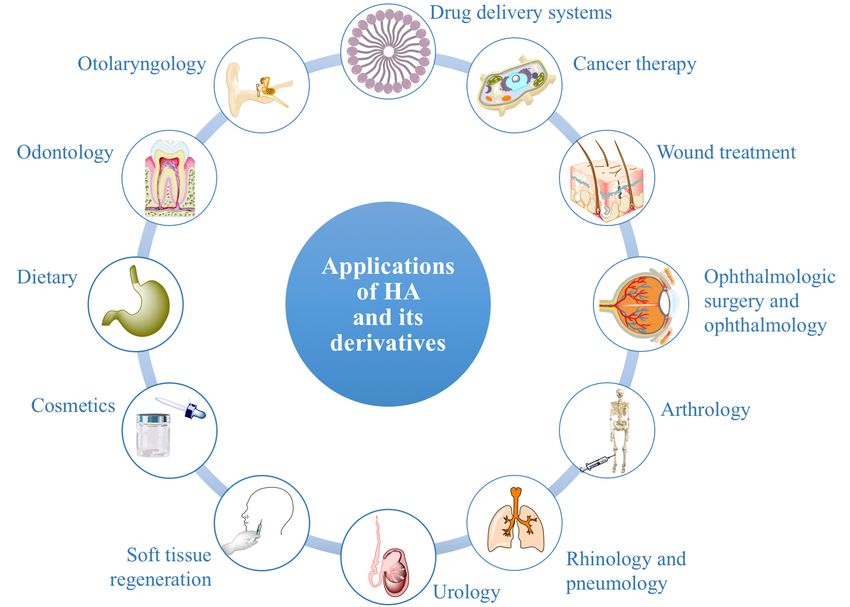
HA interactions with its cell surface receptors mediate three biological processes: signal
transduction, formation of pericellular coats and receptor-mediated internalization [60]. The present
subsection describes HA cell surface receptors and the biological actions that they control when linked
by HA (Figure 4).
The principal receptor for HA is CD44, which is a multifunctional transmembrane glycoprotein.
It is expressed in many isoforms diffused in almost all human cell types. CD44 can
interact not only with HA, but also with different growth factors, cytokines and extracellular
matrix proteins as fibronectin [96]. CD44 intracellular domain interacts with cytoskeleton;
hence, when its extracellular domain binds ECM hyaluronan, a link between the cytoskeletal
structures and the biopolymer is created [46]. HA-CD44 interaction is involved in a variety of
intracellular signaling pathways that control cell biological processes: receptor-mediated hyaluronan
internalization/degradation, angiogenesis, cell migration, proliferation, aggregation and adhesion
to ECM components [46,51,60,100,122]. Hence, CD44 plays a critical role in inflammation and
wound healing [46,96]. However, abnormal activation of HA-CD44 signaling cascades, as well as
overexpression and upregulation of CD44 (due to pro-inflammatory cytokines such as interleukin-1,
and growth factors such as epidermal growth factors) can result into development of pathological
lesions and malignant transformation [60,100]. Indeed, CD44 is overexpressed in many solid tumors,
such as pancreatic, breast and lung cancer [54].Polymers 2018, 10, 701 9 of 36
Polymers 2018, 10, x FOR PEER REVIEW 9 of 35
Figure
Figure 4. 4. SummaryofofHA
Summary HAcell
cellsurface
surfacereceptors
receptors and
and of the actions
actions that
thatthey
theycontrol
controlwhen
whenlinked
linked
by
by HA.HA.
The receptor for HA-mediated cell motility (RHAMM) is also known as CD168, and it was the
The receptor for HA-mediated cell motility (RHAMM) is also known as CD168, and it was the
first isolated cellular hyaloadherin. It exists in several isoforms, which can be present not only in the
first isolated cellular hyaloadherin. It exists in several isoforms, which can be present not only in the
cell membrane, but also in the cytoplasm and in the nucleus [96]. When liked by HA, cell surface
cellRHAMM
membrane, but also
mediates andinpromotes
the cytoplasm and in the
cell migration, nucleus
while [96]. When
intracellular RHAMMliked modulates
by HA, cell thesurface
cell
RHAMM mediates and promotes cell migration, while intracellular RHAMM
cycle, the formation and the integrity of mitotic spindle [46,60]. Interactions of HA with RHAMM modulates the cell cycle,
theplay
formation
important and roles
the integrity of mitoticand
in inflammation spindle
tissue[46,60].
repair,Interactions
by triggering of HA with RHAMM
a variety of signaling play
important roles in inflammation and tissue repair, by triggering
pathways and thus controlling cells such as macrophages and fibroblasts [96]. a variety of signaling pathways and
thus controlling cells such as macrophages and fibroblasts [96].
Hyaluronan receptor for endocytosis (HARE) was initially isolated from endothelial cells in the
Hyaluronan
liver, lymph nodes receptor for endocytosis
and spleen (HARE)
and successively was also
found initially isolated from
in endothelial cellsendothelial
of eye, brain, cells in the
kidney
liver,
andlymph
heart nodes
[96]. It and spleen
is able andnot
to bind successively
only HA, but foundalsoalso in endothelial
other GAGs, with cells of eye, brain,
the exception kidney
of keratin
andsulfate,
heart heparin
[96]. It sulfate
is able and heparin.
to bind It is involved
not only HA, butinalso the other
clearanceGAGs,of GAGs
withfrom circulationof
the exception [96].
keratin
sulfate, Furthermore,
heparin sulfate lymphatic
and heparin. vessel endothelial
It is involved in hyaluronan
the clearance receptor
of GAGs 1 (LYVE1, a HA-binding
from circulation [96].
protein expressed
Furthermore, in lymph
lymphatic vascular
vessel endothelium
endothelial hyaluronanand macrophages)
receptor 1 (LYVE1, controls HA turnover
a HA-binding by
protein
mediating its adsorption from tissues to the lymph [60,96]. In
expressed in lymph vascular endothelium and macrophages) controls HA turnover by mediating its this way, LYVE1 is involved in the
regulation
adsorption fromof tissues
tissue hydration
to the lymph and[60,96].
their biomechanical
In this way, LYVE1 properties [46,96].
is involved in Additionally,
the regulationLYVE1 of tissue
forms complexes with growth factors, prostaglandins and
hydration and their biomechanical properties [46,96]. Additionally, LYVE1 forms complexes other tissue mediators, which are with
implicated in the regulation of lymphangiogenesis and intercellular
growth factors, prostaglandins and other tissue mediators, which are implicated in the regulation of adhesion [46,96].
Finally, HA is involved in the regulation of the activity of TLRs that, recognizing bacterial
lymphangiogenesis and intercellular adhesion [46,96].
lipopolysaccharides and lipopeptides, are able to initiate the innate immune response [96]. Two
Finally, HA is involved in the regulation of the activity of TLRs that, recognizing bacterial
possible mechanisms have been proposed to explain how HA can influence TLRs. According to the
lipopolysaccharides and lipopeptides, are able to initiate the innate immune response [96].
first theory, LMW hyaluronan acts as an agonist for TLR2 and TLR4, thus provoking an
Two possible mechanisms have been proposed to explain how HA can influence TLRs. According to the
inflammatory reaction [51,114]. On the contrary, according to the second theory, hyaluronan does
firstnot
theory, LMW hyaluronan acts as an agonist for TLR2 and TLR4, thus provoking an inflammatory
bind to TLRs, but it is able to regulate TLRs interactions with their ligands through the
reaction
pericellular jellyOn
[51,114]. the contrary,
barrier according
that it forms to the second
[52]. Indeed, theory, hyaluronan
in physiological conditions,does HMW notHA bind to TLRs,
creates a
butdense
it is able to regulate TLRs interactions with their ligands through the
and viscous protective coat around the cells, thus covering surface receptors such as TLRs pericellular jelly barrier that
it forms [52]. Indeed,
and limiting their in physiological
interactions withconditions,
ligands. During HMWinflammation,
HA creates a dense and viscous
an imbalance protective
between HA
coatsynthesis
around the cells, thus covering surface receptors such as TLRs and limiting
and degradation occurs, and this alters the thickness and the viscosity of HA pericellular their interactions with
ligands.
barrierDuring inflammation,
[52]. More precisely, an HAimbalance
is rapidly between
degradedHA due synthesis and degradation
to pH reduction, ROS increaseoccurs,andandthethis
alters the thickness
possible presence and the viscosity
of pathogens of HAHYAL
producing pericellular
[46,109]. barrier
Hence, [52].
HAMore precisely, reducing
MW decreases, HA is rapidly
the
degraded
polymerdue water to binding
pH reduction,
ability andROS theincrease
thicknessand andthethepossible
viscositypresence of pathogens
of its pericellular producing
shield [52]. This
HYAL [46,109]. Hence, HA MW decreases, reducing the polymer water binding ability andthethe
results in an increased accessibility of the cell receptors to their ligands, in the initiation of
thickness and the viscosity of its pericellular shield [52]. This results in an increased accessibility of thePolymers 2018, 10, 701 10 of 36
cell receptors to their ligands, in the initiation of the innate immune response and in the enhancement
of the inflammatory reaction [52]. For this reason, HA can also be involved in the pathogenesis of
diseases sustained by immunological processes [46].
4. Industrial Production of HA
The plethora of activities of a food-contained molecule has raised important interest for public
health: the global market of HA was USD 7.2 billion in 2016, and it is expected to reach a
value of USD 15.4 billion by 2025 [123]. Indeed, hyaluronan is gaining an exponentially growing
interest for many pharmaceutical, medical, food and cosmetic applications, due to its important
activities—anti-inflammatory, wound healing and immunosuppressive—and its numerous and
incomparable biological and physico-chemical properties, such as biocompatibility, biodegradability,
non-immunogenicity, mucoadhesivity, hygroscopicity, viscoelasticity and lubricity. Hence, there is
a strong interest in optimizing HA production processes to obtain products that fulfill high quality
standards and are characterized by great yield and accessible costs. Both the source and the purification
process co-occur to determine the characteristics of the produced HA in terms of purity, MW, yield
and cost [124,125]. Therefore, producing high quality HA with high yield and less costly methods
represents one of the biggest challenges in the field of hyaluronan applied research.
The first production process applied at an industrial scale consisted of HA extraction from
animal sources, such as bovine vitreous and rooster combs [46,49,124]. Despite the extraction
protocols being improved over the years, this methodology was always hampered by several
technical limitations, which led to the production of highly polydispersed HA (MW ≥ 106 Da) with
a low yield [1,46]. This was due to the polymer intrinsic polydispersity, its low concentration in
tissues and its uncontrolled degradation caused by the endogenous HYAL and the harsh isolation
conditions [46,49]. Additional disadvantages of animal-derived HA were represented by the risk
of biological contamination—the presence of proteins, nucleic acids and viruses—and by the high
purification costs [46,49,124]. Therefore, alternative methodologies for the industrial production of HA
have been developed.
Currently, commercial hyaluronan is principally produced with biotechnology (microbial
fermentation). Microorganism-derived HA is biocompatible with the human body because the HA
structure is highly conserved among the different species [1,49]. Streptococci strains A and C were the
first bacteria used for HA production, and nowadays, many commercial products are derived from
Streptococcus equi (such as Restylane® by Q-med AB and Juvederm® by Allergan). Optimum bacterial
culture conditions to obtain HMW HA (3.5–3.9 × 10⁶ Da) have been determined at 37 ◦ C, pH 7, in the
presence of lactose or sucrose [125,126]. Hyaluronan yield has been optimized up to 6–7 g/L, which is
the upper technical limit of the process due to mass transfer limitation caused by the high viscosity of
the fermentation broth [1]. As streptococci genera include several human pathogens, an accurate and
expensive purification of the produced HA is necessary [46,49]. Hence, other microorganisms have
been and are currently investigated to synthesize HA. An ideal microorganism for HA biosynthesis
should be generally regarded as safe (GRAS), not secrete any toxins and be able to produce at
least 106 Da HA, as the polymer quality and market value increase with its purity and MW,
which affect rheological and biological properties and define suitable applications [49,127]. Since the
natural hyaluronan-producing organisms are mostly pathogenic, metabolic engineering currently
represents an interesting opportunity to obtain HA from non-pathogenic, GRAS microorganisms.
Endotoxin-free HA has already been synthesized by recombinant hosts including Lactococcus lactis [128],
Bacillus subtilis [129], Escherichia coli [130] and Corynebacterium glutamicum [131]. However, up to now,
there has been no heterologous bacterial host producing as much HA as the natural ones. Hence,
there is an increasing effort to find an ideal bioreactor for HA production: in addition to bacteria,
also eukaryotic organisms such as yeasts, like Saccharomyces cerevisiae [132] and Pichia pastoris [133],
and plant cell cultures, like transformed tobacco-cultured cells [134], have been explored in the last
few years.Polymers 2018, 10, 701 11 of 36
Finally, to obtain HA of defined MW and narrow polydispersity, other approaches have been used.
For example, to produce monodisperse oHA, chemoenzymatic synthesis has been performed [135].
This technique has successfully led to a product commercialized under the name Select HA™ (Hyalose
LLC), characterized by a low polydispersity index value. Moreover, other studies have shown the
possibility to prepare HA monodisperse fragments by controlling the degradation of HMW hyaluronan
using different methods, including acidic, alkaline, ultrasonic and thermal degradation [110].
5. Synthetic Modifications of HA
HA has several interesting medical, pharmaceutical, food and cosmetic uses in its
naturally-occurring linear form. However, chemical modifications of the HA structure represent
a strategy to extend the possible applications of the polymer, obtaining better performing products
that can satisfy specific demands and can be characterized by a longer half-life. During the design of
novel synthetic derivatives, particular attention is paid to avoiding the loss of native HA properties
such as biocompatibility, biodegradability and mucoadhesivity [46].
5.1. General Introduction of the Chemical Approaches to Modify HA
HA chemical modifications mainly involve two functional sites of the biopolymer: the hydroxyl
(probably the primary alcoholic function of the N-acetyl D glucosamine) and the carboxyl groups.
Furthermore, synthetic modifications can be performed after the deacetylation of HA N-acetyl groups,
a strategy that allows one to recover amino functionalities [136]. All these functional groups of HA
can be modified through two techniques, which are based on the same chemical reactions, but lead
to different products: conjugation and crosslinking (Figure 5). Conjugation consists of grafting a
monofunctional molecule onto one HA chain by a single covalent bond, while crosslinking employs
polyfunctional compounds to link together different chains of native or conjugated HA by two
or more covalent bonds [136]. Crosslinked hyaluronan can be prepared from native HA (direct
crosslinking) [56,58,137] or from its conjugates (see below). Conjugation and crosslinking are generally
performed for different purposes. Conjugation permits crosslinking with a variety of molecules;
to obtain carrier systems with improved drug delivery properties with respect to native HA; to develop
pro-drugs by covalently linking active molecules to HA [136]. On the other hand, crosslinking is
normally intended to improve the mechanical, rheological and swelling properties of HA and to
reduce its degradation rate, in order to develop derivatives with a longer residence time in the site of
application and greater release properties [58,138,139]. A recent trend is to conjugate and crosslink
HA chains using bioactive molecules in order to develop derivatives with improved and customized
activities [58] for a variety of applications in medicine, aesthetics and bioengineering, including cell
and molecule delivery, tissue engineering and the development of scaffolds [46,56,58,140–144].
A number of synthetic approaches have been developed to produce conjugated or crosslinked
hyaluronan [136].
Generally, HA is chemically modified in the liquid phase. Since it is hydrophilic, several
reactions are performed in aqueous media also from its conjugates [145–147]: however, they are
pH dependent and, therefore, require acidic or alkaline conditions, which if too strong, can determine
HA degradation [75,145]. Other synthetic methods, involving the use of reagents sensitive to
hydrolysis, are performed in anhydrous organic solvents such as dimethylsulfoxide (DMSO) [146] or
dimethylformamide (DMF) [148]. These approaches necessarily introduce a preparation step to convert
native HA into tetrabutylammonium (TBA) salt, soluble in organic ambient [148]: this increases the
reaction time and cost, as well as the chances of HA chain fragmentation due to physico-chemical
treatments. Additionally, when HA modifications take place in organic solvents, longer final
purification processes are necessary [136]. The basic and classic chemistry that underlies the
possible modifications of HA functional groups in the liquid phase is overviewed in the following
Sections 5.2–5.4.Polymers 2018, 10, 701 12 of 36
Polymers 2018, 10, x FOR PEER REVIEW 12 of 35
Figure 5. Chemical modifications of HA: conjugation and crosslinking (A). HA forms used for
Figure 5. Chemical modifications of HA: conjugation and crosslinking (A). HA forms used for
pharmaceutical, medical, food and cosmetic applications: native, conjugated and crosslinked (B).
pharmaceutical, medical, food and cosmetic applications: native, conjugated and crosslinked (B).
Since HA derivatives of high quality and purity are necessary to develop injectable products,
Generally,
implantableHAscaffolds,
is chemically modified
drug delivery systems inandthe3Dliquid phase.
hydrogel Since
matrices it is hydrophilic,
encapsulating living cells, several
reactionstechniques
are performedfor efficient, low-cost
in aqueous mediaand alsosafe from
modification of HA [145–147]:
its conjugates are continuously
however, beingthey are
explored
pH dependent [46,147,149].
and, therefore, Hence,
requirein the last or
acidic fewalkaline
years, several efforts which
conditions, have been made
if too to introduce
strong, can determine
one-pot reactions that preferably proceed in an aqueous environment, under mild and, possibly,
HA degradation [75,145]. Other synthetic methods, involving the use of reagents sensitive to
environmentally-friendly conditions, without the use of toxic catalysts and reagents [147,149].
hydrolysis, are performed
Additionally, in anhydrous
alternative approaches organic solvents
to efficiently modify such
HA as dimethylsulfoxide
have (DMSO) [146] or
been introduced: solvent-free
dimethylformamide
methods, i.e.,(DMF) [148].
reactions These
in solid approaches
phase [57], “clicknecessarily
chemistry”introduce
syntheses,awhich
preparation
are simplestepand
to convert
native HA chemoselective, proceeding with fast
into tetrabutylammonium (TBA)kinetics
salt,insoluble
an aqueous environment,
in organic ambientunder mild this
[148]: conditions,
increases the
reaction leading
time and to quantitative
cost, as well yields,
as thewithout
chances appreciable amounts
of HA chain of side products,
fragmentation duei.e.,
tothe thiol-ene
physico-chemical
reaction [150], the Dies–Alder cycloaddition [151] and the azide-alkyne cycloaddition [152]; in situ
treatments. Additionally, when HA modifications take place in organic solvents, longer final
crosslinking of functionalized HA through air oxidation [153]; photo-crosslinking of functionalized
purification
HA inprocesses
the presence are necessary [136].
of photosensitizers The basic and classic chemistry that underlies the
[154,155].
possible modifications of HA functional groups in the liquid phase is overviewed in the following
Sections 5.2. Modification of HA Hydroxyl Groups
5.2–5.4.
Since HA By derivatives
modifying HA’s of hydroxyl
high quality
groups, andthepurity
carboxylare necessary
groups to developthus
remain unchanged, injectable
preservingproducts,
HA’s natural recognition by its degradative enzymes [136]. Over
implantable scaffolds, drug delivery systems and 3D hydrogel matrices encapsulating the years, different derivatives of living
HA (ethers, hemiacetals, esters and carbamates) have been produced
cells, techniques for efficient, low-cost and safe modification of HA are continuously being through reactions that occur
between the polymeric hydroxyl groups and mono- or bi-functional agents.
explored [46,147,149]. Hence, in the last few years, several efforts have been made to introduce
Epoxides and bisepoxides like butanediol-diglycidyl ether (BDDE) [137], ethylene
one-pot glycol-diglycidyl
reactions that preferably proceed in polyglycidyl
ether, polyglycerol an aqueous ether environment, under mild and,
[156], epichlorohydrin andpossibly,
environmentally-friendly
1,2,7,8 diepoxyoctane conditions,
[157] have been without
widely used the touse of toxic
synthesize catalysts
ether derivativesand reagents in
of hyaluronan [147,149].
alkaline
Additionally, aqueous solution.
alternative Currently,
approaches HA-BDDE ether
to efficiently modify represents
HA have one been
of the introduced:
most marketedsolvent-free
HA
methods, i.e., reactions in solid phase [57], “click chemistry” syntheses, which are simple and
chemoselective, proceeding with fast kinetics in an aqueous environment, under mild conditions,
leading to quantitative yields, without appreciable amounts of side products, i.e., the thiol-ene
reaction [150], the Dies–Alder cycloaddition [151] and the azide-alkyne cycloaddition [152]; in situ
crosslinking of functionalized HA through air oxidation [153]; photo-crosslinking of functionalized
HA in the presence of photosensitizers [154,155].Polymers 2018, 10, 701 13 of 36
5.2. Modification of HA Hydroxyl Groups
By modifying HA’s hydroxyl groups, the carboxyl groups remain unchanged, thus preserving
HA’s natural recognition by its degradative enzymes [136]. Over the years, different derivatives of HA
(ethers, hemiacetals, esters and carbamates) have been produced through reactions that occur between
the polymeric hydroxyl groups and mono- or bi-functional agents.
Epoxides and bisepoxides like butanediol-diglycidyl ether (BDDE) [137], ethylene glycol-
diglycidyl ether, polyglycerol polyglycidyl ether [156], epichlorohydrin and 1,2,7,8 diepoxyoctane [157]
have been widely used to synthesize ether derivatives of hyaluronan in alkaline aqueous solution.
Currently, HA-BDDE ether represents one of the most marketed HA derivative: it can be obtained
through simple synthetic procedures in an aqueous environment, and it is degraded into non-cytotoxic
fragments [136]. Other efficient methods to form ether derivatives of HA involve the use of divinyl
sulfone (DVS) [158] or ethylene sulfide [159] in basic water.
Many studies showed that hemiacetal bonds can be formed between the hydroxyl groups of HA
and glutaraldehyde in an acetone-water medium. Since glutaraldehyde is toxic, particular handling is
required during the reaction and purification of the final product [160,161].
The hydroxyl groups of HA can be also esterified by reacting with octenyl succinic anhydride [162]
or methacrylic anhydride [163] under alkaline conditions. Alternatively, HA can be converted into a
DMSO-soluble salt, which can undergo esterification with activated compounds such as acyl-chloride
carboxylates [164].
Finally, the activation of HA hydroxyl groups to cyanate esters, and the subsequent reaction
in basic water with amines, allows one to synthesize carbamate derivatives with high degrees of
substitution, in a reaction time of only 1 h [165].
5.3. Modification of HA Carboxyl Groups
Strategies for the derivatization of HA also involve esterification and amidation, which can be
performed after the activation of the polymeric carboxyl groups using different reagents. By modifying
HA’s carboxyl groups, derivatives more stable to HYAL degradation can be synthesized: hence, if a
drug is conjugated on the carboxyl groups of HA, a slow drug release may occur [136].
Esterification can be performed by alkylation of HA carboxyl groups using alkyl halides [166]
or tosylate activation [167]. Moreover, HA esters can be synthesized using diazomethane as the
activator of the carboxyl groups [168]. All these reactions proceed in DMSO from the TBA salt
of HA. Alternatively, HA can undergo esterification also in water using epoxides such as glycidyl
methacrylate and excess trimethylamine as a catalyst [169]. The conversion of HA carboxyl groups
into less hydrophilic esters represents a strategy to decrease the water solubility of HA, with the
aim to reduce its susceptibility to HYAL degradation and enhance its in situ permanence time [46].
A well-known biopolymer synthesized to this end is HA benzyl ester (HYAFF 11), the properties of
which are finely regulated by its degree of functionalization [36,170].
Amidation represents a further approach to modify HA: over the years, several synthetic
procedures have been developed. However, some of these present important drawbacks: for example,
Ugi condensation (useful to crosslink HA chains through diamide linkages) requires a strongly acidic
pH (3), the use of formaldehyde, which is carcinogenic, and cyclohexyl isocyanide, which determines
a pending undesired cyclohexyl group in the final product [145,160]. HA amidation with
1,10 -carbonyldiimidazole [171] or 2-chloro-1-methylpyridinium iodide [148] as activating agents
are performed in DMSO and DMF, respectively: hence, HA conversion into TBA salt and longer
purification steps are needed. On the contrary, other methods are based on reaction conditions that meet
the modification requirements for HA. Particularly efficient is the activation of HA carboxyl groups
by carbodiimide (i.e., N-(3-dimethylaminopropyl)-N 0 -ethylcarbodiimide hydrochloride) (EDC) and
co-activators such are N-hydroxysuccinimide (NHS) or 1-hydroxybenzotriazole in water: proceeding
under mild conditions, this reaction does not lead to HA chains’ cleavage, and it is suitable
also for the derivatization with biopolymers easily susceptible to denaturation, such as proteinYou can also read