Current Trends in Diagnostics of Viral Infections of Unknown Etiology - MDPI
←
→
Page content transcription
If your browser does not render page correctly, please read the page content below
viruses
Review
Current Trends in Diagnostics of Viral Infections of
Unknown Etiology
Daniel Kiselev 1,2 , Alina Matsvay 1,3 , Ivan Abramov 1 , Vladimir Dedkov 4,5 ,
German Shipulin 1 and Kamil Khafizov 1,3, *
 1    FSBI “Center of Strategic Planning” of the Ministry of Health, 119435 Moscow, Russia;
      neurolynx13@gmail.com (D.K.); arity767@gmail.com (A.M.); abriv@bk.ru (I.A.);
      shipgerman@gmail.com (G.S.)
 2    I.M. Sechenov First Moscow State Medical University, 119146 Moscow, Russia
 3    Moscow Institute of Physics and Technology, National Research University, 117303 Moscow, Russia
 4    Pasteur Institute, Federal Service on Consumers’ Rights Protection and Human Well-Being Surveillance,
      197101 Saint-Petersburg, Russia; vgdedkov@gmail.com
 5    Martsinovsky Institute of Medical Parasitology, Tropical and Vector Borne Diseases, Sechenov First Moscow
      State Medical University, 119146 Moscow, Russia
 *    Correspondence: kkhafizov@gmail.com; Tel.: +7-917-597-2085

 Received: 16 December 2019; Accepted: 12 February 2020; Published: 14 February 2020

 Abstract: Viruses are evolving at an alarming rate, spreading and inconspicuously adapting to
 cutting-edge therapies. Therefore, the search for rapid, informative and reliable diagnostic methods
 is becoming urgent as ever. Conventional clinical tests (PCR, serology, etc.) are being continually
 optimized, yet provide very limited data. Could high throughput sequencing (HTS) become the
 future gold standard in molecular diagnostics of viral infections? Compared to conventional clinical
 tests, HTS is universal and more precise at profiling pathogens. Nevertheless, it has not yet been
 widely accepted as a diagnostic tool, owing primarily to its high cost and the complexity of sample
 preparation and data analysis. Those obstacles must be tackled to integrate HTS into daily clinical
 practice. For this, three objectives are to be achieved: (1) designing and assessing universal protocols
 for library preparation, (2) assembling purpose-specific pipelines, and (3) building computational
 infrastructure to suit the needs and financial abilities of modern healthcare centers. Data harvested
 with HTS could not only augment diagnostics and help to choose the correct therapy, but also facilitate
 research in epidemiology, genetics and virology. This information, in turn, could significantly aid
 clinicians in battling viral infections.

 Keywords: viruses; viral infections; diagnostics; sequencing; PCR; NGS; HTS; single-molecule
 sequencing; bioinformatics

1. Introduction
      As some statistical models suggest, there are over 320,000 mammalian viruses in existence [1],
a little over 200 of which are known to infect humans [2], with the number increasing steadily over
decades [3]. The recent years have been marked by a rising frequency of emerging viral infections,
e.g., SARS [4], Zika [5], or a more recent Coronavirus 2019-nCoV outbreak [6]. Bearing that in mind, the
range of available diagnostic tools seems disproportionate to the growing number of diseases, because
they detect only known pathogens, which constitute ~0.07% of viral entities. Metaphorically, we are
attempting to look at a vast sea of threats through the eye of a needle.
      As of this day, a multitude of clinical tests are available for detection of viruses, e.g., FISH, ELISA
(enzyme-linked immunosorbent assay), and PCR- and their numerous modifications. The latter has

Viruses 2020, 12, 211; doi:10.3390/v12020211                                       www.mdpi.com/journal/viruses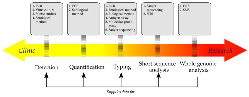
Viruses 2020, 12, 211                                                                               2 of 28

become a gold standard, for example, in cases of gastrointestinal [7] and respiratory infections [8].
Indeed, the presence of viral nucleic acids in biological media, particularly in blood plasma, while
not definitive, is highly suspect of an ongoing infection, because it strongly implies viral replication.
Furthermore, PCR not only enables us to detect pathogens, but also quantify them with great precision,
based on the number of copies of a pathogen’s DNA/RNA templates in a sample.
     As effective as they are, PCR tests are sometimes prone to major pitfalls, chiefly owing to high
mutability of a viral genome, which significantly perplexes primer design and requires researchers to
update primer sequences continually. With the entirety of human-infecting viruses, most of which are
yet to be revealed, it becomes increasingly difficult to maintain up-to-date primer panels while also
ensuring the correct reaction conditions and high specificity for differentiating closely related viral
species. This situation is aggravated by the low rates of correct pathogen identification [9].
     Immunoassays have been extensively used in clinical laboratories for over 60 years now. Fast
and relatively reliable screening tests for infectious diseases have been successfully introduced into
healthcare practice, e.g., for HIV, HBV, HEV, HAV, HCV, and Rubella virus. Although ELISA does
not quantify pathogens with great precision, it effectively detects pathogens based on their antigen
structure. Useful as they are, immunoassays are also subject to biases and fundamental pitfalls,
although they are often disregarded for the sake of low cost and convenience.
     Problems with conventional diagnostic methods have been stated in several studies. For example,
Arnold et al. [10] found that only 62% of viral respiratory infections among children could be confidently
attributed to known pathogens, mostly to human metapneumovirus (63%) and adenoviruses (45%),
while almost a third of all cases remained undiagnosed. In another study by Vu et al. [11], it was
demonstrated that in 42.7% of cases of viral gastroenteritis the pathogen could not be identified with
conventional methods. Kennedy et al. [12] outline that up to 40% of viral encephalitis infections remain
undiagnosed with modern clinical tests.
     High throughput sequencing (HTS) represents a range of technologies based on sequencing by
synthesis, which allow retrieving of multiple nucleotide sequences at once with high reliability for
further pathogen identification. Thus, HTS may tackle the aforementioned diagnostic issues, adding
an extra layer of data for identification and further insight into a pathogen.
     In this paper, we assess modern clinical tests for viral infections and explore the ways that HTS
provides detailed data about a pathogen’s genome, allowing for a deeper analysis of its features, such
as drug resistance and phylogeny. We also hope to demonstrate how modern clinical laboratories
would benefit from adopting HTS as a routine diagnostic technique.

2. Traditional Methods of Diagnosing Infections
      Before molecular and serological diagnostics became widely accessible, detection of pathogens
primarily relied on conventional microbiological methods, e.g., cultures on growth media, in cells, and
in laboratory animals with further microscopy, identification of antigenic and pathogenic characteristics
and the metabolic profile.
      ELISA has become one of the most widely used tests, owing to high sensitivity and specificity
(both usually reported at >90% [13–15]), low price per sample, and the simplicity of sample processing
and result interpretation. Modern commercial kits are designed to cover a large variety of antigens,
bacterial and viral, even at femto- and attomole concentrations. All these factors contribute to the
popularity of ELISA, turning it into a universal tool for disease screening and prognosis. At the
same time, strong limitations restrict its use. Because the test basically relies on antigen detection,
cross-reactivity between heterologous antigens can lead to false positive results, like with influenza
virus-specific CD8+ T cell and hepatitis C virus antigens [16]. Other fundamental methodological
flaws have been described by Hoofnagle and Wener (2009) [17] and include the hook effect, possible
presence of anti-reagent and autoantibodies and lack of concordance. It is also of note that ELISA does
not provide any extra data beyond approximate viral burden and limited detection of a pathogen,
rendering the identification of new viruses impossible.
Viruses 2020, 12, 211 3 of 28
The development of molecular cloning and commercial solutions for Sanger sequencing aided
the identification of new pathogens. The core principle of this approach includes three steps:
(1) amplification of a selected region via PCR, (2) molecular cloning of the amplicons, and (3) Sanger
sequencing of the molecular clones. In some cases, hybridization is used to detect viral nucleic
acids. This requires at least some knowledge of the target sequences, as it utilizes a microarray with
oligonucleotides nested on its surface that are complementary to the conservative sequences in the
genomes of the chosen viral species. The technique never grew to become popular in diagnostics,
because (1) content of viral nucleic acids in the sample had to be high, and (2) high levels of the host’s
DNA and the DNA of the microbiome interfered with amplification [18].
A number of alternative approaches have been proposed to detect target sequences in the
pathogen’s genome. Among them are SISPA (sequence-independent single primer amplification) and
its modifications [19–23], and VIDISCA (virus discovery based on cDNA-AFLP), which is based on the
AFLP (amplified fragment length polymorphism) method that was specifically adapted for studying
viromes [23,24].
In spite of a multitude of available technologies, real-time PCR remains, as of now, one of the most
powerful and common, often regarded as a reference diagnostic test. RT-PCR has been extensively
used [25] for studying viral genera [1,25–27], for which a significant number of primers have been
designed [28–30]. Since the potential of this method became apparent, many enterprises have stepped
in, developing commercial PCR kits for reliable fast-track diagnosis of infections like enterovirus,
influenza, herpes simplex, etc. While effective and highly sensitive, these panels by design cannot
identify new strains and types with altered target sequences. Because of possible genetic similarities
between known and previously undiscovered viruses, primers might anneal incorrectly, resulting in
false positives and taxonomic misidentification. Multiplexing of primers and probes can be challenging,
as there is always a risk of cross-specificity and primer-dimer formation. While separating reactions
solves this issue, it requires greater volumes of clinical samples, which are usually limited. Furthermore,
in the case of probes, the number of targets is strictly limited to a handful of available fluorophores.
The choice of a diagnostic test is normally based on the circumstances. For instance, while popular
and simple methods (PCR, ELISA) are quick, they are less informative than Sanger or next generation
sequencing, and it is obvious that the latter could supply extra information about pathogens. (Figure 1)
Figure 1. The prevalence of clinical or scientific application depends on the method and the type of
data it yields. Classic approaches, like serology and PCR, are quick, but naturally limited to only the
known pathogens. More advanced methods, such as HTS, could supply vital data for diagnostics
(e.g., optimal target regions for PCR) and further clinical research.
3. Studying Viral Pathogens with High Throughput Sequencing (HTS)
The invention of HTS—alias next generation sequencing (NGS)—has revolutionized research
in biology and medical science, including virology, allowing new viruses to be genetically analyzedViruses 2020, 12, 211 4 of 28
without prior cultivation. Sequencing of viral genomes has already become a pivotal part of virological
research [31], aiding clinicians in identifying complex infections [32]. It is gradually growing to
become one of the main methods in molecular diagnostics [33], development of vaccines [34,35] and
searches for new therapeutic strategies [36,37]. Over the years, it has pushed the frontiers of molecular
epidemiology [38] and evolutionary genomics [39,40].
Another type of NGS-based analysis is metagenomics, which was comprehensively defined by
Chen and Pachter as “the application of modern genomics technique without the need for isolation and
lab cultivation of individual species” [41]. In essence, it offers the means to investigate the taxonomic
diversity of a microbial community in an environmental or biological sample as the entirety of present
nucleic acids is studied. Further applications of this method extend to transcriptomics and proteomics,
allowing for a deeper insight into several aspects of pathogens, such as drug resistance, adaptation
mechanisms and vast communication networks between species.
3.1. Metagenomic Approach
Metagenomics allows for identification and in-depth studies of unknown viral pathogens by
using shotgun sequencing [42–44], which has been extensively applied in clinical and environmental
research [45,46]. Nucleic acids from a sample undergo virtually unbiased sequencing, i.e., with
minimum prejudice towards specific organisms [47,48]; in theory, the method can be used to analyze
a potentially unlimited range of targets [49,50]. Nevertheless, there is evidence that metagenomic
sequencing is restricted by multiple pitfalls and biases, based on the pathogen’s structure, extraction
method, GC content, and other factors [51–53]. Therefore, there are five major limitations: (1) sequences
of interest should share at least low identity with the analogous sequences in a reference genome,
ensuring correct mapping; (2) the analytical complexity of obtained data often requires employment of
a qualified bioinformatician and the use of a specialized computational infrastructure (including both
hardware and software; (3) hidden experimental and methodological prejudices towards certain taxa;
(4) defining the method’s sensitivity and properly measuring it against a relevant reference; (5) dealing
with the abundance of host-cell, bacterial and fungal nucleic acids.
Unlike traditional tests [54], metagenomics removes the necessity of designing and synthesizing
PCR primers and probes. This reduces time consumption, which is critical during outbreaks of viral
infections, such as Zika virus [55] or Ebola virus [56], when fast unbiased pathogen identification is
crucial for effective disease containment. Unbiased HTS can also aid in investigating cases of unknown
clustered viral infections, when other diagnostic tools do not provide sufficient information, like in the
case of the new Arenavirus [57].
A metagenomic approach is deemed most helpful when heterogeneous infections share an identical
clinical presentation [58] or when genetic markers of antibiotic resistance are in question [50]. Data
supplied using NGS also augment diagnostics of respiratory infections: whereas modern multiplex
molecular assays reveal the etiology of respiratory infections in approximately 40–80% of cases [58–62],
metagenomics successfully tackles this limitation [63].
Furthermore, some respiratory viruses (e.g., Rhinoviruses) frequently appear to be the only or
the most abundant pathogens in samples from patients with respiratory infections [64]. This pattern
implies that either the diagnostic tools are erroneous [65–68] or the groups of related viruses cause
infections with a variable clinical manifestation [69]. Metagenomics reveals actual etiological factors,
allowing for genotyping, identifying antibiotic resistance markers [70] and supplying data molecular
epidemiology [71]. Recently, the applicability of HTS protocols to analyzing clinical samples has been
evaluated, demonstrating high potential benefits for clinical studies [58,59,72].
Graf et al. (2016) [47] compared the sensitivity of two pathogen identification methods:
metagenomic RNA sequencing (RNA-Seq) with further Taxonomer processing and commercial
FDA-approved respiratory virus panel (RVP) for GenMark eSensor. They used 42 virus-positive
controls from pediatric patients and 67 unidentified samples [47]. Metagenomic analysis revealed
86% of known respiratory pathogens, with supplementary PCR corroborating the finding in a mereViruses 2020, 12, 211 5 of 28
third of discordant samples. However, for unknown samples, consistency between the two methods
reached as high as 93%. Still, metagenomic analysis uncovered 12 extra viruses that were either not
targeted by the RVP or failed to bind to complementary nucleotide strands on the chip because of
significantly mutated genome sequences. A metagenomic approach not only aids in identification of a
pathogen, but also grants an insight into its nucleotide sequence. Although viral sequences constituted
only a small fraction of total reads, sufficient data for 84% of samples were gathered to allow for
high-resolution genotyping.
Therefore, despite technological encumbrances, a metagenomic approach has a great potential for
clinical application. It could provide the means to identify both known and novel pathogens, establish
phylogenetic connections and detect drug-resistant and highly virulent strains of common viruses [47].
3.2. Problems of Metagenomic Approach
Even though the cost per raw megabase of DNA sequence has plummeted over the years [73], HTS
remains a costly and time-consuming method, requiring a complex procedure of sample preparation,
expensive equipment and at least some bioinformatic training with laboratory personnel, therefore
rendering it unsuitable for large-scale screening studies [74]. For example, library preparation in
the aforementioned study by Graf et al. took no less than 14 h. A HiSeq 2500 (Illumina, San Diego,
California, USA) was used to conduct sequencing that lasted 11 consecutive days. Hypothetically,
a metagenomic approach could be adapted to a clinical lab routine, provided that library preparation
and run processing are optimized to deliver results as fast as conventional tests and present them in an
easily interpretable form. Outsourcing of data processing is one option to solve this problem. Ranges
of bioinformatics tools facilitate data analysis, e.g., Kraken [75], Kaiju [76], VirusFinder [77]. Different
workflows have recently been benchmarked and comprehensively overviewed elsewhere [78–80].
In-house analysis is another alternative, given that the algorithms for sequence processing include
criteria that would return a reliable interpretation of assessed reads. Ideally, such algorithms would
not only filter out artefacts and perform mapping to the reference genome, but also suggest the most
represented pathogen in each sample, narrowing down the spectrum of considered pathogens. This
would create a fast definitive NGS-based diagnostic tool [81,82].
Library preparation requires the use of total DNA/RNA from a sample, implying the presence
of nucleic acids from other organisms. Direct shotgun sequencing (i.e., without contaminant nucleic
acid removal) yields most reads from a host, whereas target reads constitute a mere fraction of total
data [50], leading to a significant drop in the method’s sensitivity. Researchers are forced to search for
target sequences amongst a handful of “junk” reads, employing time and computer power to solve
this “needle in a haystack” sort of challenge.
A few strategies can tackle this methodological nuisance. In principal, they are based on either
one or both of the following approaches [83]: (1) in silico separation of host sequences based on their
similarity with the hosts reference genome; (2) conducting of taxonomic classification for the whole set
of sequences using comprehensive databases which includes sequences of both target and host genome.
The former option is very demanding in terms of computational power, since millions of reads undergo
a complex analysis [84]. The latter is subject to faults of its own: partial or low-quality reference
sequences, false mapping and high genetic diversity resulting in numerous polymorphisms [79].
A pathogen’s content in a sample is unknown a priori and can only be approximated, for
example, based on severity of an onset [85]. Consequently, it becomes difficult to evaluate a minimally
required number of sequencing reads per sample (SRPS) sufficient for a reliable in-depth analysis.
Underestimation of SRPS results in a low yield of target sequences, for example, 0.008% for Epstein-Barr
virus (EBV) [86], 0.0003% for Lassa virus [87] and 0.3% for Zika virus [88]. Considering that
metagenomics focuses on relative quantities of nucleic acids rather than absolute counts, establishing
appropriate thresholds for positive and negative results becomes challenging. In cases of cell cultures
and tissues, calculating copies per cell (CPC) has been suggested [89]; however, this parameter would
not work for other types of samples, e.g., swabs and washes. High content of a host’s nucleic acidsViruses 2020, 12, 211 6 of 28
might artificially lower CPC, falsely attributing negative results to samples. As we have mentioned,
the sequencing depth is another aspect to this issue, because shallow sequencing provides little data
with poor scalability, to the point where an extra round of sequencing is required to verify the results
based on low SRPS data [54]. Therefore, taking the aforementioned factors into account and designing
a functional SRPS assessment tool poses an important objective for further research.
Furthermore, DNA/RNA samples might be subject to contamination, which creates artifacts [90].
Thus, it is crucial to keep track of any possible contamination within the laboratory. It would also help
to scan scientific magazines periodically for the discovery of new microorganisms in normal human
microbiota to adjust the sequence data accordingly.
Metagenomics was initially suggested for expanding the knowledge on existing families, rather
than discovering new ones [91], which naturally limits its application to the broad-range search.
In practice, reads very often fail to map to reference genomes due to a lack of even minimally
homologous sequences [72]. This situation is anticipated in cases of organisms that have not been
screened for viral infections before or whenever strains of known viruses in the sample possess unique
sequences that have not been described yet. Thus, a better understanding of the virosphere requires not
only expansive sets of samples, but also improvements to data processing algorithms. Paradoxically,
the more we explore the biology of viruses, the more apparent it becomes that only a negligible part of
their taxonomy is being studied and, to make matters worse, mainly that of closely related viruses,
rather than the principally new ones [92,93]. In this way, while rapidly gaining profoundness, our
knowledge of viruses is progressively narrowing.
Standardization of the method is yet another issue. Because metagenomics is aimed at describing
a multitude of microorganisms in a sample, and also due to biases and host-cell DNA and RNA
abundance, robust references and normalization methods have to be developed. A possible solution
has been proposed by Wilder et al. [94], who suggested creating reference metagenomic libraries for
sequencing with unknown samples. Indeed, this approach seems logical and reliable in cases when
an abundance of a particular virus is expected. For example, comparing reference samples with a
clinical sample, where an abundance of a particular viral species is observed, would indicate a possible
on-going infection. Nevertheless, confidence intervals have to be set to ensure that the differences are
significant, and studying more samples is required for an accurate evaluation, so further research is
required. However, it is challenging to conceptualize standards when the search for new viruses is
concerned, because there is no apparent candidate for a standard. In this case, limits on the sequencing
quality can be imposed that would help differentiate between sequencing artifacts and the actual
discovery of a new pathogen. Validation could include cultivation with supplementary PCR and
further whole genome sequencing for new pathogens.
Finally, complex techniques of sample preparation significantly drop reproducibility owing to
numerous steps and variations in protocols, resulting in higher error rates. This is a major stumbling
block for clinical application of HTS, because there is not much room for error in the medical field,
with people’s lives being at stake [95].
In summary, the most notable deterrents for clinical metagenomics are: (1) the complexity and
high costs of sample preparation, (2) the requirement for special bioinformatic education and skills,
(3) the need for a powerful data processing infrastructure and (4) the possible inconsistency between
results owing to uneven pathogen distribution in a body and/or quality of sampling. Nevertheless, the
mentioned limitations can potentially be alleviated with advances in sequencing technologies [96].
It is important to remember that nearly 40 years ago Sanger sequencing appeared cost-ineffective and
overly complex for clinical application [95]; yet, it is one of the widely celebrated diagnostic tools in
healthcare [97,98].
In most NGS-based pathogen studies, clutter reads are a problem. Whenever clinical samples are
used, the presence of a host’s nucleic acids becomes an inevitability, requiring additional computational
power for filtering them out and, consequently, adding to the gross sequencing cost. To make matters
worse, target DNA/RNA usually constitutes only a small fraction of total DNA/RNA. However, targetViruses 2020, 12, 211 7 of 28
amplification combined with enrichment become a game-changer, allowing for both concentration of
target templates and removal of unwanted sequences.
3.3. Methods for Improving Sequencing Output
Numerous methods are widely used: affinity enrichment [99], filtration [100], ultracentrifugation [101]
and depletion of the host’s nucleic acids [90], including protocols utilizing saponin [102] and propidium
monoazide [103]. Depletion is a powerful tool, but potential loss of target sequences restricts its use
for samples with a low content of viral nucleic acids. Their fraction is usually lost during ethanol
cleaning and stems from either weak probe affinity, competitive binding, low ethanol concentration
or contamination with strong bases. Additionally, listed techniques are expensive and laborious.
Unspecific amplification methods, e.g., multiple displacement amplification (MDA), utilize random
primers and Phi29 polymerase to increase reaction output, however not necessarily with high
enrichment quality [104], and may lead to additional contamination, uneven fragment distribution
and amplification errors, lowering the overall library quality [105]. Sometimes depletion protocols do
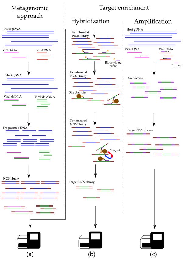
not effectively remove host-cell DNA [106]. Let us look further into specific techniques for improving
NGS library concentration. (Figure 2)
Figure 2. Unlike metagenomics (a), target HTS takes on an extra step of hybridization enrichment
(b) and/or target amplification (c), during which the concentration of selected sequences is increased.Viruses 2020, 12, 211 8 of 28
3.3.1. Nucleic Acids Depletion
Various types of clinical specimens routinely contain a large portion of host-cell nucleic acids,
which can interfere with target PCR [18] and decrease sequencing efficiency and, therefore, have to
be removed in a process called depletion. Utilizing biological, physical or chemical principles, it
significantly decreases the content of host-cell DNA and/or RNA, while only negligibly lowering
concentration of a pathogen’s nucleic acids. Most depletion kits are based on hybridization of molecular
probes with target sequences, which is a lengthy but effective process. Commercial depletion kits are
widely available (Ribo-Zero, RiboCop, HostZERO, NEBNext, MolYsis).
In a protocol by Gu et al. [107], Cas9 utilizes a set of guide RNAs to target unwanted nucleic
acids during depletion of abundant sequences by hybridization (DASH). Cleavage-mediated depletion
resulted in an up to 99% reduction of mitochondrial DNA. This approach can be used for targeting
human genomic DNA in samples with viral nucleic acids.
Despite its beneficial role in sample processing, depletion poses a few risks for the output quality.
Since it acts as a bottleneck, which allows only certain sequences through, biases are inherently imposed
on the selection process. Cross-hybridization [108] is one example: recombinations in a viral genome or
flaws in guide RNA design cause unspecific annealing to homologous non-target sequences, resulting
in an erroneous cleavage.
Although separation of target sequences from “junk” nucleic acids is the primary goal of depletion,
high levels of host-cell nucleic acids can, in theory, impede probe-sequence binding by competitive
mechanisms, in which case the effectiveness of depletion would sag. However, some modified protocols
have been developed that tackle this issue [103,109].
All points considered, nucleic acid depletion is an effective tool for decreasing concentrations
of unwanted sequences in a sample, despite it possessing a few limitations that could lower the
sequencing output by removing target nucleic acids.
3.3.2. Hybridization-Based Enrichment
Experiments that compare modern clinical assays with HTS-based tests conclude that data derived
from sequencing might be helpful in several ways. It can serve for pathogen detection and identification
and also supply clinically valuable information on the presence of resistance genes or resistance-yielding
mutations in a pathogen’s genome [110]. It could help healthcare professionals choose the correct
therapy without having to wait for microbiological tests and prescribing broad-spectrum drugs that
are constantly shown to have adverse side effects [111]. Of note is that NGS finds pathogens beyond
standard testing panels, unlike traditional tests. However, genetic libraries must be created, often from
samples with low concentrations of a pathogen’s nucleic acids and high content of nucleic acids from a
host and other microorganisms. An elegant solution has been proposed by two independent teams
of researchers, resulting in the development of a novel commercially available enrichment tool [112]
based on hybridization.
The most notable feature of this approach is that molecular probes, instead of targeting selected
viral species or families, bind to certain sequences in all known viral genera infecting vertebrates,
thus supposedly creating the most comprehensive tool for viral screening. This enrichment technique
greatly increases sequencing depth, while also raising the fraction of viral reads by 100–10,000 times
compared to standard library preparation protocols [113,114]. A diagnostic tool of such sensitivity
could serve as an alternative to traditional methods when they fail to detect the pathogen. In contrast,
total sample sequencing could at least partly detect a new pathogen’s sequences, revealing its identity.
Target probe-based enrichment (also known as hybridization-based enrichment) is used for
studying viral genomes without prior cultivation or clonal amplification [105–117]. For this, short
DNA or RNA probes are used, which are complementary to the chosen sequences in a viral genome.
The trick is to use several probes for one viral genome, thus ensuring its full coverage and guaranteed
extraction: even if the genome contains major mutations, there is a distinct possibility that some target
sequences remain unchanged, allowing for binding with a probe. Probes themselves are biotinylatedViruses 2020, 12, 211 9 of 28
at one end, binding strongly to beads covered in streptavidin. After that, a magnet separator is used to
wash the beads from any redundant DNA and RNA with further elution of target sequences [118].
Hybridization-based enrichment holds a few advantages over PCR-based techniques. Alterations
to a pathogen’s sequence impede primer annealing, whereas probes tend to bind to target sites, given
proper reaction conditions and time, even if the target sequence contains polymorphisms [112]. Besides,
hybridization works well for covering large genomes, demonstrating less coverage biases [119].
This approach has been successfully used to describe clinically significant viruses with varying
genome sizes, such as HCV [54], HSV-1 [120], VZV [115], EBV [121], HCMV [122], HHV6 [119] and
HHV7 [123]. Hybridization can be set up in a single tube and is easily automated [121]. Moreover,
the use of probes yields results more reliable than those after cultivation. This is because acquired
sequences are almost totally identical to original templates, whereas cultivation creates quasispecies,
distorting actual genetic structure of a viral population [115,122].
Sensitivity of hybridization is increased when probes are designed using a large set of reference
sequences, resulting in a better coverage of all conceivable genetic variants. This is achieved because
target enrichment is still possible when target sequences differ from a probe sequence. However, full
sequences are needed when creating hybridization probes, while designing PCR primers requires
sequences only of the flanking regions. Another advantage of probe-based methods is that if some
oligonucleotides fail to anneal during hybridization, overlapping probes might compensate [122,124].
Briese et al. [112] developed VirCapSeq-VERT—a platform for concentrating nucleic acids of
vertebrate viruses. It contains approximately two million biotinylated probes for target enrichment.
VirCapSeq-VERT detects virtually all human viruses, including new ones, featuring up to 40% of
unknown sequences. Another team of researchers created an analogous approach, called ViroCap [117],
which has been tested in a virological lab on actual clinical samples. In 26 samples, metagenomic
shotgun sequencing (MSS) with ViroCap detected all anticipated viruses plus 30 that had not been
identified before. An experiment carried out later [125] demonstrated that out of 30 viruses identified
with NGS and missed by clinical tests, 18 were identified by MSS and ViroCap, and an additional 12 with
ViroCap only. Another powerful computational tool for designing probes has been recently developed
by Metsky et al. [126]. CATCH (compact aggregation of targets for comprehensive hybridization)
analyzes target viral genomes and selects a minimum number of probes for optimal coverage of
multiple viruses. This results in an effective hybridization-based enrichment, while preserving the
original viral diversity of a sample. Reduction in the number of probes curtails experiment costs, while
increasing the efficiency of metagenomic sequencing.
Nevertheless, the price per sample remains high for this approach, and it has not gained much
popularity yet due to insufficient data on its relative performance. In addition, hybridization protocols
are time-consuming. According to official protocols, hybridization may take up to 24 h in some cases,
after which there are additional steps required for library preparation, consuming a few extra hours.
Secondly, viral genomes that differ from probe sequences for more than 40% will most likely fail to
undergo enrichment [112]. Although this does not guarantee the loss of these viruses, the effectiveness
of the procedure nosedives. For these reasons, MSS and PCR with degenerate primers might prove
more effective, albeit yielding more “noise” data in the output, which is removable during processing.
In the end, the choice of enrichment method boils down to the experiment design and quality of the
input material.
3.3.3. Target Amplification
Target PCR is an effective and simple alternative to metagenomics, working well for both individual
genes and small viral genomes [127]. PCR-based amplification is often used for whole viral genome
sequencing of samples with low viral load [54], e.g., during the investigation of the measles outbreak
during the Olympics in 2010 [128] and epidemics of Ebola [56] and Zika diseases [129]. Sequencing
of long (2.5–3.0 kb) amplified fragments has clarified the variability of a Norovirus genome and its
spreading among patients in a few hospitals in Vietnam [130,131]. A similar approach has beenViruses 2020, 12, 211 10 of 28
utilized to measure the specificity and sensitivity of the Illumina platform for detection of minor
polymorphisms in mixed HIV populations [132]. Deep sequencing of PCR-amplified viral genomes
yielded complete genome sequences for the influenza virus [133], Dengue virus [134] and HCV [135].
This approach proves apt for investigating small viral genomes that can be covered with only
a few PCR amplicons. However, vast heterogeneity of RNA viruses (e.g., HCV27, Noroviruses and
Rhabdoviruses) might require the use of multiple primer sets to ensure amplification of all known
genotypes [130,136]. Some researchers propose that this method, coupled with NGS, is used for large
viral genomes, like HCMV [19]. In this case, PCR allows long sequences to be “split” into shorter
overlapping fragments that can be assembled into a full sequence during data analysis, which increases
sequencing depth.
According to Reyes and Kim (1991) [19], another type of amplification is SISPA, a method that
serves well when “nucleotide sequence of the desired molecule is both unknown and present in limited
amounts making its recovery by standard cloning procedures technically difficult.” Recently, it has
been used for quick and reliable identification and characterization of viruses [137].
As stated above, PCR works perfectly for amplification of small viral genomes, like those of
HIV and influenza virus. For that purpose, primers are designed so that they cover a whole genome,
either in a few fragments or in a single molecule. In theory, cloning shorter sequences is simpler,
more reliable and increases sequencing depth, whilst also requiring compilation of primer panels
for each genus. Nevertheless, target amplification has its own shortcomings. For instance, it has
low data scalability, while requiring a relatively large amount of input material to ensure proper site
coverage without biases. For this reason, its application is mainly restricted to samples that meet
robust requirements (amount of input DNA, material quality, absence of PCR inhibitors, etc.). For
instance, amplification of the whole Ebola virus genome utilizes 11 or 19 primer pairs, covering over
97% of the full sequence [138]. Two experiments studying Noroviruses included amplification with 14
and 22 primer pairs, respectively [137,138]. Finally, sequencing of the Paramurshir virus genome was
conducted with a set of 60 PCR reactions and additional Sanger sequencing to cover the unamplified
fragments [139].
Apparently, sometimes amplification, as described above, overloads laboratory workflow with
numerous reactions and mandatory normalization of the products’ concentrations. There is always
room for error, e.g., primers failing to anneal to targets due to unmatching primer sequences (particularly
in rapidly mutating viruses [139]). It is also important to consider the costs of primer synthesis and
reagents, along with the amount of labor required for setting up multiple reactions per each sample [140].
Thus, even though this method allows for amplification and further sequencing of large viral genomes,
technical complexity and low cost-effectiveness render it inapt for massive clinical research, reserving
it primarily for scientific purposes.
Multiple PCRs require more input material. In clinical practice, an amount of sample drawn
from a patient is usually limited. Multiplex PCR allows for parallel amplification of target sequences
in a single tube, thus utilizing a smaller sample volume. In this case, compatibility of primers has
to be assessed beforehand using bioinformatic tools to avoid artifacts, such as primer-dimers and
false priming.
Target PCR becomes a challenge when organisms with high genome mutability are studied,
e.g., HCV [112], influenza virus or Noroviruses. Frequent changes in their genetic sequences interfere
with primer annealing [54,130,141]. A more subtle approach to designing primers could improve the
outcome, but PCR alone is by definition incapable of identifying new viruses.
We suppose that new methods based on metabarcoding might aid identification of viruses in
the future. Originally, barcoding—another type of target amplification-mediated approach—was
developed to assess bacterial associations (e.g., gut microbiota), based on reliable universal phylogenetic
markers, such as 16S, where short taxon-specific sequences consisting of a variable fragment flanked
by conservative fragments are used for tagging organisms. Metabarcoding combines a metagenomic
approach (i.e., studying multiple organisms in a sample) with barcoding, making it a powerful tool forViruses 2020, 12, 211 11 of 28
studying complex microbial associations [142], fungi and eukaryotes [143]. Usually, the scalability of a
metabarcoding approach is limited by genus level; however, this limitation is compensated for by a
significant cost reduction in comparison with metagenomics. Unfortunately, no such loci have been
described for viruses, owing primarily to their genetic diversity; however, some attempts have been
made at solving this issue with broad-range primer panels, created by computational methods [144,145]
with promising results.
3.4. Whole Viral Genome Sequencing
Once a pathogen is identified, whole genome sequencing can be engaged for further research into
its genome. This could prove beneficial for the knowledge of its genetic patterns, drug resistance and
possible targets for antiviral therapy.
Comparatively small genomes, like those of human immunodeficiency virus (HIV), influenza
virus, hepatitis B virus (HBV) and hepatitis C virus (HCV), are frequently sequenced in research
purposes, and attempts are being made to integrate this procedure into clinical practice [146]. Until
recently, detection of drug-resistant strains required sequencing of only certain parts of viral genome.
Whole genome sequencing (WGS) was considered far too expensive for this purpose; however, we are
obtaining more data on evolving mechanisms of drug resistance, including genetic mutations that
define it [147], and with the cost of WGS steadily decreasing, the use of this method becomes justified.
Routine use of NGS for diagnostic purposes is expected to hold great advantages over existing
techniques [148]. For instance, the genotyping of Coronavirus virus, which has been drawing attention
of epidemiologists worldwide, is already influencing decision-making in healthcare [149]. Additionally,
genomes of drug-resistant HIV strains are used to study the evolution of the virus [150] and complex
genotype/phenotype associations between the virus and the host [151,152].
At present, WGS remains costly, even for small viral genomes, compared to the price of target
sequencing. Nevertheless, additional data produced using WGS might supplement our knowledge of
genetic substrates for drug resistance and uncover complex intergenic associations at its core.
3.5. Methods of Sequencing Data Analysis
Whenever metagenomics is used for the detection of pathogens, it is crucial to use reliable
bioinformatic tools and specialized databases that would help decide whether discovered
microorganisms indeed have caused the infection or act merely as artifacts. Such processing normally
demands lots of computing power and knowledge and skills in bioinformatics. Typically, data analysis
mainly involves comparing obtained reads with reference genomes. Quite a few algorithms have been
developed for this purpose [153], with BLAST being the most widely used [154]. However, BLAST
works slowly for the analysis of NGS data [155], and processing times can take several days, or even
weeks, especially when calculations are performed for amino acids. Other often-used programs are
Bowtie [156] and BWA (Burrows-Wheeler Aligner) [157], which are usually employed as filtration
tools, and DIAMOND as an alternative to BLAST [158].
As for the published bioinformatic pipelines, in general they adapt either of the two strategies:
(1) firsthand removing the host’s reads by mapping them to the host’s or another non-target reference
genome and subsequently analyzing the remaining sequences; (2) assembling short reads into larger
contigs, and only then comparing assembled sequences to reference genomes, including those of
viruses [80]. (Figure 3)Viruses 2020, 12, 211 12 of 28
Figure 3. Pipelines for processing reads utilize two main strategies for filtering out “junk” data:
(a) mapping raw reads onto the reference genome of the host and removing them, while preserving
unmapped sequences for further analysis; (b) assembling short reads into contigs and comparing them
against the host’s reference genome. Annotation follows both strategies.
First filtering of the host reads (Figure 3a) works well only when the host’s genome has been
thoroughly studied and described in detail, although it is not a problem for detection of human viral
infections. As HTS technologies advance, more complete genome sequences become publicly available.
Another problem is that viruses may contain nucleotide fragments that are similar to certain regions in
the human (or other host) genomes, leading to false negative results. The second approach requires
high coverage to work, thus significantly increasing both the processing time and the experiment
cost. Moreover, whenever input data are insufficient, spaces might form between reads, impairing
their assembly into contigs, thus halting virus classification. In principal, two pipelines utilize the
same concept of comparing sequences that “survived” filtering against reference viral genomes [80].
Depending on the sample type and expected contaminants, the filtering step may also include rRNA,
mtRNA, mRNA, bacterial or fungal sequences or non-human host genomes.
Apart from the differences in the methodology between first filtering out the host reads or leaving
them included, there are other serious challenges, such as the proper taxonomic assignment of viral
reads or contigs. Homology or alignment-based similarity search methods are often used, but a
composition search, in which oligonucleotide frequencies or k-mer counts are matched to referenceViruses 2020, 12, 211 13 of 28
sequences, is also an option. At the same time, a composition search requires the program to be
“trained” on reference data and it is not used much in viral genomics.
Dismissing the host’s sequences is an important step that has been introduced into numerous
processing algorithms, such as VirusFinder [77], VirusSeq [159] or Vy-PER [160]. This step helps
remove false positives caused by similarities between some regions of the human and viral genomes.
One popular pipeline for analysis of viral reads is by Petty et al. [161]—ezVIR, which was designed
to process HTS data from any of the sequencing platforms, and which initially compares raw reads
with reference human genome, subsequently removing them and analyzing the remaining sequences
against the viral database.
VirusFinder, for instance, first utilizes Bowtie2 [156,162] to detect and remove sequences derived
from human genome. Next, it engages BLAT (BLAST-like alignment tool) [163] to align the remaining
sequences to the database of viral genomes. During the final step, the short reads, supposedly viral,
are assembled into longer sequences—contigs—using Trinity [164]. VirusFinder, developed primarily
to identify viral integration sites within the human genome, produces the best results when given
sequencing reads with the maximum depth possible. In the original article [77], coverage varies
between 31× and 121×. Acquired contigs are then used for phylogenetic analysis.
VirusHunter [165], on the other hand, utilizes BLASTn to filter out human-related sequences after
quality evaluation. Sequences that pass filtration are taxonomically classified according to BLASTn
and BLASTx algorithms. Thus, VirusHunter requires a high-quality host’s genome for sorting the
sequences and significant computational power to run.
VirusSeq [159] is intended for detection of viral sequences in tumor tissues. First, it removes
human sequences by comparing them against the reference. The MOSAIK program [166] is used
both prior to filtration and afterwards to ensure quality sequence detection. VirusSeq sets limits
on the minimally acceptable number of reads and coverage based on the size of viral genome. For
example, it demands at least 1000 reads per virus given that the sequencing depth equals at least 30×.
Although the threshold can be adjusted, this tool has been developed for reads with high coverage and
is consequently not recommended for processing data with a low percentage of viral reads.
Vy-Per [160] is another bioinformatic instrument that utilizes reference human genome for filtering
out reads of a host’s DNA. Sequences that are not dismissed during this step are compared with the
data in the NCBI database using BLAT tool [163]. Although experiments set up to test Vy-Per use
samples with a rather high coverage (80× for samples and 40× for controls), it is not mandatory, but
lowers the risk of false positives.
PathSeq [167] is a powerful computational tool for analyzing the non-host part of sequencing data,
which is able to detect the presence of both known and new pathogens. The PathSeq approach begins
with a subtraction phase in which reads are aligned with human reference genome to subsequently
exclude them, in an attempt to concentrate pathogen-derived data. This is followed by the analytical
phase, in which the remaining reads are aligned to the microbial reference sequences and assembled de
novo. The formation of large contigs, including several unmapped reads that do not have a significant
similarity to alignment with any sequence in the referenced databases, may suggest a previously
undetected organism.
SURPI (“sequence-based ultrarapid pathogen identification”) [168] is one more example of the
pipelines for complex metagenomic NGS data generated from clinical samples that first filter out the
non-target reads using 29 databases and then identify viral, bacterial, parasitic and fungal reads, which
also involves de novo contig assembly.
There are many other tools available, and we refer to recent reviews by Nooij et al. (2018) [80] and
Fonseca et al. [169].
The search for viral pathogens is often impeded by their genetic variability, caused by a multitude
of factors: gene duplications and exchanges, frequent single-nucleotide mutations, gene-large insertions
and rapid adaptation of viruses to new hosts. These become a particular nuisance whenever large
sets of samples from various organisms are handled. Firstly, reliable reference genomes have onlyViruses 2020, 12, 211 14 of 28
been assembled for a limited number of organisms, although, as previously mentioned, this issue is
being addressed. Secondly, the amount of usable viral nucleic acids in a sample depends on numerous
factors, including the stage of a pathogen’s life cycle and the overall quality of material and its type,
all of which might cause prejudice towards certain viral species and drop the amount of usable viral
DNA and RNA. This complicates the assembly of contigs, because it requires the maximum number of
reads possible.
All search methods rely on genome reference databases, such as the NCBI GenBank, RefSeq or
BLAST nucleotide (nt) and non-redundant protein (nr) databases. The poor quality of the reference
sequences is a major obstacle to processing data. Because of this, specialized databases are being
compiled manually to ensure a strict quality control, e.g., ViPR [170], RVDB [171] and viruSITE [172].
However, they are reliable for the same reason that they are limited, because only a small fraction
of all published sequences ever make it to these databases. As a consequence, reads obtained from
supposedly new strains are frequently left out. Contrarily, a vast NCBI-based GenBank database is
brimming with viral sequences, both complete and partial; however, the price for the quantity is the
quality of the assembled data. Even so, GenBank is far from being truly comprehensive, but this is
merely a question of time. Protein databases are also used, for example, Pfam [173] and UniProt [174].
Protein-level searches can usually detect more distant homology due to the usage of sensitive amino
acid similarity matrices, which can improve the detection of divergent viruses, but untranslated regions
of the genome remain unused.
4. Long Read Sequencing
Despite being one of the most powerful research tools, the NGS sequencing, which is sometimes
called second-generation sequencing, is not flawless. Namely, not a single platform can read fragments
more than 700 bp [175], thus necessitating DNA fragmentation prior to sequencing. Additionally,
second-generation platforms amplify the signal prior to detection. PCR is used for molecular cloning
of the fragmented DNA, which inevitably results in overrepresentation of some amplicons and
uneven sequence coverage. Small length of the reads sometimes becomes a trouble for haplotyping,
significantly complicating localization of the questioned loci. It also causes problems when analyzing
repeats and recombinant regions of the DNA due to mapping issues.
In contrast, long read sequencing (LRS) technologies do not require significant DNA fragmentation
or additional template amplification. For these reasons, they are frequently referred to as the
“third-generation” sequencing technologies, although this distinction sometimes may not be accurate.
Platforms such as PacBio (Pacific Biosciences), PromethION, GridION and MinION (ONT, Oxford
Nanopore Technologies) are capable of reading exceptionally long fragments, including whole viral
genomes. Secondly, these platforms potentially allow ultra-high coverage to be easily reached and even
quantify the sample, because the viral load would be directly proportional to the number of detectable
viral genomes in the sample. Thus, samples with high viral titer can provide high-quality sequences
without prior amplification. Another feature of the ONT platforms is the sample turn-around time,
which is often critical in the clinical environment; point-of-care testing makes it possible to identify the
infectious agent virtually at the patient’s bed. In this setting, the correct therapy can be started within
hours or days of disease manifestation, prospectively improving the patient care outcome.
One of the most popular ONT sequencers—the MinION—has been praised for possessing multiple
boons: relatively low cost, when compared to platforms by Illumina and Thermo Fisher Scientific, and
mobility, owing to compact dimensions of the sequencer, simplicity of protocols, and rapid sample
processing. Another great advantage that ONT platforms hold over second-generation sequencing
is the length of analyzed fragments. Because fragments over 2 Mb can be read in a single run with
maintained quality, in metagenomic experiments, genes of interest, like those of drug resistance [176],
can be easily attributed to relevant microorganisms, as opposed to reading shorter fragments, when
identifying the resistant strains can, in theory, pose a significant challenge. Furthermore, available
protocols include not only those for metagenomics, but also for studying select organisms, e.g., viruses.Viruses 2020, 12, 211 15 of 28
Both parallel and consecutive sequencing of samples can be performed, owing to a multitude of pores
in a cell.
MinION has been extensively used in detecting known [177–181] and new [182] pathogen strains,
for instance, of Ebola virus [183], Begomovirus [184] and Papillomavirus [185]. Curiously, MinION allows
samples to be detected and genotyped within mere hours from sample collection, even “in the field”,
sometimes literally [186,187]. It has also been successfully used for rapid investigation of Dengue virus
outbreak in Angola [188] and Ebola epidemic in Guinea [189]. Another experiment [190] allowed for
deciphering full genomes of three Avipoxvirus strains in real-time, skipping extraction and enrichment
altogether. Another analogous example is Everglades Virus (EVEV), which has been detected and
subgenotyped on-site [191].
Another great advantage of Nanopore technology is that unique protocols for direct RNA
sequencing have been developed, meaning that RNA viruses could be analyzed right away, without
reverse transcription [192,193]. Even more, depending on the experiment objective, either differential
RNA sequencing (dRNA-Seq) or mRNA could be investigated, the latter being achieved by ligating an
adapter only to the poly-A tail, thus allowing for transcriptome analysis [192,194]. Direct RNA-Seq
helps avoid distortions of fragments’ representation [195] in the sample, revolutionizing the approach
to studying RNA-viruses and the intricacies of their life cycle [196].
Despite the apparent benefits of this approach, there are a few drawbacks to consider, e.g.,
significant error rates that require a large coverage for their partial compensation. This poses a problem
that is particularly important for the research of drug resistance, the substrate for which is often
a few single-nucleotide polymorphisms, particularly in RNA-viruses [132]. Xu et al. suggest that
incorrect sample differentiation and chimeric reads that emerge during sequencing with MinION
might significantly alter the experiment outcome [197].
Combining target enrichment with long-range sequencing could potentially solve the problem of
high error rate of the sequencing method [198,199] by increasing coverage and improving processing
methods [200,201]. For instance, in one metagenomic study of sea water [202], the LASL method
(long-read linker-amplified shotgun library) was used to produce enough DNA templates for successful
MinION sequencing of a sample with initially low target DNA content (VirION system).
To summarize, small physical dimensions of MinION, along with other ONT sequencers,
availability of DNA and RNA sequencing protocols, potentially high precision and high data processing
rates create promising prospects for its clinical application [179]. So far, it has already been tested
in veterinary science for detection of canine distemper virus (CDV) [203,204] and in biology for
identification of numerous pathogens in plants [205] and fish [206]. Nevertheless, prior to extrapolating
these positive results into the medical field, we need to develop and validate the workflows that
would easily integrate into the everyday work of a clinical laboratory and not require any special
bioinformatic training in the lab staff [207].
5. Obstacles to Overcome in the Nearest Future
The risk of contamination and the issues with sensitivity are important to sequencing viral
nucleic acids, because they cause false positive and false negative results, respectively. High-sensitivity
sequencing (be it metagenomic, PCR-based or probe-enriched) allows for detection of minimal quantities
of contaminating viral nucleic acids [208,209]. For example, murine leukemia virus (MLV) [198,210]
and Parvovirus-like sequences [99,211] are only two kinds of the vast range of common contaminants
found even in commercial laboratory supplies [212].
Like with other highly sensitive technologies, only robust validated protocols and their strict
implementation can effectively reduce the probability of contamination. This principle has been
observed in action for quite a long time in paleogenetics [213], although methods in this field might
prove somewhat exorbitant in everyday NGS research with samples not as valuable and unique.
Nevertheless, some practices common in this field surely will find application in clinical NGS, when
human wellbeing and life are at stake, compared to an abstract scientific interest of pure research.You can also read