The impact of molecular tumor profiling on the design strategies for targeting myeloid leukemia and EGFR/CD44-positive solid tumors
←
→
Page content transcription
If your browser does not render page correctly, please read the page content below
The impact of molecular tumor profiling on the design
strategies for targeting myeloid leukemia and
EGFR/CD44-positive solid tumors
Nikola Geskovski*1,§, Nadica Matevska-Geshkovska2,
Simona Dimchevska Sazdovska1,3, Marija Glavas Dodov1, Kristina Mladenovska1
and Katerina Goracinova*1,4,§

  Review                                                                                                                          Open Access

  Address:                                                                Beilstein J. Nanotechnol. 2021, 12, 375–401.
  1Institute of Pharmaceutical Technology, Faculty of Pharmacy,           https://doi.org/10.3762/bjnano.12.31
  University of Ss. Cyril and Methodius in Skopje, Skopje, North
  Macedonia, 2Center for Pharmaceutical Biomolecular Analyses,            Received: 06 November 2020
  Faculty of Pharmacy, University of Ss. Cyril and Methodius in Skopje,   Accepted: 12 April 2021
  Skopje, North Macedonia, 3Department of Nanobiotechnology,              Published: 29 April 2021
  Institute of Biotechnology, Brandenburg University of Technology
  Cottbus-Senftenberg, Senftenberg, Germany and 4College of               Associate Editor: A. Salvati
  Pharmacy, Qatar University, PO Box 2713, Doha, Qatar
                                                                          © 2021 Geskovski et al.; licensee Beilstein-Institut.
  Email:                                                                  License and terms: see end of document.
  Nikola Geskovski* - ngeskovski@ff.ukim.edu.mk;
  Katerina Goracinova* - kago@ff.ukim.edu.mk

  * Corresponding author
  § Institute of Pharmaceutical Technology, Faculty of Pharmacy,
  University Ss. Cyril and Methodius in Skopje, Mother Teresa 47, 1000
  Skopje, North Macedonia, Telephone: +389 2 3123 026

  Keywords:
  CD44; EGFR; liquid tumors; molecular tumor targeting; myeloid
  leukemia; solid tumors; surface-engineered nanoparticles

Abstract
Nanomedicine has emerged as a novel cancer treatment and diagnostic modality, whose design constantly evolves towards increas-
ing the safety and efficacy of the chemotherapeutic and diagnostic protocols. Molecular diagnostics, which create a great amount of
data related to the unique molecular signatures of each tumor subtype, have emerged as an important tool for detailed profiling of
tumors. They provide an opportunity to develop targeting agents for early detection and diagnosis, and to select the most effective
combinatorial treatment options. Alongside, the design of the nanoscale carriers needs to cope with novel trends of molecular
screening. Also, multiple targeting ligands needed for robust and specific interactions with the targeted cell populations have to be
introduced, which should result in substantial improvements in safety and efficacy of the cancer treatment. This article will focus on
novel design strategies for nanoscale drug delivery systems, based on the unique molecular signatures of myeloid leukemia and
EGFR/CD44-positive solid tumors, and the impact of novel discoveries in molecular tumor profiles on future chemotherapeutic
protocols.

                                                                                                                                           375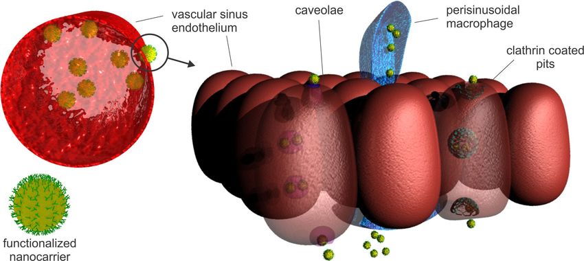
Beilstein J. Nanotechnol. 2021, 12, 375–401.

Introduction
The conventional chemotherapy regimens of both liquid (hema-          frequently reported CD44/EGFR axis, resulted in useful strate-
tological) and solid tumors are challenged by their lack of           gies for the development of dual-targeted NDDSs capable of
targeting ability, which usually results in therapy failure and       attacking multiple targets for a more efficacious cancer treat-
noticeable side effects. Nanomedicine has emerged as a novel          ment [2]. With this in mind, in this article we will review novel
cancer treatment and diagnostic modality, whose design con-           NDDS design strategies, based on the unique morphological
stantly evolves towards increasing the safety and efficacy of the     and molecular signatures of liquid (myeloid leukemia) and solid
chemotherapeutic and diagnostic protocols. Recently, a novel          tumors. The focus on myeloid leukemia and solid tumors with
generation of smart nanoscale drug delivery carriers with in-         overexpression of EGFR and/or CD44 was chosen to highlight
creased selectivity and multistage targeting capabilities has         the differences and similarities of the NDDS design strategies
emerged. Common cancer signatures and the synthesis of                for targeting two seemingly different morphological and molec-
ligands with high avidity for the overexpressed cancer cell re-       ular types of tumors and to illustrate the impact of novel discov-
ceptors are a valuable addition to the general targeting concepts.    eries in molecular tumor profiles and targeting strategies on the
Important discoveries regarding the surface-expressed recep-          future chemotherapeutic protocols.
tors and their intracellular molecular pathways have been made
during the last decade, which improved the design of targeted
anticancer nanomedicines and the targeting of specific tumor          Review
types. No doubt that the advances will further progress to the        Molecular profile and characteristics of
application of individualized tumor signatures for a personal-        myeloid leukemia
ized therapy against cancers. The greatest interest regarding the     Leukemia comprises a heterogeneous group of diseases charac-
development of targeted nanoscale drug delivery systems is            terized by abnormal proliferation and differentiation of a clonal
related to solid tumors. However, liquid tumor targeting can          population of blood progenitor cells. The proliferation of the
greatly benefit from the application of nanomedicines during          self-renewing malignant clones develops and expands in the
therapy. Unlike solid tumors, which necessitate nanoscale drug        bone marrow from where the cells start to circulate to the
delivery system (NDDSs) to reach a specific site of action,           peripheral hematopoietic tissues, leading to a severe and often
liquid tumors are mainly spread throughout the blood circula-         fatal systemic malignancy that affects hematopoiesis, immune
tion. In fact, the barriers that apply to the NDDSs for solid         defense system, and many other body systems [3]. All types of
tumor targeting usually do not exist in the case of liquid tumors,    leukemia account for 2.5% of overall cancer incidence and
since the tumor cells are generally exposed in the blood circula-     3.1% of cancer mortality worldwide. Estimated numbers for
tion. Hence, hematological tumors generally require a slightly        new cases and deaths in 2020 are 474,519 and 311,594, respec-
different approach for diagnosis and treatment. However, the          tively [4]. In adults, AML and chronic lymphocytic leukemia
discovery of the leukemic stem cells (LSCs), which are located        (CLL) are the most common types of leukemia. In children
in the bone marrow endosteal region, has introduced the possi-        aged 0 to 14 years, leukemia is the most commonly diagnosed
bility of employing similar strategies for passive targeting as for   cancer, accounting for up to 28% of all cancers, of which over
solid tumors, but these tumors also have much in common               75% are acute lymphoblastic leukemia (ALL) [5].
regarding the expression of specific molecules as viable targets
for therapy and/or homing of NDDSs. For example, overexpres-          The treatment depends on the type of leukemia, disease stage,
sion of the EGFR gene or protein kinase cascades downstream           prior history of treatment, age, overall condition, and genetic
the growth factor receptor mutations, which were detected in a        profile. The current treatment options include traditional
variety of solid tumors, in diverse acute myeloid leukemia            chemotherapy, haematopoietic stem cell transplantation
(AML) cells, and in many other leukemic cells, highlighted            (HSCT), and targeted therapy (small molecule inhibitors
their role as therapeutic targets and the importance of novel         and monoclonal antibodies). Traditional chemotherapeutic
therapeutic approaches and treatment alternatives using NDDSs         regimens are limited by two primary concerns: overall toxicity
[1]. Literature data points to combinatorial therapy, coadminis-      and lack of efficacy (resistance). Nevertheless, the application
tration, and codelivery of agents by nanomedicines as a success-      of cytotoxic chemotherapeutics (e.g., daunorubicin and cytara-
ful approach to bypass signaling inhibition, combat anticancer        bine), with or without HSCT, still remains the backbone of
drug resistance, and increase the efficacy of the clinical treat-     treatment for AML [6,7]. All chemotherapy agents interfere
ment. Further advances in molecular screening and novel               somehow with the DNA replication process or with cell mitosis.
discoveries related to unique cancer molecular signatures, in-        Anthracyclines (such as daunorubicin) interfere with topoisom-
cluding the specific biomarkers of the cancer stem cells and the      erase II and inhibit DNA replication and histone activity. Alky-
correlated action of receptors in cancer survival, such as the        lating agents (such as cyclophosphamide) introduce inter- and

                                                                                                                                       376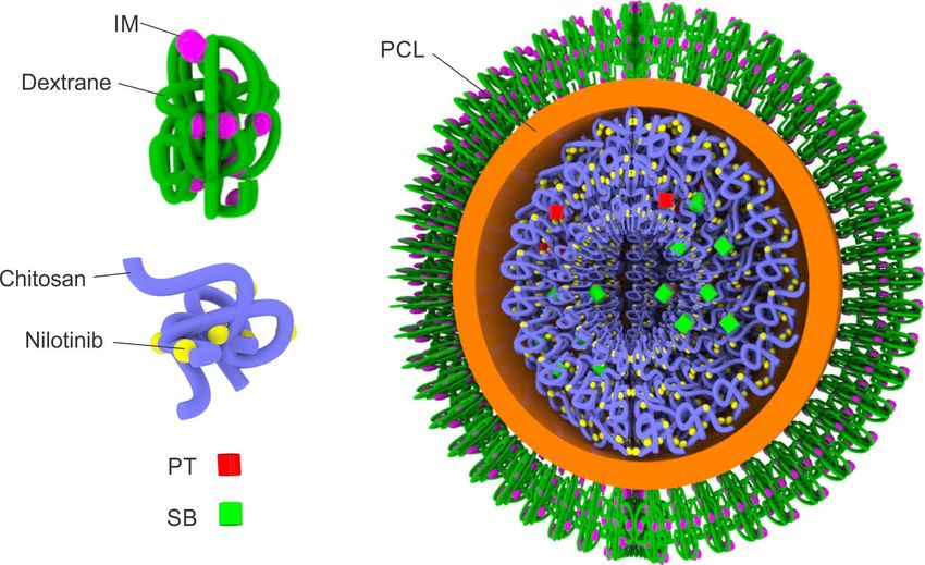
Beilstein J. Nanotechnol. 2021, 12, 375–401.

intrastrand cross-linkages and breaks in the DNA. Antimetabo-           administration of drugs, improving their bioavailability,
lites (such as 5-fluorouracil) obstruct the synthesis of nucleic        reducing side effects, and restoring the efficacy/response, espe-
acids required for DNA replication, while taxanes and vinca             cially through evading the drug transport resistance mecha-
alkaloids interfere with the polymerization/depolymerization of         nisms.
the microtubules thus inhibiting mitosis [8]. However, in addi-
tion to cancer cells, all of these drugs also affect the normal/        In contrast to traditional cytotoxic chemotherapy, targeted ther-
healthy tissues with cells that rapidly divide, such as bone            apies, such as tyrosine kinase inhibitors (TKIs), are directed
marrow stem cells and the gastrointestinal epithelium. This             towards the molecular aberrations responsible for elevated
leads to severe and sometimes life-threatening side effects and         kinase activity or towards fusion oncoproteins involved in
disruption of the normal hematopoiesis and subsequent                   proliferative or anti-apoptotic signaling pathways [13]. For
recovery, especially of elderly patients.                               instance, more than 90% of the cases of chronic myelogenous
                                                                        leukemia (CML) are characterized by a unique chromosomal
The lack of sensitivity and the development of resistance is            abnormality known as Philadelphia (Ph) chromosome [14]. This
another major drawback of the traditional “broad-spectrum”              abnormality is a reciprocal chromosomal translocation between
cytotoxic drugs. Leukemic cells generally respond well to drug          the long arms of chromosomes 9 and 22, designated as
therapy at the onset of treatment, but the drugs lose their effec-      t(9;22)(q34;q11), creating a derivative 9q+ and a shortened 22q
tiveness over a period of 6–12 months in a significant fraction         Ph chromosome. The chimeric Ph chromosome generates the
of patients [9]. In contrast to solid tumors, where cancerous           BCR-ABL1 gene by fusion of the Abelson murine leukemia
cells accumulate at defined tumor sites and the cytotoxic drugs         (ABL1) gene from chromosome 9 with the breakpoint cluster
could be passively targeted to these tumor sites through en-            region (BCR) gene from chromosome 22. Due to different
hanced permeation and retention (EPR) effect, leukemic cells            possible breakpoints on chromosome 22, several transcripts can
are prevalent in the whole circulatory system and the EPR is of         originate from this translocation. However, all BCR-ABL1 gene
no use. Moreover, the inherent plasticity of the leukemic cells         fusions described so far encode for a constitutively active BCR-
combined with diverse resistance mechanisms allows malig-               ABL1 tyrosine kinase that promotes growth and replication
nant cells to naturally adapt to drugs. Several resistance mecha-       through downstream pathways such as RAS, RAF, JUN kinase,
nisms have been acknowledged, including failure of the cell to          MYC, and STAT. This influences leukemogenesis by causing
undergo chemotherapy-induced apoptosis and failure of the               enhanced proliferation, differentiation arrest, and resistance to
drug to reach and/or affect its intracellular target due to intracel-   cell death. The use of the TKIs, the first of which was imatinib,
lular drug transport resistance mechanisms [10]. Additionally,          has improved the three-year survival to over 95%, from 50% in
the high recurrence rate in leukemia patients has been also attri-      the pre-imatinib era [7]. However, imatinib and related TKIs
buted to the existence of a rare population of LSCs capable of          (the second-generation TKIs dasatinib, bosutinib, nilotinib, and
evading drug therapies [11,12]. These CD34 + CD38 − LSCs,               the third-generation ponatinib) are not exclusively specific for
which preferentially reside in the bone marrow endosteal                the BCR-ABL fusion protein, and may also affect normal
region, have acquired abnormal self-renewal and have shown              c-ABL and other kinases such as c-KIT. This can lead to side
the capability to give rise to heterogeneous nonstem leukemic           effects, mainly diarrhea and skin toxicity [15]. Also, all TKIs
cells. Therefore, they are considered responsible for disease ini-      have short half-lives (in the case of imatinib and its main
tiation and maintenance. Moreover, the CD34+CD38− LSCs                  metabolite 18 and 40 h, respectively) and require daily dosing.
predominantly exist in the quiescent phase of the cell cycle
(G0). They, hence, have a resistance to cytotoxic drugs that            More importantly, similar to the traditional cytotoxic agents,
interfere with cell mitosis [11,12]. These observations imply           resistance to TKIs and early relapse are still major concerns.
that minimal residual disease (MRD) can be attributed to rare           For example, 15% to 40% of CML patients will develop resis-
quiescent CD34 + CD38 − LSCs remaining after therapy and                tance or intolerance to first-line imatinib. The resistance mecha-
highlight the importance of the complete eradication of the             nisms can either depend on BCR-ABL1 or not. BCR-ABL1-de-
LSCs in improving the long-term outcomes in leukemia                    pendent resistance mechanisms are primarily associated with
patients. This suggests the need for the identification of LSC-         the overexpression of the BCR-ABL1 oncogene or mutational
specific molecules, which can be employed as drug targets for           events [16]. The clinically relevant mutations arise in the kinase
the development of novel therapeutic antibodies and inhibitors          domain of BCR-ABL1 and lead to impaired TKI activity,
of LSC-specific kinases or transcription factors, but also as           mainly by preventing the fusion protein from adopting the
roadmaps for active targeting using novel nanoparticles (NPs).          correct conformation required for specific binding. More than
Furthermore, the use of nanotechnology for the delivery of              40 different point mutations have been identified in relapsed
cytotoxic drugs can also be valuable in facilitating cell-specific      CML patients receiving imatinib. However, the most frequent

                                                                                                                                         377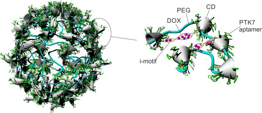
Beilstein J. Nanotechnol. 2021, 12, 375–401.

alterations occur in three different regions and seven specific     endothelial cells. It provides the appropriate microenvironment
residues, that is, in the docking site for phosphate moieties of    for efficient hematopoiesis. The blood flow is supported by the
ATP named P-loop (M244V, G250E, Y253F/H, and E255K/V),              nutrient arteries, which supply the marrow with nutrients, the
in the contact site related to selective BCR-ABL1 inhibition by     periosteal artery, arterioles, and the capillary circulation, which
IM (T315I), and in the catalytic domain (M351T and F359V)           forms a sinusoidal network of vessels comprising a single endo-
[17]. In general, second-generation TKIs are active against most    thelial cell layer lacking supporting cells. The barrier between
of the subclones resistant to imatinib mesylate (IM), except        the hematopoietic compartment and the blood circulation is
against the subclones harboring the T315I mutation. The T315I       commonly referred to as the marrow–blood barrier (MBB). The
mutation displays resistance to all currently available TKIs,       MBB is 2–3 µm thick. It is composed of the continuous
except for ponatinib, a third-generation TKI. In addition,          vascular endothelium and the discontinuous adventitial retic-
second-generation TKIs also display similar resistance mecha-       ular cell layer. Hence, it is relatively highly permeable to a wide
nisms as imatinib, but the mutation spectra are different: T315I,   variety of solutes and even particulate matter [18]. Portions of
F317L or V299L for dasatinib, E255K/V, T315I, F359C/V or            the sinusoidal endothelial cells could be noticeably reduced into
Y253H for nilotinib, and V299L or T315I for bosutinib.              small fenestrae with sizes in the range of 80–150 nm, which
                                                                    could facilitate the paracellular MBB transport. On the other
Molecular targeting of myeloid leukemia using                       hand, their clathrin-coated pits, lysosomes, clathrin-coated vesi-
surface-engineered nanoparticles                                    cles, and phagosomes could be employed as a gateway for
Nanomedicines as an efficient tool for bone marrow                  endocytosis-mediated transcellular transport across the MBB
targeted delivery of drugs: It is known that solid tumors reside    [19]. Since there is no lymphatic drainage, the BM tissue relies
in extravascular spaces. In order to reach them, the NDDSs          on these pathways for bidirectional transport of a variety of
need to extravasate the porous tumor vasculature. In contrast,      molecules across the MBB. The capability of the sinusoidal en-
leukemic cells and leukemia stem cells are settled in the blood     dothelial cells, and the present fenestrae, to remove colloid par-
vessels and bone marrow (BM) and seem readily available to          ticles from the bloodstream nurtures the possibility of targeting
the intravenously administered NDDSs. Nevertheless, the abun-       the leukemia stem cells with NDDSs.
dance of normal cell populations in the blood and BM imposes
the necessity of a targeted and potent approach in the design of    In order to reach the BM vasculature, it is of utmost importance
NDDSs for leukemia treatment.                                       that the NDDSs do not interact with the elements of the reticu-
                                                                    loendothelial system (RES) located in the liver and spleen. Such
The cancer stem cells, with their self-renewal and tumor-initi-     interactions will result in RES-induced sequestration of the
ating properties, are considered as one of the prime factors        NDDSs, drastically reducing its availability in the organ of
affecting the promotion and relapse in most cancer types. As        interest (BM). Additionally, to effectively extravasate, a circu-
described in the previous section, the enhanced DNA repair          lating NDDS firstly needs to drift to the margins of the blood
ability, the expression of anti-apoptotic proteins, and the drug    vessels, and then recognize the specific endothelial target to
efflux transporters of the leukemia stem cells are considered the   which it needs to firmly adhere. Afterward, the adhered NDDS
main contributors to the resistance to chemo- and radiotherapy      should induce endocytosis-mediated internalization into the BM
of many types of leukemia. Hence, it is logical to identify this    vascular sinus endothelium, which probably will result in
cell population as a prime target for leukemia therapy.             carrier transcytosis towards the stroma [20] (Figure 1).

The BM microenvironment provides a protective accommoda-            Illum and Davis were among the first researchers that reported
tion for the normal stem cells, and many studies suggest that the   on the possibility of guiding colloidal particles to the BM [21].
leukemia stem cells use the same shelter for their own survival.    The initial goal of the group was to evaluate the effects of dif-
Therefore, targeting leukemia cells located in the BM is not a      ferent PEO–PPO-based hydrophilic copolymer (poloxamer)
straightforward issue regarding intravenously administered          shells upon the biodistribution, primarily the circulation half-
NDDSs. The BM is a primary lymphoid tissue that is located          life of polystyrene colloidal particles [21,22]. In their experi-
inside various bone types. It can be classified as red or yellow    ments, besides the groundbreaking work on the steric barrier
marrow, depending on whether the dominant cell population is        and the so-called “stealth” effect of the poloxamer, they have
of hematopoietic origin or fat cells. There are two types of stem   further noticed that the adsorbed poloxamer 338 and 407 shells
cells in the BM, hematopoietic, which are responsible for the       directed the polystyrene beads to the sinusoidal endothelial cells
hematopoiesis, and mesenchymal, which produce the stromal,          of rabbit BM [23]. Even though the authors eliminated the pos-
fat, cartilage, and bone tissue. The BM stroma contains fibro-      sibility of a BM distribution mediated through macrophage
blasts, macrophages, adipocytes, osteoblasts, osteoclasts, and      phagocytosis, the exact mechanism of localization remained

                                                                                                                                      378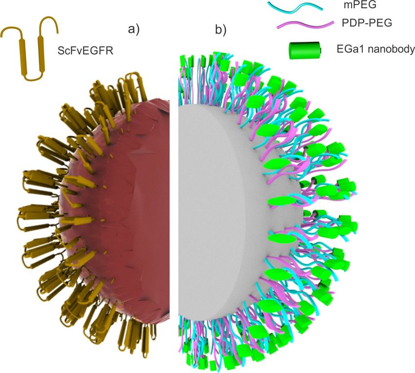
Beilstein J. Nanotechnol. 2021, 12, 375–401.

 Figure 1: Hypothetical mechanisms of extravasation of NDDSs into the BM stroma. The nanoscale carrier could induce clathrin- or caveolin-medi-
 ated endocytosis into the vascular sinus endothelium, resulting in transcytosis towards the BM stroma. Another possible mechanism of extravasation
 of NDDSs is the uptake mediated through perisinusoidal macrophages.

unknown. Moreover, the poloxamer-coated polystyrene beads                   BM-targeting liposomes were among the first formulation types
circulated in the blood for up to eight days, and regardless of             that employed this specific mechanism. Allen et al. reported
the steric barrier, somehow managed to interact with the sinu-              that the lipid composition greatly affected the bone marrow
soidal endothelial cells of the BM. One possible explanation is             affinity of the liposomes. The authors noted that phos-
that the recognition may be mediated by a plasma component                  phatidylserine and phosphatidylcholine increased the uptake of
(such as erythropoietin, transferrin, or transcobalamin) or an en-          the liposomes in cultured BM macrophages [26]. Schettini et al.
dothelial factor that specifically adsorbs onto the surface of              made an attempt at BM targeting by incorporating negatively
poloxamer 407-coated colloidal particles, thus exhibiting                   charged lipids in the liposomes. However, the employed lipid
microdomains that are specific for the sinusoidal BM endothe-               components (1,2-distearoyl-sn-glycero-3-phosphocholine,
lium.                                                                       cholesterol, and dicetylphosphate) did not promote selective
                                                                            BM uptake [27]. Moreover, the authors reported that the size
Another mechanism of BM targeting is the phagocytosis-medi-                 reduction of the liposomes produced better results. The surface
ated uptake from the perisinusoidal macrophages. It is known                modification of liposomes with an anionic amphiphile
that the perisinusoidal BM macrophages protrude through the                 (N-(3-carboxy-1-oxopropyl)-ʟ-glutamic acid 1,5-bis(hexadecyl
vascular endothelial wall to gain access and monitor blood                  ester)), reported by Sou and co-workers, resulted in significant
circulation [24]. Hussain et al demonstrated that perisinusoidal            improvement in BM uptake. The authors noted that, besides the
macrophages are responsible for the accumulation of the                     anionic amphiphile, a small amount of 1,2-distearoyl-sn-
chylomicrons in the BM and, hence, play a crucial role in the               glycero-3-phosphoethanolamine-poly(ethylene glycol) (DSPE-
delivery of lipids, as a source of energy and material for mem-             PEG) was needed to circumvent the liver and spleen sequestra-
brane biosynthesis, and fat-soluble vitamins [25]. In the past,             tion and to achieve a distribution of 60% of the injected lipo-
this pathway was generally ignored as a targeting possibility,              somes inside the BM within 6 h after administration [28]. The
mostly because the BM contribution to the RES is quite negli-               addition of larger quantities of surface-oriented DSPE-PEG
gible compared to that of spleen and liver. Also, there was a               resulted in a reduction of BM uptake, probably due to steric
lack of understanding and knowledge regarding the presence of               hindrance of the anionic amphiphile, which is considered as the
any specific BM macrophage moieties. Considering the afore-                 active targeting moiety. In their further research, the authors
mentioned, the development of an effective targeting system                 confirmed that the BM uptake mechanism relies on phagocy-
will rely on strategies that will enable evasion of liver and               tosis-mediated lipid transport. In fact, when administered in
spleen uptake and, at the same time, facilitate BM uptake using             higher doses or along with other specific lipids, the BM macro-
appropriate ligands.                                                        phages get saturated such that the phagocytosis rate drops re-

                                                                                                                                                  379
Beilstein J. Nanotechnol. 2021, 12, 375–401.

sulting in a cessation of BM transport of the liposomes [29].        members [36]. The formulation demonstrated an eightfold
Tardi and co-workers developed distearoylphosphatidylcholine/        increase in the BM localization of the encapsulated liposomes,
distearoylphosphatidylglycerol/cholesterol (DSPC/DSPG/Chol)          relative to the equivalent without ligands, in a healthy murine
liposomes with BM targeting capability [30]. The synthesized         model. Even though the authors reported an organ distribution
liposomes were able to maintain the BM levels of cytarabine          of nearly 20% per gram in the BM, most of the porous silicon
and daunorubicin for longer periods and demonstrated, respec-        particles were entrapped in the spleen, liver, and lungs, because
tively, threefold and eightfold higher BM drug concentrations        of their size and surface characteristics. In this case, the physi-
relative to the plasma concentration in the 48th hour post-          cochemical properties of the carrier dominate the overall bio-
administration in healthy mice. In contrast, the combination of      logical behavior of the NDDS, although some specificity was
free drugs (cytarabine and daunorubicin) reached maximum             achieved through the employment of a specific targeting ligand.
BM concentrations in one hour, which decreased rapidly. The
authors estimate that the high levels of BM accumulation for         Strategies for targeting specific leukemia cells using NDDS:
this liposome formulation were probably due to the presence of       As discussed in the previous section, nanotechnology enables
20% DSPG in the liposomal bilayer. This yields an overall an-        selective delivery of a wide variety of therapeutic molecules to
ionic surface, which is known to have an affinity for the scav-      cancer cells. This allows the molecules to reach critical tissue
enger receptor on BM macrophages. Other polyanions, such as          compartments, such as the BM and lymph nodes, which are
dextran sulfate, were used as ligands for targeting this macro-      otherwise inaccessible to the drugs. Targeting specific surface-
phage receptor [31]. Yet, not all polyanions could be consid-        exposed moieties of the cancer cell subpopulations is consid-
ered as viable ligands for BM macrophage targeting [32].             ered as a general strategy of active targeting. It could be essen-
                                                                     tial in leukemia treatment, especially in the cases where a
Another mechanism of BM targeting was reported by Swami et           persistent clone dominates the leukemia cell population. Taking
al. who employed the specific affinity of bisphosphonates for        into account the molecular profile of the disease, there are a
bone tissues with higher turnover rates [33]. The authors used       plethora of overexpressed molecules in leukemia cancer cells,
alendronate as a surface-exposed ligand for directing borte-         which could be used as potential targets for a NDDS-based ther-
zomib-loaded nanoparticles consisting of poly(ᴅ,ʟ-lactic-co-         apeutic approach (Table 1).
glycolic acid) (PLGA) and poly(ethylene glycol) (PEG) to the
bone microenvironment, which resulted in a 9.6-fold increase of      The folate receptor is usually overexpressed in all cancer cells
bone localization of the functionalized nanoparticles relative to    and the high frequency (ca. 70%) of folate receptor (FR) β
the non-functionalized nanoparticles. Additionally, the loaded       expression in CML and AML relative to normal hematopoietic
PLGA–PEG–alendronate nanoparticles demonstrated satisfac-            cells suggests the feasibility of the development of FR
tory results in myeloma growth inhibition and overall survival       β-targeted delivery systems [48,49]. Even though the concept of
of the murine models. Considering the toxicity of bortezomib,        FR β targeting seems conceptually feasible, the heterogenicity
acquiring this targeted approach could enable the employment         of the FR β expression levels in CML and AML presents a
of higher doses that will result in a more effective therapy with    confounding issue, which can be usually resolved by co-treat-
fewer side effects.                                                  ment with all-trans retinoic acid (ATRA) or other agonists of
                                                                     nuclear receptors for retinoids [50]. Such approach was demon-
In vivo imaging of the epithelium of BM blood vessels revealed       strated by Pan and co-workers, who formulated doxorubicin
that the vasculature expresses the adhesion molecule E-selectin      (Dox)-loaded DSPE/cholesterol/PEG liposomes decorated with
and the chemoattractant stromal-cell-derived factor 1 in             folic acid and evaluated their effects upon leukemia cells both
discrete, discontinuous areas that influence the homing ability      in vitro and in vivo. The authors noticed that the cytotoxicity of
of a variety of tumor cell lines inside the BM, and that these       folate-functionalized liposomes was greater in FR positive cell
specific molecules determine a microenvironment for early BM         lines and that the effect could be blocked through the addition
tumor metastasis [34]. This specific feature was employed by         of 1 mM folic acid. Additionally, the authors revealed that the
Mann and co-workers, who developed E-selectin-targeted               functionalized carriers increased the median survival of the
porous silicon particles with capabilities for selective uptake to   mouse ascites leukemia models, compared to non-functionali-
the BM [35]. The authors formulated a multistage carrier             zed carriers, and that the pre-treatment of the animals with
composed of porous silica microparticles that encapsulate nano-      ATRA further improved the efficacy of the FR-targeted lipo-
scale paclitaxel-loaded liposomes. The porous silica particles       somes by up to 50% [51].
were decorated with E-selectin thioaptamer ligand (ESTA),
which binds to E-selectin with high affinity and at the same         Since AML and CML proliferation is mainly governed by
time expresses minimal cross-reactivity to other selectin family     leukemia stem cells (LSC), there is a rationale to develop

                                                                                                                                       380
Beilstein J. Nanotechnol. 2021, 12, 375–401.
Table 1: NDDSs in molecular targeting of myeloid leukemia.
type of NP type payload target targeting ligand efficacy ref.
leukemia assessment
AML
generation 7 miR-150 FMS-like tyrosine FLT3 peptide in vitro, in vivo [37]
poly(amidoamine) (PAMAM) kinase 3 (FLT3)
nanoscale dendriplex
lipopolyplex NPs antagomiR-126 miR-126 transferrin or in vivo [38]
anti-CD45.2 antibody
Gold NPs rapamycin Tim-3 immune anti-Tim-3-single in vitro [39]
(immobilized receptor and a chain antibodies
using a trafficker for its
glutathione natural ligand
linker) galectin-9
multistage vector (MSV) parthenolide E-selectin E-selectin in vivo [40]
system of mPEG-PLA thioaptamer (ESTA)
micelles in protective
degradable porous silicon
particles
mesoporous silica NPs siRNA CD44 hyaluronan in vitro [41]
mesoporous silica NPs daunorubicin B220 surface marker anti-B220 antibody in vitro, in vivo [42]
cyclodextrin-based NPs siRNA IL-3 receptor α-chain fragment in vitro, ex vivo [43]
(IL-3Rα), also known antigen-binding (Fab)
as CD123 of a monoclonal
antibody
gold NPs oligonucleotides NCL/miR-221/NF-κB/ nuclear localization in vitro, in vivo [44]
anti-221 and DNMT1 signaling signal (NLS) peptide
AS1411 pathway
CML
gold NPs tyrosine kinase e14a2 Bcr-Abl1 single-stranded DNA in vitro [45]
(AuNP@PEG@e14a2) inhibitor imatinib transcript oligonucleotide
magnetic NPs paclitaxel lectin receptor lectin glycoprotein in vitro, in vivo [46]
PEG–PLA micelles tyrosine kinase hydroxyapatite alendronate in vitro, in vivo [47]
inhibitor
ponatinib and
JAK2 inhibitor
SAR302503
multiple myeloma
PLGA-b-PEG NPs bortezomib hydroxyapatite alendronate in vitro, in vivo [33]
nanoparticulated drug delivery systems targeted to this cell loaded non-functionalized nanoparticles, and anti-B220 anti-
population. It is known that the leukemia-propagating cells in body-functionalized nanoparticles loaded with daunorubicin.
murine CALM/AF10-positive AML differ from normal The functionalized NPs demonstrated superior results (median
haematopoietic stem cells regarding the surface expression of time for onset of leukemia = 160 days) relative to the drug and
B220 [52]. To demonstrate the efficacy of targeting a specific to non-functionalized NPs (19–22 days), most probably because
leukemia cell population, Mandal and co-workers developed of the increased intracellular concentrations of daunorubicin
mesoporous silica nanoparticles decorated with an anti-B220 present in the B220+/Mac1− cells. Even though the B220 recep-
antibody intended for B220+LSC targeting [53]. The daunoru- tor is not internalized after antibody binding, the authors specu-
bicin-loaded targeted nanoparticles demonstrated selective effi- late that the NP-enriched cell membrane promotes some kind of
cacy against B220+/Mac1− cells, relative to B220− AML LSCs. “passive” internalization of the functionalized NPs.
In addition, the treated B220+/Mac1− cells were injected in
immunodeficient mice, and the median time for leukemia onset Protein tyrosine kinase 7 (PTK7) is a highly expressed receptor
in the animals was observed for daunorubicin, daunorubicin- in AML cells and is mostly assigned to granulocytic lineage dif-
381Beilstein J. Nanotechnol. 2021, 12, 375–401.
ferentiation. Therefore, it could be employed as a specific cell Co-delivery of different therapeutic agents for simulta-
surface-expressed moiety for active targeting [54]. Jang and neous targeting of several molecular pathways of the
co-workers developed a polyrotaxane-based nanoconstruct with disease: The resistance to chemotherapy remains a major chal-
pliable structure, bearing surface-oriented sliding PTK7 lenge to effectively manage diseases such as leukemia. The
aptamers for the targeting of doxorubicin to a PTK7+ cell line combination of two or more drugs into one treatment protocol
(Figure 2). The aptamer DNA–cyclodextrin (CD) component of usually increases the therapeutic outcomes. This is achieved
the polyrotaxan complex demonstrates a high degree of sliding through modifications of specific cell signaling pathways with
freedom. This unique ability allows for a more efficient binding the aim to increase the cell sensitivity to at least one agent of the
of the aptamer DNA to target molecules on cancer cells. It si- employed therapeutic drug combination. This approach is par-
multaneously allows other ligands on the same carrier to ticularly attractive because single-drug therapeutic regimens are
migrate toward other target molecules to form multimeric a rare commodity in chemotherapy [56]. The drug combination
bonds, thus enhancing the robustness of the bonding among the regimens offer the opportunity to target different molecular
nanoscale carriers and target cells. In addition to the sliding pathways that are unique for specific leukemia clones, thus
targeting ligands, the authors enabled stimuli-responsive drug overcoming possible drug resistance mechanisms and increas-
release by incorporating i-motif DNA in the nanoscale carrier ing the overall therapeutic effect. Still, it is challenging to coor-
structure. I-motifs are four-stranded quadruplex structures dinate pharmacokinetics, biodistribution, and intracellular con-
formed by cytosine-rich DNA that posses unique pH-sensitive centration profiles of individual drugs with different physio-
characteristics. The double-stranded complementary DNA chemical and biological properties [57,58]. Hence, current clini-
responds to a drop of the pH value in the environment by cal combinatorial therapy regimens, which simply combine dif-
forming i-motifs and releasing the intercalated doxorubicin. The ferent drugs in a conventional dosage form, are far from a
targeting potential of the developed formulation was confirmed perfect chemotherapy approach for leukemia patients. It is
both with in vitro experiments on the PTK7+ T lymphoblast cell logical to assume that loading multiple drugs onto a single
line CCRF–CEM and on in vivo immunodeficient BALB/c nanoscale carrier will synchronize the corresponding thera-
mice harboring the PTK7+ T lymphoblast cell line. The results peutic activities, thus maximizing the synergistic effect of the
demonstrated that the sliding targeting aptamers had, respec- combinational chemotherapy regimen.
tively, a three and a six times greater affinity for binding the
PTK 7 receptor, relative to the non-sliding and non-targeted Clinical observations have revealed that co-administration of
counterparts. Additionally, the authors demonstrated the stimu- imatinib mesylate (IM) and other TKIs, such as nilotinib or
li responsiveness of the carriers by evaluating the release of dasatinib, may yield additive/synergistic anti-leukemia effects
doxorubicin, which was completely retarded at pH 7.4 (less in CML [59,60]. Considering this, the development of a nano-
than 1% in 48 h) and almost immediate at pH 5.5 (more than scale targeting system for the co-delivery of these drugs appears
90% in 1 h) [55]. to be a rational approach for combinational chemotherapy [61].
Figure 2: Schematic representation of a polyrotaxane nanoconstruct bearing surface-oriented PTK7 aptamers for targeted delivery of doxorubicin
(PEG: poly(ethylene glycol), CD: cyclodextrin, DOX: doxorubicin). The dynamic structure allows for ligand “sliding” for efficient receptor binding and
selective DOX release from the DNA i-motifs at lower pH values. Adapted from [55].
382Beilstein J. Nanotechnol. 2021, 12, 375–401.
It has been shown that nilotinib, in the chemotherapeutic combi- Mendonca and co-workers worked on developing a targeted
nation with IM, has a critical role in inhibiting or eradicating the NDDS for а combination of siRNA and TKI in CML treatment
several subclones that are resistant to IM. Cortese and [63]. The authors developed sterically stabilized liposomes
co-workers developed wool-like hollow polymeric nanoparti- decorated with transferrin for co-delivery of siRNA and IM for
cles loaded with the abovementioned drug combination for the specifically silencing the BCR-ABL oncogene. In this case, IM
treatment of CML (Figure 3) [62]. The authors developed enables the pre-sensitization of tumor cells, as a prerequisite for
core–shell nanoparticles from polycaprolactone (PCL). The effective gene silencing, mediated by the siRNA. The results
core of the nanoparticles was loaded with chitosan-complexed demonstrate that the siRNA/IM ratio affected the efficacy in
nilotinib, while the shell was loaded with a protease-sensitive BCR-ABL silencing and reflected on the evaluated IC50 values
dextrane–IM complex. In addition, the core was loaded with so- of IM on different cell lines. Additionally, the comparative
dium bicarbonate and potassium tartrate, a mixture that quickly analysis of the IC50 values on different cell lines revealed that
generates CO2 in an acidic environment and, thus, produces the transferrin receptor expression and the cellular levels of
large pores in the nanoparticles, resulting in a burst release of BCR-ABL mRNA affected the efficacy of the formulation. The
the core contents. In this way, the authors managed to achieve a cell lines with higher levels of transferrin receptor expression
stimuli-responsive sequential drug release pattern. IM will be and lower BCR-ABL mRNA levels were more sensitive to the
released in the cytoplasm from the dextrane complexes due to prepared liposomal formulation. The importance of co-delivery
the activity of intracellular protease while the nilotinib–chitosan of both agents was also observed as lower siRNA/imatinib
complexes will be released in the acidic environment of the ratios were needed to achieve IC50 values equivalent to those of
lysosomal compartment following the generation of CO2. The the experiments in which the cells were exposed to a combina-
first released IM should induce a partial inactivation of BCR- tion of encapsulated siRNA and free IM. This research high-
ABL oncoprotein, while the second released nilotinib should lights the need of achieving specific intracelullar concentra-
complete the oncoprotein inactivation, thus substantially tions and drug ratios in order to increase the efficacy of
reducing the probability of resistance. The authors reported that chemotherapeutic regimens, indicating the importance of
the combinatorial delivery of IM and nilotinib demonstrated a co-delivery systems in the improvement of the overall thera-
substantial reduction of the IC50 value regarding the KU812 peutic efficacy.
CML cell line, compared to the free drugs. Also, it was noticed
that the blockade of the G2/M phase played the main role in cell A targeted NDDS for the co-delivery of ponatinib and
cycle arrest, demonstrating a sensitiveness of the cells to nilo- SAR302503 (selective JAK2 inhibitor) for efficient CML treat-
tinib that is attributable to IM. ment was developed by Mu and co-workers [47]. The authors
Figure 3: Wool-like hollow polycaprolactone (PCL) NDDS for the sequential co-delivery of imatinib mesylate (IM) and nilotinib (PT: potassium tartrate,
SB: sodium bicarbonate). Nilotinib is released at low pH values as pores are formed in the PCL shell due to CO2 generated by PT and SB (preferably
in the lysosomes), followed by IM release from the dextrane complexes in the cytoplasm due to the activity of the intracellular protease. Adapted from
[62].
383Beilstein J. Nanotechnol. 2021, 12, 375–401.
employed alendronate-decorated PLA–PEG micelles for BM cols due to unfavorable physicochemical and pharmacokinetic
targeting. Both ponatinib and SAR302503 are hydrophobic properties, nanotechnology offers strategies for targeting a
drugs and were co-encapsulated inside the hydrophobic core of variety of leukemia cell populations. Even though liquid tumors
PEG–PLA micelles by solvent displacement nanoprecipitation. mainly reside in the blood circulation and seem readily avail-
The results demonstrated that the targeted NDDS enabled a able for chemotherapy, the subpopulation of leukemia stem
high availability of the drugs inside the BM, which is a prereq- cells that is mainly responsible for tumor progression and resis-
uisite for overcoming the dose-dependent side effects of both tance is capable of evading treatment within the bone marrow
drugs. In part, this was also accomplished by the low-dose microenvironment and to adapt through self-renewal, clonal
synergistic effect of countering different biological signaling expansion, and additional mutations. Therefore, LSC targeting
pathways, with the goal of achieving short-term BCR–ABL1 is considered as one of the primary therapy goals in myeloid
kinase inhibition that resulted in induction of apoptosis in leukemia. Hence, the design of the nanoscale carriers needs to
BCR–ABL1-positive and primary CML progenitor cells. The integrate strategies for extravasation in the bone marrow niches
targeted NDDS significantly increased the survival rate of the and subsequent specific molecular recognition of the LSC. Cur-
murine leukemia models relative to the groups treated with rent advances in molecular diagnostics offer a glimpse into the
single drugs, non-targeted co-delivery NDDS, and the untreated molecular profile of each tumor subtype and define its common
control. In addition, the low-administered dose of both co-deliv- chromosomal translocations, shared mutations in oncogenes,
ered drugs and the NDDS itself proved to be non-toxic to gene expression profiles, and immune phenotypes. Such know-
healthy BALB/c mice. ledge is important in the development of ligands for targeting a
specific subpopulation of cells, based on their molecular profile
The co-delivery strategy has the potential to overcome therapy- and specific surface-exposed molecules. Considering that
induced niche-mediated leukemia resistance in AML. Dong et leukemia has shown a vast genomic heterogeneity, the integra-
al. developed a biomimetic NDDS composed of a mesoporous tion of the molecular profile of the tumor into the design of the
silica nanoparticulate core loaded with daunorubicin and coated multimodal nanoscale carriers will have a tremendous impact
with NALM-6 cell membrane vesicles that were further deco- for the future development of personalized nanomedicine for a
rated with aTGFβRII antibodies, attached via hypoxia-sensitive safe and efficacious treatment of myeloid leukemia.
azobenzene linker. The membrane coating plays a dual role
in the formulation. It guides the drug to the BM via In addition, the co-delivery of different therapeutic molecules
receptor–ligand interaction between CXCR4 (a chemokine re- using nanomedicines is of great importance in the treatment of
ceptor expressed on NALM-6 CM) and SDF-1 (a molecule of resistant cancer clone populations. Drug combinations for
the BM endothelium) and mediates the hypoxia-driven release targeting different molecular pathways are available in conven-
of the attached aTGFβRII antibodies in the BM, which then tional chemotherapy regimens. However, in order to achieve
interfere with the interplay among LSCs and the BM niche maximal therapeutic effects and to avoid drug resistance mecha-
cells. The remaining NALM-6-coated silica nanoparticles with nisms, both drugs need to be released inside the cells simulta-
daunorubicin are taken up by the leukemia cells, in which the neously or in a specific sequence. This can be accomplished by
drug is released, leading to high intracellular concentrations formulating combined drug therapy in a nanoscale carrier. This
[64]. The results from this study highlight the importance of the treatment modality highlights the importance of NDDSs in the
sequential order of release of the therapeutic molecules in the safety and efficacy of future chemotherapeutic regimens for
co-delivery system and the formulation design approach to myeloid leukemia.
achieve such a complicated task. When one of the employed
molecules interferes with the possible resistance mechanisms of Molecular profiles of solid tumors – the role of
the target cells, it is of prime importance to enable its release EGFR and CD44
from the co-delivery system prior to the pharmaceutically active Overexpression of epidermal growth factor receptor (EGFR) or
drug(s). its altered activity due to mutations is a common trait in many
cancers [65]. It is a compelling marker of many malignances
Future perspectives in the design of nanomedicines significantly associated with cancer progression, metastasis, and
for targeting myeloid leukemia drug resistance. Also, EGFR is a promising molecule for
Nanomedicine offers new perspectives and promising ap- targeted therapy. Different drugs against EGFR have been intro-
proaches in the therapy of myeloid leukemia. With the advances duced in the clinical practice [66]. However, the efficacy of cur-
in the selective delivery of a variety of therapeutic molecules, rently developed agents used for blocking EGFR signaling path-
from conventional drugs to proteins and nucleic acids, some of ways, such as TKIs and monoclonal antibodies (mAbs)
which were not available in the conventional treatment proto- preventing EGFR to be activated by its ligands, is constantly
384Beilstein J. Nanotechnol. 2021, 12, 375–401.
challenged by innate and acquired resistance. Therapeutic inef- cancers including lung, colorectal, and certain subtypes of
ficacy may be overcome by the design of nanoscale systems, breast cancer [69]. For instance, overexpression of EGFR and
based on receptor-mediated endocytosis, that carry several func- DNA mutations in the extracellular and intracellular portions
tional agents against multiple intracellular pathways or differ- of the protein have been observed in 43–89% of cases of
ent components of the EGFR signaling network. The systems non-small-cell lung carcinoma (NSCLC) [70], which makes this
are (i) nanoscale systems capable of downregulation or inhibi- receptor a relevant target in NSCLC treatment. Two distinct
tion of positively correlated receptors in order to improve the therapeutic approaches developed for targeting EGFR in
sensitivity of the cancer cells against the antineoplastic agents, various human malignancies are the use of mAbs (binding
(ii) functionalized nanoscale systems carrying anticancer agents to extracellular domains) and tyrosine kinase inhibitors
that target multiple receptors, or (iii) other multifunctional ap- (targeting the intracellular TK domain). In addition to the
proaches involving gene therapy for receptor knockdown monoclonal antibodies, different EGFR-specific targeting
administered with anticancer agents in targeted nanomedicines ligands were developed for the extracellular EGFR domain,
[67]. Cluster of differentiation 44 (CD44) promotes carcinogen- such as single-chain antibody fragments, antibodies, recombi-
esis and progression. It also acts as a co-receptor in the EGFR nant epidermal growth factor (EGF), and EGF peptide
signaling cascade [2,68]. The expression level of CD44 is posi- mimetics.
tively correlated with the wild-type EGFR level in cancer
tissues. Its downregulation/inhibition significantly accelerates At the site of action, mAbs against EGFR competitively inhibit
the degradation of EGFR. This increases the sensitivity of the ligands, promote receptor internalization, and prolong downreg-
cancer cells to antineoplastic agents and contributes to over- ulation induction. Antibody-dependent cell-mediated cytotoxic-
come multidrug resistance. Overwhelming evidence of mecha- ity and, to a lesser extent, complement-mediated cytotoxicity
nisms involving different receptors, signaling networks, and are the mechanisms of the therapeutic action of mAbs [71].
ligands collaborating together in cancer development and Used as specific motifs for the surface decoration of nanoscale
survival have fostered the parallel development of multifunc- carriers, mAbs generally improve the capacity of receptor-medi-
tional bioactive nanomedicines capable of attacking multiple ated active endocytosis and enhance the intracellular delivery of
targets, which hold potential to overcome current deficiencies the carrier cargo [72,73]. However, it is ambiguous whether the
of targeted therapy. downregulation effect is preserved when mAbs are used as
ligands for targeting diagnostic or therapeutic nanoparticles.
Targeting strategies with innovative ligands for Qian et al. used cetuximab (C225 mAbs), an EGFR-neutraliz-
nanoparticle surface engineering ing monoclonal antibody for cancer treatment, as a targeting
Targeted delivery of anticancer agents using nanoparticles ligand for AuNPs [74]. The authors evaluated the efficacy of
decorated with EGFR ligands: Molecular profiling of solid C225 mAbs to target EGFR and improve the internalization, the
tumors is already a valuable tool for molecular classification, chemical sensitivity of the cancer cells, and the efficacy of the
outcome prognosis, and the prediction of therapy, efficacy, and gold nanoparticles, using different types of EGFR-expressing
toxicity of current chemotherapy regimens. The screening of NSCLC cancer cell lines. C225-AuNPs showed the largest
individual cancer signatures is an increasingly important aspect inhibitory effect on cell growth and cell proliferation when the
in the design of novel targeted therapies and nanoscale carriers NSCLC cell line A549 with high EGFR expression was used. In
for personalized cancer treatment. Precision in the targeting of addition, it was pointed out that the cell proliferation was inhib-
nanoscale carriers to specific cancers or cancer populations is ited due to the significantly increased rate of apoptosis in the
gaining momentum ever since the development of simple tech- A549 cell line, while no alteration of the cell cycle distribution
niques for the prediction and synthesis of native ligands for spe- was noticed. The effects in the H1299 cells with low EGFR
cific receptors. Advances in molecular targets for cancer expression were negligible. The authors hypothesized that the
therapy and their implications in nanoscale targeting strategies interaction between EGFR and C225-AuNPs influenced cell
will be briefly presented in the following part of the article. internalization, EGFR signaling, and downstream protein levels.
Their results showed that proliferation-related p-Akt and p-Erk
EGFR is a member of the ErbB family of receptors, a subfamily levels were both significantly downregulated, and apoptosis-
of closely related receptor tyrosine kinases, which includes related Bcl-2 levels were upregulated after treatment with
ErbB1 (also known as EGFR), ErbB2 (HER2/neu), ErbB3 (Her C225-AuNPs, compared with the treatment with C225 or IgG-
3), and ErbB4 (Her 4). Two types of pathological alterations of AuNPs, in cell lines with high EGFR expression [74]. All things
EGFR in cancers, the kinase-activating mutation in EGFR and considered, EGFR ligands such as C225 may play a dual role.
the overexpression of the EGFR protein, are common traits in They may actively target the nanosystem and enhance cell
many solid tumors and validated delivery targets for several internalization, while at the same time activating the extracel-
385Beilstein J. Nanotechnol. 2021, 12, 375–401.
lular domain of membranous EGFR and influencing the [82,83]. Further, the native EGF ligand, although much smaller
EGFR signaling pathways through increased endocytosis than mAbs, will influence cell proliferation and survival
(receptor internalization) and cytoplasmic accumulation of through downstream signaling cascades. It may, therefore, acti-
EGFR. vate compensatory signaling and amplify cancer growth [84].
Today, there are various approaches of actively targeting EGFR
In the study of Yokoyama et al. paramagnetic gold-coated plas- besides using the native ligand or mAbs to enhance the delivery
monic NPs (40–50 nm) with iron core and anti-EGFR antibody of therapeutic and diagnostic agents to EGFR-overexpressing
(clone 225) functionalization were designed. Their efficacy cancers. The ligands include antibody fragments, functional
against human NSCLC cells was evaluated. Increased anti- oligonucleotides developed by combinatorial methodologies,
tumor efficacy of C225-NPs by inducing apoptosis and aptamers, and EGFR-specific peptides of low molecular weight
autophagy, compared to C225 and the carrier, was reported. The [85].
authors mentioned the importance of the three-dimensional
arrangement of macromolecular biopharmaceuticals, especially Commonly used are single-chain recombinant antibodies
of nanoscale templates, for the interaction of the nanoscale against EGFR (ScFvEGFR, MW = 25 kDa), which contain the
systems with cancer cells. Attaching C225 to NPs was impor- specific EGFR binding region but lack the Fc region. In order to
tant for the enhanced tumor cell killing. A mixture of free C225 increase their functional affinity, single-chain antibodies
and NPs did not exhibit the same degree of cell killing activity. are converted into a multivalent form, which allows for
Also, in contrast to C225-NPs, free C225 antibody did not in- targeting two antigens simultaneously and for additional func-
duce autophagy in cells [75]. Maya et al. reported the design tionalities such as effector functions, cytotoxic cells recruit-
and evaluation of EGFR-targeted cetuximab–chitosan cross- ment, or the delivery of immunodiagnostics. Further, the
linked γ-poly(glutamic acid) nanoparticles loaded with binding affinity can be controlled in order to optimize tumor
docetaxel. The NPs were prepared by cross-linking the NH2 penetration and selectivity for targeting [86]. Peng et al.
groups of chitosan with the carboxylic groups of poly(glutamic developed ScFvEGFR–heparin nanoparticles for the targeted
acid). EDC/NHS chemistry was used for conjugating the mAbs delivery of cisplatin to EGFR-positive NSCLC cells
on the surface of the nanoscale carriers. The cross-linked nano- (Figure 4a). The antitumor activity was significantly enhanced
particles showed superior antiproliferative activity compared to without weight loss or damage to kidney and spleen in nude
NPs without ligands. In vitro cell culture studies on A549 cells mice bearing EGFR-expressing non-small cell lung carcinoma
pointed to increased apoptotic and necrotic cancer cell death [81].
after treatment with nanoscale carriers with specific EGFR
targeting motifs, most probably due to improved uptake of Variable heavy homodimers (VHH), that is heavy-chain-only
docetaxel-loaded cross-linked nanoparticles and the EGFR re- antibody fragments or nanobodies, are more stable, smaller, and
ceptor internalization [76]. There is a plethora of research arti- exhibit increased solubility and specificity. They are non-
cles regarding the use of EGFR mAbs as a specific targeting immunogenic, antigen-binding fragments with considerable
motif for polymer NPs such as poly(lactic acid-co-lysine), therapeutic activity and huge potential for homing of drug-
poly(ethylene glycol-co-caprolactone), an poly(lactic acid-co- loaded nanoscale carriers and diagnostic agents to tumors [87].
glycolic acid) NPs. All of them have shown improved tumor Compared to monoclonal antibodies, which contain two heavy
targeting or internalization, as well as enhanced efficacy in vitro and two light chains, nanobodies are variable fragments of
or in vivo [77-81]. heavy-chain-only antibodies, which are considered to be non-
immunogenic due to the high similarity with human VH se-
Nonetheless, when combining a ligand with a nanoscale carrier, quences [88]. These innovative targeted therapeutic tools, pro-
a number of factors that may influence the targeting efficacy as duced by cloning the variable domain of heavy-chain anti-
well as the efficacy and safety of the drug delivery system has bodies, combine high stability and solubility, low immuno-
to be considered. The most effective ligand has to be selected, genicity and excellent affinity and specificity against all
which is complicated by the lack of standard methods for the possible targets including tumor markers. Oligoclonal
evaluation of the targeting specificity, safety, and efficacy of nanobodies, which recognize different epitopes at the same
NPs. In general, the large size and the limited number of mAbs antigen, or multivalent nanobodies can be targeted against
that might be anchored at the NP surface, their immunogenicity, extracellular targets for therapeutic purposes in order to inhibit
the risk of activation of compensatory mitogenic signaling path- receptor–ligand binding or receptor activity in vitro and in vivo.
ways, and the engagement of growth-promoting cues that Used as specific motifs at the surface of different nanoscale
compensate for inhibition of the targeted kinase, may reduce the carriers, nanobodies may contribute to improved tumor
efficacy and safety of mAbs and their use as a targeting ligands targeting and cell internalization [89-91]. Literature data for dif-
386You can also read


